Distinct effects of V617F and exon12 mutated JAK2 expressions on erythropoiesis in a human induced pluripotent stem cell (iPSC) based model - Nature
←
→
Page content transcription
If your browser does not render page correctly, please read the page content below
www.nature.com/scientificreports
OPEN Distinct effects of V617F
and exon12‑mutated JAK2
expressions on erythropoiesis
in a human induced pluripotent
stem cell (iPSC)‑based model
Nungruthai Nilsri1,2, Panchalee Jangprasert3, Jaturawat Pawinwongchai4, Nipan Israsena5 &
Ponlapat Rojnuckarin6*
Activating mutations affecting the JAK-STAT signal transduction is the genetic driver of
myeloproliferative neoplasms (MPNs) which comprise polycythemia vera (PV), essential
thrombocythemia (ET) and myelofibrosis. The JAK2p.V617F mutation can produce both erythrocytosis
in PV and thrombocytosis in ET, while JAK2 exon 12 mutations cause only erythrocytosis. We
hypothesized that these two mutations activated different intracellular signals. In this study, the
induced pluripotent stem cells (iPSCs) were used to model JAK2-mutated MPNs. Normal iPSCs
underwent lentiviral transduction to overexpress JAK2p.V617F or JAK2p.N542_E543del (JAK2exon12)
under a doxycycline-inducible system. The modified iPSCs were differentiated into erythroid cells.
Compared with JAK2V617F-iPSCs, JAK2exon12-iPSCs yielded more total CD71+GlycophorinA+
erythroid cells, displayed more mature morphology and expressed more adult hemoglobin after
doxycycline induction. Capillary Western immunoassay revealed significantly higher phospho-STAT1
but lower phospho-STAT3 and lower Phospho-AKT in JAK2exon12-iPSCs compared with those of
JAK2V617F-iPSCs in response to erythropoietin. Furthermore, interferon alpha and arsenic trioxide
were tested on these modified iPSCs to explore their potentials for MPN therapy. Both agents
preferentially inhibited proliferation and promoted apoptosis of the iPSCs expressing mutant JAK2
compared with those without doxycycline induction. In conclusion, the modified iPSC model can be
used to investigate the mechanisms and search for new therapy of MPNs.
Myeloproliferative neoplasms (MPNs) are clonal hematopoietic stem cell disorders caused by acquired activating
mutations of cytokine signal transduction resulting in excessive cellular proliferation. The BCR-ABL fusion gene
is the hallmark of chronic myeloid leukemia, while the main pathogenesis of BCR-ABL-negative MPNs is over-
activation of the JAK/STAT p athway1,2. Clinical syndromes of the latter group comprise polycythemia vera (PV)
that is characterized by erythrocytosis, essential thrombocythemia (ET) that shows isolated thrombocytosis and
primary myelofibrosis (PMF) that is typified by bone marrow fibrosis and splenomegaly from extramedullary
hematopoiesis. Clinical consequences of MPNs are increased risks of thromboembolism, excessive bleeding,
exhausted hematopoiesis and transformation to acute myeloid l eukemia3.
Janus kinase 2 (JAK2) protein is the main signal transducer from both erythropoietin (EPO) and throm-
bopoietin (TPO) receptors. The gene, JAK2, is the most common target of driver mutations with the frequencies
of approximately 98% in PV and 50–60% in ET and PMF4. The mechanisms underlying these diverse clinical
manifestations from a single gene mutation remain unclear. Clinical observations revealed that homozygous
1
Doctor of Philosophy Program in Medical Sciences, Faculty of Medicine, Chulalongkorn University, Bangkok,
Thailand. 2Department of Medical Technology, Faculty of Allied Health Sciences, Naresuan University, Phitsanulok,
Thailand. 3Interdisciplinary Program of Biomedical Sciences, Faculty of Medicine, Chulalongkorn University,
Bangkok, Thailand. 4Faculty of Medical Technology, Rangsit University, Pathum Thani, Thailand. 5Stem Cell and
Cell Therapy Research Unit, Faculty of Medicine, Chulalongkorn University, Bangkok, Thailand. 6Research Unit in
Translational Hematology, Division of Hematology, Department of Medicine, Faculty of Medicine, Chulalongkorn
University and King Chulalongkorn Memorial Hospital, Bangkok 10330, Thailand. *email: Ponlapat.R@Chula.ac.th
Scientific Reports | (2021) 11:5255 | https://doi.org/10.1038/s41598-021-83895-6 1
Vol.:(0123456789)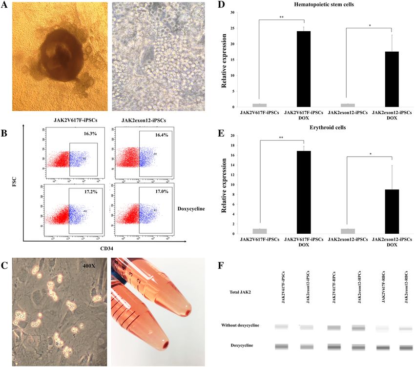
www.nature.com/scientificreports/
JAK2p.V617F mutation or, less commonly, heterozygous JAK2 exon12 mutations, e.g. JAK2p.N542_E543del, are
found in PV, while heterozygous JAK2p.V617F mutation was usually detectable in ET5. However, the differences
in signal activations responsible for these distinct MPN syndromes are still undefined.
Mouse models have been widely used to investigate the pathophysiology underlying human genetic diseases
because mice have almost similar set of genes which can be modified for studies. However, there is a limitation
as mice do not always demonstrate similar pathological changes as humans6. In order to study hematological
diseases, human hematopoietic stem cells must be expanded and maintained ex vivo which is a complicated
process. Peripheral blood mononuclear cells are as an easier alternative source, but they have a limited lifespan
in culture7,8.
Human induced pluripotent stem cells (iPSCs) are generated from mature cells via induction by four fac-
tors, Oct3/4, Sox2, c-Myc and Klf4. These stem cells can be derived from either normal subjects or patients to
be differentiated into various cell types in vitro. The iPSCs are more likely to represent normal hematopoiesis
compared with immortalized cancer cell lines. Furthermore, the cells can be genetically modified and greatly
expanded6,9. Therefore, iPSCs have potentials to be blood disease models which are probably closer to human
physiology than cancer cell lines or animals. Moreover, they may become cell sources for transfusion or immu-
notherapies in the future.
In this study, we reported the generation of two lines of iPSCs haboring doxycycline-inducible JAK2V617F
and JAK2 exon 12 mutants using viral transduction. The iPSCs were derived from normal volunteers instead
of patients to avoid interfering effects of concomitant genetic and/or epigenetic alterations in MPN patients.
The inducible system in the same iPSC line was used to exclude line-to-line variations because the difference
between mutant and control in each experiment was only the expression of the mutant JAK2 genes. They were
the same iPSCs that underwent the same transduction and selection processes followed by freezing, thawing
and culturing at the same time under the same conditions. Therefore, the controls were completely matched.
The erythrocytes generated from modified iPSCs were enumerated and characterized. Furthermore, the differ-
ences in signal activation between these two mutated JAK2 were explored. This will give us deeper insights in
the molecular pathogenesis of MPNs. Subsequently, the mutated iPSCs can be used to screen for the drugs that
preferentially affected neoplastic cells more than normal cells. This may lead to novel targeted therapy for MPNs.
Results
Generation and characterization of the genetically‑modified iPSCs. A normal human iPSC
line was modified by overexpressing two types of hyperactive JAK2 gene mutations (JAK2V617F-iPSCs and
JAK2exon12-iPSCs) by using viral transduction. The system used Tet-One inducible expression, in which the
inserted gene functioned under the doxycycline control. The modified iPSCs were tested for JAK2 gene inser-
tion by conventional polymerase chain reaction (PCR) using specific primers to JAK2-mutated vectors. Only
modified iPSCs demonstrated the PCR products representing an inserted JAK2 gene, whereas they were absent
in normal iPSCs (Fig. 1A). The DNA sequencing confirmed the JAK2p.V617F mutation, which was a change
from GTC (Valine) to TTC (Phenylalanine) in exon 14, and a deletion at the position N542_E543 of JAK2 gene
in the exon 12 mutation line (Fig. 1B).
The selected iPSCs were determined for the efficiency of doxycycline inducible system by evaluating JAK2
gene expression. After culturing normal iPSCs and modified iPSCs with and without doxycycline for 24 h,
JAK2V617F-iPSCs and JAK2exon12-iPSCs expressed the higher levels of JAK2 gene after doxycycline exposure
at approximately 17.95 ± 1.0 folds (p = 0.008) and 13.7 ± 6.4 folds (p = 0.034), respectively, when compared with
cells in the absence of doxycycline (Fig. 1C).
After transduction, JAK2V617F-iPSCs and JAK2exon12-iPSCs demonstrated normal karyotypes in both
numbers (46XY) and overall structures of chromosomes (Fig. 2A). Modified iPSC lines showed the RNA expres-
sion of five pluripotency genes that were NANOG, OCT4, SOX2, KLF4 and MYC (Fig. 2B). The germ layers from
embryoid body differentiation showed positive markers for ectoderm, mesoderm and endoderm by immuno-
fluorescence (Fig. 2C).
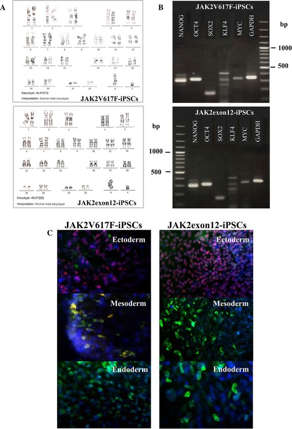
Erythroid cell differentiation. Normal and modified iPSCs that were co-cultured with irradiated
C3H10T1/2 feeder cells showed sac-like structures on day 14 of differentiation (Fig. 3A). Hematopoietic progen-
itor cells from ES-Sacs were collected, passed through a 40-micron cell strainer. The percentages of iPSC-derived
CD34+ cells from JAK2V617F-iPSCs and JAK2exon12-iPSCs representing hematopoietic stem cells from ES-
sacs were all approximately 16–17% with or without doxycycline (Fig. 3B). Hematopoietic stem cells induced by
the ES-Sac method were transferred onto fresh feeder cells and then cultured for 15 days. The cells were obtained
on day 15 after the initiation of hematopoietic cell culture derived from ES sacs. At that time, round floating cells
appeared in culture supernatant (Fig. 3C). After centrifugation, cell pellets showed the red color suggesting the
presence of hemoglobin (Fig. 3C).
The expression of JAK2 transgenes and protein after doxycycline induction. At the stage of
hematopoietic progenitor cells, JAK2V617F-iPSCs and JAK2exon12-iPSCs after induction with doxycycline
expressed JAK2 mRNA at approximately 24.1 ± 1.3 folds (p value = 0.002) and 17.6 ± 5.2 folds (p value = 0.046),
respectively (Fig. 3D). At the erythroid stage, the expression levels were 16.9 ± 1.0 folds (p value = 0.003) and
9.0 ± 4.9 folds (p value = 0.049) in JAK2V617F-iPSCs and JAK2exon12-iPSCs, respectively (Fig. 3E). Therefore,
both hematopoietic progenitor cells and erythroid cells sustained the high levels of JAK2 transgene expression.
The JAK2 protein expression was also determined by capillary Western immunoassay. JAK2V617F-iPSCs and
JAK2exon12-iPSCs expressed higher levels of total JAK2 proteins after doxycycline exposure at approximately 5
and 3 folds, respectively. In addition, JAK2V617F-iPSCs and JAK2exon12-iPSCs at the hematopoietic progenitor
Scientific Reports | (2021) 11:5255 | https://doi.org/10.1038/s41598-021-83895-6 2
Vol:.(1234567890)
www.nature.com/scientificreports/
Figure 1. Verification of JAK2 gene mutations and expression in the modified induced pluripotent stem
cells (iPSCs). (A) Conventional polymerase chain reaction (PCR) using transgene-specific primers showed
exogenous JAK2 genes in the two modified iPSC lines. The normal iPSC line was used as a negative control. The
full gel is presented in the Supplementary Fig. S1A. (B) DNA sequencing confirmed the point mutation p.V617F
in exon 14 and the p.N542_E543del in exon 12 of JAK2 gene in the respective iPSC lines. (C) Exogenous JAK2
gene expression levels in iPSCs after transfection comparing normal, JAK2V617F and JAK2 exon 12 mutation
with and without doxycycline (DOX) induction for 24 h and analyzed by real-time quantitative RT-PCR. Data
are presented as means ± standard deviations (SD) from three independent experiments. The asterisks (*) and
(**) denoted p < 0.05 and p < 0.01.
cell stage showed the elevations in total JAK2 protein levels of approximately 3 and 1.5 folds, respectively. At the
erythroid stage, the JAK2 protein increases were approximately 13 and 9 folds, respectively, compared with cells
in the absence of doxycycline (Fig. 3F).
Characterization of erythroid cells derived from the modified iPSCs. Erythroid cells which were
harvested from JAK2V617F-iPSCs and JAK2exon12-iPSCs in the condition without doxycycline on day 29
of differentiation yielded CD71/Glycophorin A (GPA) positivity at approximately 90.1% and 94.1% by flow
cytometry, respectively. Cells after doxycycline incubation dually expressed CD71/GPA at 95.4% and 96.2% in
JAK2V617F-iPSCs and JAK2exon12-iPSCs, respectively (Fig. 4A).
Regarding the total numbers of erythroid cells, JAK2V617F-iPSCs without vs. with doxycycline produced
275 ± 19.0 × 10⁵ cells/ml vs. 279 ± 1.4 × 10⁵ cells/ml, respectively, while JAK2exon12-iPSCs showed a significant
increase in the number of erythroid cells upon stimulation with doxycycline from 362 ± 18.3 × 10⁵ cells/ml (with-
out doxycycline) to 822 ± 17.2 × 10⁵ cells/ml (p = 0.007) (Fig. 4B). Only iPSCs expression JAK2 exon 12 mutation
showed increase in erythroid cell number and differentiation after doxycycline induction. On the other hand,
overexpression of wild-type JAK2 did not show significant increases in the erythroid cell number (Supplemen-
tary Fig. S3).
The experiments in the absence and a lower concentration (0 and 2.5 U/ml) of EPO were performed. They
showed that the effects of mutant JAK2 overexpression under these conditions were not prominent as those at
5 IU/ml (Supplementary Fig. S4).
Morphology of iPSC‑derived erythroid cells. During the 29 days of culture, cells from modified iPSCs
demonstrated erythroid morphology by Wright-Giemsa staining. The percentage of proerythroblasts, basophilic
erythroblasts, polychromatic erythroblasts, orthochromatic erythroblasts and erythrocytes from in vitro eryth-
roid differentiation were demonstrated in Fig. 4C and the most prevalent cells in cultures were orthochromatic
erythroblasts. JAK2exon12-iPSCs after doxycycline induction showed a significant increase in the subpopulation
of more mature orthochromatic erythroblasts when compared with JAK2V6 17F-iPSCs (p = 0.007) (Fig. 4D).
Hemoglobin analysis. Chromatograms from the Bio-Rad Variant demonstrated the peaks of embryonic
hemoglobin at the retention time approximately 0.15 min from iPSC-derived red blood cells with or without
doxycycline (Fig. 4E).
Scientific Reports | (2021) 11:5255 | https://doi.org/10.1038/s41598-021-83895-6 3
Vol.:(0123456789)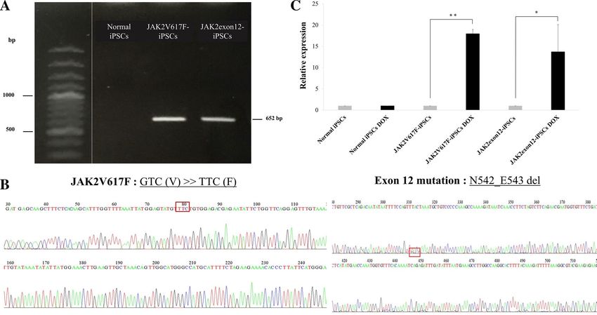
www.nature.com/scientificreports/

                                            Figure 2.  Characteristics of modified induced pluripotent stem cells (iPSCs). (A) Karyotyping of the genetically modified iPSCs:
                                            JAK2V617F-iPSCs and JAK2N542_E543del-iPSCs (JAK2exon12-iPSCs). (B) The expression of NANOG, OCT4, SOX2, KLF4 and MYC of
                                            JAK2V617F-iPSCs and JAK2exon12-iPSCs using reverse transcriptase polymerase chain reaction (RT-PCR). The full gels are presented in the
                                            Supplementary Fig. S1B. (C) Immunofluorescence of differentiated cells from JAK2V617F-iPSCs and JAK2exon12-iPSCs. Embryoid bodies
                                            were transferred onto 0.1% gelatin coverslips and cultured for 14 days for differentiation. Cells were stained with antibodies specific to ectoderm
                                            (red and green), mesoderm (green), endoderm (green) layers and DAPI (blue) for nuclei (×400 magnification). The images were captured by
                                            Axio Observer fluorescence microscopy.

          Scientific Reports |   (2021) 11:5255 |                      https://doi.org/10.1038/s41598-021-83895-6                                                                            4

Vol:.(1234567890)
www.nature.com/scientificreports/
Figure 3. Erythroid cell differentiation from modified induced pluripotent stem cells (iPSCs) via ES-Sacs.
(A) ES-derived sacs containing hematopoietic progenitor cells were generated from modified iPSCs on day
14 at ×100 and ×400 magnifications. (B) The percentages of C D34+ cells derived from JAK2V617F-iPSCs
and JAK2exon12-iPSCs were similar with or without doxycycline. (C) Erythroid cells in a culture plate and
red blood cell pellets after centrifugation. (D) Expression mRNA levels of exogenous JAK2 in hematopoietic
progenitor cells determined by real-time quantitative RT-PCR. (E) Expression mRNA levels of exogenous JAK2
in erythroid cells. (F) The capillary Western immunoassay showed total JAK2 proteins from JAK2V617F-iPSCs
and JAK2exon12-iPSCs at the induced pluripotent stem cell, hematopoietic progenitor cell (HPC) and erythroid
cell (RBC) stages in the conditions with vs. without doxycycline. Each band was electrophoresed in a separate
capillary tube. The full blot is presented in the Supplementary Fig. S2A.
Globin gene expression was also evaluated by real-time quantitative RT-PCR. After stimulation with doxy-
cycline, JAK2exon12-iPSCs derived erythroid cells showed a significant increase in the beta globin mRNA
expression when compared with JAK2V617F-iPSCs (13.35 ± 0.75 folds vs. 7.16 ± 0.62 folds, respectively, p = 0.018)
(Fig. 4F).
JAK/STAT signal transduction. Hematopoietic progenitor cells from ES-Sacs were harvested and cul-
tured with a hematopoietic cell differentiation medium supplemented with 5 IU/ml EPO, 50 ng/ml TPO and
50 ng/ml SCF for 24 h. Stimulated cells were collected and then subjected to capillary Western assays for the cell
signaling which was composed of JAK2, STAT1, STAT3, STAT5, ERK1/2 and AKT in both native and phospho-
rylated forms (Fig. 5A). The relative changes in phosphorylated JAK2 proteins in the presence of doxycycline in
JAK2V617F-iPSCs and JAK2exon12-iPSCs were 2.50 ± 0.87 and 1.25 ± 0.29 folds, respectively.
JAK2V617F-iPSCs showed significant increases in the relative changes of phospho-STAT3 and phospho-AKT
proteins when compared with JAK2exon12-iPSCs at 5.60 ± 0.69 vs. 0.20 ± 0.35 (p value < 0.001) and 1.20 ± 0.23 vs.
Scientific Reports | (2021) 11:5255 | https://doi.org/10.1038/s41598-021-83895-6 5
Vol.:(0123456789)www.nature.com/scientificreports/
Scientific Reports | (2021) 11:5255 | https://doi.org/10.1038/s41598-021-83895-6 6
Vol:.(1234567890)www.nature.com/scientificreports/
◂Figure 4. Characteristics of modified induced pluripotent stem cells (iPSC)-derived erythroid cells. (A) Flow
cytometry of erythroid-specific surface molecules on iPSC-derived erythroid cells without vs. with doxycycline.
Cells were stained with PE-conjugated anti-human CD71 and FITC-conjugated anti-human Glycophorin A
(GPA) antibodies. (B) Total CD71+GPA+ erythroid cell numbers from erythroid differentiation via ES-Sacs with
vs. without doxycycline. (C) Five stages of erythroid series were classified by Wright-Giemsa stain consisting
of proerythroblasts, basophilic erythroblasts, polychromatic erythroblasts, orthochromatic erythroblasts and
erythrocytes. The images were captured by Leica DM 1000 microscopy using LAS49 software and the scale bars
represented 10 µm for all panels. (D) The percentage of erythroid cell differentiation stages. (E) Chromatograms
from the ion exchange high performance liquid chromatography (HPLC) Bio-Rad Variant II showed mainly
embryonic hemoglobin in modified iPSC-derived erythroid cells with and without doxycycline. (F) The relative
expression of beta-similar globin genes which were epsilon, gamma and beta in iPSC-derived erythroid cells
comparing overexpression of JAK2V617F vs. JAK2 exon 12 mutants and analyzed by real-time quantitative
RT-PCR. Data were presented as mean ± SD from three independent experiments. The asterisks (**) denoted
p < 0.01.
0.40 ± 0.23 (p value = 0.008), respectively. Moreover, the phospho-STAT5 signaling protein level in JAK2V617F-
iPSCs was higher than that of JAK2exon12-iPSCs at approximately 0.63 ± 0.03 vs. 0.41 ± 0.28, but there was no
statistical significance.
On the other hand, JAK2exon12-iPSCs expressed a higher level of phospho-STAT1 and phospho-ERK1/2
(1.00 ± 0.69 and 0.65 ± 0.00) when compared with JAK2V617F-iPSCs (0.02 ± 0.14 and 0.51 ± 0.20). However, only
phospho-STAT1 reached a statistical significance (p value = 0.022) (Fig. 5B).
Interferon alpha and arsenic trioxide treatments. Interferon alpha and arsenic trioxide were tested
on modified iPSCs with vs. without doxycycline induction to examine the relative sensitivity of cells expressing
mutant JAK2 compared with wild-type cells. The hematopoietic progenitor cells were cultured in the absence
or presence of 0.5 µg/ml interferon alpha and/or 250 nM arsenic trioxide. These optimal concentrations were
obtained from cultures using various doses of interferon alpha and arsenic trioxide as demonstrated in Supple-
mentary Fig. S5.
The JAK2V617F-iPSCs showed a significant decrease in the number of erythroid cells after treatment with
arsenic trioxide, interferon alpha and the combination of both drugs from 412 ± 6.35 × 10⁵ cells/ml (untreated)
to 250 ± 6.35, 261 ± 6.35 and 172 ± 57.4 × 10⁵ cells/ml, respectively. Statistical analyses revealed the significant
p-values of 0.008, 0.010 and 0.002, respectively.
The JAK2exon12-iPSCs displayed a significant decrease in cell numbers from 686 ± 32.33 × 10⁵ cells/ml
(untreated) to 305 ± 12.7 × 10⁵ cells/ml with arsenic trioxide (p value = 0.003), 300 ± 38.68 × 10⁵ cells/ml with
interferon alpha (p value = 0.003) and 189 ± 19.05 × 10⁵ cells/ml with interferon alpha plus arsenic trioxide (p
value = 0.002). Notably, these agents did not affect the cell numbers of modified iPSCs without doxycycline
induction (Fig. 5C).
Concerning the relative changes of apoptotic cells when compared with the untreated control, JAK2V617F-
iPSCs after treatments with arsenic trioxide, interferon alpha and the combination of both drugs showed the
increases of 3.50 ± 2.10, 2.19 ± 0.42 and 4.00 ± 0.98 folds, respectively, while JAK2exon12-iPSCs displayed the
relative changes of approximately 2.40 ± 0.57, 2.67 ± 1.04 and 2.50 ± 0.51 folds, respectively. There was no apoptotic
rate difference in modified iPSCs after incubation with either one or both drugs without doxycycline stimula-
tion (Fig. 5D).
Discussion
From our study, the lentivirus-modified iPSCs retained normal karyotypes, stem cell properties and multi-lineage
potentials. The modified iPSC-derived red blood cells displayed erythroid morphology by Wright-Giemsa stain,
erythroid surface markers by flow cytometry and embryonic hemoglobin expression similar to normal iPSC-
derived cells. Interestingly, JAK2exon12-iPSCs significantly enhanced erythroid cell proliferation mimicking the
pathophysiology of JAK2 exon 12 mutations in patients with PV. In addition, expression of JAK2 with an exon
12 mutant resulted in enhanced erythroid differentiation as shown by more mature morphology and higher
expression of adult globin as determined by real-time quantitative RT-PCR. In contrast, JAK2V617F-iPSCs did
not show a significant increase in erythroid cell proliferation as enumerated by cell counting and differentiation
as evaluated by morphology and hemoglobin analysis despite the overexpression of JAK2 transgene on real-time
quantitative RT-PCR assay and JAK2 protein by Western immunoassay. This disparity may be explained by that
JAK2V617F expression in our study was more consistent with heterozygous JAK2V617F mutation in essential
thrombocythemia (ET) patients because endogenous JAK2 gene was still present. The polycythemia vera (PV)
patients usually carry homozygous JAK2V617F mutation and, less frequently, heterozygous JAK2 exon 12 muta-
tion as modeled in our s tudy10. Therefore, modified iPSCs from this study can be used as an experimental model
to investigate the molecular pathogenesis of MPN patients and possibly answer the questions why the JAK2V617F
and JAK2 exon 12 mutations attributed to different erythroid phenotypes.
To our knowledge, this is the first report comparing effects of JAK2V617F vs. JAK2 exon12 mutants (JAK2p.
N542_E543del) overexpression on erythroid development from modified iPSCs and exploring their signal-
ing pathways. Notably, JAK2V617F-iPSCs showed significantly higher relative changes of phospho-STAT3 and
phospho-AKT signaling proteins, whereas JAK2exon12-iPSCs exhibited higher amounts of phospho-STAT1.
These results were consistent with previous studies in human specimens. Bone marrow biopsies of ET patients
who had JAK2V617F mutation showed an increase in phospho-STAT3 by immunohistochemical analysis and
Scientific Reports | (2021) 11:5255 | https://doi.org/10.1038/s41598-021-83895-6 7
Vol.:(0123456789)www.nature.com/scientificreports/
Figure 5. Signal transduction and drug treatment of modified induced pluripotent stem cells (iPSCs). (A)
The capillary Western immunoassay of phosphorylated and total signaling proteins which were JAK2, STAT1,
STAT3, STAT5, ERK1/2 and AKT in the absent and presence of doxycycline in JAK2V617F-iPSCs and
JAK2exon12-iPSCs. Each band was electrophoresed in a separate capillary tube. The full blot is presented in
the Supplementary Fig. S2B. (B) The relative changes of phosphorylated signaling proteins after doxycycline
induction in JAK2V617F-iPSCs and JAK2exon12-iPSCs compared with those without doxycycline. The levels
of phosphoproteins were corrected by the amounts of respective total proteins. (C) Erythroid cell numbers after
incubations without (untreated) vs. with arsenic trioxide, interferon alpha and the combination of both drugs.
(D) The relative increases in numbers of apoptotic cells in modified iPSCs in the presence of arsenic trioxide,
interferon alpha and the combination of both drugs. Data were presented as mean ± SD from three independent
experiments. The asterisks (*), (**) and (***) denoted p < 0.05, p < 0.01 and p < 0.001, respectively.
immunoblotting11. Additionally, bone marrow trephine biopsy sections of MPN patients, JAK2V617F muta-
tion was associated with significantly increased levels of phospho-STAT5 and phospho-AKT in hematopoietic
cells, which were most prominent in m egakaryocytes12. Furthermore, cells reprogrammed from heterozygous
JAK2V617F patients showed a high level of phospho-STAT5 and displayed TPO-independent formation of
megakaryocytic colonies but not EPO-independent erythroid colony13. Supporting these data, the study in
BaF3/EPOR cells transduced with various types of JAK2 gene mutations including JAK2V617F, N542_E543del,
H538QK539L, K539L and F537_K539delinsL showed that these exon 12 mutations activated the RAS-ERK
signaling pathway. The levels of phospho-ERK1 and ERK2 were markedly higher than JAK2V617F mutation
and there were variable levels of phospho-ERK in different types of exon 12 m utations14. In addition, the STAT1
knockout mice showed reduced bone marrow-derived erythroid colony forming units and less differentiated
phenotypes associated with increased apoptosis of early erythroblasts. These data demonstrated that STAT1
played a critical role in the regulation of erythropoiesis15. On the other hand, STAT3 is probably a minor signal-
ing molecule for EPO-independent growth but may play an important role in megakaryopoiesis. The stronger
effects of the JAK2exon 12 mutation may be from the more prominent STAT1 activation compared with those of
the JAK2V617F. Therefore, dissimilar signals may explain the different phenotypes of patients with heterozygous
JAK2V617F and those with JAK2 exon 12 mutations.
In 2013, iPSCs containing heterozygous and homozygous JAK2V617F were generated from MPN patients
and studied for molecular mechanisms. However, MPN patient samples usually co-carried other genetic defects
including 20q deletion, ASXL1, FBXO15 and MATN213 mutations that can affect iPSC phenotypes. Clonal sub-
population may vary among samples depending on disease progression and treatment processes. Furthermore,
different clones of iPSCs may display distinct intracellular signaling and growth potentials. In this study, the iPSC
lines with doxycycline-inducible JAK2 mutations were constructed from normal iPSCs. The in vitro erythrocyte
generation was observed comparing between cultures with vs. without doxycycline. Therefore, the phenotype
Scientific Reports | (2021) 11:5255 | https://doi.org/10.1038/s41598-021-83895-6 8
Vol:.(1234567890)www.nature.com/scientificreports/
differences were attributed solely to the overexpressed mutated JAK2 genes without interferences by other genetic
and/or epigenetic background.
A possible limitation of our model is that iPSC-derived hematopoietic stem cells (HSCs) might show different
properties compared with the marrow-derived HSCs. Mascarenhas et al. revealed that the mouse embryonic
HSCs were relatively resistant to JAK2V617F mutation compared with adult H SCs16. Consistent with this find-
ing, we found that JAK2V617F expression also showed minor effects on erythropoiesis from human iPSCs.
However, our experiments revealed that overexpressing JAK2 with mutated exon 12 in human iPSCs significantly
increased erythropoiesis similar to the erythrocytosis phenotype in patients. Therefore, the iPSC model gave us
the opportunities to demonstrate that the different effects of mutant JAK2s were correlated with different STAT
signals and to explore the drugs that selectively inhibit cells with mutant JAKs. These yielded a deeper insight in
the pathogenesis of MPNs and may lead to future therapy.
In the past, potentially new drugs were screened in immortalized cancer cell lines and animal models which
cannot always predict efficacy and safety in h umans17. The iPSCs can be differentiated into disease specific cell
types and demonstrate the phenotypes similar to primary cells. Drug screening on these iPSC-derived cells may
be helpful for discoveries of novel treatments.
Interferon alpha that signals through the JAK/STAT pathway has been used for the treatments of ET or PV.
The mechanisms of action of interferon-alpha have been ascribed to its anti-proliferative, pro-apoptotic, anti-
angiogenic, and immunomodulatory e ffects18. Interestingly, interferon can decrease the mutated JAK2 allele bur-
dens in MPN patients. In addition, the combination with the other drugs may be more efficacious for advanced
and transformed d iseases19. There were reports that interferon alpha preferentially induced JAK2-positive cell
apoptosis which was mediated through the p53 or p38MAPK p athways20,21. Furthermore, recent data found that
the interferon-sensitivity depended on STAT2 activation22. Arsenic trioxide is the standard treatment for relapsed
acute promyelocytic leukemia (APL) through promoting apoptosis which is involved intracellular glutathione and
hydrogen peroxide23. Recent researches showed that hematologic malignancies other than APL also responded
to combination therapy containing arsenic trioxide. JAK2V617F-UT7 cell lines were generated and revealed the
synergistic effects of interferon alpha and arsenic trioxide c ombination24. Arsenic trioxide was shown to induce
acute promyelocytic cell (APL) differentiation at low concentrations and apoptosis at high concentrations of
over 500 nM partly from the specific degradation of the PML-RARα oncoprotein25. The proposed mechanisms
of arsenic trioxide in other cancers are the increases in reactive oxygen species (ROS) from mitochondria and/
or endoplasmic reticulum causing cellular a poptosis26.
According to our experiments, JAK2V617F-iPSCs and JAK2exon12-iPSCs showed a significant decrease in
the number of erythroid cells and an increase in apoptotic cells after treatment with arsenic trioxide, interferon
alpha and the combined regimen. The additive effect of these two agents was observed in our model. Interest-
ingly, arsenic trioxide and interferon alpha treatments showed the specific effects on mutated iPSCs but did not
in the condition without doxycycline induction. This disease model of overexpressing JAK2V617F and JAK2
exon 12 mutants suggests the potential roles of interferon alpha and arsenic trioxide in therapy of MPN patients.
To explore whether interferon alpha and/or arsenic trioxide also affected other lineages, the experiments were
performed during the megakaryopoiesis. Similarly, these agents preferentially suppressed the proliferation of
mutant JAK2 expressing cells as compared with cultures without doxycycline induction (Supplementary Fig. S6).
Therefore, they have a potential to eliminate the malignant clone. In the future, this modified iPSCs can be used
to test for other new therapeutic agents.
Derivation of red blood cells from iPSCs may become blood products for transfusion. Genetic engineering
can be applied to generate erythrocytes with very rare blood groups without requirement for exceptional donors.
The proteome analysis of erythroid cells differentiated from iPSC lines revealed a similar pattern to that of normal
adult erythroid c ells27. However, the challenges of erythroid production are inefficient enucleation, low expres-
sion of the adult β hemoglobin and scalable production28. From our result, JAK2exon12-iPSCs enhanced red
blood cell production with a greater number of the late-stage erythrocytes and produced more adult hemoglobin
(α2β2, HbA) at the mRNA level. Overexpression of JAK2 with exon 12 mutations may be one of the factors to
improve the blood cell production for transfusion.
Conclusions
Our study used the iPSC technology to obtain better understanding of the JAK2 mutation effects on erythropoie-
sis. The JAK2 exon 12 mutation strongly promoted erythroid cell proliferation and differentiation correlating with
STAT1 and ERK activation. Modified iPSCs provided a model to study the mechanisms of mutated JAK2, screen
for novel therapeutic agents and possibly offer a potential source for red blood cell transfusion in the future.
Materials and methods
Establishing the iPSC lines with JAK2 gene mutations. A normal human iPSC line was modified
by overexpressing two types of hyperactive JAK2 gene, which were a point mutation in exon 14 (JAK2p.V617F)
and a small deletion in exon 12 (JAK2p.N542_E543del), using viral transduction. The experimental design using
human iPSCs was approved by the Institutional Review Board of the Faculty of Medicine at Chulalongkorn
University, Bangkok, Thailand (Certificate No. 33/2018) and was conducted in accordance with the Declaration
of Helsinki. The iPSCs were derived from skin fibroblasts of a healthy subject after informed consent. The repro-
gramming process was described in a previous study29. The details on iPSC characterization of pluripotency
were described in the Supplementary Fig. S7.
Firstly, the wild-type JAK2-containing plasmid (Addgene, Cambridge, MA, USA) was altered using the Site-
directed mutagenesis kit (Thermo Fisher Scientific, Waltham, MA, USA). The constructed plasmids carrying
JAK2p.V617F (JAK2V617F) and JAK2p.N542_E543del (JAK2exon12) were inserted into the complementary site
Scientific Reports | (2021) 11:5255 | https://doi.org/10.1038/s41598-021-83895-6 9
Vol.:(0123456789)www.nature.com/scientificreports/
Genes Forward primers Reverse primers
Exogenous JAK2 CCCTCGTAAAGAATTCATGGGAATGGCC TGCCTTACGATG TCTTTGCTCGAATACATTTTGG
NANOG ATACCTCAGCCTCCAGCAGA CAGGACTGGATGTTCTGGGT
OCT4 GAAGGTATTCAGCCAAACGC GTTACAGAACCACACTCGGA
SOX2 GGGAAATGGGAGGGGTGCAAAAGAGG TTGCGTGAGTGTGGATGGGATTGGTG
KLF4 ACGATCGTGGCCCCGGAAAAGGACC TGATTGTAGTGCTTTCTGGCTGGGCTCC
MYC GCGTCCTGGGAAGGGAGATCCGGAGC TTGAGGGGCATCGTCGCGGGAGGCTG
Epsilon globin GCCTGTGGAGCAAGATGAAT GCGGGCTTGAGGTTGT
Gamma globin TGAGAACTTCAAGCTCCTGGGAAA TGCAGAATAAAGCCTATCCTTGAA
Beta globin TACATTTGCTTCTGACACAAC ACAGATCCCCAAAGGAC
Table 1. The primer sets for exogenous JAK2, pluripotency testing of stem cells and hemoglobin expression.
of pLVX-TetOne-Puro vector by which the mutated JAK2 were expressed under the Lentiviral Tet-One inducible
expression system (Clontech, Palo Alto, CA, USA). For lentiviral production, the recombinant vectors containing
either JAK2V617F or JAK2exon12 coding sequences were transfected into 293FT cells. Virus-containing super-
natants were incubated with normal iPSCs in the presence of 6 µg/ml polybrene for transduction. Transfected
iPSCs were selected by p uromycin30.
To verify the engineered cell lines, modified iPSCs were harvested and extracted for genomic DNA by using
the prepGEM kit (MicroGEM, Aotearoa, New Zealand). The DNA from modified iPSCs was assayed for JAK2
gene insertions using polymerase chain reaction (PCR) and sequencing. Each tube contained cDNA (100 ng/µl
final concentration), 10 µM forward and reverse primer mix specific for exogenous JAK2 (The primer sequences
are listed in Table 1) and GoTaq Master Mixes (Promega, Madison, Wisconsin, USA). The PCR condition was
95 °C initial denaturation for 5 min, followed by 30 cycles of denaturation (98 °C, 40 s), annealing (63 °C, 1 min),
and extension (72 °C, 1.30 min). Unmodified iPSCs were used as a negative control. DNA templates were ampli-
fied in a T gradient Biometra thermal cycler (Biometra GmbH, Göttingen, Germany). Sanger DNA sequencing
was used to confirm JAK2 gene mutations and analyzed by capillary electrophoresis at Macrogen Inc., Korea.
The selected iPSCs were examined for the efficiency of doxycycline inducible system. Cells were cultured
in the medium with 0 or 2 µg/ml of doxycycline (DOX, Stemcell Technologies, Vancouver, BC, Canada) and
incubated for 24 h before harvesting cell pellets and tested for JAK2 gene expression by using real-time quanti-
tative RT-PCR. PCR was performed using exogenous JAK2-specific primers and the following protocol: 95 °C
initial denaturation for 10 min, followed by 40 cycles of denaturation (95 °C, 15 s), annealing (59.5 °C, 30 s),
and extension (72 °C, 45 s).
Characterization of genetically‑modified iPSC properties. The genetically-modified iPSCs were
required to ensure the normal karyotypes, the pluripotent status and the capacity to differentiate into cells of the
three germ layers.
Chromosomal analysis was performed by GTG-banding analysis at the Center for Medical Diagnostic Labo-
ratories, Faculty of Medicine, Chulalongkorn University, Thailand, following the recommendations by the Inter-
national System for Cytogenetics Nomenclature (ISCN).
The pluripotency of stem cells was evaluated by reverse-transcriptase PCR (RT-PCR) of stem cell markers
including NANOG, OCT4, SOX2, KLF4 and MYC. The primer sequences are listed in Table 131–34.
The ability of stem cells to differentiate into endoderm, mesoderm and ectoderm were tested via embryoid
body (EB) formation and stained by Human three germ layer 3-color immunocytochemistry kit (R&D Systems,
Minneapolis, MN, USA)35.
To demonstrate the hematopoietic multipotency, our iPSC-derived hematopoietic progenitors were cultured
in the presence of 100 ng/ml human thrombopoietin (TPO), 50 ng/ml human stem cell factor (SCF), and 25 ng/
ml heparin for 21 days. The culture finally yielded the mixture of megakaryocytes (by morphology and flow
cytometry for CD41/CD42b surface expression) and neutrophils (by morphology) as shown in the Supplemen-
tary Fig. S8.
Reverse transcriptase polymerase chain reaction (RT‑PCR) and real‑time quantitative
RT‑PCR. Total RNA from modified iPSCs were extracted using an RNA purification kit (GeneJet kit; Thermo
Fisher Scientific, Waltham, MA, USA). Isolated RNA was reverse transcribed using a cDNA synthesis kit
(Thermo Fisher Scientific).
Real-time quantitative RT-PCR assay was performed by using Capital qPCR probe mix (Biotechrabbit GmbH,
Hennigsdorf, Germany) on 7500 Fast real-time PCR system (Applied Biosystems, Foster City, CA, USA). The
relative quantity of each target gene was normalized to glyceraldehyde-3-phosphate dehydrogenase (GAPDH)
as a house-keeping gene. Fold changes were calculated by quantifying expression using comparative CT (ΔΔCt)
method compared with those of normal unmodified iPSCs. All samples were processed in triplicate.
Differentiation of iPSCs into erythrocytes. Normal iPSCs and modified iPSCs were differentiated into
erythroid cells using the ES-sac method according to Ochi et al.36. Cells were dissociated into small pieces (> 100
Scientific Reports | (2021) 11:5255 | https://doi.org/10.1038/s41598-021-83895-6 10
Vol:.(1234567890)www.nature.com/scientificreports/
Figure 6. Schematic diagrams of in vitro differentiation protocols for erythrocytes production via ES-Sacs
formation. VEGF Vascular endothelium growth factor, TPO thrombopoietin, SCF stem cell factor, EPO
erythropoietin, HPCs hematopoietic progenitor cells.
cells) by collagenase treatment. Small clump of cells were transferred onto irradiated C3H10T1/2 feeder cells
and cultured in a hematopoietic cell differentiation medium, Iscove’s Modified Dulbecco’s Medium (IMDM)
supplemented with 10 µg/ml human insulin, 5.5 µg/ml human transferrin, 5 ng/ml sodium selenite, 2 mM l-glu-
tamine, 0.45 mM α-monothioglycerol, 50 µg/ml ascorbic acid and 15% fetal bovine serum (FBS) containing
20 ng/ml recombinant human vascular endothelial growth factor (rhVEGF, R&D Systems) with 0 or 2 µg/ml of
doxycycline from the first day of culture.
On day 14 of culture, embryonic stem cell–derived sacs (ES-Sacs) were emerged. Cells from ES-Sacs were gen-
tly crushed with a needle and passed through a 40-µm cell strainer which selected a population of hematopoietic
progenitor cells (HPCs). The hematopoietic progenitors at 5 × 10⁴ cells/ml were maintained in hematopoietic cell
differentiation medium supplemented with 50 ng/ml human thrombopoietin (TPO, R&D Systems), 50 ng/ml
human stem cell factor (SCF, R&D Systems) and 5 IU/ml erythropoietin (EPO, Eprex, Janssen Pharmaceutical,
Beerse, Belgium) and then transferred onto fresh and irradiated C3H10T1/2 cells in a six-well plate for 6 days.
After 6 days, cells were transferred to fresh irradiated C3H10T1/2 cells and cultured in hematopoietic cell dif-
ferentiation medium supplemented only with 5 IU/ml EPO for another 9 days. Non-adherent cells were harvested
and analyzed on day 29 of culture36 (Fig. 6).
Flow cytometry analysis. Erythroid cells on day 29 were incubated with PE-conjugated anti-human
CD71 (Clone CY1G4, BioLegend, San Diego, CA, USA) and FITC-conjugated anti-human Glycophorin A
(GPA, Clone HI264, BioLegend) at room temperature in the dark for 30 min. Flow cytometry was performed by
using BD FACSAria II (Becton Dickinson, Franklin Lakes, NJ, USA). The total erythroid cells were calculated by
D71+GPA+ cells.
counting the total numbers of cells and multiplying by the percentages of C
Morphological analysis. The modified iPSCs on day 29 of culture were classified by morphology into vari-
ous differentiation stages of the erythroid lineage. Cells were harvested from culture supernatant, mounted on
slides by Cellspin I 1–12 (Tharmac GmbH, Wiesbaden, Germany) and stained by Wright-Giemsa with phosphate
buffer. Subsequently, erythrocytes were observed under Leica DM 1000 microscopy (Leica, Wetzlar, Germany).
Hemoglobin typing. The Variant II Beta Thalassemia Short Program utilizing the ion-exchange high-per-
formance liquid chromatography (HPLC) principle (Bio-Rad, California, United States) was applied.
The mRNA of beta globin subtype genes were measured by real time quantitative PCR to determine the
expression levels of embryonic hemoglobin (epsilon; ε), fetal hemoglobin (gamma; γ) and adult hemoglobin
(beta; β). The primer sequences are listed in Table 137–39.
JAK/STAT signal transduction. From ES-Sacs formation, the hematopoietic progenitor cells were har-
vested and transferred onto fresh irradiated C3H10T1/2 cells and then cultured with a hematopoietic cell dif-
ferentiation medium supplemented with 50 ng/ml TPO, 50 ng/ml SCF and 5 IU/ml EPO for 24 h. Stimulated
cells were measured for protein concentrations using Micro BCA protein assay kit (Thermo Fisher Scientific).
Signaling protein analyses were performed on a capillary Western immunoassay system (WES, ProteinSim-
ple, California, USA) using antibodies to JAK2, STAT1, STAT3, STAT5, ERK1/2 and AKT in both native and
Scientific Reports | (2021) 11:5255 | https://doi.org/10.1038/s41598-021-83895-6 11
Vol.:(0123456789)www.nature.com/scientificreports/
phosphorylated forms (Cell Signaling Technology, Massachusetts, USA). The chemiluminescent signals were
detected and quantitated by Program Compass for SW. The level of each phosphoprotein was corrected by the
amount of the respective total signaling protein and later calculated for the fold changes comparing with vs.
without doxycycline induction.
Effects of drugs on erythroid development from modified iPSCs. The hematopoietic progenitor
cells generated from modified iPSCs at 5 × 10⁴ cells/ml were cultured in the absence or presence of drugs which
had potentials to treat MPN patients, 0.5 µg/ml interferon alpha-2a (Roche, New Jersey, USA) and/or 250 nM
arsenic trioxide (M&B, London, United Kingdom)40,41.
The yields of total erythroid cells were enumerated as above. The percentage and relative changes of cell deaths
were determined using FITC-conjugated anti-human Glycophorin A (Clone HI264, BioLegend) and propidium
iodide (#421301, BioLegend). The results were compared between with vs. without doxycycline to determine the
differential effects on mutated vs. normal cells, respectively.
Statistical analysis. Statistical analyses were performed using the SPSS software (version 22.0). All the con-
tinuous variables were expressed as means ± standard deviations (SD). The statistical differences between groups
(doxycycline induction vs. no doxycycline induction of mutated JAK2 transgene expression) were determined
using the paired T-test. In addition, one way ANOVA and independent-sample T-test were also used to detect
statistical significances. The probability (P) values of less than 0.05 were considered statistically significant.
Received: 16 November 2019; Accepted: 9 February 2021
References
1. Arber, D. A. et al. The 2016 revision to the World Health Organization classification of myeloid neoplasms and acute leukemia.
Blood 127, 2391–2405. https://doi.org/10.1182/blood-2016-03-643544 (2016).
2. Vainchenker, W. & Kralovics, R. Genetic basis and molecular pathophysiology of classical myeloproliferative neoplasms. Blood
129, 667–679. https://doi.org/10.1182/blood-2016-10-695940 (2017).
3. Rumi, E. & Cazzola, M. Diagnosis, risk stratification, and response evaluation in classical myeloproliferative neoplasms. Blood
129, 680–692. https://doi.org/10.1182/blood-2016-10-695957 (2017).
4. Tefferi, A. & Pardanani, A. Myeloproliferative neoplasms: A contemporary review. JAMA Oncol. 1, 97–105. https: //doi.org/10.1001/
jamaoncol.2015.89 (2015).
5. Levine, R. L., Pardanani, A., Tefferi, A. & Gilliland, D. G. Role of JAK2 in the pathogenesis and therapy of myeloproliferative
disorders. Nat. Rev. Cancer 7, 673–683. https://doi.org/10.1038/nrc2210 (2007).
6. Wattanapanitch, M. Recent updates on induced pluripotent stem cells in hematological disorders. Stem Cells Int. 2019, 5171032.
https://doi.org/10.1155/2019/5171032 (2019).
7. Ye, Z., Chou, B. K. & Cheng, L. Promise and challenges of human iPSC-based hematologic disease modeling and treatment. Int.
J. Hematol. 95, 601–609. https://doi.org/10.1007/s12185-012-1095-9 (2012).
8. Focosi, D. et al. Induced pluripotent stem cells in hematology: Current and future applications. Blood Cancer J. 4, e211. https://
doi.org/10.1038/bcj.2014.30 (2014).
9. Slukvin, I. I. Hematopoietic specification from human pluripotent stem cells: Current advances and challenges toward de novo
generation of hematopoietic stem cells. Blood 122, 4035–4046. https://doi.org/10.1182/blood-2013-07-474825 (2013).
10. Morotti, A., Rocca, S., Carra, G., Saglio, G. & Brancaccio, M. Modeling myeloproliferative neoplasms: From mutations to mouse
models and back again. Blood Rev. 31, 139–150. https://doi.org/10.1016/j.blre.2016.11.004 (2017).
11. Teofili, L. et al. Different STAT-3 and STAT-5 phosphorylation discriminates among Ph-negative chronic myeloproliferative diseases
and is independent of the V617F JAK-2 mutation. Blood 110, 354–359. https://doi.org/10.1182/blood-2007-01-069237 (2007).
12. Grimwade, L. F. et al. Phospho-STAT5 and phospho-Akt expression in chronic myeloproliferative neoplasms. Br. J. Haematol. 147,
495–506. https://doi.org/10.1111/j.1365-2141.2009.07870.x (2009).
13. Saliba, J. et al. Heterozygous and homozygous JAK2(V617F) states modeled by induced pluripotent stem cells from myeloprolifera-
tive neoplasm patients. PLoS ONE 8, e74257. https://doi.org/10.1371/journal.pone.0074257 (2013).
14. Scott, L. M. et al. JAK2 exon 12 mutations in polycythemia vera and idiopathic erythrocytosis. N. Engl. J. Med. 356, 459–468. https
://doi.org/10.1056/NEJMoa065202 (2007).
15. Halupa, A. et al. A novel role for STAT1 in regulating murine erythropoiesis: Deletion of STAT1 results in overall reduction of
erythroid progenitors and alters their distribution. Blood 105, 552–561. https://doi.org/10.1182/blood-2003-09-3237 (2005).
16. Mascarenhas, M. I. et al. Analysis of Jak2 signaling reveals resistance of mouse embryonic hematopoietic stem cells to myelopro-
liferative disease mutation. Blood 127, 2298–2309. https://doi.org/10.1182/blood-2015-08-664631 (2016).
17. Shi, Y., Inoue, H., Wu, J. C. & Yamanaka, S. Induced pluripotent stem cell technology: A decade of progress. Nat. Rev. Drug Discov.
16, 115–130. https://doi.org/10.1038/nrd.2016.245 (2017).
18. Thomas, H., Foster, G. & Platis, D. Mechanisms of action of interferon and nucleoside analogues. J. Hepatol. 39(Suppl 1), S93-98.
https://doi.org/10.1016/s0168-8278(03)00207-1 (2003).
19. Hasselbalch, H. C. & Holmström, M. O. Perspectives on interferon-alpha in the treatment of polycythemia vera and related
myeloproliferative neoplasms: Minimal residual disease and cure?. Semin. Immunopathol. 41, 5–19. https://doi.org/10.1007/s0028
1-018-0700-2 (2019).
20. Lu, M., Xia, L., Li, Y., Wang, X. & Hoffman, R. The orally bioavailable MDM2 antagonist RG7112 and pegylated interferon α 2a
target JAK2V617F-positive progenitor and stem cells. Blood 124, 771–779. https://doi.org/10.1182/blood-2013-11-536854 %JBloo
d (2014).
21. Lu, M. et al. Interferon-alpha targets JAK2V617F-positive hematopoietic progenitor cells and acts through the p38 MAPK pathway.
Exp. Hematol. 38, 472–480. https://doi.org/10.1016/j.exphem.2010.03.005 (2010).
22. Schubert, C. et al. Differential roles of STAT1 and STAT2 in the sensitivity of JAK2V617F- vs. BCR-ABL-positive cells to interferon
alpha. J. Hematol. Oncol. 12, 36. https://doi.org/10.1186/s13045-019-0722-9 (2019).
23. Miller, W. H. Jr. Molecular targets of arsenic trioxide in malignant cells. Oncologist 7(Suppl 1), 14–19. https://doi.org/10.1634/
theoncologist.7-suppl_1-14 (2002).
Scientific Reports | (2021) 11:5255 | https://doi.org/10.1038/s41598-021-83895-6 12
Vol:.(1234567890)www.nature.com/scientificreports/
24. Maslah, N. et al. Specific and synergistic targeting of JAK2V617F cells by interferon alpha and arsenic. Blood 132, 52–52. https://
doi.org/10.1182/blood-2018-99-112518 (2018).
25. Tomita, A., Kiyoi, H. & Naoe, T. Mechanisms of action and resistance to all-trans retinoic acid (ATRA) and arsenic trioxide ( As2O3)
in acute promyelocytic leukemia. Int. J. Hematol. 97, 717–725. https://doi.org/10.1007/s12185-013-1354-4 (2013).
26. Khairul, I., Wang, Q. Q., Jiang, Y. H., Wang, C. & Naranmandura, H. Metabolism, toxicity and anticancer activities of arsenic
compounds. Oncotarget 8, 23905–23926. https://doi.org/10.18632/oncotarget.14733 (2017).
27. Trakarnsanga, K. et al. Qualitative and quantitative comparison of the proteome of erythroid cells differentiated from human iPSCs
and adult erythroid cells by multiplex TMT labelling and nanoLC-MS/MS. PLoS ONE 9, e100874. https://doi.org/10.1371/journ
al.pone.0100874 (2014).
28. Focosi, D. & Amabile, G. Induced pluripotent stem cell-derived red blood cells and platelet concentrates: From bench to bedside.
Cells https://doi.org/10.3390/cells7010002 (2017).
29. Ingrungruanglert, P. et al. Wiskott–Aldrich syndrome iPS cells produce megakaryocytes with defects in cytoskeletal rearrangement
and proplatelet formation. Thromb Haemost. 113, 792–805. https://doi.org/10.1160/TH14-06-0503 (2015).
30. Sim, X., Cardenas-Diaz, F. L., French, D. L. & Gadue, P. A doxycycline-inducible system for genetic correction of iPSC disease
models. Methods Mol. Biol. (Clifton, N.J.) 1353, 13–23. https://doi.org/10.1007/7651_2014_179 (2016).
31. Brookhouser, N., Zhang, P., Caselli, R., Kim, J. J. & Brafman, D. A. Generation and characterization of human induced pluripotent
stem cell (hiPSC) lines from an Alzheimer’s disease (ASUi001-A) and non-demented control (ASUi002-A) patient homozygous
for the Apolipoprotein e4 (APOE4) risk variant. Stem Cell Res. 24, 160–163. https://doi.org/10.1016/j.scr.2017.06.003 (2017).
32. Yang, J. et al. Ghrelin promotes differentiation of human embryonic stem cells into cardiomyocytes. Acta Pharmacol. Sin. 32,
1239–1245. https://doi.org/10.1038/aps.2011.79 (2011).
33. Lu, S. J. et al. Robust generation of hemangioblastic progenitors from human embryonic stem cells. Regener. Med. 3, 693–704.
https://doi.org/10.2217/17460751.3.5.693 (2008).
34. Hasegawa, D. et al. Gorlin syndrome-derived induced pluripotent stem cells are hypersensitive to hedgehog-mediated osteogenic
induction. PLoS ONE 12, e0186879. https://doi.org/10.1371/journal.pone.0186879 (2017).
35. Afzal, M. Z. et al. Generation of human iPSCs from urine derived cells of a patient with a novel homozygous PAI-1 mutation. Stem
Cell Res. 17, 657–660. https://doi.org/10.1016/j.scr.2016.11.010 (2016).
36. Ochi, K. et al. Multicolor staining of globin subtypes reveals impaired globin switching during erythropoiesis in human pluripotent
stem cells. Stem Cells Transl. Med. 3, 792–800. https://doi.org/10.5966/sctm.2013-0216 (2014).
37. Tubsuwan, A. et al. Molecular analysis of globin gene expression in different thalassaemia disorders: Individual variation of beta(E)
pre-mRNA splicing determine disease severity. Br. J. Haematol. 154, 635–643. https://doi.org/10.1111/j.1365-2141.2011.08770.x
(2011).
38. Bai, H. et al. Definitive hematopoietic multipotent progenitor cells are transiently generated from hemogenic endothelial cells in
human pluripotent stem cells. J. Cell. Physiol. 231, 1065–1076. https://doi.org/10.1002/jcp.25199 (2016).
39. Qiu, C., Olivier, E. N., Velho, M. & Bouhassira, E. E. Globin switches in yolk sac-like primitive and fetal-like definitive red blood
cells produced from human embryonic stem cells. Blood 111, 2400–2408. https://doi.org/10.1182/blood-2007-07-102087 (2008).
40. Verger, E. et al. Ropeginterferon alpha-2b targets JAK2V617F-positive polycythemia vera cells in vitro and in vivo. Blood Cancer
J. 8, 94. https://doi.org/10.1038/s41408-018-0133-0 (2018).
41. Lunghi, P., Costanzo, A., Levrero, M. & Bonati, A. Treatment with arsenic trioxide (ATO) and MEK1 inhibitor activates the p73–
p53AIP1 apoptotic pathway in leukemia cells. Blood 104, 519–525. https://doi.org/10.1182/blood-2003-08-2743 (2004).
Acknowledgements
This work was supported by Ratchadapiseksompotch Fund, Faculty of Medicine, Chulalongkorn Univer-
sity (Grant No. RA63/084) and the 90th Anniversary of Chulalongkorn University Scholarship (Grant No.
GCUGR1125622038D).
Author contributions
P.R., N.I. and N.N. contributed to study conception, study design, data analysis, result interpretation and final
approval of the manuscript. P.R. and N.N. performed data analysis, result interpretation and wrote the first draft
of the manuscript. N.N., P.J. and J.P. performed the experiments, data collection and reviewed the manuscript.
Competing interests
The authors declare no competing interests.
Additional information
Supplementary Information The online version contains supplementary material available at https://doi.
org/10.1038/s41598-021-83895-6.
Correspondence and requests for materials should be addressed to P.R.
Reprints and permissions information is available at www.nature.com/reprints.
Publisher’s note Springer Nature remains neutral with regard to jurisdictional claims in published maps and
institutional affiliations.
Open Access This article is licensed under a Creative Commons Attribution 4.0 International
License, which permits use, sharing, adaptation, distribution and reproduction in any medium or
format, as long as you give appropriate credit to the original author(s) and the source, provide a link to the
Creative Commons licence, and indicate if changes were made. The images or other third party material in this
article are included in the article’s Creative Commons licence, unless indicated otherwise in a credit line to the
material. If material is not included in the article’s Creative Commons licence and your intended use is not
permitted by statutory regulation or exceeds the permitted use, you will need to obtain permission directly from
the copyright holder. To view a copy of this licence, visit http://creativecommons.org/licenses/by/4.0/.
© The Author(s) 2021
Scientific Reports | (2021) 11:5255 | https://doi.org/10.1038/s41598-021-83895-6 13
Vol.:(0123456789)You can also read