IRoot BP Plus promotes osteo/odontogenic differentiation of bone marrow mesenchymal stem cells via MAPK pathways and autophagy
←
→
Page content transcription
If your browser does not render page correctly, please read the page content below
Lu et al. Stem Cell Research & Therapy    (2019) 10:222
https://doi.org/10.1186/s13287-019-1345-3

 RESEARCH                                                                                                                                       Open Access

iRoot BP Plus promotes osteo/odontogenic
differentiation of bone marrow
mesenchymal stem cells via MAPK
pathways and autophagy
Jiamin Lu1†, Zehan Li1†, Xiao Wu1, Yan Chen3, Ming Yan1,2, Xingyun Ge1 and Jinhua Yu1,2*

  Abstract
  Background: iRoot BP Plus is a novel bioceramic endodontic material. Recently, it has been considered as an
  alternative to MTA which is the most popular scaffold cover during regenerative endodontic therapy. This study
  aimed to evaluate the effects of iRoot BP Plus on the osteo/odontogenic capacity of bone marrow mesenchymal
  stem cells (BMMSCs), including the underlying mechanisms.
  Methods: BMMSCs were collected by a whole marrow method and treated with iRoot BP Plus-conditioned
  medium (BP-CM). The proliferation ability was evaluated by cell counting kit 8 and flow cytometry. Complete
  medium was used as a blank control, and 2 mg/ml MTA-conditioned medium was served as a positive control.
  Alkaline phosphatase (ALP) activity assay, ALP staining, western blot, real-time RT-PCR, Alizarin Red S staining, and
  immunofluorescence staining were performed to explore the osteo/odontogenic potential and the involvement of
  MAPK pathways. Besides, autophagy was investigated by western blot, immunofluorescence staining, and
  transmission electron microscopy.
  Results:
  Keywords: iRoot BP Plus, BMMSCs, Osteo/odontogenic differentiation, Autophagy, MAPK pathways

0.2 mg/ml BP-CM showed higher ALP activity, enhanced                                 and suppression of autophagy downregulated BP-CM-
matrix mineralization, and upregulated osteo/odonto-                                 induced osteo/odontogenic differentiation.
genic-related makers without affecting the proliferation
ability of BMMSCs. In addition, there was no significant                             Conclusion iRoot BP Plus promotes the osteo/odonto-
difference between the effects of iRoot BP Plus and                                  genic differentiation of BMMSCs via the MAPK path-
MTA on the osteo/odontoblastic potential of BMMSCs.                                  ways and autophagy, which are important for its
Mechanistically, activation of the MAPK pathways was                                 application in bone and tooth tissue regeneration
observed in the BP-CM-treated BMMSCs, and the effect                                 engineering.
of BP-CM on cell differentiation was weakened by inhib-
ition of the MAPK pathways. Meanwhile, the autophagy                                 Introduction
level increased during cell-committed differentiation,                               Dental pulp exposure caused by carious or trauma in
                                                                                     immature teeth always leads to necrosis and apical peri-
                                                                                     odontitis. Meanwhile, the risk of root fracture will in-
* Correspondence: yujinhua@njmu.edu.cn                                               crease due to thin root walls and shortened root length
†
 Jiamin Lu and Zehan Li contributed equally to this work.
1
 Key Laboratory of Oral Diseases of Jiangsu Province, Institute of                   resulting from arrested root development [1]. In these
Stomatology, Nanjing Medical University, 136 Hanzhong Road, Nanjing                  cases, regenerative endodontic therapy is the most desir-
210029, Jiangsu, China                                                               able healing response because of the pulp-dentine com-
2
 Endodontic Department, School of Stomatology, Nanjing Medical University,
136 Hanzhong Road, Nanjing 210029, Jiangsu, China                                    plex regeneration and further root development after
Full list of author information is available at the end of the article               treatment. Although the protocols of regenerative
                                       © The Author(s). 2019 Open Access This article is distributed under the terms of the Creative Commons Attribution 4.0
                                       International License (http://creativecommons.org/licenses/by/4.0/), which permits unrestricted use, distribution, and
                                       reproduction in any medium, provided you give appropriate credit to the original author(s) and the source, provide a link to
                                       the Creative Commons license, and indicate if changes were made. The Creative Commons Public Domain Dedication waiver
                                       (http://creativecommons.org/publicdomain/zero/1.0/) applies to the data made available in this article, unless otherwise stated.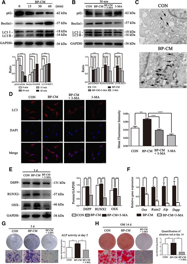
Lu et al. Stem Cell Research & Therapy   (2019) 10:222                                                       Page 2 of 14

endodontic therapy vary considerably, biomaterial is es-      downregulation of osteogenic markers and mineralization
sential in all these treatments [2]. Nowadays, mineral tri-   [16]. Besides, p-p38 and p-ERK lead RUNX2 phosphoryl-
oxide aggregate (MTA), a calcium silicate-based               ation, which increases the transcriptional potential of
biomaterial, is used extensively for scaffold cover during    RUNX2. Similarly, cooperation between the p38 and JNK
regenerative endodontic therapy. Meanwhile, our previ-        pathways promote osteogenic effects [17].
ous studies revealed that 2 mg/ml MTA-conditioned               Recently, the interaction between osteo/odontogenic
medium could boost odonto/osteogenic potential in             differentiation and autophagy, which is a significant cel-
BMMSCs [3], human periodontal ligament stem cells             lular biological process, has gained lots of interest. Au-
[4], and stem cells from apical papilla [5]. However,         tophagy is defined as a natural and self-cannibalization
MTA is not very friendly to clinicians because it has sev-    process responsible for the degradation of cytosolic pro-
eral disadvantages, such as long setting time, tooth dis-     teins and subcellular organelles in lysosomes. Nowadays,
coloration, and handling characteristics [6].                 several researchers have reported that autophagy has
   iRoot BP Plus (Innovative BioCeramix Inc., Vancouver,      critical functions in stem cell differentiation. Interest-
BC, Canada) is a laboratory-synthesized, pre-mixed,           ingly, Meng et al. verified that the impaired osteogenic
ready-to-use, injectable bioceramic-based endodontic          capacity of BMMSCs in osteoporosis model mice could
material [7]. According to the manufacturer’s instruc-        be rescued by autophagy [18]. Furthermore, autophagy
tion, the application of iRoot BP Plus includes root-end      in mesenchymal stem cells can be influenced by different
fillings, apexification, pulpotomy, and indirect or direct    factors, such as LPS, mechanical stress, and tuberous
pulp capping. iRoot BP Plus has similar cytotoxicity,         sclerosis 1 [19–21].
antibacterial efficacy, and root-end sealing in comparison      Here, the effects of iRoot BP Plus on the proliferation
with MTA [8–10]. Moreover, it overcomes the draw-             ability and osteo/odontogenic capacity of BMMSCs were
backs of MTA. Therefore, recently, iRoot BP Plus has          explored; the involvement of MAPK pathways and au-
been considered as an alternative to MTA.                     tophagy was also evaluated. Our findings revealed that
   In addition to biomaterials, stem cells are another key    iRoot BP Plus could enhance the osteogenic and odonto-
factor in tissue engineering [11]. During regenerative        genic potential of BMMSCs through the MAPK path-
endodontic therapy, blood from periapical tissues needs       ways and autophagy.
to be induced to the root canal system where different
types of stem cells, including bone marrow mesenchy-          Materials and methods
mal stem cells (BMMSCs), might help regenerating bone         BMMSCs isolation
and dentin tissues [12]. Because of their excellent           BMMSCs were collected by a whole marrow method
proliferation and multi-lineage differentiation ability,      and cultured as previously described [22]. Four-week-old
BMMSCs have been speculated to be promising cell              Sprague-Dawley rats were purchased from the Experi-
sources for stem cell-based clinical treatment. For better    mental Animal Center of Nanjing Medical University
application of BMMSCs in bone/teeth tissue regener-           and euthanized by overdose of pentobarbital, then steril-
ation, researchers identified lots of small molecules and     ized in 75% ethyl alcohol. Then, the femora and tibiae
growth factors to accelerate the osteo/odontogenic dif-       were removed aseptically, and the bone marrow was
ferentiation process of BMMSCs. For example, Lei et al.       flushed out to complete culture medium, which consists
reported that natural dentine matrix could regulate           of alpha minimum essential medium (α-MEM; Gibco,
BMMSC odontogenic differentiation [13]. Moreover,             Life Technologies, Grand Island, NY, USA), 10% fetal
0.5 μM bisphosphonates triggered the odontogenic abil-        bovine serum (FBS; Hyclone, Logan, UT, USA), 100 mg/
ity of BMMSCs [14]. However, the relationship between         ml streptomycin, and 100 U/ml penicillin. The cells were
iRoot BP Plus and osteo/odontogenic differentiation of        cultured at 37 °C with 5% CO2. When they reached 80%
BMMSCs remains elusive.                                       confluence, BMMSCs were trypsinized and passaged.
   It is generally known that multiple regulatory mecha-      BMMSCs at passage 3–5 were used in this study.
nisms take part in regulating multidirectional differenti-
ation of mesenchymal stem cells (MSCs), and MAPK              Material preparation
pathways are included. MAPK pathways consist of a             As previously reported [5], after being mixed with sterile
number of proteins and constitute a complex cascade.          water, ProRoot MTA (Dentsply, Tulsa, OK, USA) and
There are three classic MAPK subfamilies that have            iRoot BP Plus (Innovative Bioceramix, Vancouver, BC,
been characterized well in mammals: c-Jun N-terminal          Canada) were stored in a 100% humidified atmosphere
kinase (JNK), extracellular signal-regulated kinase (ERK),    at 37 °C for 3 days. After solidification, both materials
and p38 [15]. Lots of studies report the key role of          were dried for 1 day, grounded into powder, and dis-
MAPKs in osteoblast differentiation. Liu et al. verified      solved in α-MEM at a concentration of 20 mg/ml. The
that suppression of the JNK pathway resulted in               solution was then incubated at 37 °C for 3 days to obtain
Lu et al. Stem Cell Research & Therapy   (2019) 10:222                                                                         Page 3 of 14

the bioactive ingredients of both materials. Afterwards,     Real-time RT-PCR
the supernatant was filtered and different concentrations    Total cellular RNA was extracted using TRIzol reagent
of iRoot BP Plus-conditioned media (BP-CM) and 2 mg/         (Invitrogen, NY, USA). cDNA was synthesized using a
ml MTA-conditioned media (MTA-CM) were prepared              PrimeScript RT Master Mix kit (TaKaRa Biotechnology,
by mixing with fresh complete culture medium.                China). Primer sequences are shown in Table 1. Gapdh
                                                             was chosen as a housekeeping gene and the relative gene
Alkaline phosphatase (ALP) activity
                                                             expression (Osx, Alp, Runx2, Opn, and Dspp) was calcu-
According to the manufacturers’ protocols, total protein     lated using the “ΔΔCt” method.
content was evaluated by a BCA kit (Beyotime, China)
                                                             Western blot
and ALP activity of each group was investigated by an
ALP quantification kit (Jiancheng, Nanjing, China). ALP      BMMSCs were collected to evaluate the osteo/odonto-
activity was normalized to the total protein content.        genic differentiation after BP-CM treatment at days 0, 3,
                                                             and 7. BMMSCs induced with BP-CM for 0, 15, 30, and
                                                             60 min were collected to investigate the involvement of
Alkaline phosphatase staining                                the MAPK pathways and autophagy. Cell lysates were
Following the protocol of the ALP staining kit (Beyo-        harvested by RIPA buffer (Beyotime, China) containing
time, China), BMMSCs were fixed with 4% PFA, washed          1 mM phenylmethylsulfonyl fluoride (PMSF, Beyotime,
with PBS, and incubated in ALP working solution under        China) and 1 mM phosphatase inhibitor (Beyotime,
a dark condition. The results were pictured and ob-          China) on ice. Protein samples were subjected to 10% or
served under a microscope.                                   15% sodium dodecyl sulfate-polyacrylamide gels and
                                                             transferred to PVDF membranes (Millipore, USA). After
Alizarin Red S staining and cetylpyridinium chloride (CPC)   being blocked in 5% bovine serum albumin, the mem-
assay                                                        branes were incubated with polyclonal antibodies over-
Following previous studies, BMMSCs were cultured in          night [OSX (ab22552, Abcam, UK), RUNX2 (ab76956,
osteogenic-induced medium for 14 days. Osteogenic-           Abcam, UK), OPN (ab8448, Abcam, UK), DSPP
induced medium (OM) comprised complete culture               (BS70836; Bioworld, USA), JNK (#9252, Cell Signaling
medium plus 50 mg/L ascorbic acid (Sigma-Aldrich), 10        Technology, USA), p-JNK (#9255, Cell Signaling Tech-
mmol/L β-glycerophosphate (Sigma-Aldrich), and 10            nology, USA), p38 (#8690, Cell Signaling Technology,
nmol/L dexamethasone (Sigma-Aldrich). The medium             USA), p-p38 (#4511, Cell Signaling Technology, USA),
was replaced every 3 days. At day 14, cells were fixed       ERK (#4695, Cell Signaling Technology, USA), p-ERK
with 4% PFA and incubated with Alizarin Red S (Sigma-        (#4370, Cell Signaling Technology, USA), LC3 (#12741;
Aldrich). After washed with deionized water, cells were      Cell Signaling Technology, USA), Beclin1 (11306-1-AP;
photographed and observed using a microscope. Ten            Proteintech, Chicago, USA), p62 (18420-1-AP; Protein-
percent CPC (Sigma-Aldrich) was used for quantifica-         tech, Chicago, USA), and GAPDH (#2118, Cell Signaling
tion, and the spectrophotometric absorbance was exam-
ined at 560 nm. The calcium level of each group was          Table 1 Primer sequences of real-time RT-PCR
normalized to the total protein content.                     Genes                 Primers                 Sequences (5′–3′)
                                                             Osx                   Forward                 GGAGGCACAAAGAAGCCATA
Cell counting kit-8 assay                                                          Reverse                 GGGAAAGGGTGGGTAGTCAT
Cell proliferation ability was examined by cell counting     Alp                   Forward                 GGAACGGATCTCGGGGTACA
kit-8 (CCK-8, Dojindo, Tokyo, Japan). BMMSCs were
                                                                                   Reverse                 ATGAGTTGGTAAGGCAGGGT
treated with complete culture medium or BP-CM, which
was refreshed every 2 days. At days 0, 1, 3, 5, and 7,       Runx2                 Forward                 TTAACGTCAGCAGGAGCAG
CCK-8 reagent was applied and BMMSCs were incu-                                    Reverse                 CTTCACCCCCAGGACCAAG
bated at 37 °C for 1 h. The absorbance was detected at       Opn                   Forward                 ATCTGCCGACGTACCCTTTC
450 nm.                                                                            Reverse                 TCGTGGCTCTGATGTTCCAG
                                                             Dspp                  Forward                 ATCTGCCGACGTACCCTTTC
Flow cytometry (FCM)                                                               Reverse                 CCTCCTGCGTGTATCCCATC
BMMSCs cultured in different medium for 3 days were          Gapdh                 Forward                 CACTGAGCATCTCCCTCACAA
collected and fixed with 75% ethyl alcohol at 4 °C for 24
                                                                                   Reverse                 GTATTCGAGAGAAGGGAGGGCT
h. Afterwards, cells were washed and cell cycle fractions
                                                             Osx osterix, Alp alkaline phosphatase, Runx2 Runt-related transcription factor 2,
(G0G1/S/G2M phases) were analyzed by FACSCalibur             Opn osteopontin, Dspp dentin sialophosphoprotein, Gapdh
(BD Biosciences, CA, USA).                                   D-gluteraldehyde-3-phosphate dehydrogenase
Lu et al. Stem Cell Research & Therapy   (2019) 10:222                                                       Page 4 of 14

Technology, USA)]. Membranes were washed with PBST           cells were divided into three groups: complete culture
(PBS with 0.05% Tween 20) and immersed in secondary          medium, BP-CM, and BP-CM+3-MA. After being cul-
antibodies for 1 h. Finally, the visualization of the mem-   tured for 3 days, western blot and real-time RT-PCR
branes was conducted by a Western Blot Imaging Sys-          were performed to evaluate the protein and gene levels
tem (GE Healthcare). ImageJ software was used for            of osteo/odontogenic-associated markers. At day 5, the
quantification.                                              expression of ALP was investigated by ALP staining and
                                                             ALP activity assay. At day 14, mineralization of
Immunofluorescence staining                                  BMMSCs was investigated by Alizarin Red S staining.
BMMSCs were fixed with 4% PAF, permeabilized with
Triton X-100 solution (Beyotime, China), and blocked         Transmission electron microscopy (TEM)
with goat serum. Then, cells were incubated with the         TEM was performed to observe autophagosomes as de-
primary antibodies against OSX, RUNX2, p-JNK, p-p38,         scribed previously [25]. BMMSCs were harvested and
p-ERK, and LC3 overnight, then incubated with a sec-         fixed with 2.5% glutaraldehyde. Then, samples were
ondary antibody for 2 h under a dark condition. Nuclei       postfixed with 1% osmium tetroxide, dehydrated, and
were stained with DAPI (Beyotime, China). Afterwards,        embedded. Ultrathin sections (60–80 nm) were obtained
OSX, RUNX2, p-JNK, p-p38, and p-ERK were observed            by ultramicrotome, stained with uranyl acetate and lead
using the Olympus inverted fluorescence microscope,          citrate, and examined by a transmission electron micro-
while LC3 was observed by the LSM 710 laser scanning         scope (JEM-2000EX; JEOL, Ltd., Tokyo, Japan).
confocal microscope. ImageJ software was used for
quantification.                                              Statistical analysis
                                                             Data from three independent experiments was expressed
Signal blocking assays                                       as mean ± standard deviation and analyzed by SPSS 20.0.
SP600125 (specific JNK inhibitor), SB203580 (specific        Student’s t test or one-way ANOVA was used to analyze
p38 inhibitor), and U0126 (specific ERK inhibitor) were      significant difference. P < 0.05 was considered statistical
purchased from Selleck Chemicals (USA). According to         significance.
the manufacturer’s suggestion, BMMSCs were respect-
ively pretreated with a 10-μM inhibitor for 2 h before be-   Results
ing stimulated with BP-CM. Cells were divided into four      Screening for the optimal concentration of iRoot BP Plus
groups: complete culture medium, BP-CM, BP-                  Primary BMMSCs cultured after 3 days were observed as
CM+inhibitor, and complete culture medium+inhibitor.         shown in Fig. 1a. The adherent cells had different cell
After stimulation with BP-CM for 15 min, BMMSCs              morphologies. The round-shaped cells attached loosely
were collected to evaluate the inhibition of the MAPK        and could be washed out by changing the culture
pathways. Western blot and immunofluorescence stain-         medium, while the main cell type of BMMSCs at passage
ing assay were performed to detect the protein levels of     3 is spindle-forming cells (Fig. 1b). At day 5, real-time
phosphorylation of JNK, p38, and ERK. The expression         RT-PCR showed that the gene expression of Alp was
of ALP at day 5 was investigated by ALP staining and         highest in the 0.2 mg/ml BP-CM group (Fig. 1c; P <
ALP activity assay. After cultured for 7 days, western       0.001). As compared with other groups, the 0.2 mg/ml
blot and real-time RT-PCR were performed to evaluate         BP-CM group also presented the highest ALP activity
the expression of osteo/odontogenic-related markers. At      after being induced for 3, 5, and 7 days (Fig. 1d; P <
day 14, mineralization of BMMSCs was investigated by         0.01). Therefore, 0.2 mg/ml was selected to be the opti-
Alizarin Red S staining.                                     mal concentration for the following experiments.

Suppression of autophagy                                     Proliferation of BP-CM-treated BMMSCs
To confirm the influence of iRoot BP Plus on autophagy       CCK-8 assay revealed that 0.2 mg/ml BP-CM did not
in BMMSCs, 3-methyladenine (3-MA) (Sigma-Aldrich)            affect cell growth (Fig. 1e; P > 0.05). Flow cytometry
was used to inhibit cell autophagy. As previously de-        assay showed no statistical difference in the proliferative
scribed [22–24], BMMSCs were respectively pretreated         index (PI = G2M + S) between the two groups (Fig. 1f–h;
with 5 mM 3-MA for 24 h before being stimulated with         P > 0.05).
BP-CM. Cells were divided into four groups: complete
culture medium, BP-CM, BP-CM+3-MA, and complete              iRoot BP Plus induced the osteo/odontogenic capability
culture medium+3-MA. After stimulation with BP-CM            of BMMSCs
for 30 min, BMMSCs were collected to evaluate the sup-       The results of western blot indicated that BP-CM pro-
pression of autophagy. To explore the effects of autoph-     moted the protein levels of dentin sialophosphoprotein
agy on osteo/odontoblastic differentiation of BMMSCs,        (DSPP), osteopontin (OPN), runt-related transcription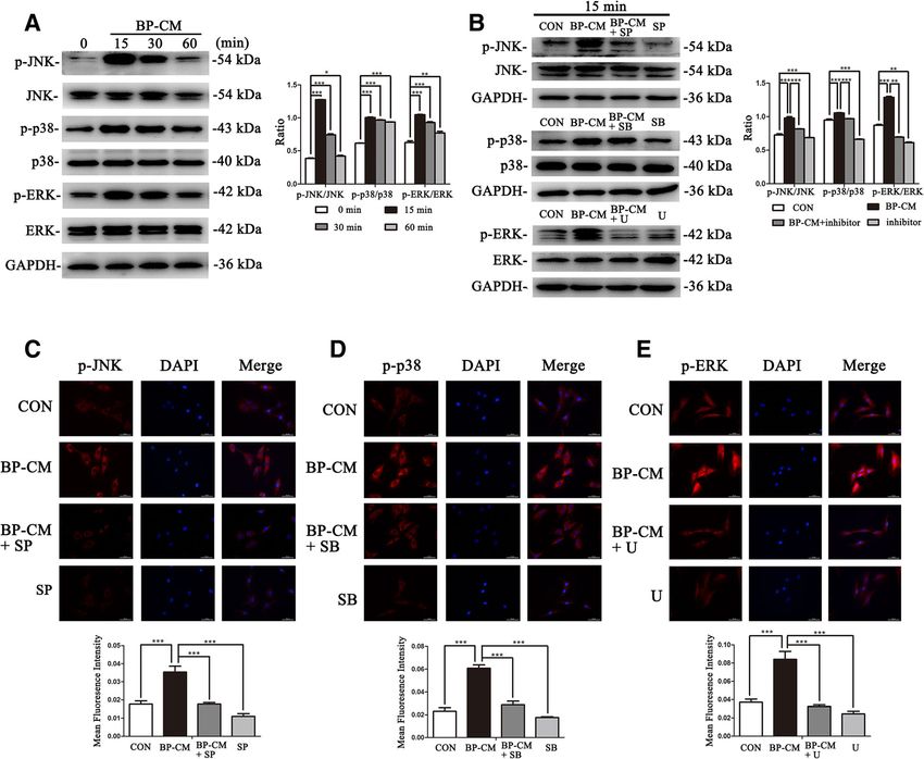
Lu et al. Stem Cell Research & Therapy      (2019) 10:222                                                                                Page 5 of 14

 Fig. 1 Screening for the optimal concentration of BP-CM and investigating proliferation ability of BP-CM-treated BMMSCs. a Primary BMMSCs at
 day 3. b BMMSCs at passage 3. Scale bar = 100 μm. c The gene expression of Alp was dramatically increased in the 0.2 mg/ml BP-CM group at
 day 5. ***P < 0.001. d ALP activity of BMMSCs cultured in different concentrations of BP-CM at days 3, 5, and 7. **P < 0.01, ***P < 0.001. e–h The
 proliferation ability of BP-CM-treated BMMSCs was measured by CCK-8 and FCM

factor 2 (RUNX2), and osterix (OSX) in BMMSCs                                indicated that iRoot BP Plus triggered osteo/odonto-
(Fig. 2a; P < 0.05). Relative gene (Dspp, Opn, Runx2, Alp,                   genic differentiation of BMMSCs.
and Osx) expression was consistent with the proteins’
trend (Fig. 2d; P < 0.05). ALP was detected to examine                       The effects of iRoot BP Plus on the osteo/odontoblastic
osteogenic differentiation of BMMSCs as an early-                            differentiation of BMMSCs were similar to those of MTA
phase marker [26]. Compared with the control group,                          According to our previous studies, 2 mg/ml was the op-
the expression of ALP of the BP-CM group was up-                             timal concentration of the MTA-conditioned medium
regulated (Fig. 2b, P < 0.01). Meanwhile, Alizarin Red                       (MTA-CM) to accelerate the osteo/odontogenic differ-
S staining demonstrated that matrix mineralization                           entiation process of different stem cells [3–5]. Therefore,
was enhanced in the OM+BP-CM group compared                                  2 mg/ml MTA-CM was selected to be the positive con-
with that in the OM group after induction for 14 days                        trol, while a complete culture medium was used as a
(Fig. 2c, P < 0.01). Besides, the images of immuno-                          blank control. After being cultured for 3 days, cells were
fluorescence staining showed that the protein levels of                      collected to extract total cellular protein and RNA.
OSX and RUNX2 were upregulated in the BP-CM                                  Western blot and real-time RT-PCR results revealed that
group (Fig. 2e, P < 0.01). Above all, these data                             both materials could upregulate the expression of osteo/
Lu et al. Stem Cell Research & Therapy   (2019) 10:222   Page 6 of 14

 Fig. 2 (See legend on next page.)
Lu et al. Stem Cell Research & Therapy (2019) 10:222 Page 7 of 14
(See figure on previous page.)
Fig. 2 The influence of iRoot BP Plus on osteo/odontogenic differentiation of BMMSCs. a The osteo/odontogenic-associated proteins (DSPP, OPN,
RUNX2, and OSX) in BMMSCs were detected by western blot at days 0, 3, and 7, respectively. ImageJ software was used to quantify the results,
*P < 0.05, **P < 0.01, and ***P < 0.001. b ALP staining and ALP activity assay were performed to evaluate the expression of ALP in BP-CM-induced
BMMSCs at day 5 and day 7. Scale bar = 100 μm. **P < 0.01. c Alizarin Red S staining and CPC assay were conducted to investigate mineralization
of BMMSCs after being cultured in complete culture medium or osteogenic-induced medium (OM) (+/− BP-CM) for 14 days. Scale bar = 100 μm.
**P < 0.01. d Relative gene expression was detected by real-time RT-PCR. *P < 0.05, **P < 0.01, and ***P < 0.001. e OSX and RUNX2 in BP-CM-
treated BMMSCs were observed by immunofluorescence staining. Scale bar = 50 μm. Quantification was done by ImageJ, **P < 0.01. f, g Western
blot and real-time RT-PCR were used to compare the changes of osteo/odontogenic-related markers in the BP-CM group with those in the MTA-
CM group. *P < 0.05, **P < 0.01, and ***P < 0.001. h The protein level of ALP in both the BP-CM- and MTA-CM-treated BMMSCs was detected by
ALP staining and ALP activity assay. Scale bar = 100 μm, *P < 0.05, **P < 0.01
odontoblastic-associated markers, but there was no sig- in BP-CM+inhibitor groups were significantly lower than
nificant difference between the two groups (Fig. 2f–g, those in the BP-CM group (Fig. 4a–c, P < 0.05). Simi-
P < 0.05). Besides, at day 5, the ALP expression was larly, the ALP expression in BP-CM+inhibitor groups
measured by ALP staining and ALP activity assay. Both were appreciably decreased in comparison with those in
BP-CM- and MTA-CM-treated BMMSCs showed higher the BP-CM group (Fig. 4d, P < 0.01). In addition, the cal-
ALP expression than the control group, and no statis- cium nodules in the BP-CM+inhibitor groups, which
tical significance was observed between the two groups were observed by Alizarin Red S staining, were also re-
(Fig. 2h, P < 0.05). According to these data, iRoot BP markably less and smaller than those in the BP-CM
Plus had a similar osteo/odontogenic capability of group (Fig. 4e, P < 0.05). Above all, these results con-
BMMSCs to that of MTA. firmed that iRoot BP Plus enhanced osteoblastic and
odontoblastic differentiation of BMMSCs through the
iRoot BP Plus enhanced osteo/odontogenic potential of MAPK pathways.
BMMSCs by activating the MAPK pathways
To explore the involvement of the MAPK pathways dur- iRoot BP Plus accelerate the osteo/odontogenic
ing the BP-CM-induced differentiation of BMMSCs, the differentiation process of BMMSCs via autophagy
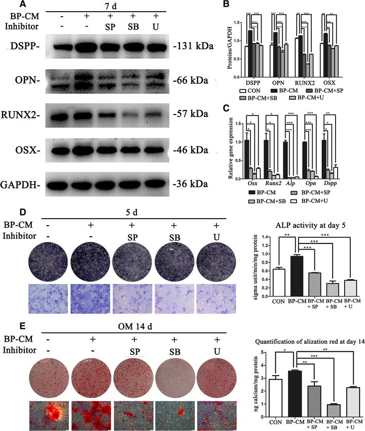
MAPK pathway-related proteins, such as p-p38, p38, p- The protein levels of autophagy-related makers (p62,
ERK, ERK, p-JNK, and JNK, were evaluated by western Beclin1, and LC3) were evaluated to determine whether
blot. The results revealed that p-JNK, p-p38, and p-ERK 0.2 mg/ml BP-CM induced autophagy in BMMSCs.
in BP-CM-treated BMMSCs increased rapidly after 15 Western blot results indicated that BP-CM led to the
min and declined subsequently (Fig. 3a, P < 0.01). To downregulation of p62, while the protein levels of LC3II
further investigate the influence of iRoot BP Plus on the and Becline1 were increased in a time-dependent man-
MAPK pathways in BMMSCs, inhibitors of the JNK, ner (Fig. 5a, P < 0.05). The images of TEM revealed that
p38, and ERK pathways (SP: SP60016, SB: SB203580, more autophagosomes could be observed in BP-CM-
and U: U0126) were used in this study. After being pre- treated BMMSCs (Fig. 5c). The data above suggested
treated with inhibitors for 2 h, BMMSCs were treated that autophagy was activated during induction of osteo/
with the complete culture medium, BP-CM, BP- odontogenic differentiation in BP-CM-treated BMMSCs.
CM+inhibitor, and inhibitor for 15 min and the total To further confirm the effects of BP-CM on autophagy
protein of each group was extracted. Western blot re- of BMMSCs, 3-MA was used to suppress BP-CM-
sults revealed that the MAPK signaling kinase, which induced autophagy. As compared with the BP-CM
was activated by BP-CM, was blocked after being pre- group, p62 was decreased in BP-CM+3-MA group, while
treated with specific inhibitors (Fig. 3b, P < 0.01). The the expression of Becline1 and the ratio of LC3II/LC3I
result of immunofluorescence staining were consistent were upregulated (Fig. 5b, P < 0.001). Meanwhile, the re-
with that of western blot (Fig. 3c–e, P < 0.001). There- sults of immunostaining staining were consistent with
fore, MAPK pathways in BMMSCs could be instantly ac- the proteins’ trend (Fig. 5d, P < 0.05). To investigate the
tivated by iRoot BP Plus treatment. involvement of autophagy during the BP-CM-induced
differentiation of BMMSCs, cells were divided into three
Blocking off MAPK pathways inhibited osteo/ groups: complete culture medium, BP-CM, and BP-
odontoblastic differentiation of iRoot BP Plus-treated CM+3-MA. Western blot and real-time RT-PCR indi-
BMMSCs cated that the protein and gene levels of osteo/odonto-
BMMSCs were divided into five groups: complete cul- genic-related markers were downregulated in the BP-
ture medium, BP-CM, and BP-CM with different inhibi- CM+3-MA group in comparison with the BP-CM group
tors (SP600125, SB203580, and U0126). The expression (Fig. 5e, f, P < 0.05). At day 5, the expression of ALP in
levels of odontogenic and osteogenic associated makers the BP-CM+3-MA group was lower than that in the BP-Lu et al. Stem Cell Research & Therapy (2019) 10:222 Page 8 of 14
Fig. 3 iRoot BP Plus activates the MAPK pathways in BMMSCs. BMMSCs of each group were cultured for an hour simultaneously, and culture
medium was replaced with BP-CM at different time points (15 min, 30 min, and 60 min). a MAPK pathway-related proteins were investigated by
western blot. Quantification was done by ImageJ. *P < 0 05, **P < 0 01, and ***P < 0 001. b BMMSCs were pretreated with the MAPK pathway
inhibitors (SP: SP600125, SB: SB203580, and U: U0126) for 2 h and then treated with BP-CM for 15 min. Western blot was performed to investigate
the MAPK pathway-related proteins. Quantitative analysis was conducted by ImageJ. **P < 0 01 and ***P < 0 001. c–e Immunofluorescence
staining indicated that p-JNK, p-ERK, and p-p38 were decreased after inhibition of the MAPK pathways. Scale bar = 50 μm. Quantification was
done by ImageJ, ***P < 0.001
CM group (Fig. 5g, P < 0.001). After cultured for 14 days, There are several studies focusing on the comparison
Alizarin Red S staining showed that BMMSCs in the BP- between MTA, the gold standard material, and iRoot BP
CM group generated more calcium nodules than the Plus. Liu et al. reported that iRoot BP Plus could en-
BP-CM+3-MA-treated cells (Fig. 5h, P < 0.001). The re- hance the formation of a calcium bridge at exposed pulp
sults above demonstrated that iRoot BP Plus promoted sites and the induction ability was superior than that of
osteo/odontogenic capability of BMMSCs through acti- MTA [7]. On the other hand,. Shi et al. demonstrated
vating autophagy. that iRoot BP Plus is comparable with MTA when
served as pulp-capping agents in dogs. Both of them
Discussion formed a complete dentine bridge without pulpal inflam-
Accumulated evidence has demonstrated that iRoot BP mation, and no statistical significance was observed be-
Plus, a novel bioceramic material, could enhance tween the two groups [29]. Besides, iRoot BP Plus
adhesion, migration, attachment, proliferation, and possesses superior capacity to the formation of apatite
mineralization ability of dental pulp stem cells [27, 28]. crystals as compared with MTA in vitro [30]. Moreover,Lu et al. Stem Cell Research & Therapy (2019) 10:222 Page 9 of 14 Fig. 4 The effects of specific MAPK pathway inhibitors on osteo/odontoblastic differentiation of BP-CM-induced BMMSCs. a, b Osteo/ odontogenic-related proteins were measured by western blot. Quantification was done by ImageJ, **P < 0.01 and ***P < 0.001. c Relative gene expression was detected by real-time RT-PCR. *P < 0.05, **P < 0.01, and ***P < 0.001. d ALP staining and ALP activity assay indicated that the expression of ALP in the BP-CM+inhibitor (SP600125, SB203580, and U0126) groups were downregulated as compared with those in the BP-CM group. **P < 0.01 and ***P < 0.001. e Alizarin Red S staining and CPC assay showed that the mineralization of BMMSCs in the BP-CM+inhibitor groups was deceased in comparison with that in the BP-CM group. Scale bar = 100 μm, **P < 0.01 and ***P < 0.001
Lu et al. Stem Cell Research & Therapy   (2019) 10:222   Page 10 of 14

 Fig. 5 (See legend on next page.)
Lu et al. Stem Cell Research & Therapy   (2019) 10:222                                                                           Page 11 of 14

 (See figure on previous page.)
 Fig. 5 BP-CM could enhance the osteo/odontogenic capability of BMMSCs through autophagy. a BMMSCs of each group were cultured for an
 hour simultaneously, and culture medium was replaced with BP-CM at different time points (15 min, 30 min, and 60 min). Western blot was
 conducted to detect the autophagy-related markers (LC3, Beclin1, and p62). Quantification was done by ImageJ, *P < 0.05 and ***P < 0.001. b
 BMMSCs were divided into four groups and treated with different medium for 30 min. The normalized expression of p62 and Beclin1, and the
 ratio of LC3II/LC3I represented the relative expression. ***P < 0.001. c TEM pictures of autophagosomes showed that BP-CM-induced BMMSCs had
 more autophagic vacuoles as compared with cells in the control group. Autophagosomes are indicated by black arrows. Scale bar = 2 μm. d The
 images of LC3 (red) and DAPI (blue) in BMMSCs cultured with different media for 30 min were observed by immunofluorescence staining. Scale
 bar = 50 μm. Quantification was done by ImageJ. ***P < 0 001. e, f The effects of autophagy inhibitor 3-MA on osteo/odontoblastic-associated
 markers of BP-CM-induced BMMSCs were investigated by western blot and real-time RT-PCR. *P < 0.05, **P < 0.01, and ***P < 0.001. g, h
 Suppression of autophagy decreased the expression of ALP and mineralization of BMMSCs, while complete culture medium was served as a
 blank control and BP-CM was served as a positive control. Scale bar = 100 μm, ***P < 0.001

iRoot BP Plus exhibited similar results to those of MTA                  odontogenic markers (OPN, RUNX2, OSX, and DSPP)
when used as root-end filling materials in endodontic                    were remarkably increased. These genes and proteins
microsurgery [31].                                                       are vital factors in regulating osteo/odontogenic differen-
   Furthermore, similar to MTA, iRoot BP Plus contains                   tiation at different stages [38–41], indicating that iRoot
carbon (C), oxygen (O), sodium (Na), Silicon (Si), phos-                 BP Plus could trigger the osteo/odontogenic differenti-
phorus (P), sulfur (S), chlorine (Cl), and calcium (Ca).                 ation process of BMMSCs. However, further research of
The major difference between the two materials is that                   the influence of iRoot BP Plus in vivo should be
iRoot BP Plus contains a significant amount of tantalum                  performed.
(Ta) and zirconium (Zr), but aluminum (Al) is not in-                       Our previous studies found that MTA-CM at a con-
cluded [30]. In addition, when exposed to PBS (pH =                      centration of 2 mg/ml upregulate odonto/osteogenic po-
7.4), abundant Ca and Si was eluted from both materials                  tential of BMMSCs [3], human periodontal ligament
[32]. Calcium takes part in the regulation of a great deal               stem cells [4], and stem cells from the apical papilla [5].
of cellular biological behavior. For example, the osteo-                 Therefore, besides the blank control group (complete
genic differentiation of BMMSCs could be promoted by                     culture medium), 2 mg/ml MTA-CM was served as a
increasing concentration of extracellular calcium [33].                  positive control in this study. According to the data
On the other hand, silicon could regulate the formation                  above, iRoot BP Plus showed similar effects on osteo/
and calcification of bone tissue [34]. Previous studies re-              odontogenic differentiation of BMMSCs to those of
ported that the release of Ca and Si ions contributed to                 MTA, which was consistent with other research.
the effects of calcium silicate-based biomaterials on cell                  Previous studies demonstrated that the MAPK path-
differentiation [35]. Notably, during regenerative end-                  ways have vital functions in regulating cell odontogenic
odontic therapy, most of the cells in the blood clot con-                and osteogenic differentiation. To date, a number of
tact the covering material indirectly. Thus, many                        stimulators have been reported to trigger osteogenic dif-
researchers selected material eluates to study the bio-                  ferentiation of MSCs through the MARK pathways. For
logical properties of biomaterials because they can be                   example, osteogenic capability of BMP9 could be upreg-
observed conveniently and analyzed easily. In the                        ulated by Win 11 via the p38 pathways [42]. Besides,
present study, iRoot BP Plus- and MTA-conditioned                        extracellular calcium is reported to be an important
media were prepared to investigate their effects on                      regulator in determining the specificity of the ERK cas-
BMMSCs.                                                                  cade [43]. In addition, several calcium silicate-based bio-
   Alkaline phosphatase (ALP) has a vital function during                materials, such as Biodentine, MTA, and Bioaggregate,
the formation of hydroxyapatite crystal, which is the ini-               could promote osteo/odontogenic potential by activating
tial phase in mineralization of osteoblasts and odonto-                  the MAPK pathways [3, 44]. In this study, p-JNK, p-
blasts [26], and its activity is upregulated at an early                 ERK, and p-p38 in BMMSCs were upregulated immedi-
stage of calcification [36]. Therefore, many studies used                ately after being treated with BP-CM. Moreover, specific
ALP as an early-stage marker of osteo/odontoblastic dif-                 inhibitors (SP600125, U0126, and SB203580) could re-
ferentiation [25, 37]. In the present study, 0.2 mg/ml was               duce the activation of BP-CM-induced MAPK pathways,
the optimal concentration of BP-CM to promote differ-                    respectively, and suppress the committed differentiation
entiation of BMMSCs through detecting the ALP activity                   of BMMSCs at the same time. These results implied that
at days 3, 5, and 7, as well as the gene level of Alp at day             iRoot BP Plus potentiated osteo/odontogenic differenti-
5. Meanwhile, CCK-8 assay and FCM results showed                         ation of BMMSCs through triggering the MAPK
that 0.2 mg/ml BP-CM had no cytotoxicity on BMMSCs.                      pathways.
The mineralization of BP-CM-induced BMMSCs was                              Autophagy is a self-cannibalization mechanism, which
significantly upregulated, and the expression of osteo/                  enables recycling of cytosolic proteins and subcellular
Lu et al. Stem Cell Research & Therapy   (2019) 10:222                                                                    Page 12 of 14

organelles to maintain cellular homeostasis. Recently,      pathways [51]. Besides, butyrate response factor 1 regu-
autophagy has been recognized to be an important fac-       lates inflammation of RAW264.7 cells by autophagy
tor in stem cell differentiation. Aged BMMSCs have a        crosstalking with the ERK pathway [52]. High glucose
significantly lower autophagy level than young ones, and    suppresses the migratory capacity of keratinocytes
activation of autophagy can restore degenerative proper-    through triggering the p38 pathway which leads to
ties of aged BMMSCs [22]. Meanwhile, osteogenesis           downregulation of autophagy [53]. However, the rela-
could be reduced by inhibition of autophagy, while adi-     tionship between autophagy and the MAPK pathways
pogenesis is upregulated [45]. Besides, autophagy could     during cell osteo/odontogenic differentiation needs fur-
be induced when cells differentiate to osteoblasts, and a   ther investigation.
decline of autophagy-essential genes might result in the
downregulation of mineralization ability. Moreover, oste-   Conclusion
oblasts use autophagic vacuoles as vehicles to secrete      In summary, 0.2 mg/ml iRoot BP Plus-conditioned
apatite crystals [46]. Furthermore, previous studies have   medium could trigger BMMSCs differentiating to osteo/
revealed that various stimulators could promote osteo-      odontoblasts through the MAPK pathways and autoph-
genic differentiation of BMMSCs through activating au-      agy without interference with cell proliferation. Our re-
tophagy, such as LPS, mechanical stress, and tuberous       sults provide theoretical foundation on the clinical
sclerosis 1 [19–21].                                        application of iRoot BP Plus in regenerative endodontic
   LC3 has a critical function in autophagy because it      therapy.
governs autophagosome biogenesis. Cytoplasm LC3I and
                                                            Acknowledgements
membrane-bound form LC3II are two forms of LC3.
                                                            Not applicable.
LCII was formed by the combination of LC3I and phos-
phatidylethanolamine during the formation of auto-          Authors’ contributions
phagosomes [24]. Thus, researchers could monitor            JL and ZL conceived and designed the study, collected and assembled data,
                                                            and wrote the manuscript. XW and XG performed the data analysis and
autophagy by detecting LC3 levels. Beclin1 is another es-   interpretation. YC and MY reviewed the data. JY conceived and designed the
sential protein in autophagosome formation. It controls     study, provided financial support and study material, performed the data
the autophagy by regulating PI3KC3-dependent gener-         analysis and interpretation, and approved the final version of the manuscript.
                                                            All authors read and approved the manuscript.
ation of PI3P and the subsequent recruitment of add-
itional ATG proteins that orchestrate autophagosome         Funding
formation [47]. p62/SQSTM1 is one of the best-              This study was supported by National Natural Science Foundation of China
                                                            (81873707), Medical Talent Project of Jiangsu Province (ZDRCA2016086), the
characterized autophagy receptors. It is a key regulator    Priority Academic Program Development of Jiangsu Higher Education
in aggrephagy, mitophagy, and xenophagy and degraded        Institutions (PAPD, 2018-87), and Science and Technology Development Pro-
in autophagolysosomes together with the cargo [48].         ject of Jiangsu Province (BE2017731).
Therefore, during the activation of autophagy, Beclin1
                                                            Availability of data and materials
and LC3II are upregulated accompanied with a decline        The datasets used and analyzed during the current study are available from
of p62. On the contrary, inhibition of autophagy leads to   the corresponding author on reasonable request.
decreased Beclin1 and LC3II and results in accumulation
                                                            Ethics approval and consent to participate
of p62. In this study, activation of autophagy was ob-      Studies were carried out in accordance with the Declaration of Helsinki and
served during the osteo/odontogenic differentiation in      got approval of the Ethical Committee of Nanjing Medical University.
BP-CM-treated BMMSCs. And suppression of autoph-
                                                            Consent for publication
agy resulted in the decrease of osteo/odontogenic-related   Not applicable.
markers, as well as ALP activity and mineralization of
BMMSCs. These findings are consistent with the previ-       Competing interests
                                                            The authors declare that they have no competing interests.
ous studies, indicating that in addition to the MAPK
pathways, autophagy was also involved in osteo/odonto-      Author details
                                                            1
blastic differentiation of BMMSCs.                           Key Laboratory of Oral Diseases of Jiangsu Province, Institute of
                                                            Stomatology, Nanjing Medical University, 136 Hanzhong Road, Nanjing
   To date, several studies have reported the interaction
                                                            210029, Jiangsu, China. 2Endodontic Department, School of Stomatology,
between the MAPK pathways and autophagy in different        Nanjing Medical University, 136 Hanzhong Road, Nanjing 210029, Jiangsu,
types of cells during different cell behaviors. In neural   China. 3Nanjing Stomatological Hospital, Medical School of Nanjing
                                                            University, 30 Zhongyang Road, Nanjing 210008, Jiangsu, China.
stem cells, heat stress-induced autophagy and apoptosis
could be reversed via inhibition of the p38 pathway [49].   Received: 17 May 2019 Revised: 30 June 2019
2-Phenyloxypyrimidine derivative E5 could activate the      Accepted: 15 July 2019
ERK pathway to induce autophagy in hepatocellular car-
                                                            References
cinoma [50]. Plakophilin 3 in ovarian cancer tissue         1. Staffoli S, Plotino G, Nunez Torrijos BG, Grande NM, Bossu M, et al.
modulates autophagy through the JNK/ERK/mTOR                    Regenerative endodontic procedures using contemporary endodontic
Lu et al. Stem Cell Research & Therapy (2019) 10:222 Page 13 of 14
materials. Materials (Basel). 2019;12:ma12060908. https://doi.org/10.3390/ 22. Ma Y, Qi M, An Y, Zhang L, Yang R, et al. Autophagy controls mesenchymal
ma12060908. stem cell properties and senescence during bone aging. Aging Cell. 2018;
2. Sachdeva GS, Sachdeva LT, Goel M, Bala S. Regenerative endodontic 17:e12709. https://doi.org/10.1111/acel.12709.
treatment of an immature tooth with a necrotic pulp and apical 23. Li Y, Su J, Sun W, Cai L, Deng Z. AMP-activated protein kinase stimulates
periodontitis using platelet-rich plasma (PRP) and mineral trioxide osteoblast differentiation and mineralization through autophagy induction.
aggregate (MTA): a case report. Int Endod J. 2015;48:902–10. https://doi. Int J Mol Med. 2018;41:2535–44. https://doi.org/10.3892/ijmm.2018.3498.
org/10.1111/iej.12407. 24. Deng XS, Meng X, Venardos N, Song R, Yamanaka K, et al. Autophagy
3. Wang JLY, Song W, Yu J. Mineral trioxide aggregate upregulates negatively regulates pro-osteogenic activity in human aortic valve interstitial
odontoosteogenic capacity of bone marrow stromal cells from craniofacial cells. J Surg Res. 2017;218:285–91. https://doi.org/10.1016/j.jss.2017.05.088.
bones via JNK and ERK MAPK signalling pathways. Cell Prolif. 2014;47:241–8. 25. Pan Y, Li Z, Wang Y, Yan M, Wu J, et al. Sodium fluoride regulates the
https://doi.org/10.1111/cpr.12099. osteo/odontogenic differentiation of stem cells from apical papilla by
4. Wang Y, Zhou Y, Jin L, Pang X, Lu Y, et al. Mineral trioxide aggregate modulating autophagy. J Cell Physiol. 2019. https://doi.org/10.1002/jcp.2
enhances the osteogenic capacity of periodontal ligament stem cells via 8269.
NF-kappaB and MAPK signaling pathways. J Cell Physiol. 2018;233:2386–97. 26. Uday S, Matsumura T, Saraff V, Saito S, Orimo H, et al. Tissue non-specific
https://doi.org/10.1002/jcp.26110. alkaline phosphatase activity and mineralization capacity of bi-allelic
5. Yan M, Wu J, Yu Y, Wang Y, Xie L, et al. Mineral trioxide aggregate mutations from severe perinatal and asymptomatic hypophosphatasia
promotes the odonto/osteogenic differentiation and dentinogenesis of phenotypes: results from an in vitro mutagenesis model. Bone. 2019;127:9–
stem cells from apical papilla via nuclear factor kappa B signaling pathway. 16. https://doi.org/10.1016/j.bone.2019.05.031.
J. Endod. 2014;40:640–7. https://doi.org/10.1016/j.joen.2014.01.042. 27. Zhang S, Yang X, Fan M. BioAggregate and iRoot BP Plus optimize the
6. Parirokh M, Torabinejad M, Dummer PMH. Mineral trioxide aggregate and proliferation and mineralization ability of human dental pulp cells. Int
other bioactive endodontic cements: an updated overview - part I: vital pulp Endod J. 2013;46:923–9. https://doi.org/10.1111/iej.12082.
therapy. Int Endod J. 2018;51:177–205. https://doi.org/10.1111/iej.12841. 28. Zhang J, Zhu LX, Cheng X, Lin Y, Yan P, et al. Promotion of dental pulp cell
7. Liu S, Wang S, Dong Y. Evaluation of a bioceramic as a pulp capping agent migration and pulp repair by a bioceramic putty involving FGFR-mediated
in vitro and in vivo. J. Endod. 2015;41:652–7. https://doi.org/10.1016/j.joen.2 signaling pathways. J Dent Res. 2015;94:853–62. https://doi.org/10.1177/
014.12.009. 0022034515572020.
8. Shi S, Bao ZF, Chen X, Zhang DD. Cytotoxicity of a novel endodontic 29. Shi S, Bao ZF, Liu Y, Zhang DD, Chen X, et al. Comparison of in vivo dental
treatment material iRoot BP Plus to human gingival fibroblasts. Shanghai J pulp responses to capping with iRoot BP Plus and mineral trioxide
Stomatol. 2014;23:681–4. aggregate. Int Endod J. 2016;49:154–60. https://doi.org/10.1111/iej.12439.
9. Damlar I, Ozcan E, Yula E, Yalcin M, Celik S. Antimicrobial effects of several 30. Zhu L, Yang J, Zhang J, Lei D, Xiao L, et al. In vitro and in vivo evaluation of
calcium silicate-based root-end filling materials. Dent Mater J. 2014;33:453–7. a nanoparticulate bioceramic paste for dental pulp repair. Acta Biomater.
10. Leal F, De-Deus G, Brandao C, Luna A, Souza E, et al. Similar sealability 2014;10:5156–68. https://doi.org/10.1016/j.actbio.2014.08.014.
between bioceramic putty ready-to-use repair cement and white MTA. Braz 31. Zhou W, Zheng Q, Tan X, Song D, Zhang L, et al. Comparison of mineral
Dent J. 2013;24:362–6. https://doi.org/10.1590/0103-6440201302051. trioxide aggregate and iRoot BP Plus root repair material as root-end filling
11. Gong T, Heng BC, Lo EC, Zhang C. Current advance and future prospects of materials in endodontic microsurgery: a prospective randomized controlled
tissue engineering approach to dentin/pulp regenerative therapy. Stem study. J Endod. 2017;43:1–6. https://doi.org/10.1016/j.joen.2016.10.010.
Cells Int. 2016;2016:9204574. https://doi.org/10.1155/2016/9204574. 32. Tian J, Zhang Y, Lai Z, Li M, Huang Y, et al. Ion release, microstructural, and
12. Lovelace TW, Henry MA, Hargreaves KM, Diogenes A. Evaluation of the biological properties of iRoot BP Plus and ProRoot MTA exposed to an
delivery of mesenchymal stem cells into the root canal space of necrotic acidic environment. J. Endod. 2017;43:163–8. https://doi.org/10.1016/j.joen.2
immature teeth after clinical regenerative endodontic procedure. J. Endod. 016.10.011.
2011;37:133–8. https://doi.org/10.1016/j.joen.2010.10.009. 33. Cheng S, Wang W, Lin Z, Zhou P, Zhang X, et al. Effects of extracellular calcium
13. Gang Lei GZ, Yu J. Differentiation of BMMSCs into odontoblast-like cells on viability and osteogenic differentiation of bone marrow stromal cells in
induced by natural dentine matrix. Arch Oralbiol. 2013;58:862–70. https:// vitro. Hum Cell. 2013;26:114–20. https://doi.org/10.1007/s13577-012-0041-8.
doi.org/10.1016/j.archoralbio.2013.01.002. 34. Odatsu T, Azimaie T, Velten MF, Vu M, Lyles MB, et al. Human periosteum
14. Hu L, Wen Y, Xu J, Wu T, Zhang C, et al. Pretreatment with bisphosphonate cell osteogenic differentiation enhanced by ionic silicon release from
enhances osteogenesis of bone marrow mesenchymal stem cells. Stem porous amorphous silica fibrous scaffolds. J Biomed Mater Res A. 2015;103:
Cells Dev. 2017;26:123–32. https://doi.org/10.1089/scd.2016.0173. 2797–806. https://doi.org/10.1002/jbm.a.35412.
15. Liang YJ, Yang WX. Kinesins in MAPK cascade: how kinesin motors are 35. Hoppe A, Guldal NS, Boccaccini AR. A review of the biological response to ionic
involved in the MAPK pathway? Gene. 2019;684:1–9. https://doi.org/10.1016/ dissolution products from bioactive glasses and glass-ceramics. Biomaterials.
j.gene.2018.10.042. 2011;32:2757–74. https://doi.org/10.1016/j.biomaterials.2011.01.004.
16. Liu H, Liu Y, Viggeswarapu M, Zheng Z, Titus L, et al. Activation of c-Jun NH 36. Li J, Zhang F, Zhang N, Geng X, Meng C, et al. Osteogenic capacity and
(2)-terminal kinase 1 increases cellular responsiveness to BMP-2 and cytotherapeutic potential of periodontal ligament cells for periodontal
decreases binding of inhibitory Smad6 to the type 1 BMP receptor. J Bone regeneration in vitro and in vivo. PeerJ. 2019;7:e6589. https://doi.org/10.771
Miner Res. 2011;26:1122–32. https://doi.org/10.1002/jbmr.296. 7/peerj.6589.
17. Rodriguez-Carballo E, Gamez B, Ventura F. p38 MAPK signaling in osteoblast 37. Wu J, Li N, Fan Y, Wang Y, Gu Y, et al. The conditioned medium of calcined
differentiation. Front Cell Dev Biol. 2016;4:40. https://doi.org/10.3389/fcell.2 tooth powder promotes the osteogenic and odontogenic differentiation of
016.00040. human dental pulp stem cells via MAPK signaling pathways. Stem Cells Int.
18. Qi M, Zhang L, Ma Y, Shuai Y, Li L, et al. Autophagy maintains the function 2019;2019:4793518. https://doi.org/10.1155/2019/4793518.
of bone marrow mesenchymal stem cells to prevent estrogen deficiency- 38. Yamakoshi Y, Kinoshita S, Izuhara L, Karakida T, Fukae M, et al. DPP and DSP
induced osteoporosis. Theranostics. 2017;7:4498–516. https://doi.org/10.715 are necessary for maintaining TGF-beta1 activity in dentin. J Dent Res. 2014;
0/thno.17949. 93:671–7. https://doi.org/10.1177/0022034514534690.
19. Li J, Wang P, Xie Z, Yang R, Li Y, et al. Elevated TRAF4 expression impaired 39. Guo JJ, Luk KD, Karppinen J, Yang H, Cheung KM. Prevalence, distribution,
LPS-induced autophagy in mesenchymal stem cells from ankylosing and morphology of ossification of the ligamentum flavum: a population
spondylitis patients. Exp Mol Med. 2017;49:e343. https://doi.org/10.1038/ study of one thousand seven hundred thirty-six magnetic resonance
emm.2017.69. imaging scans. Spine (Phila Pa 1976). 2010;35:51–6. https://doi.org/10.1097/
20. Choi HK, Yuan H, Fang F, Wei X, Liu L, et al. Tsc1 regulates the balance BRS.0b013e3181b3f779.
between osteoblast and adipocyte differentiation through autophagy/ 40. Vimalraj S, Arumugam B, Miranda PJ, Selvamurugan N. Runx2: structure,
Notch1/beta-catenin cascade. J Bone Miner Res. 2018;33:2021–34. https:// function, and phosphorylation in osteoblast differentiation. Int J Biol
doi.org/10.1002/jbmr.3530. Macromol. 2015;78:202–8. https://doi.org/10.1016/j.ijbiomac.2015.04.008.
21. Klein-Nulend J, Bacabac RG, Bakker AD. Mechanical loading and how it 41. Kim TH, Bae CH, Lee JC, Kim JE, Yang X, et al. Osterix regulates tooth root
affects bone cells: the role of the osteocyte cytoskeleton in maintaining our formation in a site-specific manner. J Dent Res. 2015;94:430–8. https://doi.
skeleton. Eur Cell Mater. 2012;24:278–91. org/10.1177/0022034514565647.Lu et al. Stem Cell Research & Therapy (2019) 10:222 Page 14 of 14
42. Zhu JH, Liao YP, Li FS, Hu Y, Li Q, et al. Wnt11 promotes BMP9-induced
osteogenic differentiation through BMPs/Smads and p38 MAPK in
mesenchymal stem cells. J Cell Biochem. 2018;119:9462–73. https://doi.org/1
0.1002/jcb.27262.
43. Shin MK, Kim MK, Bae YS, Jo I, Lee SJ, et al. A novel collagen-binding
peptide promotes osteogenic differentiation via Ca2+/calmodulin-
dependent protein kinase II/ERK/AP-1 signaling pathway in human bone
marrow-derived mesenchymal stem cells. Cell Signal. 2008;20:613–24.
https://doi.org/10.1016/j.cellsig.2007.11.012.
44. Jung JY, Woo SM, Lee BN, Koh JT, Nor JE, et al. Effect of Biodentine and
Bioaggregate on odontoblastic differentiation via mitogen-activated protein
kinase pathway in human dental pulp cells. Int Endod J. 2015;48:177–84.
https://doi.org/10.1111/iej.12298.
45. Wan Y, Zhuo N, Li Y, Zhao W, Jiang D. Autophagy promotes osteogenic
differentiation of human bone marrow mesenchymal stem cell derived
from osteoporotic vertebrae. Biochem Biophys Res Commun. 2017;488:46–
52. https://doi.org/10.1016/j.bbrc.2017.05.004.
46. Nollet M, Santucci-Darmanin S, Breuil V, Al-Sahlanee R, Cros C, et al. Autophagy
in osteoblasts is involved in mineralization and bone homeostasis. Autophagy.
2014;10:1965–77. https://doi.org/10.4161/auto.36182.
47. Galluzzi L, Baehrecke EH, Ballabio A, Boya P, Bravo-San Pedro JM, et al.
Molecular definitions of autophagy and related processes. EMBO J. 2017;36:
1811–36. https://doi.org/10.15252/embj.201796697.
48. Sanchez-Martin P, Saito T, Komatsu M. p62/SQSTM1: ‘Jack of all trades’ in
health and cancer. FEBS J. 2019;286:8–23. https://doi.org/10.1111/febs.14712.
49. Li H, Liu Y, Wen M, Zhao F, Zhao Z, et al. Hydroxysafflor yellow A (HSYA)
alleviates apoptosis and autophagy of neural stem cells induced by heat
stress via p38 MAPK/MK2/Hsp27-78 signaling pathway. Biomed
Pharmacother. 2019;114:108815. https://doi.org/10.1016/j.biopha.2019.1
08815.
50. Wang J, Sun P, Chen Y, Yao H, Wang S. Novel 2-phenyloxypyrimidine
derivative induces apoptosis and autophagy via inhibiting PI3K pathway
and activating MAPK/ERK signaling in hepatocellular carcinoma cells. Sci
Rep. 2018;8:10923. https://doi.org/10.1038/s41598-018-29199-8.
51. Lim V, Zhu H, Diao S, Hu L, Hu J. PKP3 interactions with MAPK-JNK-ERK1/2-
mTOR pathway regulates autophagy and invasion in ovarian cancer.
Biochem Biophys Res Commun. 2019;508:646–53. https://doi.org/10.1016/j.
bbrc.2018.11.163.
52. Xie W, Zheng W, Liu M, Qin Q, Zhao Y, et al. BRF1 ameliorates LPS-induced
inflammation through autophagy crosstalking with MAPK/ERK signaling.
Genes Dis. 2018;5:226–34. https://doi.org/10.1016/j.gendis.2018.04.004.
53. Li L, Zhang J, Zhang Q, Zhang D, Xiang F, et al. High glucose suppresses
keratinocyte migration through the inhibition of p38 MAPK/autophagy
pathway. Front Physiol. 2019;10:24. https://doi.org/10.3389/fphys.2019.00024.
Publisher’s Note
Springer Nature remains neutral with regard to jurisdictional claims in
published maps and institutional affiliations.You can also read