THE IN VITRO EFFECT OF FAMOTIDINE ON SARS COV 2 PROTEASES AND VIRUS REPLICATION - NATURE
←
→
Page content transcription
If your browser does not render page correctly, please read the page content below
www.nature.com/scientificreports
OPEN The in‑vitro effect of famotidine
on SARS-CoV-2 proteases and virus
replication
Madeline Loffredo 1,6, Hector Lucero1,6, Da‑Yuan Chen3,4, Aoife O’Connell3,5,
Simon Bergqvist2, Ahmad Munawar1,6, Asanga Bandara1,6, Steff De Graef1,7,
Stephen D. Weeks 1,7, Florian Douam3,5, Mohsan Saeed3,4* & Ali H. Munawar 1,6,7*
The lack of coronavirus-specific antiviral drugs has instigated multiple drug repurposing studies to
redirect previously approved medicines for the treatment of SARS-CoV-2, the coronavirus behind
the ongoing COVID-19 pandemic. A recent, large-scale, retrospective clinical study showed that
famotidine, when administered at a high dose to hospitalized COVID-19 patients, reduced the
rates of intubation and mortality. A separate, patient-reported study associated famotidine use
with improvements in mild to moderate symptoms such as cough and shortness of breath. While a
prospective, multi-center clinical study is ongoing, two parallel in silico studies have proposed one of
the two SARS-CoV-2 proteases, 3CLpro or PLpro, as potential molecular targets of famotidine activity;
however, this remains to be experimentally validated. In this report, we systematically analyzed
the effect of famotidine on viral proteases and virus replication. Leveraging a series of biophysical
and enzymatic assays, we show that famotidine neither binds with nor inhibits the functions of
3CLpro and PLpro. Similarly, no direct antiviral activity of famotidine was observed at concentrations
of up to 200 µM, when tested against SARS-CoV-2 in two different cell lines, including a human cell
line originating from lungs, a primary target of COVID-19. These results rule out famotidine as a
direct-acting inhibitor of SARS-CoV-2 replication and warrant further investigation of its molecular
mechanism of action in the context of COVID-19.
A large part of the current therapeutic discovery effort against the severe acute respiratory syndrome corona-
virus 2 (SARS-CoV)-2 is focused on drug repurposing1. Of such agents, only remdesivir has thus far shown
clinical evidence of antiviral e ffect2, while several others have not met their primary endpoints in various clini-
cal studies3,4. Recently, famotidine has gained attention as a therapeutic option against SARS-CoV-2, initially
based on anecdotal evidence of its positive effects in COVID-19 patients in China. Famotidine (PEPCID®), a
histamine-2 receptor (H2R) antagonist, is an FDA approved drug for the treatment of gastroesophageal reflux
disease (GERD) and gastric ulcers5.
Earlier reports of the beneficial effect of famotidine in China were recently supported by a retrospective
clinical study involving 1620 patients in the U.S., which noted that hospitalized COVID-19 patients receiving
a total median dose of 136 mg famotidine, in oral or IV formulation once daily, for 6 days had a reduced risk
of death or i ntubation6. Another study involving 10 non-hospitalized patients linked the use of high-dose oral
famotidine (240 mg per day for a median of 11 days) with patient-reported improvements in symptoms such as
shortness of breath and c ough7. These two reports conclude that the use of high-dose famotidine may be associ-
ated with improvements in both mild and severe symptoms of COVID-19. While a large, multi-center clinical
trial to confirm these observations is in progress, the mechanism by which famotidine purportedly improves
the clinical outcomes in COVID-19 patients is unknown. In silico modeling and molecular docking studies have
separately suggested either of the two SARS-CoV-2 proteases as potential targets of famotidine activity8,9. In one
computational study, Wu et. al. docked a library of approved drugs on to the available X-ray crystal structure of
the 3-chymotrypsin-like protease (3CLpro) of SARS-CoV-2, identifying famotidine as one of the drugs likely to
1
Bisect Therapeutics, Inc., 45 Dan Road, Canton, MA, USA. 2Biofizik, Inc., 9865 Mesa Rim Road, San Diego, CA,
USA. 3National Emerging Infectious Diseases Laboratories (NEIDL), 620 Albany Street, Boston, MA 02118,
USA. 4Department of Biochemistry, Boston University School of Medicine, Boston, MA, USA. 5Department of
Microbiology, Boston University School of Medicine, Boston, MA, USA. 6Orthogon Therapeutics LLC, 960 Turnpike
St, Canton, MA, USA. 7Pledge Therapeutics B.V., Gaston Geenslaan 1, Leuven 3000, Belgium. *email: msaeed1@
bu.edu; amunawar@bisect-tx.com
Scientific Reports | (2021) 11:5433 | https://doi.org/10.1038/s41598-021-84782-w 1
Vol.:(0123456789)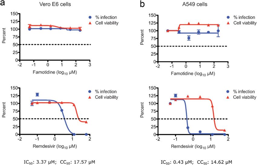
www.nature.com/scientificreports/
Figure 1. Effects of famotidine on P Lpro and 3CLpro protease activity. In-vitro inhibition assays ( IC50) of P
Lpro
(a) and 3CLpro (b) activity show that famotidine had no effect on either of the two SARS-CoV-2 proteases. I C50
values represent inhibition of viral protease activity by control compounds (black) or famotidine (green) when
tested at various concentrations. The initial slopes of protein catalytic activity were converted from RFU/min
to μmole fluorophore/min. Values are mean ± standard deviation of triplicates. The compounds tested in this
experiment neither quenched fluorescence nor produced auto-fluorescence.
act on the p rotease8. Other computational reports have predicted famotidine as an inhibitor of the Papain-like
protease (PLpro), a second SARS-CoV-2 protease9. Together, these studies have raised the prospect of a direct
antiviral effect of famotidine on SARS-CoV-2 replication. While both proteins are attractive targets for SARS-
CoV-2 drug d evelopment10–19, there are at present no clinical-stage or approved drugs targeting either protein.
The possibility of famotidine, an approved drug, acting on SARS-CoV-2 proteases is of significant clinical inter-
est. In this in-vitro study, we performed an array of biochemical, biophysical, and antiviral experiments to test
if famotidine is an effector of SARS-CoV-2 proteases and whether it inhibits virus replication in cultured cells.
Results
Famotidine is not an inhibitor of SARS‑CoV‑2 proteases. Processing of the SARS-CoV-2 polypro-
tein is critical to the generation of a functional virus replication complex11,18,20. To carry out this essential pro-
teolytic function, the SARS-CoV-2 genome encodes two cysteine proteases, called PLpro and 3 CLpro18. Due to
their critical roles in viral polyprotein processing and virus proliferation, both proteases are considered attractive
targets for drug discovery10,11,13–17,21. Since in silico docking studies have predicted these proteases as putative
molecular targets of famotidine6,8,9, we methodically investigated the effect of famotidine on the catalytic func-
tions of each protease.
First, we developed an in-vitro activity assay of P Lpro. PLpro is a protease domain found within the large multi-
domain nsp3 protein encoded by SARS-CoV-2. While many coronaviruses encode two papain-like proteases,
SARS-CoV, MERS-CoV and SARS-CoV-2 possess only one P Lpro, which processes the amino-terminal end of
the viral polyprotein liberating nsp1, nsp2 and n sp319,21. Additionally, P Lpro deubiquitinates host cell proteins by
cleaving the consensus motif of L XGG18,19 and is known to efficiently hydrolyze both diubiquitin and synthetic
peptide substrates19. We leveraged the deubiquitinating property of P Lpro to set up a functional activity assay using
ubiquitin-AMC, a fluorogenic substrate cleavable by P Lpro. Upon incubation with P Lpro, the ubiquitin is recog-
nized and cleaved at the C-terminus to liberate the AMC (amido-4-methylcoumarin) fluorophore which results
in increased fluorescence that is read using excitation and emission wavelengths of 355/460 nm. We assessed
the ability of famotidine to inhibit the proteolytic activity of P Lpro at a broad range of drug concentrations vis-à-
vis compound 6, a previously reported inhibitor of PLpro activity21. Experimental conditions including protein
and substrate concentrations, buffer composition, and assay kinetics were optimized using compound 6. While
compound 6 inhibited PLpro activity with the expected low single-digit μM IC50 values, famotidine showed no
reduction in P Lpro activity in the titrated range of 0.01–200 μM (Fig. 1a).
We next tested whether famotidine can inhibit the enzymatic activity of 3 CLpro, the second protease encoded
by the SARS-CoV-2 genome. This protein, also referred to as the main protease ( Mpro) or nsp5, cleaves the
viral polyprotein at 11 unique sites11. This proteolytic activity generates multiple individual functional proteins
required for the assembly of the SARS-CoV-2 replication/transcription complex, which drives viral genome
replication20. Owing to its central role in the coronavirus life cycle, 3 CLpro has received significant attention as a
drug target resulting in the discovery of several potent inhibitors 10,14,15,17. Native 3CLpro exists as a homodimer
and requires dimerization for its proteolytic a ctivity11. The catalytic mechanism of 3 CLpro activity is typical of
cysteine proteases, where the Cys-His catalytic dyad drives site-specific cleavage of substrates. We evaluated the
enzymatic activity of 3 CLpro using a FRET-peptide substrate that quenched fluorescence in its intact form, how-
ever, cleavage of the peptide substrate by 3CLpro produced fluorescence that could be measured at the excitation/
emission wavelengths of 490/535 nm. The inclusion of ML188, a previously reported 3 CLpro inhibitor served as
a control, also aiding assay setup and optimization. Results of the FRET assay for various ML188 and famoti-
dine concentrations are shown in Fig. 1b. Both compounds were tested between a range of 0.01–200 μM. While
ML188 produced a dose-dependent inhibition of 3 CLpro activity with an expected I C50 of 2.4 μM, famotidine
did not inhibit 3 CLpro activity.
Scientific Reports | (2021) 11:5433 | https://doi.org/10.1038/s41598-021-84782-w 2
Vol:.(1234567890)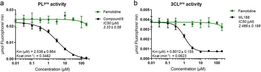
www.nature.com/scientificreports/
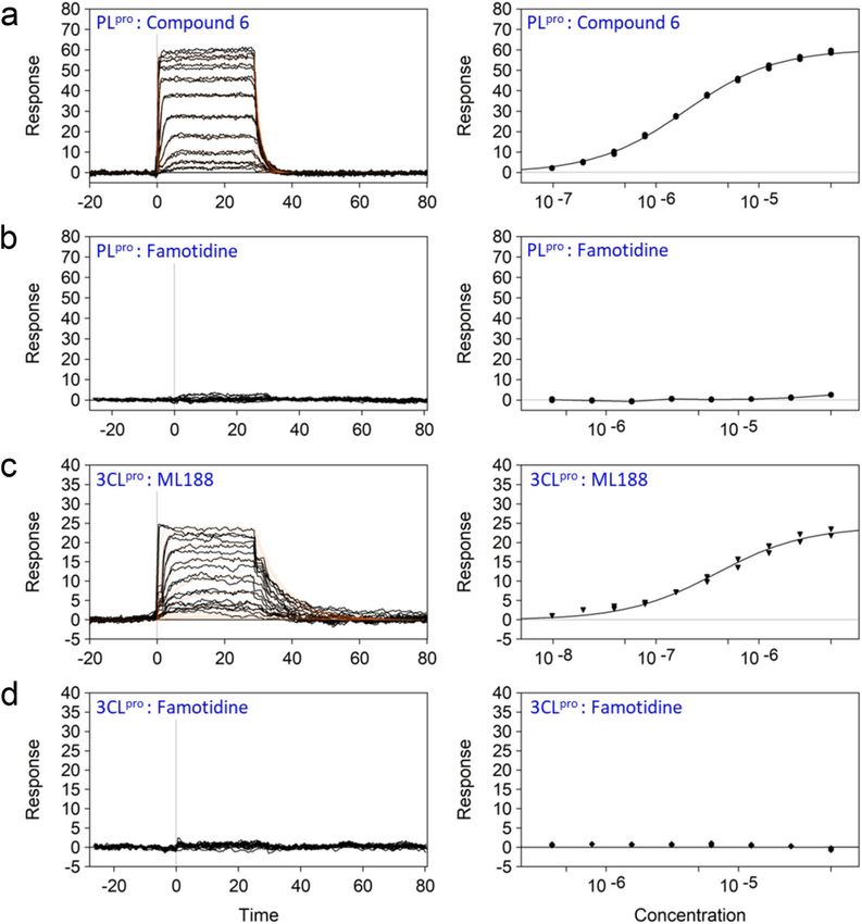
Figure 2. Binding of famotidine to P Lpro and 3CLpro analyzed by SPR. Soluble biotinylated PLpro (a, b) and
3CLpro (c, d) were immobilized on a neutravidin-coated sensor chip and a range of compound concentrations
were injected with solvent (DMSO) corrections. Both, (a) compound 6, the known PLpro inhibitor and (c)
ML188, the 3 CLpro inhibitor displayed dose-dependent binding to PLpro and 3CLpro, respectively. Whereas, (b,
d) no binding of famotidine was detected to either protein. The dissociation constant (Kd) values for the control
compounds are shown in Table S1 (Supplementry Information).
When tested alone, neither famotidine nor the other tested compounds produced autofluorescence at the
355/460 nm and 490/535 nm wavelengths, respectively. These two experiments indicate that famotidine does
not interfere with the catalytic activity of either of the two SARS-CoV-2 proteases.
Famotidine does not directly engage PLpro or 3CLpro of SARS‑CoV‑2. The function of many
enzymes, such as proteases and kinases, can extend beyond their catalytic roles and includes a wide spectrum of
non-catalytic activities such as allosteric regulation, scaffolding, protein–protein interactions, and protein-DNA
interactions22. To rule out whether famotidine could bind away from the active site of the two viral proteases,
and exert an effect through interference with non-proteolytic functions, we asked if famotidine is able to bind
directly with either of the two SARS-CoV-2 proteases. For this, we employed two distinct biophysical techniques
i.e. surface plasmon resonance (SPR) and differential scanning fluorimetry (DSF), that are routinely used to
probe drug-protein engagement.
For our SPR studies, the biotinylated viral proteases were captured to a high density on sensor chips via neu-
travidin, permitting real-time detection of small-molecule binding to the target viral proteases. Engagement of
the small-molecule compounds was recorded as an increase in dose-dependent response units (RU) during the
assay. Experimental conditions including buffer composition and temperature were optimized using the con-
trol compounds prior to conducting the famotidine studies. The equilibrium dissociation constant (Kd) values
were determined using both a kinetic analysis and fit to a binding isotherm of the dose response data (Fig. 2).
The observed Kd values for the known 3CLpro and PLpro inhibitors (Supplementary Information Table S1) were
consistent with the published IC50 data14,21 indicating the robustness of our assay methodology. Under these
Scientific Reports | (2021) 11:5433 | https://doi.org/10.1038/s41598-021-84782-w 3
Vol.:(0123456789)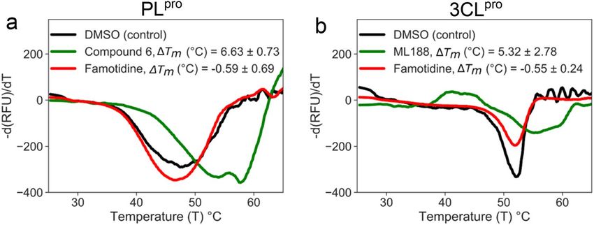
www.nature.com/scientificreports/
Figure 3. DSF assays of famotidine binding to P Lpro and 3CLpro. Fluorescence-monitored thermal denaturation
assay showing the melting curve (first-derivative of dissociation) for each of the two proteins (7 μM) in the
presence or absence of compounds (2.5 mM) . (a) PLpro melting curves for DMSO control (black), compound
6 (green) and famotidine (red) show that while compound 6 stabilizes the PLpro ΔTm by 6.6 °C, famotidine is
unable to shift the ΔTm. Similarly, in (b) while ML188 (green) stabilizes 3CLpro ΔTm, by 5.3 °C, famotidine (red)
does not shift the melting temperature of 3 CLpro. The values are mean ± standard deviation of three independent
replicates.
optimized conditions, famotidine was not found to interact with either of the two viral proteases at concentra-
tion ranges of up to 100 uM.
To validate the results obtained from our SPR analysis, we employed an orthogonal DSF assay. DSF is a
fluorescence-monitored thermal denaturing technique in which the melting temperature (Tm) of a protein is
tracked via fluorescence as the sample temperature is incrementally raised in the presence of a hydrophobic dye.
Drug binding to its target protein is known to stabilize (or destabilize) protein structure resulting in a variation
of Tm profiles in the absence or presence of a drug. DSF provides definitive confirmation of target engagement as
the increase in thermal unfolding temperature (ΔTm) is only achieved when the compounds bind to the folded
state of the protein. The ΔTm is proportional to the Kd of the interaction and concentration of the compound. We
tested the ability of famotidine and the control inhibitors to alter the thermal stability profiles of P Lpro and 3CLpro.
An optimal signal profile was obtained with 7 μM P Lpro or 3CLpro. Both proteins were tested separately in the
presence of DMSO (-ve control), their respective control inhibitors, and famotidine at concentrations of 1, 2.5
and 5 mM. In agreement with the SPR data, the control inhibitors produced a quantitative increase in observed
Tm (Fig. 3). While compound 6, the known P Lpro inhibitor, stabilized P Lpro by a Tm shift of 6.6 °C (Fig. 3a), and
ML188, the 3CLpro inhibitor, produced a Tm shift of 5.3 °C (Fig. 3b), famotidine did not alter the Tm of either of
the two viral proteases. Taken together, the biophysical data decisively rules out the possibility of famotidine
exerting its effect on P Lpro or 3CLpro through interference with catalytic or non-catalytic protein functions as it
is unable to bind with either of the two proteases.
Famotidine does not inhibit SARS‑CoV‑2 replication in cultured cells. Having established that
famotidine does not inhibit SARS-CoV-2 proteases, we investigated the ability of famotidine to block virus rep-
lication in cell culture. For this, we infected Vero E6 cells, a commonly used cell model of SARS-CoV-2 infection
derived from the African green monkey kidney. Infection efficiency was quantified through multiple, orthogo-
nal readouts, including quantitative real-time PCR (qRT-PCR), plaque formation, and immunofluorescence.
Remdesivir inhibited viral replication with an estimated half-maximum inhibitory concentration (IC50) value of
3.3 μM, as determined by immunofluorescence (Fig. 4a). In contrast, famotidine did not produce any measur-
able inhibition at concentrations of up to 200 μM at 72 h post infection. Similar results were obtained when viral
replication was examined by infectious virion production using plaque formation assays or by quantifying viral
RNA copy numbers in the cell culture medium using qRT-PCR (Supplementary Figures S1 and S2).
To confirm these results in a more physiologically relevant cell model of SARS-CoV-2 infection, we assessed
the antiviral activity of famotidine in human lung A549 cells. These cells were engineered to express essential
SARS-CoV-2 entry factors, ACE2 and TMPRSS223. The cells were infected with SARS-CoV-2 and cultured in the
absence or presence of the control or test compounds. Virus replication (infection) efficiency was measured and
reported as a function of compound concentration (Fig. 4). While remdesivir strongly inhibited virus replication
in a dose-dependent manner with an I C50 value of 0.43 µM, famotidine had no measurable effect (Fig. 4b). Our
results are consistent with previously reported studies in which remdesivir exerted a greater antiviral effect in
human lung A549 cells than in Vero E6 cells24. The fact that famotidine does not inhibit SARS-CoV-2 replication
in the A549 cells which possess the serine protease TMPRSS2 also likely points to it having no effect on host
proteases involved in viral entry.
In-parallel cytotoxicity assays, carried out in both Vero E6 and A549 cells, showed that famotidine was not
toxic up to the highest tested concentrations of 200 μM (Fig. 4). Remdesivir, on the other hand, exhibited dose-
dependent cytotoxicity at higher concentrations, well above its IC50. Together, these results show that famotidine
Scientific Reports | (2021) 11:5433 | https://doi.org/10.1038/s41598-021-84782-w 4
Vol:.(1234567890)www.nature.com/scientificreports/
Figure 4. Antiviral activity of famotidine in Vero E6 and human lung A549 cells. Percent inhibition of SARS-
CoV-2 replication and cytotoxicity are shown in the presence of a range of famotidine (top) and remdesivir
(bottom) concentrations for (a) Vero E6 cells and (b) human lung A549 cells. Percent infection values (blue)
represent the antiviral activity (IC50) of the drug compounds and cell viability values (red) represent cytotoxicity
(CC50) of the drugs. Infection was assessed through quantitation of virus-treated cells that stained positive for
the viral nucleocapsid protein, 72 h post infection. Cell viability of the corresponding compound concentrations
on the cells was measured using the CellTiter-Glo assay. Values reported are mean ± standard deviation of
triplicates.
does not inhibit SARS-CoV-2 replication in cultured cells and that its purported clinical benefit may be due to
an alternative mechanism of action.
Discussion
Two in silico studies have separately predicted the 3 CLpro or P Lpro of SARS-CoV-2 as potential molecular targets
of famotidine8,9, implying that famotidine associated improvement in COVID-19 patients may be due to a direct
antiviral mechanism of action6. Despite recent advances in computational techniques, there are several chal-
lenges associated with the use of molecular docking to predict protein–ligand interactions accurately. Some of
these challenges arise from the flexibility of the target protein, lack of prior knowledge of drug-binding sites, and
protonation states of target amino a cids25. While results obtained from molecular docking can serve as a basis
for new hypotheses, experimental validation is needed. Our ligand-binding experiments using SPR and DSF
did not support previous in silico predictions of direct binding between famotidine and SARS-CoV-2 proteases.
We further used an array of experimental approaches to show that famotidine had no effect on SARS-CoV-2
protease function or generally on viral replication. It must be noted that since the clinical studies correlated
putative clinical benefit with the use of higher doses of famotidine, we tested famotidine at significantly higher
in-vitro concentrations than the peak plasma concentrations (0.5–2 µM) achieved in the blood of patients in
both clinical studies6,7. Our data strongly suggest that the probable clinical benefit of famotidine likely arises
independently of an antiviral mechanism of action.
COVID-19 complications are associated with a severe pro-inflammatory response in the lungs of infected
patients26. The “cytokine storm” as a result of inflammation is a key pathognomonic feature of COVID-19 and
the main contributor to respiratory failure and mortality27. Severe COVID-19 cases are characterized by pulmo-
nary infiltration and extensive pulmonary edema, causing exudation of inflammatory cells in the alveolar space,
resulting in extensive pulmonary consolidation leading to pneumonia and adult respiratory distress syndrome
(ARDS)28–31. The results of the two famotidine-related COVID-19 clinical reports, when taken t ogether6,7, sug-
gest that famotidine likely helps with mitigating moderate to severe respiratory symptoms ranging from short-
ness of breath to intubation. Our data does not rule out the possibility that famotidine related improvements
in COVID-19 patients are through an anti-inflammatory action. For example, the development of the cytokine
storm in COVID-19 patients is characterized by elevation of pro-inflammatory type I cytokines, which are
secreted from a variety of cells such as polymorphonuclear cells, natural killer cells, and endothelial cells, e tc27.
Scientific Reports | (2021) 11:5433 | https://doi.org/10.1038/s41598-021-84782-w 5
Vol.:(0123456789)www.nature.com/scientificreports/
It is therefore conceivable that famotidine-related benefit in managing respiratory symptoms may be due to an
anti-inflammatory mechanism of action.
It is noteworthy that H2R, the established molecular target of famotidine, is involved in the activation of
several mediators of the adaptive immune response, such as Th1 lymphocytes, which are implicated in pro-
inflammatory cytokine production32. Histamine, the H2R ligand, also regulates bronchoconstriction, airway
inflammation, and vasodilation32. Mast cells are a major source of histamine and their activation has been
reported following viral infections of the respiratory t ract33–35. Therefore, Mast cells may represent an underap-
preciated source of pro-inflammatory cytokine release in COVID-19 p atients33. A better understanding of the
role of the H2R pathway in COVID-19 will help elucidate the molecular details of how famotidine reduces the
disease severity.
Our in-vitro study redirects the mechanism behind the potential beneficial effect of famotidine, away from
an antiviral effect to likely an anti-inflammatory action in COVID-19 patients. Given that there is an ongoing
randomized clinical trial (NCT04370262), our results may assist the investigators in reshaping their interven-
tional study to include inflammation-related outcomes. Also, it should be noted that while famotidine is one
of the relatively safer drugs, its use is not without r isk36–38, especially in elderly patients (a high-risk population
for COVID-19), in which famotidine use has been associated with CNS c omplications39. Provided the ongoing
clinical trial yields promising results, further investigation of famotidine and its safety profile in different age
brackets will be needed before the drug can be used, most likely as part of a combination therapy, for COVID-19
disease management.
Materials and methods
Compounds. Famotidine was acquired from Sigma Aldrich (Missouri, USA; cat. No. F6889). Compound
6, a previously reported inhibitor of SARS-CoV-2 P
Lpro function21 was acquired from MedChem Express, Inc.
(New Jersey, USA; cat no. HY-17542). ML188, a compound with known 3CLpro inhibitory activity15 was also
acquired from MedChem Express, Inc. (cat. no HY-136259). Similarly, remdesivir. (cat. No HY-104077) an
inhibitor of SARS-CoV-2 replication2 was purchased from the same vendor. All compounds were dissolved in
100% DMSO at 100 mM.
Cloning, expression, and protein purification. The complete sequences encoding 3CLpro and residues
746–1060 of P Lpro (Wuhan-Hu-1 isolate, GenBank accession NC_045512) were cloned into a charge modified
SUMO fusion expression vector, generated in-house. The fusion protein was expressed for 24 h in Rosetta-2
(DE3) pLysS at 18 °C in ZYP-5052 autoinducing media. Harvested cells were resuspended in 50 mM Hepes pH
7.5, containing 150 mM NaCl and lysed by sonication. The clarified supernatant was loaded onto a HiTrap HP SP
column (Cytiva, Massachusetts, USA; cat no. 17115201) and the target fusion protein was captured in a cation-
exchange chromatography step and eluted using a NaCl gradient. SUMO hydrolase was added to the pooled
fractions to liberate the target protein and the sample dialyzed against 20 mM Tris, 10% v/v glycerol, 5 mM DTT
pH 7.0 overnight at 4 °C. The protein was reloaded on the HiTrap HP SP column to remove the SUMO protein
and hydrolase in a subtractive step. The flow-through, containing 3CLpro or PLpro was further purified by anion
exchange chromatography using a HiTrap HP Q column (Cytiva; cat. no. 17115401) employing a NaCl gradient
to elute the protein. Pooled fractions were further purified by size exclusion chromatography in 20 mM Tris pH
7.4, 150 mM NaCl and 5 mM DTT. The final protein was concentrated to 4 mg/mL for PLpro and 5 mg/mL for
3CLpro and flash frozen in aliquots.
In‑vitro viral enzyme assays. PLpro proteolytic activity assay using ubiquitin‑AMC. PLpro activity was
measured in a 384 well plate format (Corning #3574) in a kinetic assay using the fluorogenic substrate Ubiquitin-
AMC (Boston Biochem, Inc. Massachusetts, USA; cat. No. U-550) with excitation and emission wavelengths
of Ex355nm/Em:460 nm. The protocol followed previously reported conditions with minor m odifications13,16.
Fluorescence was monitored at 25 °C, every 5 min for 50 min in a Victor X5 (Perkin Elmer) multimode plate
reader. Optimal enzyme and substrate concentrations were found to be 550 nM P Lpro titrating the substrate in
the range of 0.2 – 3 μM. The assay buffer (20 μL) contained 25 mM HEPES pH 7.5, 100 mM NaCl, 0.1 mg/ml
BSA, and 550 nM PLpro. The test inhibitor, famotidine and the PLpro control inhibitor (compound 6) were both
titrated in the concentration range of 0.01 μM – 200 μM. Compounds were incubated with the enzyme in the
plate for 30 min at 25 °C before the reaction was started by the addition of 1 μM Ub-AMC. All samples were run
in triplicates and their initial slopes were converted from relative fluorescence units (RFU)/ min to μmol AMC/
min using an AMC standard curve and plotted against compound concentrations tested.
3CLpro proteolytic activity assay. 3CLpro activity was assayed in a 384 well plate using the 3 CLpro FRET substrate
(AnaSpec, Inc. California, USA; cat. no. AS-65599) with excitation and emission wavelengths of Ex: 490 nm/
Em: 535 nm. A previously reported protocol was used with some modifications12,17. The kinetics of fluorescence
change were monitored every minute for 25 min. Optimal concentrations for 3 CLpro and substrate were 150 nM
and 600 nM respectively. A previously reported 3CLpro inhibitor14, ML188, was used as a positive control for
inhibition, both control and test compounds were titrated in the concentration range of 0.01 μM – 200 μM.
Initial slopes of RFU/min were converted to μM hydrolyzed substrate/ min using a standard curve of HiLyte
Fluor488 amine, TFA salt.
Biochemical data analysis. After subtraction of background fluorescence readings, values of Km and EC50
were obtained by fitting the experimental data with the Michaelis–Menten (y = (Vmax*x)/(Km + x)) and the four
Scientific Reports | (2021) 11:5433 | https://doi.org/10.1038/s41598-021-84782-w 6
Vol:.(1234567890)www.nature.com/scientificreports/
parameters logistic (4PL) equations (y = min + (max–min)/(1 + (x/EC50)^Hillslope)) respectively, using Graph-
Pad Prism 8.
Dynamic scanning fluorimetry (DSF). Thermal unfolding of proteins was monitored in a 20 uL volume
in Micro-Amp EnduraPlate Optical 384-well Clear Reaction Plates (ThermoFisher: cat no. 4483285 ). Reactions
contained 50 mM HEPES pH 7.5, 62.5 mM NaCl, 7 μM 3 CLpro or PLpro, 5% DMSO, and 4 × SYPRO-orange pro-
tein gel stain (ThermoFisher Scientific, Massachusetts, USA; cat no. S6651). Famotidine and the positive controls
ML188 and compound 6 for 3 CLpro and P Lpro, respectively, were incubated with the protein for 15 min before
the addition of SYPRO orange. Plates were covered with Micro-Amp Optical Adhesive Film (ThermoFisher
Scientific, Massachusetts, USA; cat no. 4360954) and run on Applied Biosystems 7900HT (California, USA) real
time PCR instrument. Samples were incubated at 25 °C for 2 min followed by an increase in temperature of 1 °C/
min up to 95 °C. Fluorescence was monitored continuously. Each sample was run in triplicate and compounds
were tested at 1 mM, 2.5 mM, and 5 mM. The melting temperature (Tm) was obtained from the first derivative of
the raw thermal denaturing data were determined and smoothed to calculate melting temperature (Tm) values40.
Surface plasmon resonance (SPR). SPR studies were performed on a Biacore 3000 instrument (Cytiva,
Massachusetts, USA) at 10 °C. The P Lpro and 3CLpro proteins were biotinylated by minimal biotinylation approach
with the EZ-LINK Sulfo-NHS-LC-LC-biotin reagent (ThermoFisher Scientific, Massachusetts, USA; cat no.
A35358) and immobilized on a neutravidin coated CM5 sensor chip to a level of 4000 response units (RU). The
protein used during immobilization was at 1 μM for P Lpro and 1 μM for 3 CLpro. During the course of the assay
different concentrations of compounds were injected. The Compounds were serially diluted (twofold) in a run-
ning buffer of 25 mM HEPES pH 7.0, 200 mM NaCl, 2 mM TCEP, 0.005% P20 and 1% DMSO. Famotidine, and
the control inhibitors, compound 6 and the ML188 were tested up to a maximal dose of 100 μM, 50 μM and
5 μM, respectively. The final response was obtained by subtracting the blank channel (without protein) and a
buffer injection across the sample channel. Raw data were analyzed in the Scrubber2 program (BioLogic Soft-
ware) by fitting the data to a simple 1:1 equilibrium and kinetic model.
Antiviral assays. Viruses and titration. Virus infectivity assays were carried out using the 2019-nCoV/
USA-WA1/2020 isolate of SARS-CoV-2 (NCBI accession number: MN985325), obtained from the Centers for
Disease Control and Prevention and BEI Resources (Virginia, USA). The virus stock was propagated in Vero E6
reviously41.
cells and virus titers determined using plaque formation assays, as described p
Antiviral assays. Human lung A549 cells expressing SARS-CoV-2 entry factors and African Green Monkey
kidney Vero E6 cells were maintained in DMEM supplemented with 10% fetal bovine serum (FBS). The cells
were seeded into poly-L-lysine-coated 96-well plates at a density of 15,000 cells per well. The cells were then
treated for 4 h with five-fold serial dilutions of famotidine, ranging between 0.32 µM and 200 µM. DMSO served
as a negative control, while fivefold serial dilutions of remdesivir, ranging between 0.1 µM and 62.5 µM, served
as a positive control. The cells were then infected with SARS-CoV-2 at a multiplicity of infection (MOI) of 0.1.
To infect cells, the compound-containing medium was removed, and the cells were incubated with the virus for
1 h at 37ºC. The virus inoculum was then removed, and the cell monolayer was rinsed twice with 1X PBS. The
compounds were added back followed by incubation for 72 h, after which the cell culture medium was harvested
for quantitative real-time PCR (qRT-PCR) and plaque assays, while the cells were fixed with 4% paraformalde-
hyde for immunofluorescence microscopy.
Virus RNA extraction and quantitative real‑time PCR (qRT‑PCR). RNA was isolated from the cell culture super-
natant of SARS-CoV-2-infected cells using the Quick-RNA Viral Kit (Zymo, California, USA cat no. R1035)
according to the manufacturer’s instructions. Viral RNA was quantified using single-step RT-quantitative real-
time PCR using the qScript One-Step RT-qPCR Kit (Quantabio, Massachusetts, USA; cat no. 95058) with prim-
ers and Taqman probes targeting the SARS-CoV-2 E gene as previously d escribed42. Data were acquired using
a Quantstudio3 Real-Time PCR System (Applied Biosystems) using the following conditions: 55 °C for 10 min,
denaturation at 94 °C for 3 min, 45 cycles of denaturation at 94 °C for 15 s, and annealing at 58 °C for 30 s. The
primers and probe used were as follow: E_Sarbeco_Forward: ACAGGTACGTTAATAGTTAATAGCGT; E_Sar-
beco_Probe: FAM-ACACTAGCCATCCTTACTGCGCTTCG-BBQ; E_Sarbeco_Reverse: ATATTGCAGCAG
TACGCACACA. For absolute quantification of viral RNA, a 389 bp fragment from the SARS-CoV-2 E gene was
cloned onto pIDTBlue plasmid under an SP6 promoter using NEB PCR cloning kit (New England Biosciences,
Massachusetts, USA; cat no. E1202S). The cloned fragment was then in-vitro transcribed using the mMessage
mMachine SP6 transcription kit (ThermoFisher, Massachusetts, USA; cat no. AM1340) to generate the qRT-
PCR standard.
Immunofluorescence microscopy. Virus-infected cells were fixed with 4% paraformaldehyde for 30 min. The
fixative was removed, and the cell monolayer washed twice with 1X PBS. The cells were then permeabilized
and stained with an anti-SARS-CoV Nucleocapsid (N) antibody (Rockland Inc., Pennsylvania, USA; cat. no.
200–401-A50; 1:2,000 dilution). Incubation with the primary antibody was performed overnight at 4ºC. The
cells were then washed 5 times with 1X PBS and stained with Alexa 568-conjugated anti-rabbit antibody (1:1000
dilution) in the dark at room temperature for 1 h and counterstained with DAPI. Images were captured using
EVOS M5000 Imaging System (ThermoFisher Scientific, Massachusetts, USA). Quantitation and analysis of the
fixed cell images was carried out using the MuviCyte Live-Cell Imaging System (PerkinElmer, Massachusetts,
Scientific Reports | (2021) 11:5433 | https://doi.org/10.1038/s41598-021-84782-w 7
Vol.:(0123456789)www.nature.com/scientificreports/
USA). At least 7–10 microscopic fields were imaged per well using a 10X objective lens, the number of cells posi-
tive for the SARS-CoV-2 N protein and the nuclear DAPI stain, were counted. For each image, the percentage
of DAPI-positive cells expressing the viral N protein were calculated, and the mean ± SD of multiple images for
each condition was plotted.
Cytotoxicity/cell viability assay. The CellTiter-Glo Luminescent Cell Viability Assay (Promega, Wisconsin,
USA; cat no. G7570) was used to determine the cytotoxic effects of the compounds. Briefly, the cells were incu-
bated with five-fold serial dilutions of famotidine or remdesivir for 72 h, after which the CellTiter-Glo Reagent
was added to each well in a volume equal to the volume of the culture medium. The contents were mixed by
shaking the plate on an orbital shaker for 2 min, followed by a 10 min incubation at room temperature. Lumi-
nescence was recorded using a Varioskan LUX multimode plate reader (ThermoFisher Scientific, Massachusetts,
USA).
Data availability
The datasets generated during and/or analysed during the current study are available from the corresponding
author on reasonable request.
Received: 2 November 2020; Accepted: 1 February 2021
References
1. Harrison, C. Coronavirus puts drug repurposing on the fast track. Nat. Biotechnol. 38, 379–381. https://doi.org/10.1038/d4158
7-020-00003-1 (2020).
2. Beigel, J. H. et al. Remdesivir for the treatment of Covid-19 - preliminary report. N. Engl. J. Med. https://doi.org/10.1056/NEJMo
a2007764 (2020).
3. Boulware, D. R. et al. A randomized trial of hydroxychloroquine as postexposure prophylaxis for covid-19. N. Engl. J. Med. https
://doi.org/10.1056/NEJMoa2016638 (2020).
4. Cao, B. et al. A trial of lopinavir-ritonavir in adults hospitalized with severe covid-19. N. Engl. J. Med. 382, 1787–1799. https://doi.
org/10.1056/NEJMoa2001282 (2020).
5. Keithley, J. K. Histamine H2-receptor antagonists. Nurs Clin North Am 26, 361–373 (1991).
6. Freedberg, D. E. et al. Famotidine use is associated with improved clinical outcomes in hospitalized COVID-19 patients: a pro-
pensity score matched retrospective cohort study. Gastroenterology https://doi.org/10.1053/j.gastro.2020.05.053 (2020).
7. Janowitz, T. et al. Famotidine use and quantitative symptom tracking for COVID-19 in non-hospitalised patients: a case series.
Gut https://doi.org/10.1136/gutjnl-2020-321852 (2020).
8. Wu, C. et al. Analysis of therapeutic targets for SARS-CoV-2 and discovery of potential drugs by computational methods. Acta
Pharm. Sin. B https://doi.org/10.1016/j.apsb.2020.02.008 (2020).
9. Shaffer, L. 15 drugs being tested to treat COVID-19 and how they would work. Nat. Med. https://doi.org/10.1038/d41591-020-
00019-9 (2020).
10. Zhang, L. et al. Crystal structure of SARS-CoV-2 main protease provides a basis for design of improved alpha-ketoamide inhibi-
tors. Science 368, 409–412. https://doi.org/10.1126/science.abb3405 (2020).
11. Muramatsu, T. et al. SARS-CoV 3CL protease cleaves its C-terminal autoprocessing site by novel subsite cooperativity. Proc. Natl.
Acad. Sci. USA 113, 12997–13002. https://doi.org/10.1073/pnas.1601327113 (2016).
12. Tomar, S. et al. Ligand-induced dimerization of middle east respiratory syndrome (MERS) coronavirus nsp5 protease (3CLpro):
implications for nsp5 regulation and the development of antivirals. J. Biol. Chem. 290, 19403–19422. https://doi.org/10.1074/jbc.
M115.651463 (2015).
13. Baez-Santos, Y. M., Mielech, A. M., Deng, X., Baker, S. & Mesecar, A. D. Catalytic function and substrate specificity of the papain-
like protease domain of nsp3 from the Middle East respiratory syndrome coronavirus. J. Virol. 88, 12511–12527. https://doi.
org/10.1128/JVI.01294-14 (2014).
14. Jacobs, J. et al. Discovery, synthesis, and structure-based optimization of a series of N-(tert-butyl)-2-(N-arylamido)-2-(pyridin-
3-yl) acetamides (ML188) as potent noncovalent small molecule inhibitors of the severe acute respiratory syndrome coronavirus
(SARS-CoV) 3CL protease. J. Med. Chem. 56, 534–546. https://doi.org/10.1021/jm301580n (2013).
15. Jacobs, J. et al. Probe Reports from the NIH Molecular Libraries Program (2010).
16. Ratia, K. et al. A noncovalent class of papain-like protease/deubiquitinase inhibitors blocks SARS virus replication. Proc. Natl.
Acad. Sci. USA 105, 16119–16124. https://doi.org/10.1073/pnas.0805240105 (2008).
17. Lu, I. L. et al. Structure-based drug design and structural biology study of novel nonpeptide inhibitors of severe acute respiratory
syndrome coronavirus main protease. J. Med. Chem. 49, 5154–5161. https://doi.org/10.1021/jm060207o (2006).
18. Lindner, H. A. et al. The papain-like protease from the severe acute respiratory syndrome coronavirus is a deubiquitinating enzyme.
J. Virol. 79, 15199–15208. https://doi.org/10.1128/JVI.79.24.15199-15208.2005 (2005).
19. Barretto, N. et al. The papain-like protease of severe acute respiratory syndrome coronavirus has deubiquitinating activity. J. Virol.
79, 15189–15198. https://doi.org/10.1128/JVI.79.24.15189-15198.2005 (2005).
20. van Hemert, M. J. et al. SARS-coronavirus replication/transcription complexes are membrane-protected and need a host factor
for activity in vitro. PLoS Pathog. 4, e1000054. https://doi.org/10.1371/journal.ppat.1000054 (2008).
21. Freitas, B. T. et al. Characterization and noncovalent inhibition of the deubiquitinase and deISGylase activity of SARS-CoV-2
papain-like protease. ACS Infect. Dis. https://doi.org/10.1021/acsinfecdis.0c00168 (2020).
22. Kung, J. E. & Jura, N. Structural basis for the non-catalytic functions of protein kinases. Structure 24, 7–24. https: //doi.org/10.1016/j.
str.2015.10.020 (2016).
23. Ma, D. et al. Expression of SARS-CoV-2 receptor ACE2 and TMPRSS2 in human primary conjunctival and pterygium cell lines
and in mouse cornea. Eye 34, 1212–1219. https://doi.org/10.1038/s41433-020-0939-4 (2020).
24. Xie, X. et al. A nanoluciferase SARS-CoV-2 for rapid neutralization testing and screening of anti-infective drugs for COVID-19.
bioRxiv https://doi.org/10.1101/2020.06.22.165712 (2020).
25. Palacio-Rodriguez, K., Lans, I., Cavasotto, C. N. & Cossio, P. Exponential consensus ranking improves the outcome in docking
and receptor ensemble docking. Sci. Rep. 9, 5142. https://doi.org/10.1038/s41598-019-41594-3 (2019).
26. Tay, M. Z., Poh, C. M., Renia, L., MacAry, P. A. & Ng, L. F. P. The trinity of COVID-19: immunity, inflammation and intervention.
Nat. Rev. Immunol. 20, 363–374. https://doi.org/10.1038/s41577-020-0311-8 (2020).
27. Ye, Q., Wang, B. & Mao, J. The pathogenesis and treatment of the `Cytokine Storm’ in COVID-19. J. Infect. 80, 607–613. https://
doi.org/10.1016/j.jinf.2020.03.037 (2020).
Scientific Reports | (2021) 11:5433 | https://doi.org/10.1038/s41598-021-84782-w 8
Vol:.(1234567890)www.nature.com/scientificreports/
28. Carsana, L. et al. Pulmonary post-mortem findings in a series of COVID-19 cases from northern Italy: a two-centre descriptive
study. Lancet Infect. Dis. https://doi.org/10.1016/S1473-3099(20)30434-5 (2020).
29. Bhatraju, P. K. et al. Covid-19 in critically Ill patients in the seattle region—case series. N. Engl. J. Med. 382, 2012–2022. https://
doi.org/10.1056/NEJMoa2004500 (2020).
30. Tian, J. et al. Clinical characteristics and risk factors associated with COVID-19 disease severity in patients with cancer in Wuhan,
China: a multicentre, retrospective, cohort study. Lancet Oncol. https://doi.org/10.1016/S1470-2045(20)30309-0 (2020).
31. Yang, K. et al. Clinical characteristics, outcomes, and risk factors for mortality in patients with cancer and COVID-19 in Hubei,
China: a multicentre, retrospective, cohort study. Lancet Oncol. https://doi.org/10.1016/S1470-2045(20)30310-7 (2020).
32. Thangam, E. B. et al. The role of histamine and histamine receptors in mast cell-mediated allergy and inflammation: the hunt for
new therapeutic targets. Front. Immunol. 9, 1873. https://doi.org/10.3389/fimmu.2018.01873 (2018).
33. Marshall, J. S., Portales-Cervantes, L. & Leong, E. Mast cell responses to viruses and pathogen products. Int. J. Mol. Sci. https://
doi.org/10.3390/ijms20174241 (2019).
34. Zarnegar, B. et al. Influenza infection in mice induces accumulation of lung mast cells through the recruitment and maturation
of mast cell progenitors. Front. Immunol. 8, 310. https://doi.org/10.3389/fimmu.2017.00310 (2017).
35. Hu, Y. et al. Mast cell-induced lung injury in mice infected with H5N1 influenza virus. J. Virol. 86, 3347–3356. https://doi.
org/10.1128/JVI.06053-11 (2012).
36. Kirch, W., Halabi, A., Linde, M., Santos, S. R. & Ohnhaus, E. E. Negative effects of famotidine on cardiac performance assessed
by noninvasive hemodynamic measurements. Gastroenterology 96, 1388–1392. https://doi.org/10.1016/0016-5085(89)90503-9
(1989).
37. Lee, Y. C. & Wang, C. C. Famotidine-induced retinopathy. Eye (Lond) 20, 260–263. https://doi.org/10.1038/sj.eye.6701839 (2006).
38. Kallal, S. M. & Lee, M. Thrombotic thrombo-cytopenic purpura associated with histamine H2-receptor antagonist therapy. West.
J. Med. 164, 446–448 (1996).
39. Cantu, T. G. & Korek, J. S. Central nervous system reactions to histamine-2 receptor blockers. Ann. Intern. Med. 114, 1027–1034.
https://doi.org/10.7326/0003-4819-114-12-1027 (1991).
40. Sun, C., Li, Y., Yates, E. A. & Fernig, D. G. SimpleDSFviewer: a tool to analyze and view differential scanning fluorimetry data for
characterizing protein thermal stability and interactions. Protein Sci. 29, 19–27. https://doi.org/10.1002/pro.3703 (2020).
41. Ogando, N. S. et al. SARS-coronavirus-2 replication in Vero E6 cells: replication kinetics, rapid adaptation and cytopathology. J.
Gen. Virol. https://doi.org/10.1099/jgv.0.001453 (2020).
42. Corman, V. M. et al. Detection of 2019 novel coronavirus (2019-nCoV) by real-time RT-PCR. Euro Surveill https://doi.
org/10.2807/1560-7917 (2020).
Author contributions
Conceptualization: A.H.M.; data curation and analysis: H.L., A.B., S.B., M.S., A.H.M., methodology and inves-
tigation: M.L., H.L., D.Y.C., S.D.G., S.B., A.M., S.D.W., A.O., F.D. M.S., A.H.M.; writing (original draft): A.H.M.;
writing (review and editing): A.H.M., M.S., F.D., S.D.W., H.L., S.B.
Funding
This work was partially supported by the Evergrande MassCPR award to MS and DYC.
Competing interests
The authors declare no competing interests.
Additional information
Supplementary Information The online version contains supplementary material available at https://doi.
org/10.1038/s41598-021-84782-w.
Correspondence and requests for materials should be addressed to M.S. or A.H.M.
Reprints and permissions information is available at www.nature.com/reprints.
Publisher’s note Springer Nature remains neutral with regard to jurisdictional claims in published maps and
institutional affiliations.
Open Access This article is licensed under a Creative Commons Attribution 4.0 International
License, which permits use, sharing, adaptation, distribution and reproduction in any medium or
format, as long as you give appropriate credit to the original author(s) and the source, provide a link to the
Creative Commons licence, and indicate if changes were made. The images or other third party material in this
article are included in the article’s Creative Commons licence, unless indicated otherwise in a credit line to the
material. If material is not included in the article’s Creative Commons licence and your intended use is not
permitted by statutory regulation or exceeds the permitted use, you will need to obtain permission directly from
the copyright holder. To view a copy of this licence, visit http://creativecommons.org/licenses/by/4.0/.
© The Author(s) 2021
Scientific Reports | (2021) 11:5433 | https://doi.org/10.1038/s41598-021-84782-w 9
Vol.:(0123456789)You can also read