Tityus serrulatus (Scorpion): From the Crude Venom to the Construction of Synthetic Peptides and Their Possible Therapeutic Application Against ...
←
→
Page content transcription
If your browser does not render page correctly, please read the page content below
ORIGINAL RESEARCH
                                                                                                                                                     published: 20 July 2021
                                                                                                                                            doi: 10.3389/fcimb.2021.706618

                                             Tityus serrulatus (Scorpion): From
                                             the Crude Venom to the Construction
                                             of Synthetic Peptides and Their
                                             Possible Therapeutic Application
                                             Against Toxoplasma gondii Infection
                                             Diego Rodney Rodrigues de Assis 1, Pollyana Maria de Oliveira Pimentel 1,
                                             Pablo Victor Mendes dos Reis 1, Rayane Aparecida Nonato Rabelo 1,
                                             Ricardo Wagner Almeida Vitor 2, Marta do Nascimento Cordeiro 3,
                         Edited by:
                                             Liza Figueiredo Felicori 1, Carlos Delfin Chávez Olórtegui 1, Jarbas Magalhães Resende 4,
                  Michail Kotsyfakis,
                                             Mauro Martins Teixeira 1, Márcia Helena Borges 3, Maria Elena de Lima 5,
   Academy of Sciences of the Czech
          Republic (ASCR), Czechia
                                             Adriano Monteiro de Castro Pimenta 1 and Fabiana Simão Machado 1,6*

                           Reviewed by:      1Department of Biochemistry and Immunology, Instituto de Ciências Biológicas, Universidade Federal de Minas Gerais, Belo
                 William Harold Witola,      Horizonte, Brazil, 2 Department of Parasitology, Instituto de Ciências Biológicas, Universidade Federal de Minas Gerais, Belo
        University of Illinois at Urbana-    Horizonte, Brazil, 3 Fundação Ezequiel Dias-FUNED, Minas Gerais, Belo Horizonte, Brazil, 4 Department of Chemistry, Instituto de
           Champaign, United States          Ciências Exatas, Universidade Federal de Minas Gerais, Belo Horizonte, Brazil, 5 Faculdade Santa Casa de Belo Horizonte:
            Fernanda Gobbi Amorim,           Programa de Pós Graduação em Medicina-Biomedicina, Belo Horizonte, Brazil, 6 Program in Health Sciences: Infectious
          University of Liège, Belgium       Diseases and Tropical Medicine, Faculdade de Medicina, Universidade Federal de Minas Gerais, Belo Horizonte, Brazil
                   *Correspondence:
              Fabiana Simão Machado
                                             Toxoplasmosis, caused by Toxoplasma gondii, is a major public concern owing to its
               machadofs@icb.ufmg.br
                                             neurotropic nature and high morbidity and mortality rates in immunocompromised
                    Specialty section:       patients and newborns. Current treatment for this disease is inefficient and produces
          This article was submitted to
                                             side effects. Inflammatory mediators produced during T. gondii infection (e.g., cytokines
                      Parasite and Host,
                a section of the journal     and nitric oxide) are crucial in controlling parasite replication. In this context, Tityus
               Frontiers in Cellular and     serrulatus venom (TsV) induces the production of inflammatory mediators by immune
                 Infection Microbiology
                                             cells. Thus, this study aimed to isolate and identify the components of TsV with potential
             Received: 07 May 2021
             Accepted: 02 July 2021
                                             anti-T. gondii activity. TsV was extracted from scorpions and lyophilized or loaded onto a
             Published: 20 July 2021         column to obtain its fractions. TsV subfractions were obtained using chromatography,
                                 Citation:   and its amino acid sequence was identified and applied to peptide design using
       de Assis DRR, Pimentel PMdO,
                                             bioinformatics tools. The C57BL/6 mice and their harvested macrophages were used
           dos Reis PVM, Rabelo RAN,
Vitor RWA, Cordeiro MdN, Felicori LF,        to test the anti-Toxoplasma activity of TsV components and peptides. TsV and its fraction
          Olórtegui CDC, Resende JM,        F6 attenuated the replication of tachyzoites in macrophages and induced nitric oxide and
Teixeira MM, Borges MH, de Lima ME,
     Pimenta AMdC and Machado FS
                                             cytokine (IL-12, TNF, and IL-6) production by infected cells, without host cell toxicity.
                  (2021) Tityus serrulatus   Moreover, Su6-B toxin, a subfraction of F6, demonstrated anti-T. gondii activity. The
   (Scorpion): From the Crude Venom
                                             partially elucidated and characterized amino acid sequence of Sub6-B demonstrated
       to the Construction of Synthetic
            Peptides and Their Possible      93% similarity with T. serrulatus 2 toxin (Ts2). Ts2 mimetic peptides (“Pep1,” “Pep2a,” and
       Therapeutic Application Against       “Pep2b”) were designed and synthesized. Pep1 and Pep2a, but not Pep2b, reduced the
           Toxoplasma gondii Infection.
Front. Cell. Infect. Microbiol. 11:706618.
                                             replication of tachyzoites in macrophages. In vivo, treatment of T. gondii-infected mice
       doi: 10.3389/fcimb.2021.706618        with Pep1, Pep2a, or Pep2b decreased the number of cerebral cysts and did not induce

Frontiers in Cellular and Infection Microbiology | www.frontiersin.org                1                                             July 2021 | Volume 11 | Article 706618
de Assis et al.                                                                                                T. serrulatus Venom Against Toxoplasma

                                            hepatotoxicity in the animals. Taken together, our data show promising
                                            immunomodulatory and antiparasitic activity of TsV that could be explored and applied
                                            in future therapies for treating infectious parasitic diseases such as toxoplasmosis.
                                            Keywords: Toxoplasma gondii, toxoplasmosis, Tityus serrulatus venom, immunoregulation, synthetic
                                            peptides, treatment

INTRODUCTION                                                                  (Cologna et al., 2009), and presents immunomodulatory
                                                                              activities. TsV induces a pro-inflammatory response,
Toxoplasmosis, caused by the parasite Toxoplasma gondii, affects              stimulating cells, such as MOs, to produce TNF/IL-12/IFN-g/
one-third of the global population. This infection is most often              NO/ROS. Moreover, TsV stimulation increases the phagocytic
not characterized as a lethal disease in humans, but it can be fatal          capacity of MOs and activates T lymphocytes (Petricevich et al.,
in immunocompromised individuals, such as HIV/AIDS,                           2008; Zoccal et al., 2011; Casella-Martins et al., 2015), which are
transplanted, or cancer patients, and pregnant women (fetus).                 stimulators of immunity.
Importantly, there is increasing concern about this infection in                 Several natural compounds have been found from diverse
immunocompetent individuals owing to the neurotropic nature                   sources, including scorpion venoms, which are candidates for the
of this parasite, associated development of mental and behavioral             development of new drugs, for example, more effective
disorders, ocular toxoplasmosis causing focal necrotizing                     antitumor, antimicrobial and antichagasic agents (Ortiz et al.,
retinochoroiditis, and potential myocarditis or polymyositis                  2015; Lichota and Gwozdzinski, 2018; Herzig et al., 2020;
development (Montoya and Liesenfeld, 2004; Weiss and                          Pimentel et al., 2021). This study aimed to explore the
Dubey, 2009).                                                                 immunomodulatory and antiparasitic activities of TsV and its
    The main line of defense against T. gondii infection involves             components as potential alternative chemotherapies against T.
cells of the innate and acquired immune responses, including                  gondii infection.
dendritic cells, natural killer cells, macrophages (MOs), and
lymphocytes. These cells produce inflammatory mediators,
such as interleukin (IL)-12, tumor necrosis factor (TNF), and                 METHODS
interferon-gamma (IFN-g), triggering the production of
microbicidal/microbiostatic molecules (e.g., nitric oxide; NO                 Animals and Ethics Statement
and reactive oxygen species; ROS) and indoleamine 2,3-                        C57Bl/6 female mice (8–10 weeks old) were obtained from the
dioxygenase enzyme activity that limits parasite replication,                 animal care facilities of Universidade Federal de Minas Gerais
highlighting the central role of MOs in the control of parasite               (UFMG), Minas Gerais, Brazil. This research was conducted
growth and dissemination (Denkers and Gazzinelli, 1998; Sasai                 according to the Brazilian Guidelines on Animal Work and the
et al., 2018).                                                                Guide for the Care and Use of Laboratory Animals of the
    New strategies/drugs for toxoplasmosis treatment are                      National Institutes of Health (NIH). The UFMG Animal Ethics
working slowly owing to the complexity of the parasite, which                 Committee (CEUA) approved all experiments and procedures
is protected from external agents in the host cell, limiting the              (Permit Number 449/2015). The research activity was registered
action of the drugs. Additionally, drugs that are used currently in           at Sistema Nacional de Gestão do Patrimônio Genético (SIsGEN)
clinics are not effective in eradicating the latent bradyzoite stage          (numbers AF3F637, A5685EA, and A885487) and at Conselho
of the parasite, produce considerable side effects (Alday and                 Nacional de Desenvolvimento Cientı ́fico e Tecnológico/Conselho
Doggett, 2017), and present reduction in effectiveness possibly               de Gestão do Patrimôn io Genét ico (CNPq/CGEN/MMA;
due to the appearance of drug-resistant strains (Montazeri et al.,            authorization number 010815/2015-5).
2018), which underlines the need for new therapeutic approaches
against this infection.                                                       TsV Production
    Modulation of the immune system is characterized as a tool                TsV was extracted from scorpions at the Ezequiel Dias
for the treatment of some diseases (van Kasteren et al., 2018),               Foundation (FUNED) serpentarium. For assays, lyophilized
including toxoplasmosis, where T. gondii can restrict immune                  TsV was resuspended in sterile water for injection (SAMTEC®,
cell activation, inhibit the production of pro-inflammatory                    Ribeirão Preto, SP, BR), filtered (membranes/0.22 µ; MILLEX®,
mediators, and stimulating the production of anti-                            Merck-Millipore), and stored at -20°C.
inflammatory/resolving mediators, favoring its establishment in
the host organism, thereby leading to chronic infection (Lüder                Fractionation of TsV (F1 to F7)
et al., 2001; Bannenberg et al., 2004; Machado et al., 2006;                  To obtain F1, F2, F3, F4, F5, F6, and F7 fractions, TsV was
Laliberté and Carruthers, 2008; Hunter and Sibley, 2012).                    separated by a combination of gel filtration, ion-exchange and
    Tityus serrulatus (Brazilian yellow scorpion) venom (TsV), is             reverse phase chromatography. Initially, 250 mg of the sample
composed of a mixture rich in proteins, peptides, and minor                   was solubilized in 2 mL of ammonium bicarbonate buffer (0.5 M/
components, such as salts, hormones, amino acids, and sugars                  pH 7.4) and centrifuged at 5000 × g for 20 min. The supernatant

Frontiers in Cellular and Infection Microbiology | www.frontiersin.org    2                                    July 2021 | Volume 11 | Article 706618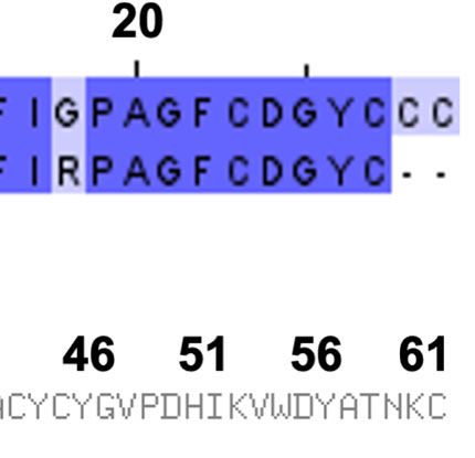
de Assis et al.                                                                                                 T. serrulatus Venom Against Toxoplasma

was collected and loaded onto a sephadex G-50 (Superfine)                     fragmentation). Briefly, a matrix (CHCA; 1:1, v/v) was used to
column, pre-equilibrated with the aforementioned buffer. Peaks               crystallize peptide samples on MTP AnchorChip 600/384 plates
(F1 – F7 fractions) were eluted at a flow rate of 0.5 mL/min based            (Bruker Daltonics, Billerica, MA, USA). Autoflex III was operated in
on protein detection at 280 nm, followed by lyophilization and               the positive reflector mode, using external calibration. The obtained
storage at -20°C until use.                                                  spectra were analyzed by using FlexAnalysis 3.1 (Bruker Daltonics,
                                                                             Billerica, MA, USA). The lyophilized peptides were diluted in
Subfractionation of F6 (Obtention of                                         phosphate-buffered saline solution (1× PBS), filtered, and
Sub-A and Sub-B)                                                             maintained at -20°C until use.
To obtain the subfractions 6A and 6B, the peak corresponding to
fraction F6 was subjected to HiTrap-SP HP cation exchange                    Macrophage Culture and
chromatography with a gradient of 0.01 M sodium acetate buffer               T. gondii Infection
(pH 4.7, solution A) and solution B (0.01 M sodium acetate buffer,           Mice were injected with 2 mL of sterile sodium thioglycolate
pH 4.7, 0.5 M NaCl) at flow rate 1mL/min, resulting in 17 peaks               solution at 3% v/v (DifcoTM® fluid), and after 72 h, MOs were
detected at 214 nm. Each fractionation peak was subjected to reverse         harvested from the peritoneal cavity by washing with sterile and
phase chromatography (Vydac C18 column, flow rate 1 mL/min) in                cold 1× PBS. The cells were centrifuged (290 × g, 10 min), counted,
a gradient system containing 0.1% v/v trifluoroacetic acid solution           plated (2 x10⁶ cells/well) onto culture plates (24 wells; TPP®), placed
in water (Solution A) and 0.1% v/v trifluoroacetic acid solution in           or not a round coverslip onto bottom of each of the wells, and
acetonitrile (Solution B) to obtain the homogeneous molecules,               maintained in an incubator at 37°C and 5% CO2 in the presence of
followed by MALDI-TOF mass spectrometry analysis. To further                 Roswell Park Memorial Institute 1640 culture medium (RPMI,
characterize the molecules (peptides), their partial sequence was            Gibco, Grand Island, NY, USA) supplemented (RPMIc) with 5%
elucidated by Edman degradation using a Shimadzu PPS-21A                     v/v fetal bovine serum (Cultilab, Campinas, SP, BR), 1% v/v L-
automated protein sequencer.                                                 glutamine (Gibco), and 1% v/v penicillin/streptomycin (Gibco) for
                                                                             3 h, followed by a wash, addition of RPMIc, and overnight
Toxin Identification and Peptide Design                                       incubation for subsequent infection/stimulation. The tachyzoite
The Sub6-B molecule was sequenced (de novo sequencing method)                forms of T. gondii (RH strain) were cultured in the kidney
and the following partial amino acid composition (~47%) was                  epithelial cell line LLCMK2 (Macaca mulatta– “Rhesus”) in the
found: (KEGYAMDHEGCCFSCFIGPAGF CDGYCCC…); to                                 presence of RPMIc. The parasites were centrifuged twice (50 × g for
identify it, a protein BLAST search was performed using the                  5 min, and then 2438 × g for 10 min), counted, and used to infect
default parameters against the non-redundant (nr) database                   the MOs (at a 1:1 parasite/host cell ratio), for 2 h, followed by a wash
(Altschul et al., 1990). The alignment was visualized using Jalview          (RPMIc). The uninfected and infected MOs were stimulated or not
2.11.0t (Waterhouse et al., 2009). First, a three-dimensional model          with TsV (100, 200 and 400µg/mL) or TsV fractions 6 and 7 (100 or
of the identified TsV alpha-mammal toxin Ts2 (Uniprot: P68410.1)              50 mg/mL, respectively) or Sub6-A, Sub6-B (100 and 50 mg/mL) or
was obtained via homology modeling using the software package                peptides (25, 50 and 100 mg/mL) for 4, 24 or 48 h. Intracellular
Modeller 9v16 (Webb and Sali, 2016). To build the model, the X-ray           tachyzoites counting was determined by light microscopy in
structure of Neurotoxin (Ts1) (PDB accession code: 1B7D) from                macrophages/coverslips fixed and stained with hematoxylin/eosin
TsV (Polikarpov et al., 1999) was used as a template. Modeller script        method (Panótico Rápido; LB Laborclin, Pinhais, PR, BR), and
salign was used for the alignment, QMEAN for model evaluation                mounted onto microscope slides (triplicates) using Entellan
(Benkert et al., 2008), and image rendering was performed using the          mounting medium (Merck, KGaA, Darmstadt, Germany). Total
Pymol package (DeLano, 2002). The bioinformatics tool PEPOP                  300 cells were counted for each coverslip discriminating between
(Demolombe et al., 2019) was used to design peptides representing            uninfected and infected cells. Moreover, at 24 and 48 h after
potential discontinuous epitopes from Ts2. The optimized nearest             stimulation, supernatants were collected for subsequent viability
neighbor path (“ONN”) method was used to build (extend) the                  assay and nitric oxide and cytokine quantification. For parasite pre-
peptide primary structure. Thus, two peptides carrying an acetylated         stimulation experiments, the tachyzoites were incubated in the
(CH3CO) N-terminus and C-terminal carboxamide (NH2) were                     presence of TsV or F6, both at 100 µg/mL, or RPMIc alone for
generally named “Pep1” and “Pep2a”; a third peptide sequence,                1 h, followed by washing with RPMIc, and used for MO infection
named “Pep2b”, was designed by removing the N-terminal acetyl                (as described above).
group of Pep2a.
                                                                             Cell Viability Assay
Peptide Synthesis                                                            Peritoneal macrophages were obtained as described above, and
The C-terminal amidated peptides were synthesized on a Rink                  plated (2 × 10⁶ cells/well into 24-well culture plate), and
amide resin using the Fmoc (9-fluorenylmethyloxycarbonyl)                     incubated for 3 h (37°C, 5% CO2). Then, the cells were washed
strategy as described elsewhere (Gomes et al., 2018). N-terminal             with RPMIc and incubated overnight. Subsequently, the cells
acetylation of Pep1 and Pep2a was promoted by the reaction of the            were first washed with RPMIc medium either treated with 2% v/v
respective peptidyl-resins with acetic anhydride before the peptide-         triton X100 (Sigma-Aldrich Corp., St. Louis, MO, USA) (death
resin cleavage. Peptide quality control was evaluated by mass                cell positive control), TsV (100 µg/mL), F6 (100 µg/mL), or F7
spectrometry (MALDI-ToF/ToF; Autoflex III smartbeam Bruker                    (50 µg/mL). At 48 h after incubation, cell viability was performed
Daltonics, Billerica, USA) and by both MS and MS/MS (peptide                 using the colorimetric method MTT (3-(4,5-Dimethyl-2-

Frontiers in Cellular and Infection Microbiology | www.frontiersin.org   3                                      July 2021 | Volume 11 | Article 706618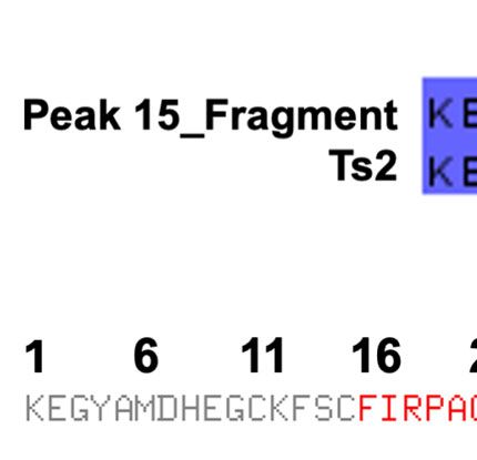
de Assis et al.                                                                                              T. serrulatus Venom Against Toxoplasma

thiazolyl)-2,5-diphenyl-2H-tetrazolium bromide) in adherent                  Next, C57Bl/6 female mice, 9–10 weeks old, were
cells, as previously described (Plumb, 2004). The reading was                intraperitoneally infected with 20 cysts/animal. Treatment with
performed using a spectrophotometer (Bio Tek- Elx800) at 490                 Pep1, Pep2a, or Pep2b (at a dose of 1 mg/kg, intraperitoneally)
nm. The percentage of viable cells was calculated compared to                started 8 h after infection and lasted until 7 dpi, with a 24/24 h
the average ratio of control cells (incubated with RPMIc only).              interval. The animals were monitored throughout the
                                                                             experiment (weight loss and survival) at 5, 7, and 30 dpi.
Measurement of Nitric Oxide                                                  Blood samples were collected for cytokine analysis in serum
and Cytokines                                                                (as described above). Additionally, the presence of liver enzymes
Macrophages was plated, infected and/or stimulated with TsV, F6              (glutamic oxaloacetic transaminase [GOT] and glutamic pyruvic
or F7 as described above. The supernatants were harvested at 24 and          transaminase [GPT]) was quantified (Bioclin® kinetic assay, Belo
48 h after infection and/or stimulation and assayed for nitrite              Horizonte, MG/BR). At 30 dpi, the animals were euthanized and
concentration using the Griess colorimetric method, as previously            the brains cysts were quantified as described above.
described (Green et al., 1982). Briefly, 50 mL of culture supernatant
was mixed with 50 mL of Griess reagent. Absorbance at 540 nm was             Statistical Analysis
read using a spectrophotometer (Elx800 – Bio Tek) 10 min later.              Statistical analyses were performed using Prism v. 8.2.1
The NO−2 concentration was determined by reference to a standard             (GraphPad software, San Diego, CA, USA) by comparing
curve of 500 mM (initial concentration) undergoing serial dilutions          the means ± SEM from controls to the treated samples using
(factor 2). Additionally, the cytokines (IL-12p70, TNF, and IL-6)            the Student’s t-test and one- or two-way ANOVA with the
were assayed in the supernatants of the macrophage cultures at 24            Bonferroni test. Differences were considered significant when
and 48 h after infection and/or stimulus with TsV, F6, or F7 by              p ≤ 0.05.
enzyme-linked immunosorbent assay (ELISA) method, according
to the manufacturer’s instructions (kit, R&D Systems Inc.,
Minneapolis, Minnesota, MN, USA). Briefly, 100 µL of                          RESULTS
monoclonal antibody specific for a particular cytokine (IL-12p70,
TNF or IL-6) was pre-coated onto a 96-well microplate and                    TsV Induces the Production of
incubated overnight at room temperature. Then, the plate was                 Inflammatory Mediators by Macrophages,
washed (wash buffer) and blocked by adding 300 µl/well of reagent            Reducing T. gondii Replication
diluent, followed incubation at room temperature for 1h. After               First, was evaluated the effect of TsV on nitric oxide production,
washing, 100 µL of standards and samples (culture supernatant)               an important antiparasite mediator. As demonstrated in
were added to the wells and incubated for 2h at room temperature.            Figure 1A, cells incubated with TsV at different concentrations
After washing the unbound substances, the detection antibody                 (100, 200, or 400 µg/mL) were stimulated to produce significant
(specific for the cytokine of interest; 100 µL) was added to the              levels of NO. Notably, T. gondii-infected MOs stimulated with
wells and incubated for 2h at room temperature. Following a wash,            TsV inhibited the production of NO (Figure 1A). However, at
100 µL of streptavidin-HRP solution was added and the plate was              24 h, NO production remained significantly higher in the
incubated for 20 min at room temperature. Then, the plate was                stimulated than the non-stimulated infected cells. Since there
washed and the substrate solution (100 µL) was added to the wells,           was no significant difference in the induction of NO levels among
incubated for 20 min at room temperature, and 50 µL of stop                  the concentrations tested, the 100 µg/mL concentration was
solution was added to the wells, and the color development was               chosen for the subsequent tests. Cell viability assay (MTT)
measured as optical density at 450 nm using a spectrophotometer              demonstrated that TsV at 100 µg/mL was not toxic to MOs
(BioTek - Elx800). Moreover, to determine serum levels of IL-12p70           48 h after incubation compared with the unstimulated cells
and IFN-g, 5, 7 and 30 days after infection (dpi) and/or treatment           (Figure 1B). Moreover, TsV also stimulated the production of
with Pep1, Pep2a, or Pep2b, blood was collected from mice and                IL-12p70, TNF, and IL-6 (the critical cytokines for an efficient
allowed to clot at room temperature, and the sera were separated             immune response against T. gondii), at 24 and 48 h after
and stored at -20 °C. The sera were brought to ambient temperature           stimulation (Figure 1C). As demonstrated in Figure 1C,
and cytokines were measured by ELISA (kit, R&D Systems Inc.) as              T. gondii infection did not induce significant production levels
described above.                                                             of these pro-inflammatory cytokines by MOs, which is described
                                                                             as an immune suppression mechanism induced by the parasite to
Treatment of T. gondii-Infected Mice With                                    evade the host response. Besides, the infection resulted in
Ts2-Derived Peptides                                                         decreased production of IL-12 and TNF, but not IL-6, at 24 h
For in vivo experiments, infective T. gondii cysts (ME49 strain)             and 48 h. However, the TsV stimulus induced the production of
were obtained from the brains of chronically infected mice (35 –             higher levels of IL-12 and TNF compared with infected-
45 dpi). Briefly, C57Bl/6 mice were euthanized, their brains were             unstimulated cells (Figure 1C). TsV anti-protozoal activity by
removed and washed with sterile 1× PBS, macerated, and                       the infected MOs was assessed within 24 h after infection and/or
homogenized with a needle and syringe in sterile 1× PBS (1                   stimulation. Figure 1D shows that the TsV-stimulated cells
mL). This suspension (10 µL) was placed on a glass microscope                presented a reduced number of intracellular tachyzoites
slide and the number of cysts was determined microscopically.                compared with the unstimulated infected cells. Additionally,

Frontiers in Cellular and Infection Microbiology | www.frontiersin.org   4                                    July 2021 | Volume 11 | Article 706618
de Assis et al. T. serrulatus Venom Against Toxoplasma
A B
C
D E F
FIGURE 1 | TsV induces the production of inflammatory mediators by macrophages, reducing T. gondii replication. Peritoneal macrophages from C57Bl/6 female
mice were cultured and infected (1:1 T. gondii: cell ratio) and/or stimulated with TsV (100, 200, or 400 µg/mL). After 24 and 48 h, the culture supernatants were
collected for nitric oxide (A) and IL-12p70, TNF, and IL-6 cytokine (C) measurements. Viability assay was performed on the adhering cells at 48 h after TsV (100 µg/
mL) stimulation (B). Intracellular parasite counts were performed on fixed and stained cells at 24 h (D) or 4 h (E) post-infection/stimulus. Macrophages were infected
with TsV (100 µg/mL)-pre-incubated tachyzoites forms (“PI”) and 48 h after infection, the cells were fixed and stained for intracellular parasite quantification (F). “CT”=
control (non-stimulated cells). “Tg”= T. gondii-infected macrophage. Each point represents means ± SEM of one of two independent experiments, *p ≤ 0.05; ***p <
0.001; ****p < 0.0001.
the venom did not change the invasion of host cells by parasites and 48 h) and IL-6 (24 h) compared to the uninfected F6-stimulated
(Figure 1E). To examine the direct effect of TsV on the parasite, cells. However, the levels of IL-12 (24 h), TNF (24 and 48 h), and IL-
MOs were infected with tachyzoites that were previously 6 (24 and 48h) remained higher than that of the unstimulated
incubated with TsV 100 µg/mL. As demonstrated in infected cells (Figure 2E). Additionally, T. gondii decreased the
Figure 1F, incubation of the parasite with TsV prior to levels of IL-12 (48h), but not TNF or IL-6 production triggered by
infection did not affect its replication capacity. F7. Notably, F6 was a better inducer of cytokine production than F7.
In summary, our results indicate that F6 and F7, mainly F6, retain
The F6 Fraction From TsV Triggers the molecules that might participate in the control of T. gondii
the Production of Pro-Inflammatory replication, inducing a pro-inflammatory profile in MOs. Thus, the
Mediators and Induces Macrophage F6 fraction was fractionated to isolate and characterize the
Anti-Protozoal Activity molecules with possible anti-T. gondii activity.
Recently, our group isolated and characterized the key protein
fractions present in TsV that were involved in the induction of Subfraction Sub6-B From F6 Induces
NO in MOs (Pimentel et al), here named F1 – F7, according to the Potent Control of T. gondii Replication
order of elution from the column (Figure 2A), and only F6 and F7 by Macrophages
induced significant production of NO (Pimentel et al., 2021). The potential of F6 anti-T. gondii activity in MOs was confirmed
Accordingly, F6 at 100 µg/mL and F7 at 50 µg/mL induced by the reduced number of parasites in the stimulated cells
significant production of NO by MOs (Figure 2B). F6 at 100 µg/ compared to the unstimulated cells (Figure 3A). Notably in
mL (Figure 2C) and F7 at 50 µg/mL (Figure 2D) were not toxic to contrast to TsV (Figure 1E), when F6 was simultaneously
MOs and both induced the production of IL-12, TNF, and IL-6 incubated with the parasites and macrophages, increased
(Figures 2E, F). Notably, T. gondii-infected MOs did not produce parasite invasion was observed (Figure 3B). Similar to TsV
significant levels of the analyzed cytokines (Figures 2E, F). T. gondii (Figure 1F), F6 pre-incubated parasites did not alter their
infection resulted in decreased production of IL-12 and TNF (24 replication capacity (Figure 3C). These data confirmed the
Frontiers in Cellular and Infection Microbiology | www.frontiersin.org 5 July 2021 | Volume 11 | Article 706618de Assis et al. T. serrulatus Venom Against Toxoplasma
A
B C D
E
F
FIGURE 2 | F6 fraction from TsV triggers the production of pro-inflammatory mediators and induces macrophage anti-protozoal activity. TsV was separated by gel
filtration chromatography in which the components were eluted at a flow rate of 0.5 mL/min and protein detection was performed at 280 nm. The fractions obtained
were listed from 1–7 (A) Peritoneal macrophages were cultured and stimulated with F6 or F7 fractions (100 or 50 µg/mL, respectively), and at 48 h post-stimulation
the culture supernatants were collected for nitric oxide measurement (B). Viability assay (MTT) was performed on the adhering cells at 48 h after F6 (100 µg/mL) (C)
or F7 (50 µg/mL) stimulation (D). The cells were infected (1:1 parasite: cell ration) and/or stimulated with F6 (100 µg/mL) or (F7 50 µg/mL) and the culture
supernatants were collected at 24 and 48 h post-stimulus; (E) F6 and (F) F7, for cytokine measurements (IL-12p70, TNF, and IL-6). Each point represents means ±
SEM of one of the two independent experiments, *p ≤ 0.05; **p < 0.01; ***p < 0.001; ****p < 0.0001.
promising immunomodulatory ability of the components present 17) (Figure 3E). Because of the small amounts obtained, only two
in F6, leading to T. gondii replication control. Hence, the next step compounds, named Sub6-A and Sub6-B (related to peaks 12 and
was to further fractionate F6 to obtain purified toxins. For this, F6 15, respectively), were subjected to the antiparasitic activity assay
was subjected to HiTrap-SP HP cation exchange chromatography in MOs. To test the antiprotozoal activity of Sub6-A and Sub6-B,
(flow 1 mL/min, absorbance at 214 nm) with a gradient of 0.01 M infected MOs were stimulated with these compounds at 100 or 50
sodium acetated buffer (% B), resulting in 17 new chromatography µg/mL. As demonstrated in Figure 3F, Sub6-B at 100 µg/mL was
peaks that were listed according to the order of elution (peaks 1– able to significantly control tachyzoite replication compared to the
17) (Figure 3D). Subsequently, peak 13, with the best activity, was unstimulated and groups stimulated with the other subfractions.
subjected to reversed-phase chromatography (Vydac C18 column,
flow 1 mL/min, absorbance at 214 nm) in a gradient system Characterization of Sub6-B Subfraction
containing 0.1% v/v trifluoroacetic acid solution in water and 0.1% and Mimetic Peptide Production
v/v trifluoroacetic acid solution in acetonitrile to isolate pure To characterize the molecule/peptide present in Sub6-B, peak 15
molecules, followed by MALDI-TOF mass spectrometry and of this subfraction was sequenced and its partial amino acid
Edman degradation analyses, from which 17 molecules were sequence was elucidated: KEGYAMDHEGCCFSCFIGPAG
isolated and listed according to the order of elution (peaks 1– FCDGYCCC(…). This amino acid sequence was searched
Frontiers in Cellular and Infection Microbiology | www.frontiersin.org 6 July 2021 | Volume 11 | Article 706618de Assis et al. T. serrulatus Venom Against Toxoplasma
A B C
D E F
FIGURE 3 | Subfraction Sub6-B from F6 induces potent control of T. gondii replication by macrophages. Peritoneal macrophages were cultured and infected (1:1
parasite: cell ration) and/or stimulated by F6 (100 µg/mL). Intracellular parasite counts were performed on fixed and stained cells at 48 h (A) or 4 h (B) post-infection/
stimulus. Macrophages were infected with F6 (100 µg/mL)-pre-incubated tachyzoite forms (“PI”) and the intracellular parasites were quantified 48 h after infection (C).
Each point represents means ± SEM of one of the two independent experiments,*p ≤ 0.05. For purification of molecules, fraction F6 was subjected to cation
exchange chromatography in which the components were eluted at a flow rate of 1 mL/min and peaks detected at 214 nm (D), and the resulting peak 13 was
subjected to reversed-phase chromatography with a flow rate of 1 mL/min and detection at 214 nm, followed by MALDI-TOF mass spectrometry and Edman
degradation analyses, resulting in subfractions listed from 1–17 (E). The infected-macrophages were stimulated with homogeneous molecules (toxins): Sub6-A (peak
12) or Sub6-B (peak 15) (100 or 50 µg/mL). Intracellular parasite growth was analyzed on fixed and stained cells at 48 h post-infection/stimulus (F). Each point
represents means ± SEM of one experiment, *p ≤ 0.05.
against the NCBI nr protein databases using BLASTp. The concentrations (Figure 5A) and Pep2a at 50 µg/mL (Figure 5B)
highest BLASTp hit was Ts2 toxin (found in TsV – UniProt reduced the intracellular replication of tachyzoites compared with
ID: P68410 [SCX2_TITSE]), with 93% similarity (of note, 27 out the infected non-stimulated cells. In contrast, stimulation with
of 29 amino acids of the fragment aligned with Ts2 toxin) Pep2b at 50 or 100 µg/mL resulted in increased parasite
(Figure 4A). As the identified sequence is composed of several replication compared to the unstimulated infected cells
amino acids, including cysteine, which is prone to disulfide bond (Figure 5C). The antiparasitic activity of the peptides was also
formation, the synthesis of the entire segment might be rather evaluated in vivo by T. gondii-infected mice. Our results showed that
challenging and costly. Therefore, we used the PEPOP tool to the treatment of mice with Pep1 reversed from the 14th dpi, the
design two discontinuous peptides mimicking the Ts2 toxin. The weight loss caused by the infection. In contrast, untreated-infected
regions from Ts2 used to generate the peptides are highlighted in animals lost weight from the 5th–30th dpi (end of the study)
red and green (Figure 4B): Pep1: (CH3CO- DAYKTHLKSS- (Figure 5D). To a lesser extent, peptides 2a and 2b also reduced
NH2) and Pep2a (CH3CO-FIRPAGFKYSWP-NH2), whereas an the weight loss caused by T. gondii infection (Figure 5D). The
extra third compound Pep2b (FIRPAGFKYSWP-NH2) was also ability of Pep1 and Pep2a to reduce weight loss correlated with a
proposed by the N-terminal deacetylation of Pep2a. These three higher survival rate in the group of animals treated with these
peptides were obtained using chemical synthesis, purified by peptides (Figure 5E). Despite some differences in weight loss and
reversed-phase HPLC, and their purity was determined using survival, curiously, all peptides tested were able to reduce the
mass spectrometry. Figure 4C depicts representative MS spectra number of brain cysts in infected mice at 30th dpi (Figure 5F),
of the synthetized peptides. Observed molecular masses (MS) emphasizing the best ability of Pep1 to improve all the analyzed
and amino acid sequences obtained by peptide fragmentation parameters. In addition, our results suggest that the protection
(MS/MS) are in accordance with the expected theoretical values. (reduction in the number of cysts and/or higher survival rate)
offered by Pep1, Pep2a, or Pep2b treatment was independent of the
Pep1 and Pep2a, but Not Pep2b, Induce systemic production of cytokines IL-12 (Figure 5G) or IFN-g
Potent Anti-Protozoal Activity In Vitro and (Figure 5H), since all treated groups presented similar levels of
Improved the Control of T. gondii Infection these cytokines correlating to the untreated-infected group at all
in Mice analyzed time points. Importantly, the analysis of liver enzymes
To determine whether the synthetic peptides could control parasite present in the serum (GOT; Figure 5I and GPT; Figure 5J) at 30th
replication, MOs were infected and stimulated with Pep1, Pep2a, or dpi revealed that the treatment of animals with these peptides did
Pep2b at concentrations of 100, 50, or 25 µg/mL. Pep1 at all not induce late hepatotoxicity.
Frontiers in Cellular and Infection Microbiology | www.frontiersin.org 7 July 2021 | Volume 11 | Article 706618de Assis et al. T. serrulatus Venom Against Toxoplasma
A
B
C
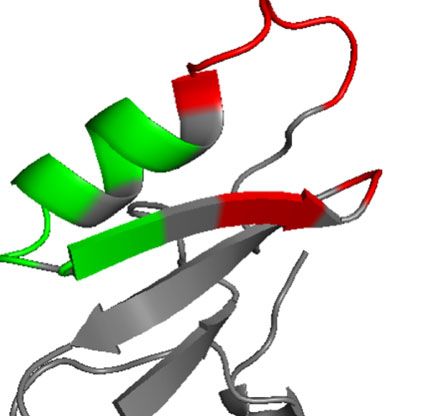
FIGURE 4 | Characterization of Sub6-B subfraction and mimetic peptide production. In order to characterize the molecule (peptide) present in Sub6-B, it was
sequenced by Edman degradation, elucidating its partial amino acid sequence: KEGYAMDHEGCCFSCFIGPAGFCDGYCCC…. The next step was to compare this
sequence with other sequences available on the online public data bank (BLAST). Sub6-B (Peak_15) sequence had a high (93%) identity with T. serrulatus Ts2 toxin
(showing conserved amino acids in dark blue and different amino acids in light blue, of which, 27 out of 29 aa of the fragment aligned with Ts2 toxin) (A). Primary
and tertiary structures of conformation peptides were designed based on the Ts2 structure. Peptide 1 is highlighted in red and Peptide 2 in green (B). The purity of
Pep1, Pep2a, and Pep2b was analyzed using MALDI-TOF-TOF mass spectrometry (C).
DISCUSSION studies using cell lines (Zoccal et al., 2011) and murine
macrophage (Pimentel et al., 2021). Moreover, despite the well-
Current therapies for treating toxoplasmosis are not yet effective known ability of T. gondii to suppress or evade host immunity,
in eradicating the parasite from the host and present toxic side TsV significantly increased NO production by controlling T.
effects. The ability of TsV to activate immune cells (Petricevich gondii replication.
et al., 2008; Zoccal et al., 2011; Casella-Martins et al., 2015; TsV was described to stimulate MOs through a complex
Pimentel et al., 2021) allowed us to hypothesize that its interaction involving membrane receptors (e.g., Toll-like
compounds could act as anti-toxoplasma agents. Herein, MOs receptor 2/4) (Zoccal et al., 2014), ion channels (e.g., Na+, Ca+,
were highlighted owing to their anti-protozoal capacity during and K+) (Lowry et al., 1998; Oliveira et al., 2019), and Mitogen-
parasite infection (Stafford et al., 2002; Pimentel et al., 2021). Activated Protein Kinases (MAPK) (Pimentel et al., 2021) which
We demonstrated that crude TsV and its F6 and F7 fractions culminate in the production of inflammatory mediators.
induced NO production by MOs without causing host cell Cytokines play a key role in amplifying the response/
toxicity. It was of great relevance since NO is a potent communication among cell types, building an efficient
microbicidal/microbiostatic agent controlling T. gondii response against pathogens. Herein, TsV, F6, and F7 fractions
replication (Seabra et al., 2002) and corroborated previous were potent inducers of IL-12p70/TNF/IL-6 in MOs, which is
Frontiers in Cellular and Infection Microbiology | www.frontiersin.org 8 July 2021 | Volume 11 | Article 706618de Assis et al. T. serrulatus Venom Against Toxoplasma
A B C
D E
F G H
I J
FIGURE 5 | Evaluation of the anti-T. gondii activity of Pep1, Pep2a, and Pep2b peptides in vitro and in vivo. Peritoneal macrophages from C57Bl/6 female mice
were cultured and infected (1:1 T. gondii: cell ratio) and/or stimulated by Pep1 (A), Pep2a (B), or Pep2b (C) at 25, 50, or 100 µg/mL. Intracellular parasite counts
were performed on fixed and stained cells at 48 h post-infection/stimulus (Each point represents means ± SEM of one of the two independent experiments, *p ≤
0.05; **p < 0.01). C57Bl/6 mice (n= 6/group) were infected with 20 cysts of T. gondii (ME49 strain) and treated or not with Pep1, Pep2a, or Pep2b (1 mg/kg) at 8 h
after infection until the 7th day post infection (dpi). The weight loss (D) and survival (E) were monitored from the 5th–30th dpi. The cytokines IL-12p70 (G) and IFN-g
(H) were quantified in the serum of the animals on 5th, 7th, and 30th dpi. The mice were euthanized on 30th dpi for the brain cyst count (F) and measurement of liver
enzymes in the serum [sGOT (I) and sGPT (J)]. “CT” = control (non-infected mice). “Tg + Veic”= infected-mice vehicle-injected➔ *p ≤ 0.05; **p< 0.01; ***p < 0.001;
****p < 0.0001 (CT x Tg + Veic)! +p ≤ 0.05; +++p < 0.001; ++++p < 0.0001 (CT x Tg + Pep1)! #p ≤ 0.05; ##p < 0.01 (CT x Tg + Pep2a)! §p ≤ 0.05; §§§p < 0.001
(CT x Tg + Pep2b). Each point represents means ± SEM of two independent experiments.
consistent with previous studies demonstrating that TsV- inflammatory response. Reinforcing this hypothesis, pre-
stimulated MOs secrete IL-1/IL-6/TNF/IFN-g (Petricevich, stimulation of the tachyzoites with TsV or F6 before infection
2002; Oliveira et al., 2019; Pimentel et al., 2021). Of great did not alter the replicative capacity of the parasite, excluding a
relevance, we demonstrated that TsV compounds induced the direct action on the parasite. Unlike TsV, the F6 fraction was able
production of IL-12 and TNF by T. gondii-infected MOs, to stimulate tachyzoite invasion in MOs. We speculate that this
abolishing the capacity of the parasite to evade host immunity. was due to the modifications on the membrane of the host cell,
The ability of this parasite may be higher or lower owing to the T. facilitating the parasite invasion and/or by activation of the host
gondii strain and protein secretion from specialized organelles receptors that sense parasite factors, favoring parasite
such as micronemes, rhoptries, and dense granules (Melo et al., internalization (Bonhomme et al., 1999; Sweeney et al., 2010).
2011; Hunter and Sibley, 2012). Because of the sustained ability of Sub6-B to control both
Hitherto, our data led us to hypothesize that TsV and F6 were parasite replication and immune response, the partial amino acid
able to reduce tachyzoite replication in MOs, activating a pro- sequence that makes up this toxin was identified and compared
Frontiers in Cellular and Infection Microbiology | www.frontiersin.org 9 July 2021 | Volume 11 | Article 706618de Assis et al. T. serrulatus Venom Against Toxoplasma
using BLASTp. The Sub6-B partial sequence has 93% similarity CONCLUSION
with the Ts2 toxin sequence, suggesting that this molecule could
be the Ts2 toxin or has high homology to it. Ts2 has been Collectively, our results reveal for the first time that molecules
described as having 72% identity with Ts1 (Mansuelle et al., from T. serrulatus venom could have promising potential as
1992), being able to prolong the duration of the action potential therapeutic agents in toxoplasmosis. Of great relevance, we
of rabbit vagus-nerve, as an a-toxin that inhibits the inactivation demonstrate that synthetic peptides treatment in vivo reduces
of some Na+ channels (e.g., NaV1.2/NaV1.3/NaV1.5/NaV1.6) the number of brain cysts number without inducing
(Cologna et al., 2012), and presents immunomodulatory actions hepatotoxicity, indicating a highly required characteristic
(Zoccal et al., 2011). Ts2 was demonstrated to induce pro- and for molecules to be used in the treatment of toxoplasmosis:
anti-inflammatory profiles in vivo (Zoccal et al., 2013). Ts2- acting at the focal point of the infection, mainly in the central
intraperitoneal injection-induced recruitment of leukocytes (e.g., nervous system.
neutrophils, mononuclear cells, CD4, and CD8 lymphocytes)
associated with increased production of pro-inflammatory (IL-6/
TNF/IFN-g/IL-1b/leukotriene-B4/prostaglandin-E2) and anti- DATA AVAILABILITY STATEMENT
inflammatory/pro-resolving (IL-10/IL-4) mediators. Thus, our
results suggest that Sub6-B increased anti-T. gondii ability, The raw data supporting the conclusions of this article will be
inducing the production of inflammatory molecules. made available by the authors, without undue reservation.
To optimize peptide production using chemical synthesis,
two Ts2 toxin mimetic peptides were designed using the PEPOP
program. After in silico analysis, we selected and combined some
amino acid sequences from the Ts2 toxin that gave rise to two ETHICS STATEMENT
peptides, namely Pep1 and Pep2a as well as the derivative Pep2a The animal study was reviewed and approved by The UFMG
sequence, Pep2b, which carries no acetylation at its N-terminus. Animal Ethics Committee (CEUA) approved all experiments
These peptides are approximately five times smaller than the and procedures (Permit Number 449/2015).
original sequence and are easily obtained by solid-phase
synthesis. The predicted activity of peptides in silico was
checked using both in vitro and in vivo models of T. gondii
infection. Pep1 and Pep2a stimulation reduced parasite AUTHOR CONTRIBUTIONS
replication in macrophages, whereas Pep2b improved it. By
adding an acetyl group at the N-terminal amino acid of a DA, ML, AP, MB, and FM conceived experiments. DA, MT, ML,
peptide, it is possible to change some of its physico-chemical AP, RV, JR, LF, CO, and FM wrote the manuscript. DA, PP, PR,
properties such as hydrophobicity and charge distribution (by RR, LF, JR, MB, and MC performed experiments. LF, CO, RV,
quenching the N-terminus positive charge). As a result, some MB, ML, AP, MT, ML, AP, and FM supervised and provided
structural or biochemical features of the original peptide might expertise and funding. All authors contributed to the article and
be affected, including (but not restricted to) folding, hydrolysis approved the submitted version.
prevention, protein interactions, and cell permeation or
subcellular location (reviewed by Ree et al., 2018). Although
any of the above-mentioned explanations (or even a sum of FUNDING
them), could explain the different activities between Pep2a and
Pep2b, further studies are needed to better understand this This work was supported by Pró -Reitoria de Pesquisa/UFMG,
interesting finding. In vivo, although only Pep1 improved the Conselho Nacional de Desenvolvimento Cientı́fico e Tecnoló gico
tested clinical parameters of the disease, the treatment with all (CNPq: 305894/2018-8; 438054/2018-0) and Fundação de Amparo
peptides showed decreases in cerebral parasitism and did not a Pesquisa de Minas Gerais (FAPEMIG: APQ-02331-18; Rede
induce chronic/late liver injury. Such discrepant effects of Pep2b Mineira de Imunobioló gicos, REDE-00140-16), Coordenação de
on parasite growth, in vitro and in vivo, may be explained by the Aperfeiçoamento de Pessoal de Nı́vel Superior (CAPES: CAPES/
fact that only MOs were stimulated in vitro, and in the in vivo COFECUB 57914) (Brazil) and the National Institute for Science
system, the peptides may activate different cell types that act and Technology in Dengue and Host-microbial interactions
cooperatively to reduce parasite replication/dissemination. (465425/2014-3). DA, PP, PR, RR, RV, LF, CO, JR, MT, AP, ML,
Moreover, differences between T. gondii strains used in vitro and FM acknowledge grants from CNPq.
(RH strain) and in vivo (ME49 strain) should also be considered
(Hwang et al., 2018). Here, the anti-T. gondii therapeutic effect of
the peptides was independent of IL-12 and IFN-g systemic levels. ACKNOWLEDGMENTS
However, the participation of IL-12 and IFN-g cannot be totally
excluded, since the peptides may reach specific cells, inducing the The authors would like to thank Jacqueline Barbosa de
production of these cytokines in an organ-specific manner, such Oliveira Viana and Rosá lida Estevam Nazar Lopes for
as in the lymphoid and brain, thereby reducing parasite load. technical assistance.
Frontiers in Cellular and Infection Microbiology | www.frontiersin.org 10 July 2021 | Volume 11 | Article 706618de Assis et al. T. serrulatus Venom Against Toxoplasma
REFERENCES Translocation of STAT1a. Eur. J. Immunol 31, 1475–1484. doi: 10.1002/1521-
4141(200105)31:53.0.CO;2-C
Alday, P. H., and Doggett, J. S. (2017). Drugs in Development for Toxoplasmosis: Machado, F. S., Johndrow, J. E., Esper, L., Dias, A., Bafica, A., Serhan, C. N., et al.
Advances, Challenges, and Current Status. Drug Des. Devel. Ther 11, 273–293. (2006). Anti-Inflammatory Actions of Lipoxin A4 and Aspirin-Triggered
doi: 10.2147/DDDT.S60973 Lipoxin are SOCS-2 Dependent. Nat. Med. 12, 330–334. doi: 10.1038/nm1355
Altschul, S. F., Gish, W., Miller, W., Myers, E. W., and Lipman, D. J. (1990). Basic Mansuelle, P., Martin-Eauclaire, M., Chavez-Olortegui, C., de Lima, M. E., Rochat,
Local Alignment Search Tool. J. Mol. Biol. 215, 403–410. doi: 10.1016/S0022- H., and Granier, C. (1992). The b-Type Toxin Ts II From the Scorpion Tityus
2836(05)80360-2 Serrulatus: Amino Acid Sequence Determination and Assessment of Biological
Bannenberg, G. L., Aliberti, J., Hong, S., Sher, A., and Serhan, C. (2004). Exogenous and Antigenic Properties. Nat. Toxins 1, 119–125. doi: 10.1002/nt.2620010211
Pathogen and Plant 15-Lipoxygenase Initiate Endogenous Lipoxin A4 Melo, M. B., Jensen, K. D. C., and Saeij, J. P. J. (2011). Toxoplasma Gondii
Biosynthesis. J. Exp. Med. 199, 515–523. doi: 10.1084/jem.20031325 Effectors are Master Regulators of the Inflammatory Response. Trends
Benkert, P., Tosatto, S. C. E., and Schomburg, D. (2008). QMEAN: A Parasitol. 27, 487–495. doi: 10.1016/j.pt.2011.08.001
Comprehensive Scoring Function for Model Quality Assessment. Proteins Montazeri, M., Mehrzadi, S., Sharif, M., Sarvi, S., Tanzifi, A., Aghayan, S. A., et al.
Struct. Funct. Genet. 71, 261–277. doi: 10.1002/prot.21715 (2018). Drug Resistance in Toxoplasma Gondii. Front. Microbiol. 9:2587.
Bonhomme, A., Bouchot, A., Pezzella, N., Gomez, J., Le Moal, H., and Pinon, J. M. doi: 10.3389/fmicb.2018.02587
(1999). Signaling During the Invasion of Host Cells by Toxoplasma Gondii. Montoya, J. G., and Liesenfeld, O. (2004). Toxoplasmosis. in. Lancet 636, 1965–
FEMS Microbiol. Rev. 23, 551–561. doi: 10.1016/S0168-6445(99)00021-2 1976. doi: 10.1016/S0140-6736(04)16412-X
Casella-Martins, A., Ayres, L. R., Burin, S. M., Morais, F. R., Pereira, J. C., Faccioli, Oliveira, I. S., Ferreira, I. G., Alexandre-Silva, G. M., Cerni, F. A., Cremonez, C. M.,
L. H., et al. (2015). Immunomodulatory Activity of Tityus Serrulatus Scorpion Arantes, E. C., et al. (2019). Scorpion Toxins Targeting Kv1.3 Channels:
Venom on Human T Lymphocytes. J. Venom. Anim. Toxins Incl. Trop. Dis 21, Insights Into Immunosuppression. J. Venom. Anim. Toxins Incl. Trop. Dis
46. doi: 10.1186/s40409-015-0046-3 25, 148118. doi: 10.1590/1678-9199-jvatitd-1481-18
Cologna, C., Marcussi, S., Giglio, J., Soares, A., and Arantes, E. (2009). Tityus Ortiz, E., Gurrola, G. B., Schwartz, E. F., and Possani, L. D. (2015). Scorpion
Serrulatus Scorpion Venom and Toxins: An Overview. Protein Pept. Lett. 16, Venom Components as Potential Candidates for Drug Development. Toxicon
920–932. doi: 10.2174/092986609788923329 93, 125–135. doi: 10.1016/j.toxicon.2014.11.233
Cologna, C. T., Peigneur, S., Rustiguel, J. K., Nonato, M. C., Tytgat, J., and Arantes, Petricevich, V. L. (2002). Effect of Tityus Serrulatus Venom on Cytokine
E. C. (2012). Investigation of the Relationship Between the Structure and Production and the Activity of Murine Macrophages. Mediators Inflamm.
Function of Ts2, a Neurotoxin From Tityus Serrulatus Venom. FEBS J. 279, 11, 23–31. doi: 10.1080/09629350210308
1495–1504. doi: 10.1111/j.1742-4658.2012.08545.x Petricevich, V. L., Reynaud, E., Cruz, A. H., and Possani, L. D. (2008). Macrophage
DeLano, W. L. (2002). Pymol: An Open-Source Molecular Graphics Tool. CCP4 Activation, Phagocytosis and Intracellular Calcium Oscillations Induced by
Newsl. Protein Crystallogr. 40, 82–92. Scorpion Toxins From Tityus Serrulatus. Clin. Exp. Immunol 154, 415–423.
Demolombe, V., De Brevern, A. G., Felicori, L., Nguyen, C., Machado De Avila, doi: 10.1111/j.1365-2249.2008.03754.x
R. A., Valera, L., et al. (2019). PEPOP 2.0: New Approaches to Mimic non- Pimentel, P. M., de, O., de Assis, D. R. R., Gualdró n-Lopez, M., Barroso, A., Brant,
Continuous Epitopes. BMC Bioinf. 20, 387. doi: 10.1186/s12859-019-2867-5 F., et al. (2021). Tityus Serrulatus Scorpion Venom as a Potential Drug Source
Denkers, E. Y., and Gazzinelli, R. T. (1998). Regulation and Function of T-Cell- for Chagas’ Disease: Trypanocidal and Immunomodulatory Activity. Clin.
Mediated Immunity During Toxoplasma Gondii Infection. Clin. Microbiol. Immunol. 226:108713. doi: 10.1016/j.clim.2021.108713
Rev. 11, 568–588. doi: 10.1128/cmr.11.4.569 Plumb, J. A. (2004). Cell Sensitivity Assays: The MTT Assay. Methods Mol. Med.
Gomes, K. A. G. G., dos Santos, D. M., Santos, V. M., Piló -Veloso, D., Mundim, 88, 165–169. doi: 10.1385/1-59259-687-8:25
H. M., Rodrigues, L. V., et al. (2018). NMR Structures in Different Membrane Polikarpov, I., Junior, M. S. M., Marangoni, S., Toyama, M. H., and Teplyakov, A.
Environments of Three Ocellatin Peptides Isolated From Leptodactylus (1999). Crystal Structure of Neurotoxin TS1 From Tityus Serrulatus Provides
Labyrinthicus. Peptides 103, 72–83. doi: 10.1016/j.peptides.2018.03.016 Insights Into the Specificity and Toxicity of Scorpion Toxins. J. Mol. Biol. 290,
Green, L. C., Wagner, D. A., Glogowski, J., Skipper, P. L., Wishnok, J. S., and 175–184. doi: 10.1006/jmbi.1999.2868
Tannenbaum, S. R. (1982). Analysis of Nitrate, Nitrite, and [15N]Nitrate in Ree, R., Varland, S., and Amesen, T. (2018). Spotlight on Protein N-Terminal
Biological Fluids. Anal. Biochem. 126, 131–138. doi: 10.1016/0003-2697(82) Acetylation. Exp. Mol. Med 50, 1–13. doi: 10.1038/s12276-018-0116-z
90118-X Sasai, M., Pradipta, A., and Yamamoto, M. (2018). Host Immune Responses to
Herzig, V., Cristofori-Armstrong, B., Israel, M. R., Nixon, S. A., Vetter, I., and Toxoplasma Gondii. Int. Immunol 10, 113–119. doi: 10.1093/intimm/dxy004
King, G. F. (2020). Animal Toxins — Nature’s Evolutionary-Refined Toolkit Seabra, S. H., De Souza, W., and DaMatta, R. A. (2002). Toxoplasma Gondii
for Basic Research and Drug Discovery. Biochem. Pharmacol. 181, 114096. Partially Inhibits Nitric Oxide Production of Activated Murine Macrophages.
doi: 10.1016/j.bcp.2020.114096 Exp. Parasitol. 100, 62–70. doi: 10.1006/expr.2001.4675
Hunter, C. A., and Sibley, L. D. (2012). Modulation of Innate Immunity by Stafford, J. L., Neumann, N. F., and Belosevic, M. (2002). Macrophage-Mediated
Toxoplasma Gondii Virulence Effectors. Nat. Rev. Microbiol. 10, 766–778. Innate Host Defense Against Protozoan Parasites. Crit. Rev. Microbiol. 28,
doi: 10.1038/nrmicro2858 187–248. doi: 10.1080/1040-840291046731
Hwang, Y. S., Shin, J. H., Yang, J. P., Jung, B. K., Lee, S. H., and Shin, E. H. (2018). Sweeney, K. R., Morrissette, N. S., Lachapelle, S., and Blader, I. J. (2010). Host Cell
Characteristics of Infection Immunity Regulated by Toxoplasma Gondii to Invasion by Toxoplasma Gondii is Temporally Regulated by the Host
Maintain Chronic Infection in the Brain. Front. Immunol. 9, 158. doi: 10.3389/ Microtubule Cytoskeleton. Eukaryot. Cell 9, 1680–1689. doi: 10.1128/
fimmu.2018.00158 EC.00079-10
Laliberté , J., and Carruthers, V. B. (2008). Host Cell Manipulation by the Human van Kasteren, S. I., Neefjes, J., and Ovaa, H. (2018). Creating Molecules That
Pathogen Toxoplasma Gondii. Cell. Mol. Life Sci 65, 1900–1915. doi: 10.1007/ Modulate Immune Responses. Nat. Rev. Chem. 2, 184–193. doi: 10.1038/
s00018-008-7556-x s41570-018-0023-9
Lichota, A., and Gwozdzinski, K. (2018). Anticancer Activity of Natural Waterhouse, A. M., Procter, J. B., Martin, D. M. A., Clamp, M., and Barton, G. J.
Compounds From Plant and Marine Environment. Int. J. Mol. Sci 9, 3533. (2009). Jalview Version 2-A Multiple Sequence Alignment Editor and Analysis
doi: 10.3390/ijms19113533 Workbench. Bioinformatics 25, 1189–1191. doi: 10.1093/bioinformatics/
Lowry, M. A. R., Goldberg, J. I., and Belosevic, M. (1998). Induction of Nitric btp033
Oxide (NO) Synthesis in Murine Macrophages Requires Potassium Channel Webb, B., and Sali, A. (2016). Comparative Protein Structure Modeling Using
Activity. Clin. Exp. Immunol. 111, 597–603. doi: 10.1046/j.1365- MODELLER. Curr. Protoc. Bioinf. 54, 5.6.1–5.6.37. doi: 10.1002/cpbi.3
2249.1998.00536.x Weiss, L. M., and Dubey, J. P. (2009). Toxoplasmosis: A History of Clinical
Lüder, C. G. K., Walter, W., Beuerle, B., Maeurer, M. J., and Gross, U. (2001). Observations. Int. J. Parasitol 39, 895–901. doi: 10.1016/j.ijpara.2009.02.004
Toxoplasma Gondii Down-Regulates MHC Class II Gene Expression and Zoccal, K. F., Bitencourt, C., da, S., Secatto, A., Sorgi, C. A., Bordon, K., et al.
Antigen Presentation by Murine Macrophages via Interference With Nuclear (2011). Tityus Serrulatus Venom and Toxins Ts1, Ts2 and Ts6 Induce
Frontiers in Cellular and Infection Microbiology | www.frontiersin.org 11 July 2021 | Volume 11 | Article 706618de Assis et al. T. serrulatus Venom Against Toxoplasma Macrophage Activation and Production of Immune Mediators. Toxicon. 57, Conflict of Interest: The authors declare that the research was conducted in the 1101–1108. doi: 10.1016/j.toxicon.2011.04.017 absence of any commercial or financial relationships that could be construed as a Zoccal, K. F., Bitencourt, C., da, S., Sorgi, C. A., Bordon, K., de, C. F., et al. (2013). potential conflict of interest. Ts6 and Ts2 From Tityus Serrulatus Venom Induce Inflammation by Mechanisms Dependent on Lipid Mediators and Cytokine Production. Copyright © 2021 de Assis, Pimentel, dos Reis, Rabelo, Vitor, Cordeiro, Felicori, Toxicon. 61, 1–10. doi: 10.1016/j.toxicon.2012.10.002 Oloŕ tegui, Resende, Teixeira, Borges, de Lima, Pimenta and Machado. This is an open- Zoccal, K. F., Bitencourt, C. D. S., Paula-Silva, F. W. G., Sorgi, C. A., De Castro access article distributed under the terms of the Creative Commons Attribution License Figueiredo Bordon, K., Arantes, E. C., et al. (2014). TLR2, TLR4 and CD14 (CC BY). The use, distribution or reproduction in other forums is permitted, provided the Recognize Venom-Associated Molecular Patterns From Tityus Serrulatus to original author(s) and the copyright owner(s) are credited and that the original publication Induce Macrophage-Derived Inflammatory Mediators. PloS One 9, 88174. in this journal is cited, in accordance with accepted academic practice. No use, distribution doi: 10.1371/journal.pone.0088174 or reproduction is permitted which does not comply with these terms. Frontiers in Cellular and Infection Microbiology | www.frontiersin.org 12 July 2021 | Volume 11 | Article 706618
You can also read