6 Control of Intestinal Protozoa in Dogs and Cats - ESCCAP
←
→
Page content transcription
If your browser does not render page correctly, please read the page content below
Control of Intestinal Protozoa
6     in Dogs and Cats
ESCCAP Guideline 06 Second Edition – February 2018

                                                     1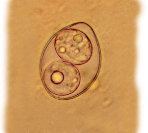
ESCCAP
                   Malvern Hills Science Park, Geraldine Road, Malvern,
                       Worcestershire, WR14 3SZ, United Kingdom

                    First Edition Published by ESCCAP in August 2011
                        Second Edition Published in February 2018

                                      © ESCCAP 2018

                                     All rights reserved

    This publication is made available subject to the condition that any redistribution or
     reproduction of part or all of the contents in any form or by any means, electronic,
         mechanical, photocopying, recording, or otherwise is with the prior written
                                     permission of ESCCAP.

    This publication may only be distributed in the covers in which it is first published
                   unless with the prior written permission of ESCCAP.

        A catalogue record for this publication is available from the British Library.

                                 ISBN: 978-1-907259-53-1

2
TABLE OF CONTENTS

INTRODUCTION                                                                             4

1: CONSIDERATION OF PET HEALTH AND LIFESTYLE FACTORS                                     5

2: LIFELONG CONTROL OF MAJOR INTESTINAL PROTOZOA                                         6

2.1 Giardia duodenalis                                                                   6

2.2 Feline Tritrichomonas foetus (syn. T. blagburni)                                     8

2.3 Cystoisospora (syn. Isospora) spp.                                                   9

2.4 Cryptosporidium spp.                                                                11

2.5 Toxoplasma gondii                                                                   12

2.6 Neospora caninum                                                                    14

2.7 Hammondia spp.                                                                      16

2.8 Sarcocystis spp.                                                                    17

3: ENVIRONMENTAL CONTROL OF PARASITE TRANSMISSION                                       18

4: OWNER CONSIDERATIONS IN PREVENTING ZOONOTIC DISEASES                                 19

5: STAFF, PET OWNER AND COMMUNITY EDUCATION                                             19

APPENDIX 1 – BACKGROUND                                                                 20

APPENDIX 2 – GLOSSARY                                                                   21

FIGURES

Figure 1: Toxoplasma gondii life cycle                                                  12

Figure 2: Neospora caninum life cycle                                                   14

TABLES

Table 1: Characteristics of apicomplexan oocysts found in the faeces of dogs and cats   10

            Control of Intestinal Protozoa
  6         in Dogs and Cats
 ESCCAP Guideline 06 Second Edition – February 2018

                                                                                             3
INTRODUCTION
A wide range of intestinal protozoa commonly infect dogs and cats throughout Europe; with a few exceptions
there seem to be no limitations in geographical distribution. The group covers flagellates (Giardia and
Tritrichomonas) and apicomplexan parasites (Cystoisospora, Cryptosporidium, Hammondia, Neospora,
Toxoplasma and Sarcocystis).
These infections share common characteristics:
Signs of disease are often associated with developing stages in the intestine and are in most cases
non-specific.
Younger animals are most commonly affected.
Their pathogenicity is variable both within and between genera and infections are often subclinical and
can be self-limiting.
The onset of clinical signs usually occurs several days after infection.
Severe clinical signs may sometimes be related to co-infections with other pathogens, for example viruses
and bacteria.
Diagnosis and differential diagnosis can be difficult and often require repeated sampling and molecular typing.
Treatment is often complicated due to lack of effective drugs or the need for off-label use of existing drugs.
Several agents are zoonotic, such as Toxoplasma, Cryptosporidium, and potentially Giardia.
This guideline focuses on the following common, and often clinically important, intestinal infections:
1) Giardia duodenalis
2) Feline Tritrichomonas foetus (syn. T. blagburni)
3) Cystoisospora (syn. Isospora) spp.
4) Cryptosporidium spp.
5) Toxoplasma gondii
6) Neospora caninum
7) Hammondia spp.
8) Sarcocystis spp.
Entamoeba histolytica is a human and primate pathogen that infects dogs only sporadically and has not been
included because of very limited relevance to our pets.
This guideline aims to give an overview of intestinal protozoa and their significance and, importantly, to suggest
rational control measures for the most important species in order to prevent animal and/or human infection.
The guideline is divided into five sections:
1. Consideration of pet health and lifestyle factors
2. Lifelong control of major intestinal protozoa
3. Environmental control of parasite transmission
4. Pet owner considerations in preventing zoonotic diseases
5. Staff, pet owner and community education
4
1: CONSIDERATION OF PET HEALTH AND LIFESTYLE FACTORS

Animals require care tailored to their individual needs. Certain factors may dictate comprehensive monitoring
and/or treatment, while others may suggest a less intensive approach. When recommending a parasite
management programme, veterinarians should consider the following:

Animal
Clinical signs due to infection with the mentioned protozoa are predominantly seen in young animals. Older
animals are mostly immune after previous infections and seldom show signs of disease, with the exceptions of
geriatric, chronically sick or immunocompromised animals, severely stressed animals, and perhaps pregnant
animals. Old animals, however, may still be a source of infection and thus pass on infections to their offspring.
Health status and background of the animal have to be considered.

Environment
Dogs and cats living in kennels/catteries, animal shelters or in crowded conditions with poor sanitation may
have a high risk of acquiring infections with protozoa that are transmitted directly, for example, Giardia,
Tritrichomonas, Cryptosporidium and Cystoisospora, and these may require special consideration. Access to
the outdoors may also influence the risk of infection.

Diet
Dogs and cats with access to rodents and raw meat, including viscera and/or foetal or placental material,
may be at risk of acquiring infections with cyst-forming coccidia, i.e. Neospora, Hammondia, Toxoplasma and
Sarcocystis.

Location and travel
Most infections are widespread in Europe and travel is not a major risk factor.

                                                                                                                    5
2: LIFELONG CONTROL OF MAJOR INTESTINAL PROTOZOA
2.1 Giardia duodenalis
2.1.1 Basic biology
Species
Giardia duodenalis (syn. G. intestinalis, G. lamblia) infects a range of vertebrates, including dogs and cats, and
is currently classified into assemblages (strains or genotypes) A–H, of variable host specificity. Assemblages
C and D are commonly found in dogs, while F has been isolated from cats and other animals. Assemblage
A has been found in both dogs and cats on occasions and assemblage B only rarely. Human infection with
Giardia is almost always with either assemblage A or B. This indicates the potential zoonotic role of Giardia
infection in pets when one or both of these assemblages are involved.
Life cycle
Giardia has a direct life cycle with repeated, asexual reproduction of trophozoites (i.e. active motile stages) in
the small intestine and intermittent production of resistant cysts that are passed in the faeces, initially often in
large numbers. Infection is by the oral uptake of cysts. Trophozoites attach to the epithelial cells after infection
and cause reduced absorptive capacity, and altered intestinal permeability. The prepatent period is 4–16
days. Patency usually persists for several weeks or months.
Epidemiology
In Europe the overall prevalence in dogs and cats is around 3–7%, however this is significantly higher in young
animals below one year of age, making it one of the most frequent endoparasites in this age group. Cyst
shedding is seen in both healthy and diseased animals. The infection is believed to induce partial immunity
resulting in loss of symptoms and, in some cases, elimination of the agent but with limited resistance to
re-infection. The transmission is faeco–oral by cysts in water, food or from the environment and only a few
cysts are needed to cause infection. Cysts may survive in the environment for months but are susceptible to
desiccation and are greatly reduced in numbers by freeze–thawing.
2.1.2 Clinical signs
Infection mostly remains subclinical, but may also cause chronic intermittent pasty diarrhoea rich in mucus,
anorexia, vomiting, weight loss and lethargy, particularly in immunocompromised animals, puppies/kittens
with coexisting infections or severely stressed animals, e.g. working sled dogs. In dogs, Giardia can often be
commensal; in cats, almost always pathogenic.
2.1.3 Diagnosis
In faeces, ovoid translucent cysts measuring 8–15 x 7–10 µm can be detected by direct wet mounts of faeces
or after concentration by sedimentation–flotation. Cysts may become deformed if flotation is attempted using
hyperosmotic flotation solutions. This will be prevented if sodium acetate acetic acid formalin is being used
for concentration. In very fresh faeces from animals with clinical signs, motile trophozoites (pear-shaped, 9–21
x 5–12 µm) may occasionally be detected. Due to fluctuating excretion, and in order to improve detection,
three samples over 3–5 days are recommended. Sedimentation techniques, in combination with MIF staining
(merthiolate iodine formalin), are also suitable methods for detecting cysts in faecal samples. Detection of
Giardia-specific coproantigen by means of commercially available assays (e.g. ELISA, immunochromatography
or in-clinic tests) is also possible. The various tests may differ in their target, and are often significantly
more sensitive than methods for the microscopic detection of Giardia cysts. This means that a diagnosis is
possible, even if cyst excretion has temporarily halted. PCR and direct IFAT (immuno-fluorescent antibody
test) may be used in diagnostic laboratories. The latter test, in particular, may be used in conjunction with IFAT
for Cryptosporidium (see section 2.4). PCR (plus multi-locus genotyping) may be of particular relevance for
identifying the infecting assemblage in order to determine zoonotic potential.
6Unfortunately, in the case of a positive Giardia test, no general statement can be made regarding a
therapy decision (see section 2.1.4) since the Giardia infection is often sub-clinical, even over a long period
of time. There is no evidence of an association between cyst count in faeces and the occurrence or strength
of clinical signs.
2.1.4 Control
Treatment
Dogs and cats with gastrointestinal symptoms and testing positive for Giardia cysts or coproantigens need to
be treated. Animals with persistent diarrhoea with no other aetiology identified should be re-tested. It is not
generally advisable to treat clinically inconspicuous Giardia positive animals for the reasons mentioned above.
In such cases a risk assessment is needed before reaching a decision on treatment. In high risk environments
like kennels, catteries or with breeders, particularly with a high incidence of diarrhoea in puppies, or where
there are small children or immunocompromised humans, treatment is advised. Strict hygiene measures are
needed – see prevention.
Fenbendazole and metronidazole are effective against Giardia. Treatment is aiming for cessation of clinical signs
and not for complete elimination of the agent. Fenbendazole (50 mg/kg bodyweight orally once daily for 3–5
days) can be used, and the treatment can be repeated after 2 weeks if clinical signs reoccur. Fenbendazole is
registered for the treatment of giardiosis in dogs in most European countries and can also be recommended
for cats. Often in cats the same dose of fenbendazole is used for 5 days followed by a 3 day break and then
another 5-day course of treatment, if necessary repeated after 2 weeks. Metronidazole (25 mg/kg bodyweight
orally twice daily or 50 mg/kg bodyweight once daily) is licensed in most European countries for dogs and cats.
Another option for dogs is to use a combination tablet containing febantel/pyrantel/praziquantel at the
standard deworming dose (15.0 mg/kg of febantel, 14.4 mg/kg pyrantel, 5.0 mg/kg praziquantel) repeated
once daily for three days. This treatment is licensed for dogs in most European countries and countries
outside the EU.
A therapy control should be carried out with one of the above-mentioned methods approximately 5 days
after treatment ends. If the sample remains positive AND if the clinical symptoms persist treatment should
be continued. Often, reinfection can occur immediately after treatment therefore it is advisable to inform the
pet owner that recurrences are possible or even likely. Other reasons for apparent treatment failure include
co-infections or other underlying disease, which should be addressed, or by incomplete parasite removal
following treatment. Drug resistance against metronidazole has been described in human isolates. Long-
lasting success of treatment is often hampered by reinfection pressure from the contaminated environment,
thus additional measures to reduce infection pressure are critical. Shampooing dogs and perhaps cats (e.g.
with a product containing chlorhexidine digluconate) at the beginning and the end of antiprotozoal treatment
may assist in reducing re-infections.
Prevention
Cleaning and drying of the environment (including blankets, bedding etc.), the use of clean utensils for feed
and water, bathing with chlorhexidine shampoos to remove adhering faeces or cysts and proper disposal of
faeces are prerequisites to avoid animal–animal transmission. There are indications that cysts on surfaces are
killed by hot water high pressure cleaning (>65°C) or quaternary ammonium compounds, but no disinfectants
are registered for this purpose. Surfaces should be left to dry completely. Personal hygiene of animal carers
to avoid the spreading of cysts is mandatory. Food and water containers should be cleaned daily with boiling
water. This also applies to litter boxes which should subsequently be thoroughly dried before being refilled.
Coproantigen tests should be performed on new puppies or kittens when introduced to households with
other pets free of infection or animals entering breeding establishments. Diarrhoeic animals and carriers
should always be quarantined and diagnosed appropriately.
2.1.5 Public health considerations
As mentioned above, humans are seldom infected by dog or cat specific assemblages, but human assemblages
may circulate in a dog or cat population.
In cases where family members as well as their dogs or cats have a Giardia infection, the pet owner should
be advised to consult a physician.
72.2 Feline Tritrichomonas foetus (syn. T. blagburni)
2.2.1 Basic biology
Species
Feline Tritrichomonas foetus (suggested: T. blagburni) has been identified as a cause of chronic diarrhoea in
cats and other felids. T. foetus is occasionally/rarely isolated from dogs.
Life cycle
The life cycle is direct, with trophozoite formation in the large intestine and ileum, and there is no known
cyst stage. Pathogenicity is related to the cytotoxic effects of trophozoites on the intestinal epithelium via
secretion/excretion of proteases and other factors; occasionally invasion deeper into the mucosa is seen.
Trophozoites can be detected after 7–14 days and the infection is often long-lasting but generally self limiting.
Epidemiology
The infection route is considered to be faeco–oral. Prevalences may be relatively high in restricted environments
like catteries and shelters but otherwise can be expected to be low, although surveys are limited in many
countries. There are indications that pure-bred cats are more commonly affected. At present, there is no
evidence to suggest any link between feline infections and bovine reservoirs with the closely related bovine
T. foetus.
2.2.2 Clinical signs
Infections are often subclinical but typically kittens or otherwise naïve animals may exhibit clinical signs of
T. foetus infection including semi-formed (“cowpat”) faeces with blood and/or mucus and faecal incontinence
with irritation and pain around the anus. The clinical course often fluctuates with transient remission after
therapy (see below). Disease is seldom observed in dogs.
2.2.3 Diagnosis
Pear-shaped trophozoites (10–25 x 3–15 µm) are detected in FRESH, still warm faeces by direct wet mounts
but sensitivity is generally low. The trophozoites are similar to Giardia in size but the rapid “jerky” movement and
the presence of an undulating membrane in T. foetus are different from the “falling leaf” motion and the typical
“eyes” (two large nuclei) of Giardia. It also needs to be differentiated from the commensal Pentatrichomonas
hominis, which can be seen in both cats and dogs, and occasionally other trichomonads. Direct detection by
PCR is the preferred option, which can also be used to provide speciation. In contrast to other protozoans,
T. foetus can be cultured, e.g. in a commercially available test system (InPouchTF-Feline™, BioMed
Diagnostics) which will not propagate P. hominis or Giardia.
2.2.4 Control
Treatment
There are no drugs registered for use in cats against T. foetus and treatment recommendations are often based
on case histories. Ronidazole (30 mg/kg bodyweight daily for 2 weeks) has been used off-label in severe cases
of diarrhoea with some success. Care should be taken when administering ronidazole and gloves worn as it
is a known mutagenic, carcinogenic and embryotoxic substance. For the same reason, treatment of pregnant
and nursing cats and kittens less than 12 weeks old is not recommended. Cats must be closely monitored
for drug-induced neurotoxicity (lethargy, ataxia, seizures) and treatment withdrawn immediately should this
happen. The signs seem reversible when the drug is withheld. Metronidazole and fenbendazole only cause
temporary remission. A change in diet may also alleviate clinical signs, and spontaneous resolution of clinical
signs often occurs.
Prevention
As clinical problems are often associated with closed environments where there is a high density of cats, many
of the precautions recommended for Giardia should be observed. Cases are often chronic and refractory to
treatment, and will contaminate the environment.
82.2.5 Public health considerations
T. foetus has at present no documented zoonotic potential, although care must always be taken with
immunocompromised individuals. Care must be taken when administering ronidazole.
P. hominis is observed in humans but little is known regarding its pathogenicity and transmission.
2.3 Cystoisospora (syn. Isospora) spp.
2.3.1 Basic biology
Species
The genus Cystoisospora is host-specific: Cystoisospora canis, C. ohioensis and C. burrowsi are the common
species infecting dogs; the latter two are often referred to as the C. ohioensis-complex because they are not
readily separated morphologically. Cystoisospora felis and C. rivolta infect cats.
Life cycle
Infection commonly takes place via the faeco–oral route by the ingestion of sporulated oocysts. Multiplication
of the intestinal stages takes place intracellularly throughout the small and large intestines. After a prepatent
period of 6–10 days, oocysts are shed in the faeces and then complete their development to the infective
stage in the environment usually within several days. Different animals, including rodents and ruminants, can
act as paratenic hosts after oral uptake of oocysts and subsequently harbour resting stages (“dormozoites”
or “hypnozoites”) in internal organs. After ingestion of dormozoites, the prepatent period is slightly shorter.
The excretion period is variable but most animals shed oocysts for 5–10 days.
Epidemiology
Cystoisospora species of dogs and cats are ubiquitous and oocysts can be found in the faeces of subclinically
infected, as well as sick, animals. Primary infections usually take place during the suckling period from the
third to the eighth week of age. Consequently, the majority of clinical cases are diagnosed in puppies/kittens
of less than four months old. At that age, most infections are acquired via ingestion of oocysts from the
environment. Oocysts remain infective in the environment for several months and can accumulate in breeding
kennels or catteries with a high density of suitable hosts. Dormozoites in paratenic hosts are infective for
several years.
2.3.2 Clinical signs
Cystoisosporosis is associated with diarrhoea in puppies and kittens. In severe cases, the faeces can contain
blood and cause morbidity or even mortality. Often, clinical presentation is associated with viral, helminth or
bacterial co-infections and, where there have been changes in their diet (e.g. puppies receiving solid food
for the first time), animals seem to be more affected by diarrhoea. As with many other coccidial infections,
diarrhoea often occurs shortly before the onset of oocyst excretion. After reinfection, animals usually shed
few oocysts and do not show clinical signs. Cross-immunity between Cystoisospora species in the same host
seems unlikely.
During the patent period, oocysts are shed in the faeces and can be detected by concentration flotation. The
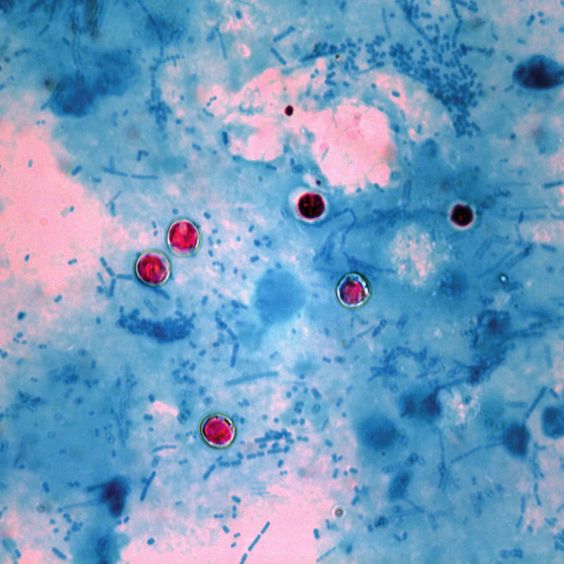
morphology of oocysts that can be found in the faeces of infected dogs and cats are described in Table 1.
9Table 1: Characteristics of coccidian oocysts found in the faeces of dogs and cats
Host Affected
Genus Species Average size (µm) Shape Shell
DOG CAT
Cystoisospora* C. burrowsi 21 x 18 round–oval thin, colourless or brownish
C. canis 39 x 32 round–oval thin, colourless or brownish
C. felis 45 x 33 ovoid thin, colourless or brownish
C. ohioensis 24 x 20 round–oval thin, colourless or brownish
C. rivolta 26 x 24 round–oval thin, colourless or brownish
Cryptosporidium C. canis 5.0 x 4.7 round–oval thin, colourless unless stained****
C. felis 3.2–5.0 x 3.0–4.5** round–oval thin, colourless unless stained****
C. parvum 5.0 x 4.5 round–oval thin, colourless unless stained****
Toxoplasma T. gondii 12.4 x 10.5 round thin, colourless
Neospora N. caninum 12.0 x 10.5 round thin, colourless
Hammondia H. hammondi 11.4 x 10.6 round thin, colourless
H. heydorni 11.9 x 11.1 round thin, colourless
Sarcocystis*** Oocyst – round very thin, colourless
Sporocyst 11 x 8 ovoid thick, colourless
Sporocyst 14 x 10 ovoid thick, colourless
* the oocysts of Cystoisospora spp. in fresh faeces contain a large sporoblast; in older faecal samples (> 12 hrs) two round
sporocysts may be seen
** variable information is available
*** several species in dogs and cat with morphologically indistinguishable sporocysts; round oocysts with very thin walls,
rupture during intestinal passage and release two fully sporulated sporocysts which can be found in the faeces
**** using the modified Ziehl Neelsen technique
2.3.4 Control
Treatment
Due to the fast replication of the pathogenic intestinal stage, followed by excretion of oocysts in large numbers,
it is critical to treat infections at an early stage. Litter mates of an affected puppy have a high risk of being
infected even though they may not yet be shedding parasites. Hence, treatment should include all susceptible
animals, i.e. all litter mates and in-contact puppies.
Toltrazuril and diclazuril are currently the drugs of choice against cystoisosporosis. Toltrazuril (9–20 mg/
kg bodyweight) or diclazuril (2.5–5.0 mg/kg bodyweight) in a single application significantly reduce oocyst
shedding in excreting animals; application in the pre-patent period largely prevents oocyst excretion and
reduces diarrhoea in affected litters. In dogs, a combination of toltrazuril/emodepside (9 mg/0.45 mg/kg
bodyweight) is registered for co-infections of coccidia and roundworms. This product is not licensed for use
in cats and it is necessary to use it off-label.
Prevention
Due to the ubiquitous nature of the parasites, eradication is not normally feasible. The risk of acquiring
an infection can be reduced by hygiene measures including the daily removal of faeces from kennels and
thorough cleaning and disinfection of litter areas in breeding units. Since heat (steam cleaning) and chemical
disinfection using cresols are necessary to inactivate oocysts, floors and walls of boarding kennels, animal
shelters and larger breeding units etc. should be chosen to resist such treatment. Surfaces should be left to
dry completely since this also reduces the survival of oocysts in the environment. Personal hygiene of animal
handlers is very important to avoid spreading oocysts through faecal material.
2.3.5 Public health considerations
Cystoisosporosis of cats and dogs has no zoonotic implication, as the parasites are strictly host specific.
102.4 Cryptosporidium spp.
2.4.1 Basic biology
Species
Cryptosporidium oocysts are very small and do not allow species differentiation based on morphology.
Cryptosporidium canis and C. felis infect dogs and cats, respectively, and these species have only very rarely
been found in humans or other animals. Cryptosporidium parvum is a species with low host specificity and
parasitises mainly calves but can also infect a range of other mammals, including humans, and occasionally
dogs and cats. Since species differentiation relies on molecular typing, the exact distribution amongst positive
cats and dogs is still unknown.
Life cycle
Infection with Cryptosporidium is initiated when oocysts from the environment are ingested and the released
sporozoites invade the epithelium of the small intestine and begin intracellular multiplication. Endogenous
replication ends with the production of sexual stages that fuse to form an oocyst that sporulates in the
intestines and is excreted with the faeces already in the infective form. Autoinfection with ruptured oocysts
before excretion is common and can result in the shedding of large parasite numbers within a short period of
time. The prepatent period varies from 2–14 days for C. canis and 3–7 days for C. felis. Excretion lasts from
25–80 days.
Epidemiology
Cryptosporidium oocysts are immediately infective when excreted with the faeces, so faeco–oral infections
are common. They are also very small and do not sediment readily in water; they are therefore frequently
water-borne and the parasite can remain infective in this environment for several months. Unlike the other
apicomplexa species described here, Cryptosporidium is strictly monoxenous, and paratenic or intermediate
hosts are not described.
2.4.2 Clinical signs
In immunocompetent adult animals, infection is usually subclinical. Clinical signs appear to be most severe in
immunocompromised individuals.
Kittens, and less commonly puppies, can develop watery, sometimes foul-smelling, diarrhoea; this can last
for days or sometimes weeks, and is frequently accompanied by abdominal pain, vomiting and an elevated
body temperature. Diarrhoea usually starts several days after the onset of oocyst excretion.
2.4.3 Diagnosis
IFAT (direct immunofluorescence antibody test) in specialised laboratories is the golden standard. Antibodies
are usually labelled with fluorescein isothiocyanate (FITC) that fluoresces apple green under appropriate
(blue) filters on a fluorescence microscope. DAPI (4’, 6-diamidino-2-phenylindole) can be used to stain nuclei
and can be a useful confirmatory stain if oocyst excretion is low (UV filters necessary for visualisation on a
fluorescent microscope). In many diagnostic laboratories, oocysts are detected by direct coproscopy (Table 1)
of a faecal smear after staining (Ziehl-Neelsen, Heine, safranin). Oocysts are presented as small, round, pink
or orange bodies when stained, depending on the staining procedure. As with Giardia, coproantigen tests for
C. parvum are commercially available and also validated for dogs. Molecular detection is both sensitive and
specific, and speciation by PCR can be performed if risk assessment raises concern.
2.4.4 Control
Treatment
There is no registered treatment available for cryptosporidiosis in cats and dogs. Since the infection usually
resolves spontaneously, only supportive treatment (fluid replacement, spasmolytic medication) can be
considered.
Prevention
Cryptosporidium oocysts are highly persistent in the environment and directly infectious so strict hygiene
measures must be taken to avoid the spread of infection (see Cystoisospora).
112.4.5 Public health considerations
Due to the rather low host-specificity of C. parvum, this parasite is infectious to humans, while zoonotic
infections with C. felis or C. canis are usually, but not always, restricted to immunocompromised individuals.
Owners of young animals should generally be advised to adhere to effective hygiene protocols and
immunocompromised individuals should not be in close contact with sick cats and/or dogs.
2.5 Toxoplasma gondii
2.5.1 Basic biology
Species
Toxoplasma gondii is the only valid species in the genus Toxoplasma. It infects only cats and a few other felids
as definitive hosts, while probably all mammals (including humans, cats and dogs) as well as birds can act as
intermediate hosts. T. gondii is globally present in at least three genotypes and multiple mixed forms thereof.
T. gondii is a major cause of abortion in sheep.
Life cycle
Cats usually acquire the infection by ingestion of tissue cysts, most commonly by predation on rodents and
birds, by feeding on raw or undercooked meat from infected livestock or, less commonly, on aborted material
(Figure 1). Although felids can become infected with oocysts via the faeco–oral route, this appears to be a
less common route of infection for the definitive host. The prepatent period is 3–10 days after ingestion of
tissue cysts and 18–36 days after uptake of oocysts. Excretion of oocysts can last up to 20 days, but is most
intensive 2–5 days after the onset of shedding. Oocysts are not infective immediately after excretion, but
require at least 24 hours and usually 2–5 days for sporulation in the environment.
transplacental
transmission:
fatal infection
cats infected in kittens
humans infected by
by ingesting ingesting tissue cysts
intermediate hosts in undercooked meat
or oocysts
unsporulated
tissue cyst oocysts passed
bradyzoites in faeces
develop
oocysts sporulate humans can also
in the environment be infected via
blood transfusion,
organ transplantation
oocysts transform or transplacentally
into tachyzoites intermediate hosts from mother to foetus
ingest oocysts
humans infected by ingesting
contaminated food or water
Figure 1: Toxoplasma gondii life cycle
Epidemiology
Cats can excrete an impressive number of oocysts (hundred thousands to millions) for a few days after primary
infection, but subsequently they excrete few or no oocysts, even after reinfection or if immunosuppressed.
Due to the ubiquitous nature of the parasite, the distribution of T. gondii is broad in free-ranging intermediate
hosts. The small oocysts can be distributed easily and get into surface water where they can survive for
several months, making water, as well as humid soil or feedstuffs contaminated with cat faeces, the prime
source of infection for herbivorous intermediate hosts. In contrast, carnivorous hosts most often acquire the
infection via ingestion of tissue cysts in meat from infected hosts. Rodents, particularly mice, act as very
efficient reservoir hosts.
122.5.2 Clinical signs
Acute toxoplasmosis is rare in cats. Kittens infected in utero can show signs of infection after birth and prenatal
infections of kittens are frequently fatal. The reasons for clinical manifestations in adult cats are unclear; it
is presumed that immunosuppression by viral pathogens (FeLV, FIV) may play a role. Affected animals show
signs of systemic infection including fever, anorexia, abdominal pain, dyspnoea, ocular inflammation (uveitis)
and rarely central nervous disorders. Clinical signs are seldom related to the enteric stage of development.
Occasionally acute disease that may be accompanied by fever and neuromuscular signs is observed in dogs
infected with T. gondii.
2.5.3 Diagnosis
Excreting cats shed the small oocysts in high numbers, but due to the short patent period and infrequent re-
shedding, infection is usually not detected by faecal examination. The oocysts are morphologically similar to
Hammondia oocysts (Table 1).
Diagnosis is based on clinical signs and by the detection of specific antibodies in the blood. In some cases
serology may be questionable, e.g. in early infections before antibodies are present, and retesting after 3
weeks is advised. Not all cats with subclinical infections may display antibody titres.
Clinical toxoplasmosis in dogs is diagnosed by serology, supported by PCR on cerebrospinal fluid.
2.5.4 Control
Treatment
Cats with clinical disease can be treated with clindamycin (oral treatment: clindamycin hydrochloride 10–12
mg/kg bodyweight, twice daily for four weeks; parenteral treatment: clindamycin phosphate 12.5–25 mg/kg
bodyweight by intramuscular injection (i.m.), twice daily for four weeks). Treatment of cats after infection has
not been shown to prevent oocyst excretion. Affected dogs may be treated with clindamycin or sulphonamide/
trimethoprim.
Prevention
Control measures aim at the prevention of oocyst shedding in order to reduce the infection of humans and
livestock with T. gondii. Cats should not be fed raw meat or allowed to eat prey animals. However, since animals
with outdoor access presumably prey on mice and other potential hosts of T. gondii, accidental infections
cannot be completely avoided and where possible cat faeces should not be introduced in the environment.
2.5.5 Public health considerations
T. gondii is one of the most prevalent parasitic zoonoses worldwide. Although there are differences in virulence
associated with genotype, healthy adults generally have a low risk of developing severe toxoplasmosis after
infection. However, immunocompromised individuals or children infected in utero can suffer from severe or
even fatal local (mostly ocular or cerebral) or generalised toxoplasmosis. Prenatal infections occur as a result
of a primary infection of the mother during pregnancy.
In humans, the infection can be acquired either by ingestion of infected raw or undercooked meat or by
ingestion of sporulated oocysts from the contaminated environment (ingesting fruit, vegetables, soil or sand
from sandpits contaminated by the faeces of infected cats). It is therefore recommended (especially for
high-risk individuals e.g. previously unexposed pregnant women or the immunocompromised) that meat
is consumed only after thorough cooking or freezing (-20°C for two or more days) and personal hygiene
measures are observed whilst handling meat. Pregnant women should avoid contact with expectant ewes
and does. They should not assist in lambing or kidding due to the risk of hand–mouth contamination by
contact with recently-infected dams during delivery. Neither should they handle lambs or kids. Working in
the meat industry (abattoir, cutting plant) is significantly associated with acquiring infection (occupational
disease). Similarly, drinking unfiltered surface water or accidental ingestion of soil as well as contact with
cat faeces in general must be avoided. All fruit and vegetables (especially from gardens) should be washed
thoroughly and gloves should be worn when gardening or handling soil or sandpits.
Litter trays should be thoroughly cleaned every day so that, in case of defaecation by shedding cats, oocysts
do not have time to sporulate. This task should not be performed by pregnant women or other persons at risk.
132.6 Neospora caninum
2.6.1 Basic biology
Species
Neospora caninum is the type species of the genus. In Europe, dogs are currently the only identified definitive
hosts and they also act as intermediate hosts. It is likely that other wild canids such as wolves can also act
as definitive hosts. Cattle, sheep, goats and other domestic and wild ungulates, as well as rodents and birds,
are natural intermediate hosts of the parasite, harbouring tachyzoites and cysts with bradyzoites in various
tissues. N. caninum is a major cause of abortion in cattle.
Life cycle
Dogs acquire the infection mainly by ingesting cysts containing bradyzoites located in tissues of infected
intermediate hosts, in particular cattle (Figure 2). In natural infection, the prepatent period is 5–9 days and
patency generally lasts 11–20 days. Oocysts are not immediately infective for other hosts after excretion in
the faeces, but require sporulation for 1–3 days in the environment. Repeated transplacental transmission
of tissue-dwelling parasites from chronically infected dams to the foetus is possible, even though highly
variable. It has been reported, however, that up to 50% of pups of N. caninum-positive dams might become
infected transplacentally, with 25% developing clinical signs (neonatal neosporosis).
wild carnivores
may also be
definitive hosts
transplacental
transmission
(tachyzoites)
oral transmission
(oocysts from
the environment)
transplacental oral transmission
transmission (bradyzoites
(tachyzoites) from tissues)
potentially infected/
diseased puppies
non-infected
or persistently Oral transmission to dogs and other
infected calf abortion of carnivores by oocysts remains to be
infected calf demonstrated in natural cases
Figure 2: Neospora caninum life cycle
Epidemiology
Age-related prevalence data indicate that the majority of dogs become infected after birth. Higher prevalences
have been documented in old dogs compared to young dogs. It has been reported that placentas from aborting
cattle are the main source of infection for dogs and feeding of raw beef has also been identified as a risk
factor for canine neosporosis. Not surprisingly, hunting dogs fed raw bovine meat have high seroprevalence
rates. N. caninum oocysts have been found in faeces from dogs ranging in age from 45 days to 13 years and
the number of oocysts per gram of faeces varies from only a few to over 100,000.
In most cases of neonatal neosporosis, clinical signs are not apparent until 5–7 weeks after birth. This suggests
that N. caninum is transmitted from the dam to the pups towards the end of gestation.
142.6.2 Clinical signs
The systemic phase can cause clinical disease whereas there are no signs associated with enteric
development. Most cases of clinical neosporosis are reported in puppies less than six months old (neonatal
neosporosis) which were infected transplacentally, but N. caninum can cause illness in dogs of any age.
Clinical signs which should arouse suspicion of neosporosis include hind limb paresis and ataxia which
becomes progressively more severe. Muscle atrophy, quadriceps contracture, signs of pain on palpation
of the lumbar and/or quadriceps muscles and later signs of head and neck involvement (head tilt); ocular
abnormalities and dysphagia may all be signs of neosporosis. Other neurological conditions not readily
attributable to other causes should also be investigated as possible neosporosis, particularly in mature dogs.
In older dogs, ulcerative dermatitis, myocarditis, pneumonia and pancreatitis have been reported. In puppies,
the ascending paralysis caused by Neospora can often be fatal and several litter mates can be affected,
although not necessarily simultaneously.
2.6.3 Diagnosis
Unsporulated oocysts in faeces measure on average 12 x 10.5 µm and are microscopically identical to
Hammondia oocysts (Table 1). Differential recognition can be obtained by specific PCR. Because clinical
disease is caused by the tissue-dwelling forms of the parasite, faecal examination for oocyst detection
does not play a role in the diagnosis of canine neosporosis. Clinical suspicion of canine neosporosis can be
confirmed by demonstrating the presence of the parasite through molecular methods: PCR can be carried
out on cerebrospinal fluid or muscle biopsies. Most cases however, are diagnosed through serology. Puppies
usually seroconvert about 2–3 weeks following infection and antibody levels are usually (but not always) high
in clinically affected animals. Therefore, diagnosis of canine neosporosis can be based on clinical signs and
positive serology (ELISA, IFAT).
2.6.4 Control
Treatment
Treatment of clinical neosporosis in dogs is difficult and only partially effective; it tends to be most effective
in the early stages, before muscular contraction has occurred. Indeed, when clinical signs are suggestive
of N. caninum, it is recommended to initiate treatment immediately rather than wait for serologic results.
Treatment with clindamycin (20 mg/kg bodyweight twice daily for 30–60 days) has been reported to improve
clinical recovery in naturally infected dogs with neurological signs. Alternatively sulphonamide/trimethoprim
may be used.
Prevention
As mentioned above, seropositive dams can transmit N. caninum to puppies. It is therefore recommended
that chronically infected females are excluded from any breeding programme. Furthermore, farm dogs should
be fed cooked meat and prevented access to calving pens and areas on farms with aborted calves, placentas,
amniotic fluid, and raw meat offal. Contamination of water and cattle feedstuff with dog faeces should be
avoided.
2.6.5 Public health considerations
As Neospora is not known to infect humans there is no zoonotic potential although antibodies have been
reported in some individuals.
152.7 Hammondia spp.
2.7.1 Basic biology
Species
Two species of Hammondia parasitise cats and dogs, i.e. H. hammondi and H. heydorni respectively.
Life cycle
The life cycle resembles that of other cyst-forming coccidia (Sarcocystis, Neospora, Toxoplasma). Dogs and
cats are the definitive hosts and acquire the infection after ingestion of infected prey; they shed oocysts after
a prepatent period of 5–13 days (H. hammondi) or 7–17 days (H. heydorni). Excretion periods are variable but
usually limited to around 20 days and sporulation takes place in the environment. Intermediate hosts (mostly
rodents and ruminants) ingest oocysts and subsequently develop tissue cysts, predominantly in muscle and
brain tissue.
Epidemiology
Very little is known about the geographic distribution of Hammondia but it is found sporadically in the faeces
of cats and dogs in Europe. Since the differentiation from Toxoplasma (in cats and occasionally in dogs after
coprophagia) or Neospora (in dogs) is only possible with molecular methods, the true prevalence of these
parasites is unknown.
2.7.2 Clinical signs
Hammondia infections in the definitive hosts usually take a subclinical course. Very rarely, anorexia and
severe diarrhoea, unresponsive to antibacterial therapy, have been described in infected puppies.
2.7.3 Diagnosis
During the patent stage of infection, the small oocysts can be found in the faeces. Morphological differentiation
from Toxoplasma or Neospora is not possible (see Table 1) but differentiation can be achieved with PCR.
2.7.4 Control
Treatment is not necessary. Prevention of infection can be achieved by avoiding the ingestion of raw food or
prey containing tissue cysts from intermediate hosts (warm-blooded animals).
2.7.5 Public health considerations
As Hammondia is not known to infect humans, there is no zoonotic potential. However, since their oocysts are
indistinguishable from those of T. gondii, care should be taken in cases of oocyst-positive animals.
162.8 Sarcocystis spp.
2.8.1 Basic biology
Species
Within the genus Sarcocystis, several species parasitise cats or dogs as definitive hosts. The faecal stages, so-
called sporocysts, are morphologically indistinguishable. Differentiation is based on tissue cyst morphology in
the different intermediate hosts (omnivorous or herbivorous animals) and molecular methods. This genus can
be responsible for the condemnation of meat from infected cattle.
Life cycle
Carnivorous animals become infected by ingestion of meat containing tissue cysts. In the intestinal epithelium
of the definitive host, sexual development takes place which results in the production of an oocyst which
sporulates before excretion. The oocyst wall is very thin and ruptures during passage through the intestine so
that usually fully infective sporocysts can be found in the faeces; these are then ingested by the intermediate
host and develop extra-intestinally into tissue cysts. The prepatent period is 8–33 days in dogs and 10–14
days in cats. Patency is long (several months) due to the slow release of parasites from the epithelium.
Epidemiology
The sporocysts in the faeces are infective on excretion and remain so for months, even years, since they have
a prolonged survival rate in the environment. The prevalence rates in the intermediate hosts (sheep, cattle,
pigs with outdoor access) are up to 100% due to the ubiquitous distribution of the parasites.
2.8.2 Clinical signs
In the definitive host, the development of the parasite is restricted to the final stages and does not cause
clinical signs under natural conditions. The clinical and hygienic importance of infection with Sarcocystis is
restricted to the intermediate host where outbreaks due to faecal contamination of feedstuff or water have
been reported and can result in clinical signs. Cysts in carcasses can lead to meat condemnation. After
reinfection, dogs and cats usually develop some degree of immunity which is species-specific.
2.8.3 Diagnosis
Sporocysts (see Table 1) can be found in faeces in low numbers. Tools for species diagnosis are not available.
2.8.4 Control
Treatment
Treatment of dogs or cats is not necessary.
Prevention
Since Sarcocystis is strictly heteroxenous, infection can be avoided by feeding meat that has been either
previously frozen (-20°C for at least 4 days) or cooked. To interrupt transmission, dogs and if possible also
cats should not be allowed to defaecate on animal feed or pastures.
2.8.5 Public health considerations
None of the Sarcocystis spp. involving dogs and cats are zoonotic. Most human infections with Sarcocystis
take place via ingestion of infected beef or pork.
173: ENVIRONMENTAL CONTROL OF PARASITE TRANSMISSION
A number of actions which will assist in the environmental control of intestinal protozoan infections of dogs
and cats have been suggested in the relevant sections above.
Environment
Environmental stages of protozoa (oocysts, cysts, sporocysts) may survive in a contaminated environment
for prolonged periods. Regular intensive cleaning of potentially contaminated areas will reduce the number
of infective stages. Premises such as breeding facilities or sanctuaries should be equipped with surfaces that
are easy to decontaminate (smooth, chemically resistant) and should be generally kept as dry as possible.
Efficient chemical disinfection depends on dry and clean surfaces. A number of products have been certified
in the EU as active against environmental parasite stages based on results of highly standardised tests e.g. in
Germany (www.dvg.net). Most of these commercial products contain cresols and all should be handled with
care. Manufacturers’ instructions should be strictly followed to ensure maximum efficacy and to minimise
environmental and health risks.
Efficient chemical disinfection in households where pets live in close proximity to people is an unrealistic
approach because efficient products contain rather aggressive chemicals. Prevention of patent infection
in pets (see previous chapters) will avoid environmental contamination with infective stages of parasites.
Transmission of zoonotic parasites to persons living in the same household is avoidable by observance of
reasonable hygiene.
Disinfection of bare ground or grassed areas is not possible. Prevention of contamination of such areas should
be strictly observed by collection of faeces followed by adequate disposal in firmly-closed plastic bags.
Dependent on the quality of surfaces, physical disinfection (heat) is the most effective means of inactivating
infective parasite stages. This however will not be appropriate in many circumstances.
Quarantine
New dogs/cats introduced into kennels/catteries should be quarantined whenever possible. As the prepatent
period of protozoa is usually shorter than that of helminths, it is realistic to quarantine cat/dogs in shelters or
catteries to control the possible onset of parasite excretion. This can aid in the prevention of those infections
that are directly transmitted via environmental stages. Faecal samples should be taken every other day and
measures undertaken for adequate isolation, environmental hygiene and, in indicated cases, treatment to
prevent parasite spreading.
Comment on feeding raw meat to pets
Diets for dogs and cats containing raw meat products, e.g. BARF, are becoming increasingly popular. Feeding
fresh, raw meat increases the risk of meat-borne protozoan parasites i.e. Toxoplasma, Neospora, Sarcocystis
and less importantly, Cystoisospora. Freezing at -20°C for 7–10 days prior to use can inactivate stages in
raw meat and reduce the risk of transmission. Meat should be of the same quality as intended for human
consumption.
184: OWNER CONSIDERATIONS IN PREVENTING ZOONOTIC DISEASES
The most important advice for the prevention of transmission of zoonotic agents, including certain intestinal
protozoa, is personal hygiene. Washing hands after contact with dogs, cats and other animals should be
routine. Since many of the intestinal protozoan infections mentioned do little or no harm to dogs and cats
(especially adult animals) and, in many cases, to pet owners, these infections go unnoticed. Fortunately
the majority of intestinal protozoan infections in dogs and cats are host-specific. Human infections with
Toxoplasma are mainly either food, water or soil-borne. Direct contact with cats is not considered a risk
factor however, contact with cat faeces and contaminated food/water are risk factors. Human infections with
Sarcocystis in Europe are almost exclusively transmitted between human–cattle and human–pig respectively.
There is no known association with dogs or cats. Although Cryptosporidium and Giardia are also largely
species-specific, some genotypes are zoonotic. Consequently, strict hygiene is the only way to prevent
transmission. This is particularly important for individuals with immune deficiency disorders or individuals
undergoing immunosuppressive treatments. In these patients, opportunistic species or rare genotypes of
otherwise non-zoonotic parasites can occasionally establish, and these, as well as other zoonotic pathogens,
frequently cause severe or even fatal diseases that would otherwise resolve in immunocompetent individuals.
5: STAFF, PET OWNER AND COMMUNITY EDUCATION
Cryptosporidium and Giardia are potentially zoonotic (see specific sections above) but only genotyping can
give definitive information. Toxoplasma is a well-known zoonotic agent and can be transmitted to humans
by several routes, including via infective oocysts from the faeces of excreting cats. It has to be kept in
mind, however, that while the stage of Giardia and Cryptosporidium are immediately infective in fresh faeces,
Toxoplasma requires external sporulation that takes at least 1–2 days, so fresh faecal material does not
contain infective oocysts of Toxoplasma. Thorough daily cleaning of litter boxes therefore greatly minimises
the risk of transmission via cat faeces. Garden soil or sand areas where cats bury their faeces may represent a
greater infection risk than litter boxes that are cleaned daily. As mentioned above, the vast majority of human
Toxoplasma infections are acquired from food (mainly meat, fresh produce and rarely raw goats’ milk) or the
environment (water/soil).
The information in this guideline deserves to be widely disseminated in veterinary practices including all
auxiliary personnel. Veterinarians working with cats do not have a higher risk of acquiring Toxoplasma
infections than other professions. Correct knowledge of protozoan infections is a prerequisite for proper
understanding, which, in turn, will help allay unjustified fear in pet owners and the general public. As in other
parasitic, bacterial or viral infections, personal hygiene is the most effective preventive measure and emphasis
of this fact should be given a very high priority in all educational programmes dealing with zoonotic disease.
Additional information and resource materials can be obtained at www.esccap.org
19APPENDIX 1 – BACKGROUND
ESCCAP (European Scientific Counsel Companion Animal Parasites) is an independent, not-for-profit
organisation that creates guidelines based on up-to-date scientific information and promotes good practice
for the control and treatment of parasites in and on companion animals. With application of the proper advice,
the risk of diseases and parasitic transmission between animals and humans can be minimised. ESCCAP
aspires to see a Europe where companion animal parasites no longer threaten the health and well-being of
animals and humans.
There is a great diversity in the range of parasites and their relative importance across Europe and the ESCCAP
guidelines summarise and highlight important differences which exist in different parts of Europe and, where
necessary, specific control measures are recommended.
ESCCAP believes that:
Veterinarians and pet owners must take measures to protect their pets from parasitic infections.
Veterinarians and pet owners must take measures to protect the pet population from risks associated with
travel and its consequent potential to change local parasite epidemiological situations through the export
or import of non-endemic parasite species.
Veterinarians, pet owners and physicians should work together to reduce the risks associated with zoonotic
transmission of parasitic diseases.
Veterinarians should be able to give guidance to pet owners regarding risks of parasite infestation and
diseases and measures which can be taken to minimise these risks.
Veterinarians should attempt to educate pet owners about parasites to enable them to act responsibly
not only for their own pet’s health but for the health of other pet animals and people in their communities.
Veterinarians should wherever appropriate undertake diagnostic tests to establish parasite infestation
status in order to provide the best possible advice.
To achieve these objectives, ESCCAP produces:
Detailed guidelines for veterinary surgeons and veterinary parasitologists.
Translations, extracts, adaptations and summarised versions of guidelines which address the varied
requirements of European countries and regions.
Versions of each guideline can be found at www.esccap.org
Disclaimer:
Every effort has been taken to ensure that the information in the guideline, which is based on the authors’
experience, is accurate. However the authors and publishers take no responsibility for any consequence
arising from the misinterpretation of the information herein nor is any condition or warranty implied. ESCCAP
emphasises that national, regional and local regulations must be borne in mind at all times before following
ESCCAP advice. All dosages and indications are provided for guidance. However, vets should consult
individual data sheets for details of locally approved treatment regimens.
20APPENDIX 2 – GLOSSARY
Asexual reproduction multiplication of parasite stages by binary or multicellular fission without production
of sexually differentiated stages
Bradyzoite slow-dividing tissue stage contained within a pseudocyst or maturating tissue cyst
Cyst a) environmentally-resistant stage of Giardia excreted with faeces able to survive
outside the host;
b) mature stage of heteroxenous protozoa in the extraintestinal tissues (= tissue
cysts)
Definitive/Final host a host in which the sexual development (production of sexually differentiated stages)
is completed (in contrast to intermediate hosts)
Dormozoites sleeping cells – non-dividing tissue stages until they are transmitted to a carnivorous
host (typically in Cystoisospora)
Excystation escape of parasite stages from the multilayered shell which covers the environmental
stages (see cyst, oocyst)
Heteroxenous infecting several host species in the life cycle
Monoxenous infecting only one host species in the whole life cycle
Hypnozoites see dormozoites
Intermediate host a host in which asexual reproduction or development occurs
Oocyst a robust transmission stage produced by sexual reproduction in the apicomplexan
and capable of surviving outside the host
Paratenic host a host which serves to maintain the life cycle of the parasite; no parasite development
or reproduction takes place
Sporocyst a multilayered stage within oocysts that contains the sporozoites
Sporozoite the cellular infective unit that emerges during excystation of oocysts and sporocysts
Sporulation development of sporozoites from the stages of sexual development
Tachyzoite fast-reproducing parasite stages within the host cell
Tissue cyst see cyst
Trophozoite motile, active stage in the host e.g. within the life cycle of Giardia, Tritrichomonas
and other protozoa
Zoonosis any infectious disease that can be transmitted between animals (usually vertebrates)
and humans
Zoonotic transmissible between animals (usually vertebrates) and humans
21ISBN: 978-1-907259-53-1
ESCCAP Secretariat
Malvern Hills Science Park, Geraldine Road, Malvern,
Worcestershire, WR14 3SZ, United Kingdom
0044 (0) 1684 585135
info@esccap.org
www.esccap.org
Control of Intestinal Protozoa
6 in Dogs and Cats
ESCCAP Guideline 06 Second Edition – February 2018
22You can also read