FIRST RECORD OF ASCOSPHAERA APIS ISOLATED FROM COMMERCIAL POLLEN FROM NORTHERN ARGENTINIAN PROVINCES - ResearchGate
←
→
Page content transcription
If your browser does not render page correctly, please read the page content below
DOI: 10.2478/JAS-2021-0007 J. APIC. SCI. VOL. 65 NO. 1 2021
                                                        J. APIC. SCI. Vol. 65 No. 1 2021
    Original Article

FIRST RECORD OF ASCOSPHAERA APIS ISOLATED FROM COMMERCIAL
POLLEN FROM NORTHERN ARGENTINIAN PROVINCES
    Marcos R. Tejerina*
    Marcelo R. Benitez-Ahrendts
    Universidad Nacional de Jujuy, Facultad de Ciencias Agrarias
    *corresponding author: tejerina.marcos@yahoo.com
    Received: 16 July 2020; accepted: 14 February 2021

                                                   Abstract
            In recent years, increased deforestation and replacement of areas intended for intensive
            cultivation have caused pathogens and parasites of Apis mellifera bees to emerge in
            regions where they had not previously been reported. Such is the case of the advancement
            of Ascosphaera apis, a fungus that causes a drop in colonies’ bee larvae population and
            great economic losses to beekeeping activity during severe cases. In northern Argentina,
            few studies have focused on this important entomopathogen. This is the first study on
            A. apis isolated from commercial pollen in Entre Ríos, Jujuy, and Misiones provinces, where
            its prevalence in bee colonies has not been reported. We identified this entomopathogen
            using macroscopic, microscopic, and molecular analyses, focusing on its botanical origin
            as a possible transmission route. We found the pathogenicity of each strain in A. mellifera
            bees to below 30%. The results of this study contribute to the promotion of sanitary
            measures that may reduce this disease’s progression in local apiaries.

            Keywords: Apis mellifera, chalk brood, pathogenicity, pollen

INTRODUCTION                                              larvae when nurse bees feed them (Aronstein
                                                          & Murray, 2010). The entomopathogen A. apis
Bees play an important ecological role worldwide          causes chalk brood, a disease which manifests
since they pollinate numerous crops and wild              in high humidity conditions and temperatures
plants and an economic role in agriculture. The           below 30ºC, which increase the pathogenicity of
pollination of such fruit and vegetable crops             the fungus (Borum & Ulgen, 2008). Sex spores
as apple, pear, melon, strawberry, onion, pea,            (ascospores) are the main cause of infection
asparagus, celery, cotton and alfalfa depend on           for 3- and 4-day-old bee larvae because they
populations of domestic bees or other native,             remain viable for years (Aronstein & Murray,
wild, solitary, or social species (Ollerton et al.,       2010; Albo et al., 2017). When they are ingested
2010; Koh et al., 2016). Bees in hive are continu-        along with the food supplied by the nurse bees,
ously exposed to pathogens affecting their pop-           they germinate and pass through the intestinal
ulations in winter, low temperatures, increased           wall of the larvae, spread throughout the body,
humidity, shortages of pollinic resources, hive           appear on the surface when the larva reaches
movements and deficient health management                 the prepupal stage, and then the fruiting
carried out by beekeepers. These factors                  bodies (sporocysts) develop and produce a new
predispose bees to the action of pathogens                generation of ascospores (Bailey, 1981; Jensen
and parasites that affect the colonies of such            et al., 2013; Maxfield-Taylor et al., 2015). The
species as Varroa spp., Nosema spp., Paenibacil-          spores pass through the intestine due to the co-
lus larvae (American foulbrood), Melissococcus            ordinated production of extracellular enzymes,
pluton (European foulbrood), and A. apis (Shin et         which are secreted into the environment
al., 2016).                                               together with the mechanical pressure of the
A. apis is a fungus spread by bees when they              hyphae in the exoskeleton and/or the peritroph-
collect pollen and nectar from the plants they            ic membrane of the intestine (Wang & Granados,
visit and transport spores to the hives and infect        2000; Teerayut & Panuwan, 2008; Cornman et

                                                                                                          147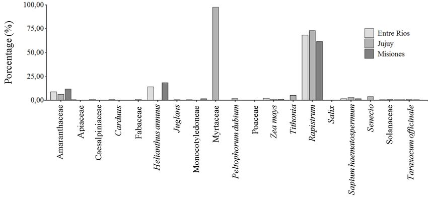
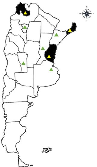
Tejerina et AL.          Ascosphaera apis isolated from commercial pollen

al., 2012).                                           have developed physical and biological defense
Although the Ascosphaera species are spread           mechanisms against pathogens, including the
throughout the hive by larva cells and feces          secretion of antimicrobial peptides, phagocy-
within the brood nest (Albo et al., 2017), pollen     tosis, melanization, enzymatic degradation of
reservoirs are known to contribute to the distri-     pathogens (Evans et al., 2006). The physical
bution of ascospores (Wynns et al., 2013; Pereira     barriers involved in the defense against
et al., 2019). Many winter beekeepers feed            pathogens are associated with the intestinal en-
their hives commercial pollen and sugar syrup         vironment of the insect what acts as a filter for
to strengthen their colonies after the season         microorganisms seeking access to the epithelial
(Ahmed, 2008). Pathogens and parasites are            cells of the midgut (Siva-Jothy et al., 2005;
transmitted with contaminated pollen among            Lundgren & Jurat-Fuentes, 2012).
hives and cause serious damage to apiaries not        Although entomopathogenic strains have been
previously exposed to them.                           registered in such countries as the United
The fungus spores are known to be viable for          States, Spain, New Zealand, Australia and
about fifteen years; they are transported in the      Canada (Albo & Reynaldi, 2010). They have been
pollen brought by the foraging bees to the colony,    reported in southern Argentina (Reynaldi et al.,
causing greater risk to the brood (Flores et al.,     2003; Reynaldi et al., 2015) and the neighboring
2005; Lopes et al., 2015). Furthermore, pollen        countries of Chile (Reynaldi et al., 2003), Brazil
bread and preserved pollen were reported to be        (Castagnino et al., 2006) and Uruguay (Rivas
reservoirs of entomopathogenic spores, which          & Bettucci, 2007), but there have been no
remain viable for months (Menapace, 1980; Hale        reports for northern Argentina. We investigat-
Wang & Granados, 2000). Ascospores of Spanish         ed pollen from Argentinian provinces because
strains remain viable in commercial pollen after      it is available on the local market and many
two years of conservation (Tejerina et al., 2019).    producers use commercial pollen to feed their
Larvae are the most susceptible between the           bee colonies in winter.
fourth and fifth days of age, although some           The objective of this work was to isolate,
authors argue that this happens between the           identify, and characterize the A. apis strains of
second and fourth days before the cells are           commercial pollen grains from different Argen-
sealed. Therefore, larvae must have consumed          tinian provinces and their potential pathogenic-
A. apis spores for the disease to start (Flores       ity in local bees.
et al., 1996; Jensen et al., 2013; Getachew et al.,
2018).                                                MATERIAL AND METHODS
This disease not only affects the larvae of
worker bees but also those of drones and              Isolation of A. apis
queens (Wynns, 2012). While it has not been           Commercial pollen was obtained dried in 2014,
proven to affect adult bees, they would transmit      2015 and 2016; one random sample from each
the disease among hives. Twenty-four hours            province was acquired from herbalists from
after the larvae have ingested the ascospores,        the Argentinian provinces of Buenos Aires,
the first signs of the disease appear, starting       Corrientes, Entre Ríos, Jujuy, Misiones, San
with a reduction in the diet (food consumption).      Luis, Santa Fe and Tucumán (Fig. 1). Although
The larvae die within 48 h, and fungal mycelia        the treatment that each sample received is
become visible on the surface of the larvae           not known, the norms for commercialization
after 72 h (Aronstein & Murray, 2010). Enzyme         proposed by the National Administration of
machinery involved in the virulence of A. apis        Drugs, Food and Medical Technology had to be
or genes involved in toxin biosynthesis could         followed, which guarantees that Argentinian
act as virulence factors helping the pathogen in      products are effective, safe and of good quality.
the invasion of the host (Cornman et al., 2012;       The eight samples received from herbalists
Getachew et al., 2018). Thus, A. mellifera bees       were stored at 20ºC in the dark until they

 148
J. APIC. SCI. Vol. 65 No. 1 2021
                                                     samples were prepared with lactophenol blue
                                                     and observed at 40X and 100X under a Zeiss
                                                     microscope; 1000 sporoscysts and the average
                                                     size of asci and ascospores were measured (µm).

                                                     Analysis and identification of A. apis strains
                                                     Genomic DNA was obtained from the collected
                                                     samples, using the mycelium grown in MY20
                                                     liquid medium. The mycelium was washed twice
                                                     with 1 mL of cold solution (Tris-HCl 0.1M, EDTA
                                                     0.02M) and spun at 12,000 rpm for 3 min. Sub-
                                                     sequently, the mycelium was crushed with
                                                     a sterile glass rod in the presence of 1 mL of
                                                     extraction buffer (Tris-HCl 100mM pH 8, NaCl
                                                     1.5M, EDTA 50mM pH 8, Proteinase K, 0.1mg/
                                                     ml, β-mercaptoethanol 10 mM, SDS 2%) and
                                                     digested at 60ºC by agitation with vortex every
                                                     10 min. The DNA was purified from the superna-
                                                     tant of this digestion using chloroform:isoamyl
                                                     alcohol (24:1) twice and potassium acetate 3M
                                                     only once. The DNA was subsequently precipi-
                                                     tated with 100% isopropanol, 70% cold ethanol
                                                     wash, spun at 12,000 rpm for 5 min, dried at
                                                     room temperature, resuspended in distilled nu-
                                                     clease-free water and stored at -20ºC until use.
                                                     Genomic DNA was revealed in agarose gel at
                                                     1%, with 3 μL of ethidium bromide (0.008 mg/
                                                     μL) for every 75 mL of agarose solution.
Fig. 1. Distribution of pollen samples collected
from apiaries in Argentinian provinces. The yellow   Amplification and sequence of analysis of
triangles denote the isolated samples of A. apis,    the internal transcribed spacer (ITS) region
while the green triangles denote the provinces
                                                     To characterize the fungal isolates, the ITS1-
where the samplings were carried out but the
fungus A. apis was not isolated.
                                                     5.8S-ITS2 region of the ribosomal DNA was
                                                     amplified using the primers described by White
were processed in the laboratory. Then, 1 g of       et al. (1990), ITS1 5’-TCCGTAGGTGAACCTGCGG
each pollen sample was seeded on a selective         and ITS4 5’-TCCTCCGCTTATTGATATGC. The PCR
medium containing agar malt and yeast extracts       was carried out in 20 μl of the final volume
with 20% glucose (hereafter MY20) (Takatori &        containing 1X buffer, magnesium chloride 2.5
Tanaka, 1982) and incubated under microaero-         mM, dNTPs 200 μM, 10ρM of each primer and
philia conditions at 30±2°C for fourteen days.       0.5 U Taq polymerase, using a cycle composed
                                                     of an initial denaturation for 4 min at 94ºC and
Characterization of the isolated strains             thirty-five cycles of 40 s at 94ºC, 40 s at 52ºC,
The fungal strains were selected according to        and 40 s at 72ºC, with a final extension of 10
their macroscopic characteristics in the MY20        min at 72ºC.
culture medium. The shape, size and color of         The PCR products were checked on agarose gels
the colony as well as the microscopic features,      at 2%, sequenced using the Macrogen Korea
namely shape and size of sporocysts, asci, and       service and analyzed with the MEGA6 program.
ascospores (µm) were taken into account. The         The sequences were contrasted against the

                                                                                                  149
Tejerina et AL. Ascosphaera apis isolated from commercial pollen
database of the NCBI (http://www.ncbi.nlm.nih. The bees used for the assay were maintained
gov/) using the BLASTn tool. The sequences in Langstroth hives on an experimental farm
were checked and corrected before being in Severino (Faculty of Agrarian Sciences of
deposited in the GenBank database the National University of Jujuy), located in El
Carmen department, Jujuy province.
Determination of the phylogenetic relation-
ships of the strains Assay preparation
All sequences were analyzed using BioEdit and We used three blind cores (no initiating queen),
Clustal W before tree construction. A phyloge- that had been prepared in the spring of 2016
netic analysis was carried out using the program and contained a standard open breeding frame,
TNT (tree analysis using New Technology), a breeding frame operculated with nurse bees
which performs phylogenetic analysis of and a frame with honey, as proposed by Ahmed
parsimony (Goloboff et al., 1999). The gaps (2008). We waited until the birth and posture of
(indels) were treated as a fifth state since they the queen. When the nuclei grew and consoli-
represented insertion-deletion events. The dated, with about 10,000 bees per frame, they
analysis included twenty-four sequences of were transferred to three standard brood boxes
the genus Ascosphaera spp. Aspergillus flavus and allowed to grow again until we obtained a
(NG_055742) was used as an outgroup. The population of approximately 50,000 bees, as
data set was reduced in the heuristic search proposed by Audisio & Benítez-Ahrendts (2011).
by implementing 100RAS and guarding a tree Above this brood nest, we placed a medium
by TBR. To evaluate the support in identifying super frame (without a queen excluder, to allow
groups, we conducted bootstrap and jackknife the queen to pass), which contained twenty-
parsimony analyses. Both analyses included four experimental frames specially designed for
1000 resampled matrices. For each resampled assays, measuring 20x10 cm. When the worker
matrix, we performed one hundred RAS-TBR bees stretched the beeswax, the posture of
cycles (Fonseca et al., 2016). the queen was observed, and then when the
first bees were born, the state of the hives was
Palynological analysis monitored monthly. Trials were conducted in the
Argentinian samples of corbicular pollen were late spring of 2017 and 2018 when tempera-
disintegrated and homogenized with a mortar tures were below 20ºC, the morning tempera-
and 0.5 g of the total was taken for additional ture registered in this season in Jujuy province.
treatment with acetolysis (Erdtman, 1960). The The three hives used for the study were
pellet was glycerin mounted and paraffin sealed. eliminated at the end of the trial to prevent the
Observations were made with a Boeco optical spread of the disease.
microscope. To determine frequency classes,
600 pollen grains were counted (Louveaux Activation of A. apis
et al., 1978). Pollen types were compared and Portions of each fungal strain inoculated with
identified to the palynological deposits from the agar and mycelium (about 36 mm2) were placed
palynology laboratory of the Faculty of Agrarian on Petri dishes in the middle of the MY20 culture
Sciences, National University of Jujuy, and by and the fungi were grown for ten days until the
consulting the palynological atlas. maturation of sporocysts, under microaerophilia
conditions at 30±1ºC. Microscopic observation
Pathogenicity assay was performed to confirm the presence of
Bees and apiary mature spores with a 40 X objective.
The bees used to carry out the assays belong
to the hybrid subspecies A. m. scutellata, Preparation of the inoculum
present in the province of Jujuy, Argentina, as An aliquot was taken from each A. apis strain
reported by Porrini et al. (2020) in this region. and suspensions were made in tubes with 5 mL
150J. APIC. SCI. Vol. 65 No. 1 2021
of sterile water-peptone (10 g of peptone, 5 g of minimum concentration and the maximum con-
sodium chloride in 1 liter of distilled water). To centration, respectively, while the third group
release the asci and ascospores contained in the was used as control (food without ascospores)
sporocysts, each tube containing glass beads (See Tab. 3). After feeding, the frames were
was vortexed for thirty seconds. Counts were taken to the original hive so that the cells could
performed using a Neubauer chamber, with the be covered by worker bees. At 48 h, the frames
concentration adjusted between 5x10³ spores/ were removed from the apiary and incubated in
mL (minimum) and 5x10⁶ spores/mL (maximum). the laboratory for ten days in hives designed for
These suspensions were kept at 5ºC until their the frames. The mummified larvae in each cell
administration (Jensen et al., 2013; Ansari et al., were then counted. To replicate the humidity
2017). and temperature environmental conditions in
Before being administered to the hives, the the laboratory, we used a stove at 30±1ºC with
ascospore suspensions were spun at 4000 60% humidity.
rpm for 5 min and the cell pellets were resus-
pended in 5 mL of sterile water and honey (1:1). Statistical analysis
For the control groups (without ascospores), a Data on the number of infected larvae were
suspension of 5 mL of sterile water and honey expressed as mean ± standard deviation of
was used (1:1). each assay. Statistical analysis of results was
performed using ANOVA and Tukey’s mean
Selection and feeding of larvae with comparison test, with a 0.5 probability of making
ascospores a type I error, using Infostat as the statistical
The brood chambers of the three assayed package (Di Rienzo et al., 2008).
colonies were checked before the administration
of the ascospores and showed no symptoms or RESULTS
signs of the disease. The uniform posture of the
queen and the presence of larvae were verified. Isolation and characterization of A. apis.
Ten open breeding trial frames containing 5- to strains
6-day-old larvae were selected for the inocula- Three strains of the genus A. apis were isolated
tion of the fungal strains. Two repetitions per from pollen grains of the provinces of Jujuy,
area of 20x10 cm were performed and areas Misiones, and Entre Rios, Argentina. They were
with capped and uncapped cells were counted identified by their macro and microscopic char-
before the administration of the strains. acteristics (Tab. 1, Fig. 2). The development of
Subsequently, all larvae were fed using a sterile the sporocysts was observed at five days of
2.5 mL syringe. They were divided into three incubation. The mycelium exhibited a white
groups; the first two were administered a cottony, aerial, mostly bright consistency,
drop of approximately 5 μL of each ascospore and its hyphae were covered with droplets of
suspension at different concentrations: at the “exudate”. When the asci ripened, they looked
Table 1.
Macroscopic description of isolated Ascosphaera spp. strains
Sporocyst
Ascus diameter Ascospores length
Ascosphaera spp. strains Origins diameter
average (µm) average ( µm)
(µm)
A. apis PMis Misiones 60-75 9 1-2
A. apis PJuy Jujuy 65-70 9 1-2
A. apis PER Entre Ríos 60-70 8 1-2
151Tejerina et AL. Ascosphaera apis isolated from commercial pollen
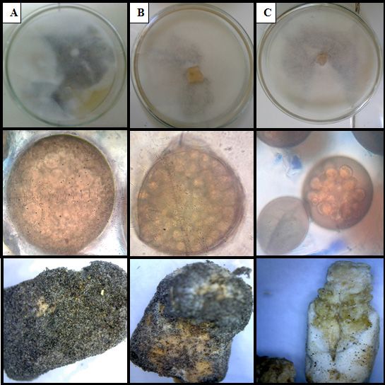
Fig. 2. Macroscopic and microscopic observation in MY20 culture medium.
A) Growth and sporulation of A. apis strain PMIS (Misiones), sporocyst observed at 100X and mummified
larvae observed at 10X.
B) Growth and sporulation of A. apis strain PJUY (Jujuy), sporocyst observed at 100X and mummified larvae
observed at 10X.
C) Growth and sporulation of A. apis strain PER (Entre Ríos), sporocyst observed at 40X and mummified
larvae observed at 10X (without sporocyst).
black. We observed microscopic spherical brown Ríos, and 610bp for Jujuy, and shared 100%,
sporocysts, containing globose asci whose 98.3%, and 99.02% identity, respectively, with
ascospores were hyaline and ellipsoid. The other strains from the NCBI database. They were
provinces of Tucumán, Buenos Aires, San Luis, registered with the following access numbers:
Corrientes and Santa Fe had not registered the MH633694 (Misiones), MH633695 (Entre Ríos),
presence of A. apis in commercial pollen, but and MH633693 (Jujuy).
strains belonging to the genera Aspergillus, In addition, when the botanical origin of each
Fusarium, Penicillium, Rhizopus and Mucor were pollen sample was identified, interestingly,
isolated and identified. the predominant pollen belonged to the genus
Rapistrum and the family Amaranthaceae. While
Molecular identification of A. apis strains and the species Helianthus annus was only recorded
registration of their botanical origin in the pollen of Entre Ríos and Misiones, a large
The A. apis strains corresponding to the three percentage of pollen from Jujuy corresponded
provinces yielded polymorphic fragments of to the family Myrtaceae (Tab. 2, Fig. 3).
304bp for the Misiones strain, 482bp for Entre
152J. APIC. SCI. Vol. 65 No. 1 2021
Table 2.
Botanical origin associated with the three strains of Ascosphaera apis isolated from commercial
pollen in Argentina
Porcentage by provincie
Pollen types
Entre Ríos Jujuy Misiones
Amaranthaceae 8.87 6.42 12.05
Apiaceae 0.55 0.28 0
Caesalpiniaceae 0.92 0 0
Carduus 1.11 0 0.17
Fabaceae 0 1.4 0
Helianthus annuus 14.23 0 18.59
Juglans 0 0.56 0
Monocotyledoneae 0.74 0 1.55
Myrtaceae 0 97.6 0
Peltophorum dubium 0 1.96 0
Poaceae 0 0 0.34
Zea mays 2.22 1.4 1.38
Tithonia 0 5.31 0
Rapistrum 68.58 73.18 61.96
Salix 0 0 0.34
Sapium haematospermum 1.66 3.07 1.72
Senecio 0 3.91 0
Solanaceae 0.55 1.12 0.69
Taraxacum officinale 0.55 1.4 0.52
Phylogenetic relationships 100% support in the bootstrap and jackknife
A total of 872bp were aligned, 299 sites were processes. The A. atra and A. duoformis strains
considered monomorphic, 504 sites were also shared a common ancestor with the species
considered polymorphic or phylogenetically in- A. subcuticulata, A. solina, and A. osmophila
formative segregants, and 359 polymorphic with 75% jackknife support and 70% bootstrap
sites had more than two variants. The parsimony support (Fig. 4).
analysis resulted in two parsimonious trees, The A. apis strain and other species of the As-
with 1889 steps. cosphaera genus have a common ancestor with
Ascosphaera spp. strains were grouped into 100% jackknife support, while for the bootstrap
different phylogenetically related groups. Thus, process it is located next to the strains of
all A. atra strains shared a common ancestor with Russia, Australia, the United States and all other
153Tejerina et AL. Ascosphaera apis isolated from commercial pollen Fig. 3. Percentage of pollen containing A. apis. Fig. 4. Phylogenetic relationship of A. apis strains. The tree was carried out using 1000 replications and bootstrap and jackknife parsimony. The numbers in parentheses correspond to the jackknife support. 154
J. APIC. SCI. Vol. 65 No. 1 2021
Spanish strains. This group contains the strainscentages close to 11%, while those from Jujuy
from Entre Ríos and Misiones. By contrast, the  and Misiones registered infection values close to
strain from Jujuy belongs to another monophy-   27% and 23%, respectively (Tab. 3). The strains
letic group with strains from the Netherlands   from Misiones and Entre Ríos produced 2% of
and share an immediate common ancestor with     mummified larvae without sporocysts (white
those of the United States and the Spanish      mummies), while the strain from Jujuy caused
strains A. apis M05 and M10. Furthermore, the   6%. Closed cells previously capped by bees were
A. celerrima and A. flava strains formed a mono-not significant for the trial.
phyletic group with similar supports separating The larvae inoculated with A. apis strains
from the species of A. apis and A. atra (Fig. 4).
                                                were mummified between days 7 and 10 of
                                                incubation. The size of the mummified larvae of
Pathogenicity assays                            worker bees was between 7.5 mm and 8 mm,
The minimum concentration of ascospores while that of drones was between 11 and 13
administered in sugar syrup did not differ sig- mm (Fig. 2).
                                                                                               Table 3.
                       Number of larvae mummies with A. apis and no mummies.
                               Values are mean ± standard deviation

                 Number of mummies                                             Number of
                                                Number of    Number of                         Total
                                                                                infected
                                               non-mummi- infected larvae                    number
  Strains     (5x10³     (5×10⁶                                                  larvae
                                    Without     fied larvae   (5x10³                        of capped
             spores/     spores/                                                 (5x10⁶
                                   sporocyst     (Witness)  spores/mL)                         cells
                mL)        mL)                                                 spores/mL)
 Misiones        0        30±7       3±3        100±40            138±1         127±29        43±6
Entre Ríos       0        10±1       2±2         112±2           128±12          83±9         28±11
  Jujuy         1±1       23±3       5±1        102±32           134±10         85±43         54±8

nificantly (ANOVA, p=0.83) in the number of
mummified larvae. No mummified larvae were
detected in connection with the Entre Rios
or Misiones strains, and only the Jujuy strain
showed the presence of 1±1 mummified larva
(Tab. 3). In contrast, when the suspension with
the maximum concentration of ascospores
was administered to the larvae, the registered
number of mummified larvae with each of the
fungal strains was higher (Misiones 30±7, Entre
Rios 10±1 and Jujuy 23±3) than the number of
mummified larvae fed the minimum concen-
tration of ascospores (Tab. 3). The number of
mummified larvae with high concentrations of
ascospores exhibited significant differences
between the pathogenic effects of the strains
from each province (ANOVA, p
Tejerina et AL. Ascosphaera apis isolated from commercial pollen
DISCUSSION Therefore, the presence of Ascosphaera spp.
strains could be explained as a deficiency in the
In this study, we isolated strains belonging to treatment of the collected pollen.
the genus Ascophaera from commercial pollen The morphological observation indicated that
of three Argentinian provinces, where local the size of the strains’ sporocysts and the
circulation of the fungal strain identified as ascospores were similar to those reported in
A. apis had not been reported. The presence other works from both southern Argentina
of the entomopathogen was also recorded in and other parts of the world (Albo & Reynaldi,
commercial pollen of Spanish provinces, where 2010; Aronstein & Murray, 2010; Wynns et al.,
the presence of this fungus had been reported 2013; Chen et al., 2018; Pereira et al., 2019). The
before (Tejerina et al., 2019). In other studies, phylogenetic analysis allowed us to establish
Ascosphaera spp. has been isolated from A. relationships between strains found in some
mellifera larvae, Apis cerana, Xylocopa californi- countries and isolates of commercial pollen
ca arizonensis, and adult bumblebees (Reynaldi from Argentina. The A. apis strains from pollen
et al., 2015; Chen et al., 2018). The presence of grains from the provinces of Misiones and Entre
A. apis has also been reported in honey samples Ríos were grouped together, while the strain
from the province of Buenos Aires, Argentina isolated from pollen grains from Jujuy was
(Reynaldi et al., 2003), but this province yielded placed in another group, evidencing polymorphic
negative results for the pollen sample analyzed variations within the group of Argentina. This
in this study. intraspecific variation was also observed by
Commercial pollen subjected to different Jensen et al. (2012), who described twelve poly-
treatments after harvesting, in which it is dried morphic variants of A. apis in Denmark and the
at temperatures below 40-45ºC, eliminates United States. In the same way, Tejerina et al.
excess humidity (6 to 7%) and preserves the (2019) showed that Spanish strains are grouped
bromatological characteristics required for its in a monophyletic clade and phylogenetically
commercialization (Aranda-Escribano et al., move away from other strains. In this study, we
1999; Baldi-Coronel et al., 2004). Although compared Ascosphaera spp. isolated from pollen
these treatments eliminate most of the microor- grains in some countries, checking for genetic
ganisms, an incorrect procedure could transport variation between regions.
diseases to the bee colonies, as demonstrat- Although multifloral pollen is known to contain
ed by Graystock et al. (2016), who recorded ascospores (Pereira et al., 2019), the palynologi-
the presence of Ascosphaera spp., Nosema cal origin associated with its transmission has
spp., among others, in pollen with different not been determined. In this work, botanical
treatments. Similarly, Zuluaga et al. (2015) determinations of pollen grains from Argentin-
recorded the presence of 105 UFC/gr of filamen- ian provinces are reported to corroborate their
tous fungi and yeasts in commercial pollen but provenance. In addition, the most representative
did not report the genus found. In the provinces percentages of each floral resource used by A.
where the presence of A. apis had not been mellifera in its diet are reported, with the genus
previously recorded, we found strains belonging Rapistrum, the families Myrtaceae and Ama-
to different genera, as also recorded by Bucio- ranthaceae, and the species Helianthus annus
Villalobos et al. (2010), who observed different being found in higher proportions in the three
percentages of the genus in commercial pollen: provinces, coinciding with Burgos & Sánchez
Aspergillus (3.6%), Alternaria (3.6%), Mucor (2015) and Méndez et al. (2018) (Jujuy), Aquino
(3.1%), Fusarium (2.9%), Penicillium (2.9%) and et al. (2015) (Misiones), and Ciappini & Vitelleschi
Rhizopus (0.7%). These authors attributed their (2013) (Entre Ríos). The reported plant groups
findings to a deficiency in the drying and con- could be reservoirs of the fungal strains found,
servation processes of pollen, although they did but more research is needed for confirmation.
not report the presence of Ascosphaera spp. Our pathogenicity assays of the different
156J. APIC. SCI. Vol. 65 No. 1 2021
strains in bee larvae allowed us to assess their that the subspecies A. m. scutellata is suscepti-
potential to infect and mummify larvae of local ble to contracting the disease caused by A. apis,
bees that have never been exposed to this en- this subspecies is known to be more resistant
tomopathogen. Flores et al. (2004) evaluated to infestation than other more susceptible
the percentage of mummified larvae that were European bee subspecies, including the Varroa
stressed at 18ºC for 24 h. These larvae came destructor ectoparasite (Principal et al., 2008;
from hives which were treated with water Nganso et al., 2017). The behavior of A. apis in
spray with ascospores, pollen with ascospores other populations of Argentinian bees needs
or sugar syrup containing the ascospores; further investigation.
the third technique was the least effective in Strains of A. apis were isolated and identified
achieving a pathogenic effect. These assays from commercial pollen of Argentinian regions,
were performed with a concentration of 1,250 where the prevalence of this entomopathogen
spores/hives. In this work, a concentration of had not been recorded, showing that the pollen
5x10⁶ spores/mL was used and the percentage commercialized in these areas is not treated
of mummies was lower than that found by Flores properly and therefore is a reservoir of different
et al. (2004) and Palacios et al. (2007), when pathogenic fungal strains of bees. The phylo-
they used sugar syrup. Both authors warned genetic analysis differentiated A. apis strains
that the percentage of mummification depends of Misiones and Entre Ríos from those of Jujuy,
on the stress conditions of the larvae when suggesting genetic variation between regions.
they receive the ascospores or on their suscep- The botanical origin of the pollen was found
tibility to contract the ectoparasite according to to belong to the mentioned regions, and it
the period of the year. indicates that certain pollen resources used by
The administration of the minimum concentra- A. mellifera could be reservoirs of fungal strains,
tion of ascospores to the larvae did not result including the A. apis fungus. The first results
in the disease. Jensen et al. (2013) indicated were obtained on the pathogenicity of Argen-
that the minimum concentration for successful tinian fungal strains on larvae.
infection in healthy hosts should be 5x10⁵
ascospores/mL, which could lead to the death of ACKNOWLEDGMENTS
the population. The pathogenicity also depends
on the origin of the inoculum with ascospores. Part of the experimental work was funded
Flores et al. (2004) performed the infection by Secretaría de Ciencia y Técnica de Estudios
techniques with ascospores from macerated Regionales, Universidad Nacional de Jujuy
bee mummies, while Palacios et al. (2007) (SeCTER-UNJu) and Consejo Nacional de Investi-
maintained ascospores on integral rice kernels gaciones Científicas y Técnicas (CONICET-Argen-
(IRK) medium and MY20. The lower percentages tina).
of infection of the larvae achieved by the direct
method of feeding than those obtained in other REFERENCES
works could be attributed to the ascospores
coming from the cultivation of commercial Ahmed, A., (2008). Manual Apícola del Norte Argen-
pollen. tino. Magna, Tucumán, Argentina.
In addition, this low percentage of mummified
larvae in comparison to other studies can also Albo, G.N., & Reynaldi, F.J. (2010). Ascosphaera apis,
be attributed to the genetic characteristics of agente etiológico de la cría yesificada de las abejas.
the bee colonies kept in Jujuy, since hybrids of Revista Argentina de Microbiología, 42(1), 80.
Africanized bees belonging to the subspecies
of A. m. scutellata have been reported to be Albo, G., Córdoba, S., Reynaldi, F. (2017). Chalkbrood:
present in this region, as shown by Porrini et pathogenesis and the interaction with honeybee
al. (2020). Although few reports have indicated defenses. International Journal of Environmental &
157Tejerina et AL. Ascosphaera apis isolated from commercial pollen
Agriculture Research, 3(1), 71-80. Fernández, G. (2004). Caracterización bromatológica
del polen apícola argentino. Ciencia, Docencia y Tec-
Alonso, J.M., Rey, J., Puerta, F., Hermoso de Mendoza, nología, 15(29), 145-181. Retrieved July 8, 2020, from
J., Hermoso de Mendoza, M., Flores, J.M. (1993). Enzy- https://www.redalyc.org/articulo.oa?id=14502906
matic equipmet of Ascosphaera apis and the devel-
opment of infection by this fungus in Apis mellifera. Borum, A.E., & Ulgen, M. (2008). Chalkbrood (As-
Apidologie, 24(4), 383-390. https://doi.org/10.1051/ cosphaera apis) infection and fungal agents of
apido:19930404 honey bees in north-west Turkey. Journal Apicultural
Research, 47(2), 170-171. https://doi.org/10.3896/
Ansari, M.J., Al-Ghamdi, A., Usmani, S., Khan, K.A., ibra.1.47.2.16
Alqarni. A.S., Kaur, M., Al-Waili, N. (2017). In vitro
evaluation of the effects of some plant essential Bucio-Villalobos, C.M., López-Preciado, G., Martínez
oils on Ascosphaera apis, the causative agent of Jaime, O.A., Torres-Morales, J.J. (2010). Micoflora aso-
Chalkbrood disease. Saudi Journal of Biological Sci- ciada a granos de polen recolectados por abejas
ences, 24(5), 1001-1006. https://doi.org/10.1016/j. domésticas (Apis mellifera L). Nova scientia, 2(4), 93-
sjbs.2016.04.016. 103. https://doi.org/10.21640/ns.v2i4.212
Aquino, D., Pellizzer N., Miranda D., Salgado C. Burgos, M.G., & Sánchez A.C. (2014). Preferencias ali-
(2015). Contenido polínico de mieles de Apis mel- menticias en las mieles inmaduras de Apis mellifera
lifera L. producidas en Misiones, Argentina. Revista en el chaco serrano (Jujuy, Argentina). Boletín de la
Forestal Yvyraretá, 22, 7-12. Sociedad Argentina de Botánica, 49(1), 41-50. http://
dx.doi.org/10.31055/1851.2372.v49.n1.7820
Aranda-Escribano, M.L. Cardenal Galvan, J.A., Álvarez
Gomez, J.A., Pozovera, J. (1999). El polen, controles Castagnino, G.L.B., Funari, S.R.C., Blume, E., Arboitte,
sanitarios. Normas Legales. Vida Apícola (España), M.Z., Weber, M. (2006). Doença Cria Giz Ascosphaera
94, 56-59. apis (Maassen ex Claussen) Olive & Spiltoir em abelhas
Apis mellifera L. na Depressão Central do Rio Grande
Aronstein, K., & Murray, K. D. (2010). Chalkbrood do Sul. Ciência Rural, 36(6), 1909-1911. https://doi.
disease in honey bees. Journal Invertebral Patho- org/10.1590/S0103-84782006000600038
loly, 103(1), S20-S29. https://doi.org/10.1016/j.
jip.2009.06.018 Chen, D., Guo, R., Xiong C., Zheng, Y., Hou, C., Fu Z.
(2018). Morphological and molecular identification
Aronstein, K., & Holloway B. (2013). Honey bee of chalkbrood disease pathogen Ascosphaera apis in
fungal pathogen, Ascosphaera apis; current under- Apis ceranae. Journal of Apicultural Research, 57(4),
standing of host-pathogen interactions and host 1-6. https://doi.org/10.1080/00218839.2018.14759
mechanisms of resistance. Microbial Pathogenesis, 43
13(1), 402-410.
Ciappini, M.C., & Vitelleschi, M.S. (2013). Característi-
Audisio, M.C., & Benítez-Ahrendts, M.R. (2011). Lac- cas palinológicas de mieles de eucalipto (Eucalyptus
tobacillus johnsonii CRL1647, isolated from Apis mel- sp.) y tréboles ( Trifolium sp.) provenientes de la pro-
lifera L. bee-gut, exhibited a beneficial effect on vincia fitogeográfica pampeana argentina. Revista
honeybee colonies. Beneficial Microbes, 2(1), 29-34. FCA UNCuyo, 45(1), 247-258.
https://doi.org/10.3920/BM2010.0024
Cornman R.S., Bennett A.K., Murray K.D., Evans J.D.,
Bailey, L. (1981). Honey Bee Pathology. (pp. 40–44) Elsik C.G., Aronstein, K. (2012). Transcriptome analy-
London: Academic Press. sis of the honey bee fungal pathogen, Ascosphaera
apis: implications for host pathogenesis. BMC
Baldi Coronel, B., Grasso, D., Chaves Pereira, S., Genomics, 13(1), 285. http://doi.org/10.1186/1471-
158J. APIC. SCI. Vol. 65 No. 1 2021
2164-13-285
Goloboff, P.A., (1999). Analyzing large data sets in
Di Rienzo, J.A., Casanoves, F., Balzanini, M.G., Gonza- reasonable time: solution for composite optima.
lez, L., Tablada, M., Robledo, C.W. (2008). Grupo Infos- Cladistics, 15(4), 415-428. https://doi.org/10.1006/
tat, versión 2008, FCA, Univ. Nacional de Córdoba, clad.1999.0122
Argentina.
Graystock, P., Jones, J.C., Pamminger, T., Parkinson,
Erdtman, G. (1960). The acetolysis method, a revised J.F., Norman, V., Blane, E.J., … Hughes, W.O.H. (2016).
description. Svensk Botanisk Tidskrift. 54, 561-564. Hygienic food to reduce pathogen risk to bumble-
bees. Journal of Invertebrate Pathology, 136, 68-73.
Evans, J.D., Aronstein, K., Chen, Y.P., Hetru, C., Imler, https://dx.doi.org/10.1016/j.jip.2016.03.007
J-L., Jiang, H., … Hultmark D. (2006). Immune path-
ways and defence mechanisms in honey bees Apis Hale, P.J., & Menapace, D.M. (1980). Effect of time and
mellifera. Insect Molecular Biology, 15(5), 645-656. temperature on the viability of Ascosphaera apis.
https://doi.org/10.1111/j.1365-2583.2006.00682.x Journal Invertebral Patholology, 36(3), 429-430. htt-
ps://doi.org/10.1016/0022-2011(80)90050-6
Flores J.M., Gutiérrez I., Puerta F. (2004). A compari-
son of methods to experimentally induce chalkbrood Holloway, B., Tarver, M.R., Rinderer, T.E. (2013). Fine
disease in honey bees. Spanish Journal of Agricul- mapping identifies significantly associating markers
tural Research, 2(1), 79-83. https://doi.org/10.5424/ for resistance to the honey bee brood fungal disease,
sjar/2004021-63 chalkbrood. Journal of Apicultural Research, 52(3),
134-140. https://doi.org/10.3896/ibra.1.52.3.04
Flores, J.M., Gutiérrez, I., Espejo R. (2005). The role of
pollen in chalkbrood disease in Apis mellifera: trans- Jensen, A.B., Aronstein, K., Flores, J.M., Vojvodic, S.,
mission and predisposing conditions. Mycologia, Palacio, M.A., Spivak, M. (2013). Standard methods
97(6), 1171-1176. https://doi.org/10.3852/mycolo- for fungal brood disease research. Journal of Apicul-
gia.97.6.1171 tural Research, 52(1), 1-21. https://doi.org/10.3896/
IBRA.1.52.1.13
Flores, J.M., Ruiz, J.A., Ruz, J.M., Puerta, F., Busto, M.,
Padilla, F., Campano, F. (1996). Effect of temperature Jensen, A.B., Welker, D.L., Kryger, P., James, R.R. (2012).
and humidity of sealed brood on chalkbrood develop- Polymorphic DNA sequences of the fungal honey
ment under controlled conditions. Apidologie, 27(4), bee pathogen Ascosphaera apis. FEMS Microbiology
185-192. https://doi.org/10.1051/apido:19960401 Letters, 330(1), 17-22. https://doi.org/10.1111/j.1574-
6968.2012.02515.x
Fonseca, M.I., Tejerina, M.R., Sawostjanik-Afanasiuk,
S.S., Giorgio, E.M., Barchuk, M.L., Villalba, L.L., Zapata, Koh, I., Lonsdorf, E.V., Williams, N.M., Brittain, C., Isaacs,
P.D. (2016). Preliminary studies of new strains of R., Gibbs, J., Ricketts, T.H. (2016). Modeling the status,
Trametes sp. from Argentina for laccase production trends, and impacts of wild bee abundance in the
ability. Brazilian Journal of Microbiology, 47(2), 287- United States. Proceedings of the National Academy
297. https://doi.org/10.1016/j.bjm.2016.01.002 of Sciences of the United States of America, 113(1),
140-145. https://doi.org/10.1073/pnas.1517685113
Getachew, A., Wubie, A.J., Wu, J., Xu, J., Wu, P., Abejew,
T.A., ... Xu, S.F. (2018). Molecular identification of Lopes, L.Q.S., Quatrin, P.M., De Sou-
pathogenicity associated genes in honeybee fungal za M.E., De Almeida Vaucher, R., Vianna
pathogen, Ascosphaera apis, by restricted enzyme- Santos, R.C. (2015). Fungal Infections In Honey
mediated integration (REMI) constructed mutants. Bees. Fungal Genomics & Biology 4(1), 1-4. https://
International Journal of Agriculture and Biology, doi.org/10.4172/2165-8056.1000118
20(12), 2879-2890.
159Tejerina et AL. Ascosphaera apis isolated from commercial pollen
Louveaux, J., Maurizio, A., Vorwhol, G. (1978). Methods Porrini, P.L, Quintana, S., Brasesco, C., Porrini, M., Gar-
of melisso-palynology by International Commission rido, M., Eguaras, M., Fernandez Iriarte, P. (2020).
of Bee Botany of IUBS. Bee World, 59(3), 139-157. Southern limit of Africanized honey bee in Argen-
https://doi.org/10.1080/0005772x.1970.11097312 tina inferred by mtDNA and wing geometric mor-
phometric analysis. Journal of Apicultural Research,
Lundgren, J., & Jurat-Fuentes, J. L. (2012). Physiology 59(4), 648-657. https://doi.org/10.1080/00218839.
and ecology of host defense against microbial in- 2019.1681116
vaders. In Insect Pathology. (pp. 461-480). 2nd Ed.
University of Tennessee, Knoxville. Principal, J., D’Aubeterre, Barrios, R, Carlos Puzzar, C.,
García de la Rosa, S., Fuselli, S.R., (2008). Hygienic
Maxfield-Taylor, S.A., Mujic, A.B., Rao, S. (2015). First behavior of africanized honeybees (Apis melliera
detection of the larval chalkbrood disease pathogen scutellata Lepeletier) in apiaries of Lara State. Ven-
Ascosphaera apis (Ascomycota: Eurotiomycetes: ezuela. Zootecnia Tropical, 26(2), 167-173.
Ascosphaerales) in adult bumble bees. PLoS ONE,
10(4), e0124868. https://doi.org/10.1371/journal. Reynaldi, F.J., López, A.C., Albo, G.N., Alippi, A.M. (2003).
pone.0124868 Differentiation of Ascosphaera apis isolates by rep-
PCR fingerprinting anddetermination of chalkbrood
Méndez, M.V., Sánchez, A.C., Flores, F.F., Lupo, L.C. incidence in Argentinean honey samples, Journal of
(2016). Análisis polínico de mieles inmaduras en el Apicultural Research, 42(4), 68-76. https://doi.org/10
sector oeste de las yungas de Jujuy Argentina. Bo- .1080/00218839.2003.11101096
letín de la Sociedad Argentina de Botánica. 51(3),
449-462. https://doi.org/10.31055/1851.2372.v51. Reynaldi, FJ, Lucia, M., Garcia Genchi, M.L. (2015).
n3.15390 Ascosphaera apis, the entomopathogenic fungus
affecting larvae of native bees (Xylocopa augusti):
Nganso, B.T., Fombong, A.T., Yusuf, A.A., Pirk, C.W., First Report in South America. Revista Iberoameri-
Stuhl, C., Torto, B. (2017). Hygienic and grooming cana de Micología. https://dx.doi.org/10.1016/j.
behaviors in African and European honeybee, New riam.2015.01.001
damage categories in Varroa destructor. PLoS ONE,
12(6), e0179329. https://doi.org/10.1371/journal. Rivas, F., & Bettucci L. (2007). Frecuencia y abun-
pone.0179329 dancia de Ascosphaera apis y otras especies de
hongos en las colmenas de Apis mellifera de
Ollerton, J., Winfree R., Tarrant S. (2011). How many Uruguay, Journal of Apicultural Research, 46
flowering plants are pollinated by animals? Oikos, (1), 1-2. https://doi.org/10.1080/00218839.2007.
120(3), 321-326. https://doi.org/10.1111/j.1600- 11101358
0706.2010.18644.x
Shin, Y.K., & Kim, K.Y. (2016). Macelignan inhibits
Palacios, M.A., Clemente, G., Ruffinengo, S., Escande, bee pathogenic fungi Ascophaera apis growth
A.R., Peña, N. (2007). Viability and pathogenicity of through HOG1 pathway. Brazilian Journal of Medical
Ascosphaera apis preserved in integral rice cultures. and Biological Research, 49(7), e5313. https://doi.
Spanish Journal of Agricultural Research, 5(4), 481- org/10.1590/1414-431X20165313
486. https://doi.org/10.5424/sjar/2007054-284
Siva-Jothy, M.T., Moret, Y., Rolff, J. (2005). Insect im-
Pereira, K.S., Meeus, I., Guy, S. (2019). Honey bee- munity: An evolutionary ecology perspective. In Ad-
collected pollen is a potential source of Ascosphaera vances in Insect Physiology (pp. 1–48). https://doi.
apis infection in managed bumble bees. Scien- org/10.1016/s0065-2806(05)32001-7
tific Reports, 9(4241), 1-9. https://doi.org/10.1038/
s41598-019-40804-2 Takatori, K., & Tanaka, I. (1982). Ascosphaera apis
isolated from chalkbrood in honey bees. Nihon Chi-
160J. APIC. SCI. Vol. 65 No. 1 2021
kusan Gakkaiho. 53, 89-92. https://doi.org/10.2508/ Wynns, A.A., Jensen, A.B., Eilenberg J. (2013). As-
chikusan.53.89 cosphaera callicarpa, a New Species of Bee Loving
Fungus, with a Key to the Genus for Europe. PLoS
Teerayut, T., & Chantawannakul, P. (2008). Pro- ONE, 8(9), e73419. https://doi.org/10.1371/journal.
tease and β-N-acetylglucosaminidase of honey pone.0073419
bee chalkbrood pathogen Ascosphaera apis. Journal
of Apicultural Research, 47(1), 68-76. https://doi. Zuluaga, C.M., Quicazán, M.C., Serrato, C.J. (2015).
org/10.3896/ibra.1.47.1.11 Valoración de la calidad microbiológica de polen apí-
cola sometido a diferentes tratamientos térmicos.
Tejerina, M.R., Cabana, M.J., Flores, J.M., Benítez-Ah- Retrieved July 8, 2020, from http://investigacion.
rendts, M.R. (2019). Studies of Ascosphaera apis bogota.unal.edu.co/fileadmin/recursos/direcciones/
strains isolated from commercial pollens of different investigacion_bogota/documentos/enid/2015/me-
Spanish provinces and their enzymatic production morias2015/ingenieria_tecnologias/valoracion_de_
capacity. Archivos de Zootecnia, 68(263), 324-330. la_calidad_microbiologica_de_.pdf
https://doi.org/10.21071/az.v68i263.4189
Wang, P., & Granados, R.R. (2000). Calcofluor dis-
rupts the midgut defense system in insects. Insect
Biochemistry and Molecular Biology, 30(2), 135-143.
https://doi.org/10.1016/s0965-1748(99)00108-3
Wynns, A.A. (2012). The bee specialist fungus family
Ascosphaeraceae and its allies: systematics, ecolo-
gy and co-evolution with solitary bees. Ph. D. thesis,
Department of Agriculture and Ecology, University
of Copenhagen. Denmark.
161You can also read