Analysis of Spatiotemporal Urine Protein Dynamics to Identify New Biomarkers for Sepsis-Induced Acute Kidney Injury - Frontiers
←
→
Page content transcription
If your browser does not render page correctly, please read the page content below
ORIGINAL RESEARCH
                                                                                                                                                   published: 03 March 2020
                                                                                                                                             doi: 10.3389/fphys.2020.00139

                                                Analysis of Spatiotemporal Urine
                                                Protein Dynamics to Identify New
                                                Biomarkers for Sepsis-Induced
                                                Acute Kidney Injury
                                                Yiming Li 1† , Junke Long 2† , Jiaquan Chen 3 , Jing Zhang 1 , Yi Qin 1 , Yanjun Zhong 4 , Fen Liu 3
                                                and Zhiyong Peng 1,5*
                                                1
                                                  Department of Critical Care Medicine, Zhongnan Hospital of Wuhan University, Wuhan, China, 2 Department
                                                of Cardiovascular Medicine, The Second Xiangya Hospital, Central South University, Changsha, China, 3 Department
                                                of Critical Care Medicine, The First Affiliated Hospital of Nanchang University, Nanchang, China, 4 ICU Center, The Second
                                                Xiangya Hospital, Central South University, Furong, China, 5 Center of Critical Care Nephrology, Department of Critical Care
                                                Medicine, University of Pittsburgh School of Medicine, Pittsburgh, PA, United States

                                                Acute kidney injury (AKI) is a frequent complication of sepsis and contributes to
                         Edited by:             increased mortality. Discovery of reliable biomarkers could enable identification of
                    Crystal A. West,
Georgetown University, United States
                                                individuals with high AKI risk as well as early AKI detection and AKI progression
                     Reviewed by:
                                                monitoring. However, the current methods are insensitive and non-specific. This study
                   Xiao-ming Meng,              aimed to identify new biomarkers through label-free mass spectrometry (MS) analysis
     Anhui Medical University, China            of a sepsis model induced by cecal ligation and puncture (CLP). Urine samples
           Carolyn Mary Ecelbarger,
Georgetown University, United States            were collected from septic rats at 0, 3, 6, 12, 24, and 48 h. Protein isolated
                   *Correspondence:             from urine was subjected to MS. Immunoregulatory biological processes, including
                        Zhiyong Peng            immunoglobin production and wounding and defense responses, were upregulated
                zn001590@whu.edu.cn
                                                at early time points. Kyoto Encyclopedia of Genes and Genomes (KEGG) pathway
     † These   authors have contributed
                    equally to this work
                                                enrichment analyses identified 77 significantly changed pathways. We further examined
                                                the consistently differentially expressed proteins to seek biomarkers that can be used
                   Specialty section:           for early diagnosis. Notably, the expression of PARK7 and CDH16 were changed in
         This article was submitted to
       Renal and Epithelial Physiology,
                                                a continuous manner and related to the level of Scr in urine from patients. Therefore,
               a section of the journal         PARK7 and CDH16 were confirmed to be novel biomarkers after validation in sepsis
               Frontiers in Physiology
                                                human patients. In summary, our study analyzed the proteomics of AKI at multiple time
      Received: 19 September 2019
                                                points, elucidated the related biological processes, and identified novel biomarkers for
       Accepted: 11 February 2020
         Published: 03 March 2020               early diagnosis of sepsis-induced AKI, and our findings provide a theoretical basis for
                             Citation:          further research on the molecular mechanisms.
        Li Y, Long J, Chen J, Zhang J,
     Qin Y, Zhong Y, Liu F and Peng Z           Keywords: sepsis-induced acute kidney injury, mass spectrometry, proteomics, biomarker, early diagnose
    (2020) Analysis of Spatiotemporal
    Urine Protein Dynamics to Identify
                                                Abbreviations: AKI, acute kidney injury; AUC, area under curve; CLP, cecal ligation and puncture; DAMPs, damage-
  New Biomarkers for Sepsis-Induced             associated molecular patterns; KDIGO, kidney disease improving global outcomes; KEGG, kyoto encyclopedia of genes
                   Acute Kidney Injury.         and genomes; LC-MS, liquid chromatography–mass spectrometry; MS, mass spectrometry; NF-κB, nuclear factor-kappa
                Front. Physiol. 11:139.         B; NGAL, neutrophil gelatinase-associated lipocalin; PAMPs, pathogen-associated molecular patterns; PCA, principal
      doi: 10.3389/fphys.2020.00139             components analysis; PPI, protein-protein interactions; ROC, receiver operating characteristic.

Frontiers in Physiology | www.frontiersin.org                                          1                                                March 2020 | Volume 11 | Article 139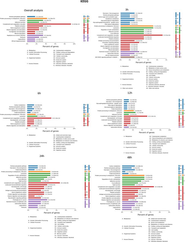
Li et al.                                                                                                        AKI Biomarkers Identified by MS

HIGHLIGHTS                                                              Unlike blood samples, urine samples can be collected non-
                                                                        invasively; moreover, the protein compositions of urine
- New biomarkers were identified by mass spectrometry analysis          samples are relatively simple, stable and easier to analyze
  in sepsis-induced AKI.                                                (Guo et al., 2018).
- Proteomics dynamic changes were exhibited at multiple time               Given the benefits offered by LC-MS methodology, the
  points.                                                               purpose of this study was to identify and validate early
- Immunoregulatory biological processes were upregulated at             biomarkers of sepsis-induced AKI using label-free proteomics,
  early time point.                                                     thus providing the possibility of early diagnosis. Bioinformatic
- PARK7 and CDH16 could accurately discern sepsis-induced               analysis of differentially expressed proteins suggested the
  AKI patients with high AUC.                                           involvement of these proteins in inflammation, immune
                                                                        responses, and metabolic regulation. In addition, we found
                                                                        new, specific biomarkers that can predict septic AKI. Our
INTRODUCTION                                                            work will have major impacts on the early prediction of and
                                                                        intervention for septic AKI.
Sepsis is associated with up to 50% of AKIs, and up to 60%
of septic patients have AKI complications (Uchino et al., 2005;
Bagshaw et al., 2009). The morbidity of AKI was found to                MATERIALS AND METHODS
be associated with the severity of sepsis in a cohort study
of 315 patients (Lopes et al., 2009). Because AKI has a                 Animal Model of AKI
high incidence and is always associated with poor outcomes,             Cecal ligation and puncture surgery was performed to induce
development of new approaches for the prevention and early              sepsis as previously described (Rittirsch et al., 2009). This model
diagnosis of AKI is of great importance (Vanmassenhove                  was modified based on the ligated percentage of the cecum,
et al., 2017). In addition, several notable drug trial failures         the number of punctures and contents of squeezed feces to
raised concerns that AKI was not diagnosed early enough                 induce AKI (Peng et al., 2014). We followed the methods of Li
for effective intervention and that a rise in serum creatinine          et al. (2019). Briefly, 10–12 weeks old male Sprague-Dawley rats
itself is not a sensitive enough marker (Griffin et al., 2018).         (400–500 g) were anesthetized with isoflurane, and a midline
Biomarkers have been proven to be important tools for                   incision (2 cm) was made below the diaphragm to expose the
current and future AKI research and clinical management                 cecum. Thirty percent of the cecum was ligated at the colon
(Griffin et al., 2019). Novel biomarkers can not only enable            juncture with a 3–0 silk ligature suture, punctured twice with
assessment of declines in kidney function but also indicate             an 18-gauge needle, and laced back in the abdomen; then, the
structural damage in the kidneys at earlier time points than            incision was closed in two layers. Sham surgery was performed
increases in serum creatinine. Early diagnosis of sepsis-induced        in the same way as the CLP surgery but without ligation and
AKI will enable appropriate and timely treatment, which                 puncture. Rats were fluid-resuscitated with 5 ml per 100 g saline
may contribute to better outcomes of AKI (Bagshaw et al.,               injected subcutaneously.
2009). Researchers have been searching for new biomarkers
to diagnose AKI with a novel detection technology. Thus,                Collection of Rats’ Urine and Blood
new biomarkers with the higher AUC values than current                  Metabolic cages (Nalgene, Thermo Fisher) were used for
biomarkers are expected to benefit the early diagnosis of AKI           collecting urine. Metabolic cages consist of a circular upper
(Bagshaw et al., 2009). The search for biomarkers associated            portion, which houses the rat, and a lower collection chamber
with early diagnosis will aid in solving the clinical problems of       with a specialized funnel that separates fecal pellets and urine
sepsis-induced AKI.                                                     that fall through the grid floor for their collection into 2 separate
    Protein quantitation is an important method for the                 tubes. Rat urine samples were collected at 0, 3, 6, 12, 24, and
identification of new biomarkers, and MS combined with                  48 h after CLP surgery and preserved at −80◦ C for proteomic
liquid chromatography is becoming a crucial tool in both                analysis. Blood (0.4 ml) was drawn from a femoral vein catheter
biological and clinical research settings (Prasad et al., 2017).        at indicated time after CLP. We collect the blood with a heparin-
Liquid chromatography-tandem mass spectrometry (LC-                     coated syringe. Heparinized blood was centrifuged at 10,000 × g
MS/MS) is a powerful technology suited for a wide range of              for 10 min to separate the plasma, which was collected for
applications, including protein and peptide biomarker analysis          serum creatinine detection. All experiments were performed in
in clinical chemistry (Grebe and Singh, 2016). Due to its               accordance with Chinese legislation on the use and care of
straightforward method development for a wide variety of                laboratory animals and were approved by the Animal Care and
candidate analytes, LC-MS can often represent a more precise            Use Committee of Nanchang University.
alternative to immunobinding assays and biomarker panel
analyses in the field of targeted proteomics (Himmelsbach,              Evaluation of the Renal Function
2012; Rauh, 2012). The use of unlabeled protein profiles to             The serum concentration of creatinine was measured using
examine differentially expressed proteins in urine samples              commercial kit reagents (Institute of Jiancheng Bioengineering,
largely eliminates the variations and biases in replicate MS            Nanjing, China). The absorbance was detected by Thermo
measurements (Atrih et al., 2014; Montoya et al., 2019).                Scientific Microplate Reader.

Frontiers in Physiology | www.frontiersin.org                       2                                        March 2020 | Volume 11 | Article 139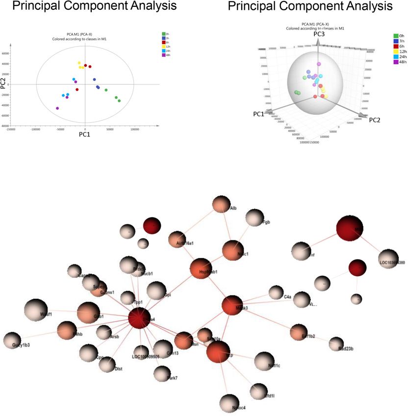
Li et al.                                                                                                          AKI Biomarkers Identified by MS

Sample Preparation for MS                                                  output increase
Li et al. AKI Biomarkers Identified by MS
the whole course of the disease were selected as the biomarkers
for early identification and diagnosis of AKI. Urine samples
from 30 patients with sepsis-induced AKI and 59 sepsis patients
without AKI were used for ELISA. The workflow is visualized in
Figure 1.
Principal Component Analysis and PPI
Analysis in Urine From Sepsis-Induced
AKI
Principal component analysis is a statistical technique for
simplifying datasets and determining principal components
(PCs) that best explain variation among samples. To obtain a
global protein profile, PCA score plots were first applied to
display the trends of the samples in the control (0 h) and
sepsis-induced AKI groups. The PCA model was based on two
principal components that summarized 75.4% of the variation
in the dataset in total (Figure 2A). The score plots revealed
that the control rats (0 h) and the rats with kidney injury were
obviously separated. There was a clear discrimination of the
positive fractions (0 and 3 h) from the negative fractions (12, 24,
and 48 h) by PC1, which contributed to most of the variance
(48%). In addition, from 0 to 12 h, the principal components
of urine were changed according to PC1, and from 12 to 48 h,
the variation was accounted for by PC2 to a large extent. PC2
contributed to 27.4% of the variance. PC3 contributed to only
5.17% of the variance (Figure 2B). PCA showed remarkable
differences among the six groups, demonstrating that sepsis
substantially influenced the urinary protein profiles.
One-way ANOVA was performed to determine the
differentially expressed proteins. There were 377 differentially
expressed proteins in urine after sepsis-induced AKI
(Supplementary Table S2). PPI network analysis of these
proteins was performed to further identify key proteins and
peptides associated with sepsis-induced AKI. The PPI network
was composed of 49 uniquely expressed nodes and 43 edges.
Slc2a4 and Mapk3 interact more with other proteins (Figure 2C).
Protein Profile Changed During
Sepsis-Induced Kidney Injury
To understand the change in the protein profile with time, one-
way ANOVA was performed. The fold change was defined as
FIGURE 1 | Overview of methods employed and results obtained. Rat urine
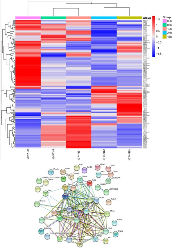
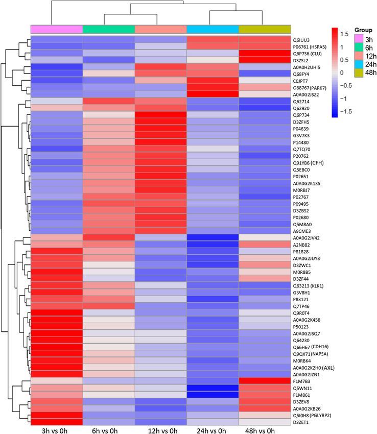
≥1.5 or ≤0.5. There were 123 differentially expressed proteins was collected at 0, 3, 6, 12, 24, and 48 h and subjected to label-free MS.
in urine after sepsis-induced AKI. Heatmap analysis showed the One-way ANOVA was performed with a p value cutoff of 0.05. For group
differentially expressed protein levels in the urine at the indicated classification, PCA, hierarchical clustering analysis and PPI were used, and
times (3, 6, 12, 24, and 48 h) compared with the levels at the significant proteins were identified. Furthermore, validation was performed on
selected proteins in human urine. AUC and a diagnostic cutoff value of
baseline (0 h) (Figure 3A).
differential expression protein were calculated by ROC curve. ANOVA, analysis
One aim of our study was to gain insight into the of variance; MS, mass spectrometry; PCA, principal component analysis; PPI,
pathways and networks altered in the urine from the onset protein-protein interaction (via OmicsNet and STRING). AUC, area under
of sepsis-induced AKI. Based on the STRING database web curve; ROC, receiver operating characteristic.
tool, PPI networks of the differentially expressed proteins
were constructed (Figure 3B). The PPI network featured
45 uniquely expressed nodes and 147 edges. The biological and defense response, were significantly changed. A vesicle,
process, cell components and molecular function in which extracellular exosome, and extracellular membrane-bound,
differentially expressed proteins were involved are shown organelle-associated proteins were excreted into the urine after
in Figure 4. The immunoregulatory biological processes, AKI. The Gene Ontology (GO) analyses of each time point
including immunoglobin production, response to wounding are shown individually in Supplementary Figures S2–S4. In
Frontiers in Physiology | www.frontiersin.org 4 March 2020 | Volume 11 | Article 139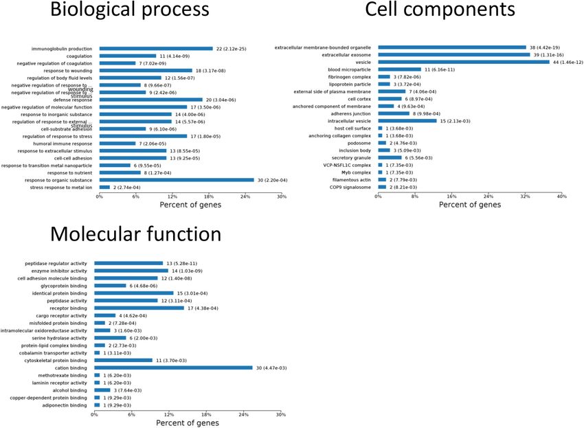
Li et al.                                                                                                                            AKI Biomarkers Identified by MS

  FIGURE 2 | PCA and PPI of urine proteomics. (A) PCA of urine proteomics. The PCA two-dimensional scatter plot represents the differential protein expression
  patterns of urine after sepsis. Axis: X = PC1: PCA Component 1 (48% variance); Y = PC2: PCA Component 2 (27.4% variance). (B) PCA were shown in the
  3-dimensional figure. (C) PPI between differentially expressed proteins was analyzed by OmicsNet.

the biological process (Supplementary Figure S2), the protein                        function of peptidase regulator activity, enzyme inhibitor activity,
regulating metabolic process was the earliest altered in the                         and peptidase activity increased in the early 6 h and remained at
first 3 h of sepsis-induced AKI, and subsequently (6 h later),                       a high level of expression (Supplementary Figure S4).
immunoglobulin production was increased significantly. From
6 to 24 h, the differentially expressed protein profiles involved
in the biological process changed slightly. Moreover, responses                      Kyoto Encyclopedia of Genes and
to organic substances, wounding, and stimulus were the                               Genomes (KEGG) Enrichment Analysis
leading biological processes. Extracellular exosomes, extracellular                  The results of significantly enriched KEGG analysis in the
membrane-bound organelles, and vesicles were the three leading                       first 48 h of the AKI pathophysiological process are shown
altered cell components during the 48 h after septic AKI                             in Figure 5. KEGG pathway enrichment analysis from 77
(Supplementary Figure S3). The proteins related to the cell                          significantly changed pathways showed that these genes were

Frontiers in Physiology | www.frontiersin.org                                    5                                              March 2020 | Volume 11 | Article 139
Li et al.                                                                                                                             AKI Biomarkers Identified by MS

  FIGURE 3 | The protein profile changed during sepsis-AKI. (A) Heatmap showed the differential expression protein in urine at the indicated time (3, 6, 12, 24, and
  48 h) compared with 0 h analyzed by one-way ANOVA. The fold change was defined as ≥1.5 or ≤0.5. (B) PPI networks of the differentially expressed proteins were
  constructed based on the STRING database web tool.

Frontiers in Physiology | www.frontiersin.org                                     6                                              March 2020 | Volume 11 | Article 139
Li et al. AKI Biomarkers Identified by MS
FIGURE 4 | Biological processes, cell components, and molecular functions in which differentially expressed proteins are involved. P < 0.05.
mainly involved in the complement and coagulation cascades, not an appropriate candidate to distinguish AKI from the high-
Staphylococcus aureus infections, protein processing in the risk population. Differentially expressed proteins changing from
endoplasmic reticulum, and platelet activation. The pathways the onset of kidney injury and lasting the whole course of the
were dramatically changed at 3 h after CLP. The immune disease are ideal biomarkers for early identification and diagnosis.
system and metabolic process changed most significantly. Proteins were sorted according to four kinds of continuous
KEGG enrichment analysis revealed that the organismal system, changing models: increase or decrease from the onset of the
including the immune system, endocrine system, and digestive kidney to 48 h all the time; increase to the peak and then decrease
system, remained the same in 12 h as in 6 h. Classes of the (higher than the expression at 0 h); decrease to baseline levels first
pathway were decreased in the urine sample at 6 h after sepsis- and then increase (lower than the expression at 0 h) (Figure 6).
induced AKI. The significantly changed pathways remained The fold change of differentially expressed protein is shown in
stable from 24 h onward. The immune system was changed Supplementary Table S3. Bioinformatics analysis demonstrated
throughout the pathological process of AKI, which influences that the immunoregulatory biological processes, including
and interacts with other cell processes, indicating that the metabolic process and response to wounding, were most
inflammation pathways play a central role in the pathophysiology significantly changed. The top three different cell components
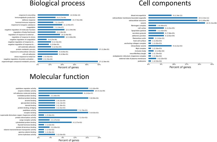
of sepsis-induced AKI. remained the same after narrowing the range via a changing trend
compared without sorting (Figure 7).
Protein Profile Changes According to
Four Continuous Changing Models in Diagnostic Accuracy of the Selected
Sepsis-Induced AKI Proteins as Sepsis-Induced AKI
To identify the biomarkers used for the early diagnosis of Fingerprints in Sepsis Human Patients
sepsis-induced AKI, we narrowed the category of differentially The continuously changing differentially expressed proteins
expressed proteins. Even though the fold change in some proteins associated with inflammation, cell death, proliferation, protein
was high, the alteration was transient. This kind of protein was transport, and bacterial hydrolysis were selected as candidate
Frontiers in Physiology | www.frontiersin.org 7 March 2020 | Volume 11 | Article 139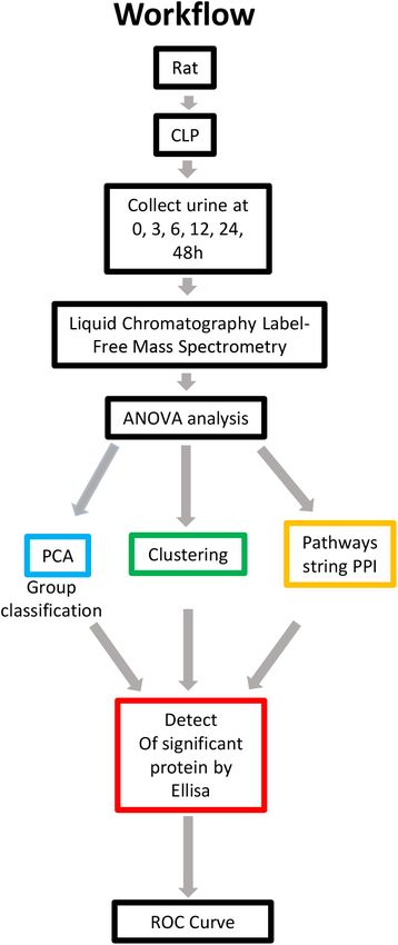
Li et al.                                                                                                                            AKI Biomarkers Identified by MS

  FIGURE 5 | The KEGG enrichment analysis of significantly changed pathways overall and at the indicated time (3, 6, 12, 24, and 48 h) compared with 0 h. P < 0.05.

Frontiers in Physiology | www.frontiersin.org                                    8                                              March 2020 | Volume 11 | Article 139
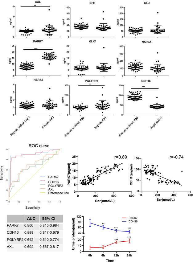
Li et al.                                                                                                                             AKI Biomarkers Identified by MS

  FIGURE 6 | Differentially expressed proteins with a continuous change, which began from the onset of kidney injury and lasted for the whole course of the disease,
  were sorted, and a heatmap showed the relative expression of these proteins in the indicated time. Protein IDs were showen in the right side of heatmap. P < 0.05.

biomarkers. After searching the biological function, nine                             AKI (n = 30) and sepsis without AKI (n = 59). Unpaired t-tests
biomarkers (AXL, CFH, CLU, PARK7, KLK1, NAPSA, HSPA5,                                 showed that in sepsis-induced AKI patients, CDH16 was 0.475-
PGLYRP2, and CDH16) were selected as potential biomarkers.                            fold of that in non-sepsis AKI patients (P < 0.01). AXL, PARK7,
We tested them in the urine from patients with sepsis-induced                         and PGLYRP2 were increased significantly (1. 35-, 2. 12-, and

Frontiers in Physiology | www.frontiersin.org                                     9                                               March 2020 | Volume 11 | Article 139
Li et al.                                                                                                                              AKI Biomarkers Identified by MS

  FIGURE 7 | Biological processes, cell components, and molecular functions in which continuously differentially expressed proteins are involved. P < 0.05.

1.37-fold, respectively) in sepsis-induced AKI patients compared                       (Coopersmith et al., 2018; Kushimoto et al., 2019). Discovery
with that in the control group (Figure 8A).                                            of biomarkers for sepsis-induced AKI could contribute to early
   To evaluate the usefulness of the selected proteins in                              diagnosis of the disease (Peerapornratana et al., 2019). This
discriminating sepsis-induced AKI patients from sepsis, we                             study identified a set of 56 statistically relevant upregulated
performed ROC curve analysis. The expression of AXL in human                           and 67 downregulated proteins from rats with CLP-induced
urine was contrary to that in rats after CLP, suggesting that AXL                      AKI using MS. These proteins were found to participate
may not be a proper biomarker. ROC curve analysis revealed                             in many biological processes, including complement and
that proteins (PARK7 and CDH16) could accurately discern                               coagulation cascades, metabolic pathways, protein processing
sepsis-induced AKI patients, with the AUCs being 0.900 and                             in the endoplasmic reticulum, and apoptosis. After further
0.898, respectively (Figure 8B). AUC analysis revealed that the                        detection of differentially expressed proteins in human urine
cutoff values of PARK7 and CDH16 for identifying patients                              samples, we found that PARK7, and CDH16 are promising
were 13.557 ng/ml (sensitivity 86.7%, specificity 96.6%), and                          candidate biomarkers.
53.29 pg/ml (sensitivity 83.3%, specificity 94.9%), respectively.                          The KDIGO guidelines highlight early AKI diagnosis and
We analyzed the correlation between expression of PARK7 and                            treatment, and serum creatinine levels are used as diagnostic
CDH16 and Scr (Figure 8C). We found that PARK7 correlated                              criteria (Thomas et al., 2015). Although the serum creatinine
to Scr positively, while CDH16 was negatively. The r-value                             test is a practical clinical tool, renal compensation may lead
between PARK7 and Scr was 0.89, and the r-value of CDH16                               to a lag in the creatinine increase; moreover, 50% of kidney
was −0.74. The expression of PARK7 and CDH16 were detected                             injury cases occur without increases in creatinine (Bellomo
at multiple time point. They were changed in a continuous                              et al., 2004; Mishra et al., 2005), resulting in delayed diagnosis
manner (Figure 8D). These results suggest the high diagnostic                          and treatment (Alge and Arthur, 2015). Creatinine may
potential of these two proteins in distinguishing sepsis-induced                       also increase due to renal hypoperfusion caused by prerenal
AKI individuals from sepsis without AKI controls.                                      factors despite a lack of injury to the renal parenchyma
                                                                                       (Ichai et al., 2016). The effectiveness of interventions for
                                                                                       kidney injury is largely limited by a lack of sensitivity and
DISCUSSION                                                                             by the specificity of diagnostic, therapeutic, and prognostic
                                                                                       biomarkers. Biomarker discovery and validation are intrinsically
Sepsis is a type of life-threatening organ dysfunction                                 difficult processes because of issues including the emergence
caused by dysregulation of the host response to infection                              of new detection methods, the heterogeneity of patients,

Frontiers in Physiology | www.frontiersin.org                                     10                                              March 2020 | Volume 11 | Article 139
Li et al. AKI Biomarkers Identified by MS FIGURE 8 | Verification of sepsis-induced AKI-specific biomarkers in sepsis human patients. (A) The concentration of nine proteins was validated by ELISA in urine from sepsis-induced AKI patients and non-sepsis-induced AKI patients. ∗ P < 0.05; two-tailed unpaired Student’s t-test. (B) Validation of 4 differentially expressed proteins by testing their performance in differentiating injury specificity by AUC for PARK7, PGLYRP2, AXL, and CDH16. (C) Pearson correlation analysis was performed between expression of PARK7 and CDH16 and Scr. Both PARK7 and CDH16 were correlated to Scr. P < 0.01; (D) The protein expression of PARK7 and CDH16 in urine from ten patients were detected by Elisa kit at 0, 6, 12, and 24 h after diagnosis with sepsis. **P < 0.01; ***P < 0.001;Compared with 0 h. Frontiers in Physiology | www.frontiersin.org 11 March 2020 | Volume 11 | Article 139
Li et al.                                                                                                          AKI Biomarkers Identified by MS

difficulties in data analysis, and other physiological confounders        CDH16, PARK7, and PGLYRP2 were significantly changed
(Sarwal et al., 2011). However, proteomics has become a powerful          in the urine of patients suffering from sepsis-induced AKI.
tool for novel biomarker discovery in kidney disease. Novel               AXL regulates oxidative stress and inflammation by including
proteomic approaches can enable discovery of biomarkers                   nuclear factor-kappa B (NF-κB). The previous study identifies
suitable for earlier and more accurate diagnosis of renal                 that AXL is a promising therapeutic target for the prevention
pathology than traditional biomarkers, such as creatinine and             of AKI-to-CKD transition (Chen et al., 2019). However, the
urine proteins. Biomarkers specific to certain phenotypes are             expression of AXL in the rat’s urine was decreased after
robust, stable, and easily accessible by urine testing, thus              sepsis. The possible explanation was that the patients always
possessing immense clinical value and providing critical insight          suffered from multiple diseases, and the source of infection
for the advancement of AKI therapy (Sigdel et al., 2016). It              was not limited to abdominal bacterial infections. PARK7
was found that TIMP2 could ameliorate LPS-induced cytokine                acts as a positive regulator of androgen receptor-dependent
release, apoptosis, and cell injury (Li et al., 2019). Biomarker          transcription and functions as an inhibitor of cellular oxidative
assessment will also contribute to the mitigation of sepsis-              stress and a regulator of mitochondrial function, autophagy,
induced kidney injury and will help guide optimal renal                   and apoptosis (Oh and Mouradian, 2017). The protective
replacement therapy dosing.                                               effect of PARK7 signaling was identified in renal tubular
    Label-free quantification is a method used in MS that aims to         epithelial cell injury induced by ischemia (Yin et al., 2019).
determine the relative amount of proteins (Moore et al., 2004).           PGLYRP2 plays a scavenger role by digesting biologically active
The use of label-free MS to examine differentially expressed              peptidoglycan into biologically inactive fragments. CDH16 is
proteins in urine samples largely eliminates the variations and           a member of the cadherin superfamily, calcium-dependent,
biases in replicate MS measurements (Griffin et al., 2010)                membrane-associated glycoproteins. Expression is exclusively
and has allowed for the exploration of the mechanism of                   in the kidney, where the protein functions as the principal
AKI and the discovery of biomarkers. In our MS results,                   mediator of homotypic cellular recognition, playing a role in
we found that 123 proteins changed in urine after sepsis-                 the morphogenic direction of tissue development. As PARK7,
induced AKI. GO analysis revealed that the protein metabolic              PGLYRP2, AXL, and CDH16 were significantly changed and
process was the first altered in the early stage of sepsis-               CDH16 is a kidney-specific protein, we used PARK7, PGLYRP2,
induced AKI. The increase in total cellular metabolism was to             AXL, and CDH16 to perform ROC curve analysis. The
provide the energy needed to sustain immune system activation             diagnostic sensitivity and specificity of PARK7 and CDH16 were
(Pravda, 2014). Mitochondria-centered structural and metabolic            higher than those of neutrophil gelatinase-associated lipocalin
alterations occur prior to the onset of AKI, supporting a                 (NGAL), which was used to diagnose AKI, particularly infection-
causative, pathogenic role of mitochondrial damage in kidney              mediated AKI (Kim et al., 2016). PARK7 and CDH16 were
injury (Parikh et al., 2015). Possible molecular mechanisms that          changed continuously, which were suitable biomarkers for
stimulate the hypermetabolic state include altered mitochondrial          early detection.
bioenergetics, impaired peroxisomal catalase activity and fatty               There are some limitations of the present study that should
acid oxidation (Vasko et al., 2013). Six hours after induction            be clearly addressed. First, the expression of proteins cannot
of AKI, responses to organic substances, wounding and stimuli             be matched with the extent of the kidney injury. We used a
were the leading biological processes. In the cell component              standardized approach to induce sepsis and to obtain stable
category, extracellular exosomes and vesicles were increased              mortality rates. However, the extent of AKI was different,
continuously from 6 to 48 h. Exosomes, a type of nanoparticle,            and the urine output of each rat was so scarce that the
function as intercellular shuttles that bud from the host cell            urine from five rats was pooled together subjected to MS.
membrane and carry information to recipient cells (Barile                 Second, it would be helpful to study the course of PARK7
and Vassalli, 2017). Exosomes can be transferred in biological            and CDH16 expression in both serum and urine at specific
fluids to interact with remote organs and can be secreted                 time points. In vitro and ex vivo experiments are needed to
in urine (Karimi et al., 2018). In the KEGG analysis, the                 confirm the biological function of these biomarkers. Clinical trials
immune system was in the center place of AKI. The host                    evaluating diagnosis and management should focus on precise
defense against infection is an adaptive response to decrease             populations and consider biomarker enrichment strategies
the pathogen load, limit tissue injury, and restore homeostasis           (Kellum, 2016). Furthermore, to ensure the sensitivity and
(Gomez et al., 2017).                                                     specificity of biomarkers, a combination of multiple proteomics
    To identify the biomarkers used for the early diagnosis of            analysis of samples from urine, serum, and tissue will be the
sepsis-induced AKI, we narrowed the category of differentially            future direction.
expressed proteins by changing their trend to avoid transient
alterations. Proteins with stable and continuous changes are ideal
biomarkers for the early identification and diagnosis of AKI. Even        CONCLUSION
when proteins were sorted by changing trends, the biological
process and cell components remained quite consistent. Here,              In summary, we performed proteomics at multiple time points
we used ELISA to identify panels of proteins that may be                  to identify altered proteins in urine and understood the
used as fingerprints of sepsis-induced AKI. Nine proteins were            biological process in sepsis-induced AKI. We analyzed urine
detected in human urine with sepsis-induced patients. AXL,                proteins with spatiotemporal dynamics to sensitively monitor

Frontiers in Physiology | www.frontiersin.org                        12                                        March 2020 | Volume 11 | Article 139
Li et al. AKI Biomarkers Identified by MS
biological alterations in sepsis-induced AKI, and two distinctive FUNDING
biomarkers were defined in patients with sepsis-induced AKI.
The results obtained with our systems biology approach might This work was supported by the National Natural Science
provide a theoretical basis for further research to unravel the Foundation of China (Nos. 81772046, 81560131, and 81701962).
molecular mechanisms and to develop novel biomarkers for
sepsis-induced AKI.
ACKNOWLEDGMENTS
DATA AVAILABILITY STATEMENT The authors would like to thank Zhongxiang Zhang for assistance
in collecting patients’ urine samples.
All included data are available in the public domain, and all
references are included in our reference list. Extracted data and
calculations will be made available to individual scientists upon SUPPLEMENTARY MATERIAL
reasonable request.
The Supplementary Material for this article can be found
online at: https://www.frontiersin.org/articles/10.3389/fphys.
ETHICS STATEMENT 2020.00139/full#supplementary-material
The animal study was reviewed and approved by the Animal FIGURE S1 | Analysis of data distribution and quantitative abundance. (A)
Analysis of data distribution. The histogram represents the data distribution of
Care and Use Committee of Nanchang University. The studies
each sample. Experimental data basically conformed to the normal distribution,
involving human participants were reviewed and approved which could be used for subsequent statistical analysis. (B) Analysis of the
by the Research Ethics Committee at the Zhongnan Hospital quantitative abundance of protein. The abscissa represents the sample name, and
of Wuhan University. The patients/participants provided their the ordinate represents the protein classification (normalized results). Each point in
written informed consent to participate in this study. the figure represents the abundance value of a protein. A polyvine diagram was
formed by connecting the points. The black box in the middle was the boxplot of
the protein abundance of each sample. The orange polyline represents the
abundance distribution trend of the protein P3052 in different samples.
AUTHOR CONTRIBUTIONS
FIGURES S2–S4 | The biological process, cell components, and molecular
function of differentially expressed proteins in which differentially expressed
YL conceived the project, designed the project, extracted and
proteins in urine at the indicated time (3, 6, 12, 24, and 48 h) were compared with
analyzed the data, drafted the manuscript, and approved the those at 0 h. P < 0.05.
final manuscript. JL revised the manuscript and approved the
TABLE S1 | Concentration of serum creatinine in CLP-induced AKI (µmol/l).
final manuscript. JC contributed to the CLP model and urine
collection. YQ collected patients’ urine samples. JZ and YZ TABLE S2 | One-way ANOVA was performed to analyze the differential expression
drafted the part of discussion and background of the manuscript. protein in the urine after sepsis-induced AKI. P < 0.05.
YL, JC, and JZ conducted the experiments. FL and ZP designed TABLE S3 | The continuous changing protein in the urine after sepsis-induced
the project, edited the manuscript, and approved the final version. AKI. P < 0.05.
REFERENCES Coopersmith, C. M., De Backer, D., Deutschman, C. S., Ferrer, R., Lat, I., Machado,
F. R., et al. (2018). Surviving sepsis campaign: research priorities for sepsis
Alge, J. L., and Arthur, J. M. (2015). Biomarkers of AKI: a review of mechanistic and septic shock. Intensive Care Med. 44, 1400–1426. doi: 10.1007/s00134-018-
relevance and potential therapeutic implications. Clin. J. Am. Soc. Nephrol. 10, 5175-z
147–155. doi: 10.2215/CJN.12191213 Cox, J., Hein, M. Y., Luber, C. A., Paron, I., Nagaraj, N., and Mann, M. (2014).
Atrih, A., Mudaliar, M. A., Zakikhani, P., Lamont, D. J., Huang, J. T., Bray, Accurate proteome-wide label-free quantification by delayed normalization and
S. E., et al. (2014). Quantitative proteomics in resected renal cancer tissue for maximal peptide ratio extraction, termed MaxLFQ. Mol. Cell. Proteomics 13,
biomarker discovery and profiling. Br. J. Cancer 110, 1622–1633. doi: 10.1038/ 2513–2526. doi: 10.1074/mcp.M113.031591
bjc.2014.24 Gomez, H., Kellum, J. A., and Ronco, C. (2017). Metabolic reprogramming and
Bagshaw, S. M., Lapinsky, S., Dial, S., Arabi, Y., Dodek, P., Wood, G., et al. (2009). tolerance during sepsis-induced AKI. Nat. Rev. Nephrol. 13, 143–151. doi: 10.
Acute kidney injury in septic shock: clinical outcomes and impact of duration 1038/nrneph.2016.186
of hypotension prior to initiation of antimicrobial therapy. Intensive Care Med. Grebe, S. K. G., and Singh, R. J. (2016). Clinical peptide and protein quantification
35, 871–881. doi: 10.1007/s00134-008-1367-2 by mass spectrometry (MS). TrAC Trends Analyt. Chem. 84, 131–143. doi:
Barile, L., and Vassalli, G. (2017). Exosomes: therapy delivery tools and biomarkers 10.1016/j.trac.2016.01.026
of diseases. Pharmacol. Ther. 174, 63–78. doi: 10.1016/j.pharmthera.2017. Griffin, B. R., Faubel, S., and Edelstein, C. L. (2018). Biomarkers of drug-
02.020 induced kidney toxicity. Ther. Drug Monit. 41, 213–226. doi: 10.1097/FTD.
Bellomo, R., Kellum, J. A., and Ronco, C. (2004). Defining acute renal failure: 0000000000000589
physiological principles. Intensive Care Med. 30, 33–37. doi: 10.1007/s00134- Griffin, B. R., Gist, K. M., and Faubel, S. (2019). Current status of novel biomarkers
003-2078-3 for the diagnosis of acute kidney injury: a historical perspective. J. Intensive Care
Chen, D. Q., Feng, Y. L., Chen, L., Liu, J. R., Wang, M., Vaziri, N. D., et al. (2019). Med. doi: 10.1177/0885066618824531 [Epub ahead of print].
Poricoic acid A enhances melatonin inhibition of AKI-to-CKD transition by Griffin, N. M., Yu, J., Long, F., Oh, P., Shore, S., Li, Y., et al. (2010). Label-free,
regulating Gas6/AxlNFkappaB/Nrf2 axis. Free Radic. Biol. Med. 134, 484–497. normalized quantification of complex mass spectrometry data for proteomic
doi: 10.1016/j.freeradbiomed.2019.01.046 analysis. Nat. Biotechnol. 28, 83–89. doi: 10.1038/nbt.1592
Frontiers in Physiology | www.frontiersin.org 13 March 2020 | Volume 11 | Article 139Li et al. AKI Biomarkers Identified by MS
Guo, Y., Han, Z., Guo, L., Liu, Y., Li, G., Li, H., et al. (2018). Identification of urinary Peerapornratana, S., Manrique-Caballero, C. L., Gomez, H., and Kellum, J. A.
biomarkers for the prediction of gestational diabetes mellitus in early second (2019). Acute kidney injury from sepsis: current concepts, epidemiology,
trimester of young gravidae based on iTRAQ quantitative proteomics. Endocr. pathophysiology, prevention and treatment. Kidney Int. 96, 1083–1099. doi:
J. 65, 727–735. doi: 10.1507/endocrj.EJ17-0471 10.1016/j.kint.2019.05.026
Himmelsbach, M. (2012). 10 years of MS instrumental developments–impact on Peng, Z. Y., Bishop, J. V., Wen, X. Y., Elder, M. M., Zhou, F., Chuasuwan, A., et al.
LC-MS/MS in clinical chemistry. J. Chromatogr. B Analyt. Technol. Biomed. Life (2014). Modulation of chemokine gradients by apheresis redirects leukocyte
Sci. 88, 3–17. doi: 10.1016/j.jchromb.2011.11.038 trafficking to different compartments during sepsis, studies in a rat model. Crit.
Ichai, C., Vinsonneau, C., Souweine, B., Armando, F., Canet, E., Clec’h, C., et al. Care 18:R141. doi: 10.1186/cc13969
(2016). Acute kidney injury in the perioperative period and in intensive care Prasad, B., Vrana, M., Mehrotra, A., Johnson, K., and Bhatt, D. K. (2017). The
units (excluding renal replacement therapies). Ann. Intensive Care 6:48. doi: promises of quantitative proteomics in precision medicine. J. Pharm. Sci. 106,
10.1186/s13613-016-0145-5 738–744. doi: 10.1016/j.xphs.2016.11.017
Karimi, N., Cvjetkovic, A., Jang, S. C., Crescitelli, R., Hosseinpour Feizi, M. A., Pravda, J. (2014). Metabolic theory of septic shock. World J. Crit. Care Med. 3,
Nieuwland, R., et al. (2018). Detailed analysis of the plasma extracellular vesicle 45–54. doi: 10.5492/wjccm.v3.i2.45
proteome after separation from lipoproteins. Cell Mol. Life Sci. 75, 2873–2886. Rauh, M. (2012). LC-MS/MS for protein and peptide quantification in clinical
doi: 10.1007/s00018-018-2773-4 chemistry. J. Chromatogr. B Analyt. Technol. Biomed. Life Sci. 88, 59–67. doi:
Kellum, J. A. (2016). Why are patients still getting and dying from acute 10.1016/j.jchromb.2011.09.030
kidney injury? Curr. Opin. Crit. Care 22, 513–519. doi: 10.1097/MCC. Rittirsch, D., Huber-Lang, M. S., Flierl, M. A., and Ward, P. A. (2009).
0000000000000358 Immunodesign of experimental sepsis by cecal ligation and puncture. Nat.
Kellum, J. A., Lameire, N., and KDIGO AKI Guideline Work Group, (2013). Protoc. 4, 31–36. doi: 10.1038/nprot.2008.214
Diagnosis, evaluation, and management of acute kidney injury: a KDIGO Sarwal, M. M., Sigdel, T. K., and Salomon, D. R. (2011). Functional
summary (Part 1). Crit. Care 17:204. doi: 10.1186/cc11454 proteogenomics–embracing complexity. Semin. Immunol. 23, 235–251. doi:
Kim, S., Kim, H. J., Ahn, H. S., Song, J. Y., Um, T. H., Cho, C. R., et al. (2016). 10.1016/j.smim.2011.08.002
Is plasma neutrophil gelatinase-associated lipocalin a predictive biomarker for Sigdel, T. K., Gao, Y., He, J., Wang, A., Nicora, C. D., Fillmore, T. L., et al. (2016).
acute kidney injury in sepsis patients? A systematic review and meta-analysis. Mining the human urine proteome for monitoring renal transplant injury.
J. Crit. Care 33, 213–223. doi: 10.1016/j.jcrc.2016.02.014 Kidney Int. 89, 1244–1252. doi: 10.1016/j.kint.2015.12.049
Kushimoto, S., Abe, T., Ogura, H., Shiraishi, A., Saitoh, D., Fujishima, S., et al. Thomas, M. E., Blaine, C., Dawnay, A., Devonald, M. A., Ftouh, S., Laing, C., et al.
(2019). Impact of body temperature abnormalities on the implementation of (2015). The definition of acute kidney injury and its use in practice. Kidney Int.
sepsis bundles and outcomes in patients with severe sepsis: a retrospective 87, 62–73. doi: 10.1038/ki.2014.328
sub-analysis of the focused outcome research on emergency care for acute Uchino, S., Kellum, J. A., Bellomo, R., Doig, G. S., Morimatsu, H., Morgera, S., et al.
respiratory distress syndrome, sepsis and Trauma study. Crit. Care Med. 47, (2005). Acute renal failure in critically ill patients: a multinational, multicenter
691–699. doi: 10.1097/CCM.0000000000003688 study. JAMA 294, 813–818. doi: 10.1001/jama.294.7.813
Li, Y. M., Zhang, J., Su, L. J., Kellum, J. A., and Peng, Z. Y. (2019). Downregulation Vanmassenhove, J., Kielstein, J., Jörres, A., and Biesen, W. V. (2017). Management
of TIMP2 attenuates sepsis-induced AKI through the NF-kappab pathway. of patients at risk of acute kidney injury. Lancet 389, 2139–2151. doi: 10.1016/
Biochim. Biophys. Acta Mol. Basis Dis. 1865, 558–569. doi: 10.1016/j.bbadis. s0140-6736(17)31329-6
2018.10.041 Vasko, R., Ratliff, B. B., Bohr, S., Nadel, E., Chen, J., Xavier, S., et al. (2013).
Lopes, J. A., Jorge, S., Resina, C., Santos, C., Pereira, A., Neves, J., et al. (2009). Acute Endothelial peroxisomal dysfunction and impaired pexophagy promotes
kidney injury in patients with sepsis: a contemporary analysis. Int. J. Infect. Dis. oxidative damage in lipopolysaccharide-induced acute kidney injury. Antioxid.
13, 176–181. doi: 10.1016/j.ijid.2008.05.1231 Redox Signal. 19, 211–230. doi: 10.1089/ars.2012.4768
Mishra, J., Dent, C., Tarabishi, R., Mitsnefes, M. M., Ma, Q., Kelly, C., et al. (2005). Yin, J., Xu, R., Wei, J., and Zhang, S. (2019). The protective effect
Neutrophil gelatinase-associated lipocalin (NGAL) as a biomarker for acute of glutaredoxin 1/DJ-1/HSP70 signaling in renal tubular epithelial cells
renal injury after cardiac surgery. Lancet 365, 1231–1238. doi: 10.1016/s0140- injury induced by ischemia. Life Sci. 223, 88–94. doi: 10.1016/j.lfs.2019.
6736(05)74811-x 03.015
Montoya, A., Lopez, M. C., Velez, I. D., and Robledo, S. M. (2019). Label-free
quantitative proteomic analysis reveals potential biomarkers for early healing Conflict of Interest: The authors declare that the research was conducted in the
in cutaneous leishmaniasis. PeerJ 6:e6228. doi: 10.7717/peerj.6228 absence of any commercial or financial relationships that could be construed as a
Moore, L. E., Wiencke, J. K., Bates, M. N., Zheng, S., Rey, O. A., and Smith, potential conflict of interest.
A. H. (2004). Investigation of genetic polymorphisms and smoking in a bladder
cancer case-control study in Argentina. Cancer Lett. 211, 199–207. doi: 10.1016/ Copyright © 2020 Li, Long, Chen, Zhang, Qin, Zhong, Liu and Peng. This is an
j.canlet.2004.04.011 open-access article distributed under the terms of the Creative Commons Attribution
Oh, S. E., and Mouradian, M. M. (2017). Regulation of signal transduction by DJ-1. License (CC BY). The use, distribution or reproduction in other forums is permitted,
Adv. Exp. Med. Biol. 1037, 97–131. doi: 10.1007/978-981-10-6583-5_8 provided the original author(s) and the copyright owner(s) are credited and that the
Parikh, S. M., Yang, Y., He, L., Tang, C., Zhan, M., and Dong, Z. (2015). original publication in this journal is cited, in accordance with accepted academic
Mitochondrial function and disturbances in the septic kidney. Semin. Nephrol. practice. No use, distribution or reproduction is permitted which does not comply
35, 108–119. doi: 10.1016/j.semnephrol.2015.01.011 with these terms.
Frontiers in Physiology | www.frontiersin.org 14 March 2020 | Volume 11 | Article 139You can also read