Synthesis, In Vitro Evaluation and Molecular Docking of the 5-Acetyl-2-aryl-6-hydroxybenzo b furans against Multiple Targets Linked to Type 2 ...
←
→
Page content transcription
If your browser does not render page correctly, please read the page content below
biomolecules

Article
Synthesis, In Vitro Evaluation and Molecular Docking
of the 5-Acetyl-2-aryl-6-hydroxybenzo[b]furans
against Multiple Targets Linked to Type 2 Diabetes
Malose J. Mphahlele 1, * , Yee Siew Choong 2 , Marole M. Maluleka 1               and
Samantha Gildenhuys 3
 1   Department of Chemistry, University of South Africa, Private Bag X06, Florida 1710, South Africa;
     43677541@mylife.unisa.ac.za
 2   Institute for Research in Molecular Medicine (INFORMM), Universiti Sains Malaysia, Penang 11800,
     Malaysia; yeesiew@usm.my
 3   Department of Life & Consumer Sciences, College of Agriculture and Environmental Sciences, University of
     South Africa, Private Bag X06, Florida 1710, South Africa; gildes@unisa.ac.za
 *   Correspondence: mphahmj@unisa.ac.za

 Received: 24 January 2020; Accepted: 19 February 2020; Published: 7 March 2020

 Abstract: The 5-acetyl-2-aryl-6-hydroxybenzo[b]furans 2a–h have been evaluated through in vitro
 enzymatic assay against targets which are linked to type 2 diabetes (T2D), namely, α-glucosidase,
 protein tyrosine phosphatase 1B (PTP1B) and β-secretase. These compounds have also been evaluated
 for antioxidant activity using the 2,2-diphenyl-1-picrylhydrazyl (DPPH) free-radical scavenging
 method. The most active compounds against α-glucosidase and/or PTP1B, namely, 4-fluorophenyl
 2c, 4-methoxyphenyl 2g and 3,5-dimethoxyphenyl substituted 2h derivatives were also evaluated
 for potential anti-inflammatory properties against cyclooxygenase-2 activity. The Lineweaver-Burk
 and Dixon plots were used to determine the type of inhibition on compounds 2c and 2h against
 α-glucosidase and PTP1B receptors. The interactions were investigated in modelled complexes
 against α-glucosidase and PTP1B via molecular docking.

 Keywords: 5-acetyl-2-aryl-6-hydroxybenzo[b]furans; α-glucosidase; protein tyrosine phosphatase 1B;
 β-secretase; antioxidant activity; cyclooxygenase-2; molecular docking

1. Introduction
     Type 2 diabetes (T2D) is a chronic metabolic disorder that has reached epidemic proportion
throughout the world and this disease is characterized by a defect in the secretion of insulin and
resistance to insulin in its target organs [1]. The deficiency is attributed to increased glucose levels
in blood also known as post-prandial hyperglycaemia (PPHG), which has emerged as a prominent
and early deficiency in T2D [2]. Several cellular and metabolic targets are involved to ameliorate T2D
complications, such as, α-glucosidase and protein tyrosine phosphatase 1 beta (PTP1B) inhibition,
correction of abnormal insulin signalling, promotion of the action of insulin on its target tissues,
including antioxidative and anti-inflammatory action [3]. α-Glucosidase, a membrane bound enzyme
in small intestine plays a key role in inhibiting hydrolysis of carbohydrates. This enzyme acts on the
1,4-α glycosidic bonds by breaking down starch and disaccharides into single absorbable α-glucose
molecules [4]. Inhibitors of α-glucosidase act in a competitive or non-competitive manner to delay the
absorption of carbohydrates by the small intestine, thus prolonging the overall carbohydrate digestion
time ultimately lowering post-prandial blood glucose and insulin levels [5]. PTP1B, on the other hand,
is an intracellular non-receptor protein tyrosine phosphatase, which is highly expressed in insulin
targeted tissues such as liver and muscle including fats [6] and has also received much attention due to

Biomolecules 2020, 10, 418; doi:10.3390/biom10030418                        www.mdpi.com/journal/biomolecules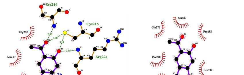
Biomolecules 2020, 10, 418                                                                         2 of 22

its role in type 2 diabetes mellitus and obesity [7]. PTP1B works specifically by dephosphorylating the
residues of phosphotyrosine both from the insulin receptor (IR) and the insulin receptor substrate [8].
α-Glucosidase and PTP1B are the key targets to treat diabetes and obesity [9] and compounds with dual
inhibitory activity against these enzymes exhibit synergistic effects to prevent hyperglycaemia, in turn,
effectively improve insulin sensitization [10]. In vivo studies have demonstrated that β-secretase
(BACE-1) regulates the amount of IR and insulin signalling in the liver making inhibitors of this
enzyme, to enhance insulin signalling during diabetes [11,12]. Likewise, oxidative stress is implicated
in the early stages and progression of T2D due to disruption of the pro-oxidant/antioxidant balance
leading to the development of insulin resistance [13,14]. Inflammatory responses, on the other hand,
contribute to T2D occurrence by causing insulin resistance, and, in turn, they are intensified in the
presence of hyperglycaemia and promote long-term diabetic complications [15]. It is envisaged that
targeting the key enzymes associated with pathogenesis and progression of T2D and oxidative stress as
well as inflammatory pathways could be a significant strategy for treating this multifunctional disease.
      Phenols and hydroxyacetophenones continue to attract considerable attention in medicinal
chemistry especially in the context of drug development. Their heterocyclic derivatives such as
benzofurans, for example, are endowed with several pharmacological properties including
antihyperglycemic, antidiabetic, anti-Alzheimer’s, anti-inflammatory, anti-oxidant and antitumor
activities and some derivatives serve as enzyme inhibitors [12,16–18]. The naturally occurring
2,4-dihydroxy-5-methylacetophenone and its derivatives have been found to exhibit α-glucosidase
inhibitory properties [19]. Likewise, the 2-acylbenzofurans were found to inhibit α-glucosidase and to
exhibit hypoglycaemic activity [20]. The analogous 2-unsubstituted 5-acetyl-4-hydroxybenzofurans
and 5-acetyl-6-hydroxybenzofurans as well as their benzoylated derivatives, on the other hand,
exhibit an inhibitory effect against PTP1B activity [21]. Several naturally occurring 2-arylbenzofurans
(sanggenofuran A, mulberrofuran D2, mulberrofuran D, morusalfuran B and mulberrofuran H),
isolated from the root bark of Morus alba, have also been found to exhibit an inhibitory effect
against PTP1B activity [22]. Based on the capability of ortho-hydroxyacetophenones to target
α-glucosidase and the PTP1B inhibitory properties of ortho-hydroxyacetylbenzofuran derivatives
and the 2-arylbenzofuran analogues, we decided to synthesize the ortho-hydroxyacetyl substituted
2-arylbenzofurans. The main aim was to evaluate these compounds through enzymatic assays
in vitro for potential dual inhibitory properties against α-glucosidase and PTP1B activities. Due to
the multifunctional origin of diabetes and contributing factors to this disease, the spectrum of targets
was extended to include β-secretase, free radical scavenging and anti-inflammatory potential of these
compounds. Their possible mode of interaction with α-glucosidase or PTP1B was investigated through
kinetic and molecular docking studies.

2. Materials and Methods

2.1. General
     The melting point (mp.) values of the prepared compounds were recorded on a Thermocouple
digital melting point apparatus (Mettler Toledo LLC, Columbus, OH, USA) and are uncorrected.
Their infrared (IR) spectra were recorded using the thin-film method on a Bruker VERTEX 70 FT-IR
Spectrometer (Bruker Optics, Billerica, MA, USA) equipped with a diamond attenuated total reflectance
(ATR) accessory. The Merck kieselgel 60 (0.063–0.200 mm) (Merck KGaA, Frankfurt, Germany) was
used as a stationary phase for column chromatography. The proton (1 H-) and carbon-13 (13 C-)
nuclear magnetic resonance (NMR) spectra of the prepared compounds were obtained as CDCl3
or dimethyl sulfoxide-d6 (DMSO-d6 ) solutions using Agilent 500 MHz NMR spectrometer (Agilent
Technologies, Oxford, UK). The chemical shifts are quoted relative to the tetramethylsilane (TMS) peak
as an internal reference standard. The high-resolution mass spectra were recorded at the University
of Stellenbosch using a Waters Synapt G2 Quadrupole Time-of-flight mass spectrometer (Waters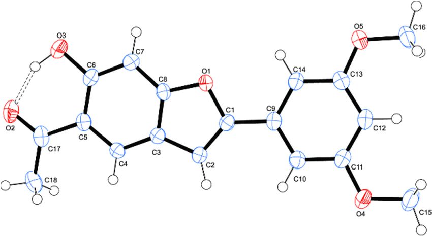
Biomolecules 2020, 10, 418                                                                               3 of 22

Corp., Milford, MA, USA) at an ionization potential of 70 eV. The synthesis and analytical data for
2,4-dihydroxy-5-iodoacetophenone, were reported in our previous investigation [23].

2.2. Typical Procedure for the Synthesis of Compounds 2a–h
     To a three necked-flask equipped with a condenser and rubber septa,
2,4-dihydroxy-5-iodoacetophenone (1.00 g, 3.60 mmol), PdCl2 (PPh3 )2 (0.13 g, 0.18 mmol),
CuI (0.07 g, 0.37 mmol), PPh3 (0.096 g, 0.37 mmol) and K2 CO3 (0.60 g, 4.34 mmol) were added followed
by 9:1 DMF-water (v/v, 50 mL). The mixture was purged for 30 min with an argon gas. A solution of
phenylacetylene derivative (1.2 equivalent) in DMF (1 mL) was introduced to the mixture using a
syringe. The stirred mixture was heated at 80 ◦ C for 3 h under an argon atmosphere and the mixture
was poured into an ice-cold water. The product was extracted with chloroform and the combined
organic layers were dried over anhydrous magnesium sulphate. The salt was filtered off and the
solvent was evaporated under reduced pressure on a rotary evaporator. The residue was purified
by silica gel column chromatography using toluene as an eluent to afford compound 2 as a solid.
The following compounds were prepared in this fashion.

2.2.1. 1-[6-Hydroxy-2-phenylbenzofuran-5-yl]ethanone (2a)
     Yellow solid (0.38 g, 84%), mp. 172-173 ◦ C; νmax (ATR) 3083 (OH), 1640 (C=O), 1611, 1446, 1366,
1288, 1247, 1140, 1012, 799, 765 cm-1 ; 1 H-NMR (500 MHz, CDCl3 ) 12.52 (1H, br s, OH), 7.96 (1H, s,
H-4), 7.85 (2H, d, J = 8.0 Hz, Ar), 7.48 (1H, t, J = 7.5 Hz, Ar), 7.42 (2H, d, J = 7.0 Hz, Ar), 7.06 (1H, s,
H-7) 6.94 (1H, s, H-3), 2.69 (3H, s, -CH3 ); 13 C-NMR (125 MHz, CDCl3 ) 204.0, 161.2, 159.6, 156.9, 129.7,
128.9, 128.8, 124.7, 123.5, 122.0, 117.0, 100.8, 96.6, 26.9; HRMS (ES+ ): m/z [M + H]+ calcd for C16 H11 O3 :
251.0708; found 251.0820.

2.2.2. 1-[2-(3-Fluorophenyl)-6-hydroxybenzofuran-5-yl]ethanone (2b)
      Brown solid (0.33 g, 69%), mp. 165-167 ◦ C; νmax (ATR) 3081 (OH), 1636 (C=O), 1618, 1573, 1487,
1446, 1370, 1331, 1270, 1154, 1141, 864,823, 780, 654 cm-1 ; 1 H-NMR (500 MHz, CDCl3 ) 12.53 (1H, br
s, OH), 7.96 (1H, s, H-4), 7.65 (1H, d, J = 8.5 Hz, Ar), 7.42 (2H, dd, J = 8.5 and 2.0 Hz, Ar), 7.14 (1H,
t, J = 8.5 Hz, Ar), 6.95 (1H, s, H-3), 2.70 (3H, s, -CH3 ); 13 C-NMR (125 MHz, CDCl3 ) 204.1, 163.0 (d,
   CF = 245.9 Hz), 161.5, 159.5, 155.5 (d, J CF = 3.2 Hz), 131.7 (d, J CF = 8.6 Hz), 130.5 (d, J CF = 8.3 Hz),
1J                                        4                         3                         3

123.8, 121.7, 120.4 (d, JCF = 3.0 Hz), 117.2, 115.6 (d, JCF = 21.3 Hz), 111.6 (d, JCF = 23.7 Hz), 101.9,
                        4                                 2                           2

96.7, 26.9; HRMS (ES+ ): m/z [M + H]+ calcd for C16 H10 FO3 : 269.0614; found 269.0605.

2.2.3. 1-[2-(4-Fluorophenyl)-6-hydroxybenzofuran-5-yl]ethanone (2c)
     Brown solid (0.35 g, 73%), mp. 194-195 ◦ C; νmax (ATR) 3082 (OH), 1626 (C=O), 1599, 1504, 1372,
1275, 1218, 1140, 844, 807, 795, 514 cm-1 ; 1 H-NMR (500 MHz, CDCl3 ) 12.52 (1H, br s, OH), 7. 95 (1H, s,
H-4), 7.79 (2H, dt, J = 8.7 Hz and, JHF = 5.7 Hz, H-20 ,60 ), 7.14 (2H, dt, JHH = 8.7 Hz and JHF = 9.6 Hz,
H-30 ,50 ), 7.05 (1H, s, H-7), 6.87 (1H, s, H-3), 2.70 (3H, s, –CH3 ); 13 C-NMR (125 MHz, CDCl3 ) 204.0,
162.9 (d, 1 JCF = 249.6 Hz), 161.2, 159.5, 156.0, 126.6 (d, 3 JCF = 8.1 Hz), 126.0 (d, 4 JCF = 3.2 Hz), 123.4,
122.0, 117.1, 116.0 (d, 2 JCF = 22.0 Hz), 100.5, 99.7, 26.9; HRMS (ES+ ): m/z [M + H]+ calcd for C16 H10 FO3 :
269.0614; found 269.0605.

2.2.4. 1-[2-(3-Chlorophenyl)-6-hydroxybenzofuran-5-yl]ethanone (2d)
     Yellow solid (0.36 g, 74%), mp.178–179 ◦ C; νmax (ATR) 3081 (OH), 1675 (C=O), 1587, 1561, 1474,
1359, 1275, 1144, 1078, 882, 788, 682,cm-1 ; 1 H-NMR (500 MHz, CDCl3 ) 12.50 (1H, br s, OH), 7.99 (1H, s,
H-4), 7.80 (1H, s, Ar), 7.70 (1H, d, J = 8.0 Hz, Ar), 7.38 (1H, d, J = 8.5 Hz, Ar), 7.32 (1H, d, J = 7.5 Hz,
Ar), 7.07 (1H, s, H-7), 6.98 (1H, s, H-3), 2.72 (3H, s, CH3 ), 13 C-NMR (125 MHz, CDCl3 ) 203.9, 161.5,
159.6, 155.3, 134.9, 131.4, 130.1, 129.7, 128.7, 124.7, 123.7, 122.7, 117.3, 102.0, 99.7, 29.6; HRMS (ES+ ): m/z
[M + H]+ calcd for C16 H11 O3 35 Cl: 285.0318; found 285.0299.
Biomolecules 2020, 10, 418                                                                                     4 of 22

2.2.5. 1-[2-(4-Chlorophenyl)-6-hydroxybenzofuran-5-yl]ethanone (2e)
     Yellow solid (0.43 g, 77%), mp. 170–173 ◦ C; νmax (ATR) 3082 (OH), 1676 (C=O), 1614, 1502, 1405,
1363, 1267, 1199, 1033, 987, 854, 746, 666 cm−1 ; 1 H-NMR (500 MHz, CDCl3 ) 12.52 (1H, br s, OH), 7.96
(1H, s, H-4), 7.81 (2H, d, J = 8.0 Hz, H-20 ,60 ), 7.42 (2H, d, J = 8.0 Hz H-30 ,50 ), 7.06 (1H, s, H-7), 7.03 (1H, s,
H-3), 2.92 (3H, s, CH3 ); 13 C-NMR (125 MHz, CDCl3 ) 202.4, 156.4, 132.6, 130.2, 128.3, 128.0, 127.9, 127.7,
126.7, 123.0, 121.5, 116.6, 101.1, 39.8; HRMS (ES+ ): m/z [M + H]+ calcd for C16 H11 35 ClO3 : 285.0312;
found 285.0318.

2.2.6. 1-[6-Hydroxy-2-(4-tolyl)benzofuran-5-yl]ethanone (2f)
     Yellow solid (0.41 g, 83%), mp. 169–172 ◦ C; νmax (ATR) 3082 (OH), 1676 (C=O), 1614, 1502, 1405,
1363, 1267, 1199, 1033, 987, 854, 746, 666 cm−1 ; 1 H-NMR (500 MHz, CDCl3 ) 12.50 (1H, br s, OH), 7.95
(1H, s, H-4), 7.71 (2H, d, J = 8.0 Hz, Ar), 7.27 (2H, d, J = 8.0 Hz, Ar), 7.06 (1H, s, H-7), 6.88 (1H, s, H-3),
2.72 (3H, s, CH3 ), 2.41 (3H, s, CH3 ); 13 C-NMR (125 MHz, CDCl3 ) 203.9, 161.2, 159.6, 138.9, 129.5, 127.0,
125.7, 124.7, 123.1, 122.2, 117.0, 100.0, 99.6, 26.8, 21.3; HRMS (ES+ ): m/z [M + H]+ calcd for C17 H14 O3 :
265.0850; found 265.0865.

2.2.7. 1-[6-Hydroxy-2-(4-methoxyphenyl)benzofuran-5-yl]ethanone (2g)
     Yellow solid (0.39 g, 78%), mp. 197-198 ◦ C; νmax (ATR) 3081 (OH), 1637 (C=O), 1609, 1505, 1369,
1274, 1252, 1164, 1140, 1015, 836, 815, 802, 659 cm−1 ; 1 H-NMR (500 MHz, CDCl3 ) 12.51 (1H, br s, OH), 7.
90 (1H, s, H-4), 7.73 (2H, d, J = 9.0 Hz, Ar), 7.03 (1H, s, H-7), 6.97 (2H, d, J = 9.0 Hz, Ar;), 6.78 (1H, s,
H-3),3.86 (3H, s, –OCH3 ), 2.69 (3H, s, –CH3 ); 13 C-NMR (125 MHz, CDCl3 ) 204.0, 160.1, 161.0, 159.5,
157.0, 126.2, 122.9, 122.5, 122.4, 116.9, 114.3, 99.5, 99.1, 55.4, 26.9; HRMS (ES): found 282.0892. C17 H13 O4
requires: 282.0926.

2.2.8. 1-[6-Hydroxy-2-(3,5-dimethoxyphenyl)benzofuran-5-yl]ethanone (2h)
     Yellow solid (0.33 g, 69%), mp.155–156 ◦ C; νmax (ATR), 1676 (C=O), 3082 (OH) 1614, 1502, 1405,
1363, 1267, 1199, 1033, 987, 854, 746, 666 cm−1 ; 1 H-NMR (500 MHz, CDCl3 ) 12.51 (1H, br s, OH), 7.95
(1H, s, H-4), 7.05 (1H, s, H-7), 6.96 (2H, s, Ar), 6.91 (1H, s, Ar), 6.48 (1H, s, H-3), 3.87 (6H, s, 2 × CH3 ),
2.70 (3H, s, CH3 ); 13 C-NMR (125 MHz, CDCl3 ) 203.9, 161.3, 161.1 (2 × C), 159.5, 156.7, 131.4, 123.5
(2 × C), 121.9, 117.1, 102.9, 101.2 (2 × C), 99.6, 55.5 (2 × C), 29.6,; HRMS (ES+ ): m/z [M + H]+ calcd for
C18 H16 O5 : 311.0909; found 311.0919.

2.3. Bioassays

2.3.1. Inhibition of α-Glucosidase Activity by Compounds by 2a–h
     α-Glucosidase type 1 from baker’s yeast, p-nitrophenyl-α-d-glucopyranoside (PNP-G) and
acarbose were purchased from Sigma Aldrich (Pty) Ltd. (Modderfontein, Johannesburg, South Africa).
The α-glucosidase inhibitory activity of compounds 2a–h was evaluated in triplicates in a 96 well
microplate following a protocol by Shi et al. [24]. In a 96-well plate, α-glucosidase (20 µL of enzyme
solution of 0.48 U/mL in 100 mM phosphate buffer of pH 6.8) was incubated with different concentrations
(0–100 µM) of the test compounds (2a–h and acarbose as positive control) in DMSO at 37 ◦ C, for 10 min.
Then 20 µL of 2 mM PNP-G was added and the absorbances were recorded at a wavelength of 405 nm
using Varioskan flash microplate spectrophotometer (Thermo Scientific, Waltham, MA, USA). The IC50
values of these compounds were calculated through the nonlinear regression analysis using Origin
Pro 9.0 (OriginLab Corporation, Northampton, MA, USA) and are expressed as the mean standard
deviation (SD) of three distinct experiments.
Biomolecules 2020, 10, 418 5 of 22
2.3.2. Inhibition PTP1B Activity by Compounds 2a–h
The PTP1B inhibitory activity of compounds 2a–h and sodium orthovanadate was evaluated in
triplicates in a 96 well microplate following a protocol by Goldstein et al. [25]. Activity of PTPase
(Sigma Aldrich, Modderfontein, South Africa) was evaluated using p-nitrophenylphosphate (PNPP) as
the substrate. A 10 µL of PTP1B solution (0.02 U/mL) in 50 mM Hepes buffer (pH 7.0) with different
concentrations (0.5, 10, 25, 50, 100 µM) of the test compounds and the reference standard were incubated
at 25 ◦ C for 10 min. After pre-incubation, 25 µL of HEPES buffer and 25 µL of 2 mM (pNPP) were
added and the mixtures were incubated at 37 ◦ C for 30 min. The reaction was stopped by the addition
of 30 µL of 3 M NaOH and absorbance was determined at wavelength of 410 nm. Control tubes
without the enzyme were ran in parallel to nullify the nonenzymatic reaction and for calculating the
concentration of p-nitrophenolate ions produced in the reaction mixture. A molar extinction coefficient
of 1.78 ×10⁴ M-1 cm-1 was used to calculate the concentration of p-nitrophenolate ions produced in the
reaction mixture.
2.3.3. In Vitro β-Secretase (BACE-1) Enzyme Assay
β-Secretase inhibitory activities of compounds 2a–h were evaluated by a fluorescence resonance
energy transfer (FRET) assay (Pan Vera) with a recombinant baculovirus-expressed β-secretase and a
specific substrate (Rh-EVNLDAEFK-Quencher), according to the manufacturer’s protocol (Thermo
Fisher Scientific Corporation, Johannesburg, Gauteng, South Africa). A mixture of human recombinant
β-secretase (1.0 U/mL), the substrate (75 µM in 50 mM ammonium bicarbonate) and the test compound
(various concentrations of 2a–h and quercetin in DMSO) dissolved in an assay buffer (50 mM sodium
acetate, pH 4.5) was incubated in a 96 well plate for 60 min room temperature. The assays were
conducted in triplicates. The increase in fluorescence intensity produced by the substrate was measured
on a fluorescence microplate reader with an excitation wavelength of 545 nm and an emission
wavelength of 590 nm. The percentage inhibition was calculated using the following equation:
1–(S –S0 )
Inhibition (%) = × 100,
(C –C0 )
where C is the fluorescence of control (enzyme, assay buffer and substrate) after 60 min of incubation,
C0 is the fluorescence of control at time 0, S is the fluorescence of tested samples (enzyme, sample
solution and substrate) after 60 min. of incubation and S0 the fluorescence of the tested samples at time
0. The IC50 values were calculated with Origin Pro 9.0. (OriginLab Corporation).
2.3.4. DPPH Radical Scavenging Activity of Compounds 2a–h
Antioxidant activities of compounds 2a–h against ascorbic acid (Sigma Aldrich, Saint Louis,
Missouri, USA) as a positive control were evaluated using 2,2-diphenyl-1-picrylhydrazyl (DPPH)
radical scavenging assay following a reported method by Zhu et al. [26]. Triplicate solutions of the
test compounds and the control in methanol (concentrations ranging from 0 µM to 50 µM) were
treated with a solution of DPPH (0.3 mM) in methanol. The mixtures were incubated in the dark
for 45 min and the absorbances were recorded at the wavelength of 512 nm using Varioskan flash
microplate spectrophotometer reader. The inhibition was calculated in terms of percentage using the
formula below,
AbC − AbS
DPPH radical scavenged (%) = × 100,
AbC
where AbC is absorbance of control and AbS the absorbance of the test sample. A graph of percentage
inhibition of free radical activity was plotted against concentration of the sample. The IC50 value, that
is, concentration of the compound required to reduce the absorbance of the DPPH control solution by
50% was obtained from the graph.Biomolecules 2020, 10, 418 6 of 22
2.3.5. COX-2 Inhibitory Assays of 2c, 2g and 2h
Solutions of compounds 2c, 2g, 2h and the reference standard drug—celecoxib—in DMSO at
concentrations (0-100 µM) were evaluated in vitro for inhibitory activity against cyclooxygenase-2
(COX-2) using a COX-2 inhibitor screening kit (cat no. K547-100) following the manufacturer’s
procedure (Biovision Inc, Milpitas, CA, USA). The absorbance was measured using a Varioskan flash
microplate spectrophotometer and the IC50 values were calculated with the aid of Origin Pro 9.0.
(OriginLab Corporation).
2.4. Kinetic Studies of 2c and 2h towards α-Glucosidase and PTP1B Inhibition
2.4.1. Kinetic Studies of 2c and 2h towards α-Glucosidase Inhibition
The kinetics studies on compounds 2c and 2h were performed according to the reaction conditions
in 2.3.1 with inhibitors of various concentrations (0.5, 5, 10, 20 µM) and the ranges of final substrate
concentration were 0–25 µM. The type of inhibition was determined by using Lineweave-Burk plot
(the inverse of velocity (1/v) against the inverse of the substrate concentration (1/[S]). The inhibitor
constant was obtained by Dixon plot (the inverse of velocity (1/v) against concentration of inhibitor at
each substrate concentration).
2.4.2. Kinetic Studies of 2c and 2h towards PTP1B Inhibition
Compounds 2c and 2h were subjected to kinetic analysis following the procedure outlined in 2.3.2
with inhibitors of various concentrations (0.5, 5, 10, 20 µM). The assay was performed with substrate of
various concentrations (0, 2.5, 6.25, 12.5, 25.0 µM). The absorbance due to the p-nitrophenol produced
by dephosphorylation of PTP1B was measured at 405 nm. The kinetic parameters were determined
using Lineweaver-Burk double-reciprocal plots and the Dixon the Dixon plot method at increasing
substrate and compound concentrations.
2.5. Molecular Docking Studies into α-Glucosidase and PTP1B Active Sites
2.5.1. Protein Structure
The primary sequence of Saccharomyces cerevisiae α-glucosidase (584 amino acids) was obtained
from UniProtKB [27] with accession number P53341. The secondary structure prediction was performed
using JPred [28], PORTER [29], Predict Protein [30], PSIPRED [31], SCRATCH [32] and YASPIN [33].
Templates for comparative modelling using MODELLERv9.20 [34] was identified from BLAST search
against RCSB Protein Data Bank. A total of 200 comparative models were built and the model from
the best DOPE fold assessment score [35] was selected for docking simulation after being evaluated
via Verify_3D [36] and ProCheck Ramachandran plot [37]. On the other hand, PTP1B in complex
with catalytic inhibitor was obtained from PDB (ID 1NNY; 2.40 Å) [38] while PTB1B in complex with
allosteric inhibitor was obtained from PDB (ID 1T49; 1.90 Å) [39]. All heteroatoms and water molecules
were removed. Polar hydrogen atoms, Kollman-Amber united atom partial charges and solvation
parameters were added by utilizing AutoDockTool [40].
2.5.2. Ligand Structure
The ligands in α-glucosidase and PTB1B were used as the control docking. The initial coordinates
for test compound 2a–2h were generated using Avogadro [41]. All ligands were retained with polar
hydrogen atoms. Gasteiger charges and torsional angles were added by utilizing AutoDockTools.
2.5.3. Molecular Docking Simulation
The grid box was centred at the protein binding site (40 × 40 × 40 points with 0.375 Å grid spacing).
AutoDock4.2.6 [40] was used to perform 100 docking runs for each ligand. Together with Lamarckian
genetic algorithm [40], 2,500,000 energy evaluations and maximum 27,000 generation were employed.Biomolecules 2020, 10, 418 7 of 22
The population size was set at 150 with 0.02 and 0.8 mutation rate and crossover rate, respectively.
The ligand conformation in the most populated cluster with most negative binding free energy was
selected for
Biomolecules further
2020, interaction
10, x FOR analysis.
PEER REVIEW 7 of 22
3.3.Results
Resultsand
andDiscussion
Discussion
3.1.Chemistry
3.1. Chemistry
Theabove-mentioned
The above-mentioned compounds
compounds were
were accessible
accessible via tandem
via tandem Sonogashira
Sonogashira cross-coupling
cross-coupling and
and Cacchi-type heteroannulation of the known 2,4-dihydroxy-5-iodoacetophenone
Cacchi-type heteroannulation of the known 2,4-dihydroxy-5-iodoacetophenone (1) [23]. The latter
(1) [23].subjected
was The latter to wasphenylacetylene
subjected to phenylacetylene
derivatives derivatives
in the in the presence ofof
presence
dichlorobis(triphenylphosphine)palladium(II)-copper iodide catalyst mixture,
dichlorobis(triphenylphosphine)palladium(II)-copper iodide catalyst mixture, triphenylphosphine triphenylphosphine
◦ C (Scheme 1).
asasligand
ligandandand potassium
potassium carbonate
carbonate as as a base
a base in aqueous
in aqueous dimethylformamide
dimethylformamide at 70at°C70(Scheme 1). We
We isolated
isolated in each
in each casecase
afterafter
3 h 3byh aqueous
by aqueous work-up
work-up andand purification
purification byby column
column chromatographyon
chromatography on
silica gel the homo-coupled dimer and the cross-coupled product in sequence.
silica gel the homo-coupled dimer and the cross-coupled product in sequence. The cross-coupled The cross-coupled
productswere
products werecharacterized
characterizedusing
usingaacombination
combinationof NMR((11H-
ofNMR and1313
H-and C-),IRIRand
C-), andmass
massspectrometric
spectrometric
techniques as the ortho-hydroxyacetyl substituted 2-arylbenzofurans 2a–h. The 1 H-NMR and 13 C-NMR
techniques as the ortho-hydroxyacetyl substituted 2-arylbenzofurans 2a–h. The 1H-NMR and 13C-
spectra
NMR of these
spectra of compounds obtainedobtained
these compounds as CDClas 3 solutions (Figure S1(Figure
CDCl3 solutions in the Supplementary Information,
S1 in the Supplementary
SI) revealed the presence of an intense singlet around δ 2.70 ppm for the
Information, SI) revealed the presence of an intense singlet around δ 2.70 ppm for the methyl methyl group and increased
group
number of signals in the aromatic region as well as a broad singlet for the
and increased number of signals in the aromatic region as well as a broad singlet for the hydroxyl hydroxyl proton around
δ 12.52around
proton ppm. No deuterium
δ 12.52 ppm. No exchange
deuterium occurred
exchangein CDCl 3 –D2 O
occurred inmixture
CDCl3–D presumably due to strong
2O mixture presumably
intramolecular hydrogen bonding of this hydroxyl proton with the carbonyl oxygen. The 13 C-NMR
due to strong intramolecular hydrogen bonding of this hydroxyl proton with the carbonyl oxygen.
spectra
The of these
13C-NMR compounds,
spectra on the other on
of these compounds, hand,
the lacked a set lacked
other hand, of singlets
a setinofthe regioninδthe
singlets 80–100 ppm
region δ
typical for the acetylene moiety, which confirmed cycloisomerization.
80–100 ppm typical for the acetylene moiety, which confirmed cycloisomerization.
Ar = C6H5- (2a); 3-FC6H4- (2b); 4-FC6H4- (2c); 3-ClC6H4- (2d); 4-ClC6H4- (2e); 4-CH3C6H4- (2f); 4-
MeOC6H4- (2g); 3,5-(MeO)C6H3- (2h)
One-pottandem
Scheme1.1.One-pot
Scheme tandemSonogashira
Sonogashiracross-coupling
cross-couplingand
andCacchi-type
Cacchi-typeannulation
annulationofof1.1.
Wewere
We were able
abletotoobtain
obtainsingle
singlecrystals of the
crystals of 3,5-dimethoxyphenyl
the 3,5-dimethoxyphenyl substituted derivative
substituted 2h suitable
derivative 2h
for X-rayfor
suitable diffraction analysis by
X-ray diffraction slow evaporation
analysis of ethanol. CCDC
by slow evaporation 1975570
of ethanol. contains
CCDC the supplementary
1975570 contains the
crystallographic
supplementary data of 2h, which
crystallographic canofbe
data 2h,obtained freebe
which can ofobtained
charge from
freeThe Cambridge
of charge Crystallographic
from The Cambridge
Data Centre via www.ccdc.cam.ac.uk/data_request/cif. The structure of
Crystallographic Data Centre via www.ccdc.cam.ac.uk/data_request/cif. The structure of these these compounds was
confirmed in
compounds wastheconfirmed
solid stateinbythe
single
solidcrystal
state X-ray diffraction
by single crystal technique, which confirmed
X-ray diffraction technique, that
whichthe
confirmed that the crystal structure contains one molecule in the asymmetric unit. Single crystalis
crystal structure contains one molecule in the asymmetric unit. Single crystal structure of 2h
includedof
structure as2hFigure 1 and the
is included as crystallographic
Figure 1 and thenumbering has been
crystallographic used in the
numbering hascontext
been usedof theinX-ray
the
analysis. Compound 2h crystallizes in the P21/n
context of the X-ray analysis. Compound 2h crystallizes in the P21/n space group. The molecule isa
space group. The molecule is essentially planar with
six-membered
essentially planarintramolecular hydrogen bonding
with a six-membered interaction
intramolecular between
hydrogen the oxygen
bonding acceptorbetween
interaction (O3) andthe
the
hydroxyl
oxygen group(O3)
acceptor withand
hydrogen bond distance,
the hydroxyl group with dO(3)-H(3) ...
hydrogenO(2)bond= 1.64 Å. The
distance, 3,5-dimethoxyphenyl
dO(3)-H(3) ...O(2) = 1.64
substituent is also in co-planarity with the 5-acetyl-6-hydroxybenzofuran
Å. The 3,5-dimethoxyphenyl substituent is also in co-planarity with the 5-acetyl-6- framework.
hydroxybenzofuran framework.Biomolecules 2020, 10, 418 8 of 22
Biomolecules 2020, 10, x FOR PEER REVIEW 8 of 22
Figure 1. Oak
Figure 1. Oak Ridge
Ridge Thermal EllipsoidPlot
Thermal Ellipsoid Plot(ORTEP)
(ORTEP)diagram
diagram(50%
(50% probability
probability level)
level) of
of 2h
2h showing
showing
atom numbering scheme.
atom numbering scheme.
In addition to playing essential roles in life processes, enzymes are known to be highly responsive to
In addition to playing essential roles in life processes, enzymes are known to be highly
inhibition by small molecular weight, drug-like molecules and therefore represent attractive targets for
responsive to inhibition by small molecular weight, drug-like molecules and therefore represent
drug development and therapy [42]. The planar structure of the ortho-hydroxyacetyl 2-arylbenzofuran
attractive targets for drug development and therapy [42]. The planar structure of the ortho-
scaffold would probably facilitate non-covalent interactions with protein residues and, in turn,
hydroxyacetyl 2-arylbenzofuran scaffold would probably facilitate non-covalent interactions with
promote enzyme inhibition. With these considerations in mind, compounds 2a–h were evaluated
protein residues and, in turn, promote enzyme inhibition. With these considerations in mind,
through in vitro enzymatic assays for inhibitory effect against α-glucosidase, PTP1B and β-secretase
compounds 2a–h were evaluated through in vitro enzymatic assays for inhibitory effect against α-
activities to correlate between both structural variations and biological activity. Their antioxidant
glucosidase, PTP1B and β-secretase activities to correlate between both structural variations and
and anti-inflammatory potential have also been evaluated. Structure activity relationship (SAR) was
biological activity. Their antioxidant and anti-inflammatory potential have also been evaluated.
rationalized in terms of substitution pattern of the phenyl substituent on the furan ring.
Structure activity relationship (SAR) was rationalized in terms of substitution pattern of the phenyl
3.2. Biology on the furan ring.
substituent
3.2. Biology
3.2.1. Inhibition of α-Glucosidase, PTP1B and β-Secretase and Antioxidant Activity of 2a–h
3.2.1.Compounds
Inhibition of2a–h were evaluated
α-Glucosidase, PTP1Bfor andinhibitory
β-Secretase activity against yeast
and Antioxidant Activity of 2a–h in vitro
α-glucosidase
using acarbose, a competitive α-glucosidase inhibitor [43] as a reference standard. Acarbose has been
found Compounds
to delay the2a–h were evaluated
absorption of carbohydrate for inhibitory
from the activity against yeast
small intestine and toα-glucosidase in vitro
reduce postprandial
using acarbose, a competitive α-glucosidase inhibitor [43] as a reference
hyperglycaemia in patients with T2D than metformin and sulfonylureas [44,45]. The half maximal standard. Acarbose has been
found to delay
inhibitory the absorption
concentration (IC50of carbohydrate
) values from the small
were calculated from intestine and to reducecurves
the dose-dependent postprandial
as the
hyperglycaemia in patients with T2D than metformin and sulfonylureas [44,45].
concentrations of inhibitors required to decrease 50% of the enzyme activity (Table 1). No activity against The half maximal
inhibitory concentration
α-glucosidase was observed (ICfor
50) values were calculated from the dose-dependent curves as the
the 2-phenyl derivative 2a. The 3-fluorophenyl substituted derivative
concentrations of inhibitors required
2b exhibited moderate inhibitory effect to against
decreaseα-glucosidase
50% of the enzymewith anactivity (Table
IC50 value 1). No
of 4.65 µMactivity
when
against α-glucosidase was observed for the 2-phenyl derivative 2a. The
compared to acarbose (IC50 = 0.01 µM). The isomeric derivative 2c substituted with a 4-fluorophenyl 3-fluorophenyl substituted
derivative
group, 2b exhibited
on the other hand, moderate
displayed inhibitory
significanteffect against α-glucosidase
inhibitory activity with withan ICan IC50 value of 4.65
50 value of 0.11 µM.
μM presence
The when comparedof chlorineto acarbose
on the meta (ICposition
50 = 0.01 μM). The isomeric derivative 2c substituted with a 4-
of 2d or para position of the phenyl ring of 2e resulted in
fluorophenyl group, on the other hand,
loss or significantly reduced activity against displayed significant
this enzyme, inhibitory
respectively. Theactivity
observed with an IC50 value
inhibitory of
effect of
0.11 μM. The2bpresence
compounds and 2c canof chlorine on the
be attributed tometa position of
the presence of the
2d or para position
strongly of the
lipophilic phenyl
fluorine ring
atom of 2e
[46,47]
resulted in loss or significantly reduced activity against this enzyme,
and the ability of this atom to form strong hydrogen and halogen bonding interactions with the respectively. The observed
inhibitory
protein effect
cavity of compounds
[48]. The difference 2b and 2c can between
in activity be attributed
2b andto the
2c, presence of the
on the other strongly
hand, lipophilic
is presumably
fluorinetoatom
related [46,47] and
a substitution the ability
pattern of this atomelectron
of the moderately to formdelocalizing
strong hydrogen
fluorineand halogen
atom, whichbonding
is more
interactions with the protein cavity [48]. The difference in activity
pronounced at the para position of the benzene ring. The 4-fluorophenyl group of 2c would between 2b and 2c, on the other
probably
hand, is presumably related to a substitution pattern of the moderately electron
increase the electron density of the benzofuran scaffold to lead to increased hydrophobic interactions delocalizing fluorine
atom, which
including π-πisstacking
more pronounced
interactions at with
the para position
protein of theinbenzene
residues ring. active
the enzyme The 4-fluorophenyl
site. Compound group2f
of 2c would probably increase the electron density of the benzofuran scaffold
substituted with a non-polar 4-tolyl group on the 2-position of the furan ring was found to be slightly to lead to increased
hydrophobic
active against interactions
α-glucosidase including
with an IC π-π value
stacking interactions
of 14.07 µM. Thewith protein
presence residues
of the in thelipophilic
more polar enzyme
50
active site. Compound 2f substituted with a non-polar 4-tolyl group on the
4-methoxyphenyl group on the 2-position of the furan ring, on the other hand, resulted in significant 2-position of the furan
ring was found to be slightly active against α-glucosidase with an IC50 value of 14.07 μM. The
presence of the more polar lipophilic 4-methoxyphenyl group on the 2-position of the furan ring, onBiomolecules 2020, 10, 418 9 of 22
inhibitory effect of 2g against this enzyme (IC50 = 0.56 µM). Significant inhibitory activity against
α-glucosidase was also observed for the 3,5-dimethoxyphenyl substituted derivative 2h with an
IC50 value of 0.78 µM. Hitherto, the analogous methoxy-substituted 2-arylbenzo[b]furans have been
found to be inactive against α-glucosidase [49]. The observed activity of 2g and 2h is presumably
due to the presence of the ortho-hydroxyacetyl moiety in analogy with the literature observation for
the 2,4-dihydroxy-5-methylacetophenones that have previously been found to exhibit α-glucosidase
inhibitory properties [19].
Table 1. PTP1B, α-glucosidase and β-secretase inhibitory activities as well as antioxidant activities of
2a–h.
IC50 (µM)
Compounds
α-Glucosidase PTP1B β-Secretase DPPH
2a >100 22.99 ± 0.23 63.15 ± 0.07 12.15 ± 2.10
2b 4.65 ± 0.13 24.50 ± 0.29 >100 46.50 ± 0.24
2c 0.11 ± 0.04 17.75 ± 0.84 >100 9.82 ± 0.86
2d >100 29.29 ± 0.23 >100 15.48 ± 0.21
2e 64.90 ± 0.05 21.46 ± 0.27 > 100 11.34 ± 1.20
2f 14.05 ± 0.07 26.67 ± 0.30 > 100 26.67 ± 0.62
2g 0.56 ± 0.24 31.88 ± 0.46 27.30 ± 1.10 16.84 ± 0.38
2h 0.78 ± 0.31 11.90 ± 0.35 25.80 ± 0.08 6.28 ± 0.33
Acarbose 0.01 ± 0.02 - - -
Na2 VO4 - 38.42 ± 0.28 - -
Quercetin - - 8.76 ± 0.10 -
Ascorbic acid - - - 10.72 ± 0.42
PTP1B: protein tyrosine phosphatase 1B; IC50 ; half maximal inhibitory concentration; DPPH:
2,2-diphenyl-1-picrylhydrazyl
PTP1B knockout studies previously revealed that mice lacking this enzyme exhibit improved
sensitivity to insulin and are resistant to a high-fat diet, which induces obesity [50]. Another in vivo
study has shown that PTP1B regulates the phosphorylation state of the Y1162/Y1163 site in the activation
loop of the IR PTK catalytic domain [51]. As a result, PTP1B-specific inhibitors are expected to enhance
insulin sensitivity by prolonging the phosphorylated state of the insulin receptor and act as effective
therapeutics for the treatment of T2D and obesity. These observations endorse PTP1B as a key negative
regulator of insulin signal transduction and as an attractive target for T2D mellitus treatment [21].
With the aim to develop compounds with potential to simultaneously target α-glucosidase and
PTP1B activities, compounds 2a–h were evaluated against PTP1B activity using sodium vanadate
(Na3 VO4 ) as a reference standard. Sodium vanadate is a non-selective inhibitor of protein tyrosine
phosphatases (PTPs), which normalizes blood glucose level in diabetes [52]. Compounds 2a–h were
found to show enhanced activity profile as compared to sodium vanadate (IC50 = 38.4 µM) with
IC50 values in the range 11.9–31.9 µM. The parent compound 2a exhibited increased inhibitory effect
against PTP1B when compared to the reference standard and its corresponding IC50 value is 22.99 µM.
The presence of a fluorine or chlorine atom at the para position of the phenyl ring, for example,
resulted in increased inhibitory effect for compounds 2c (IC50 = 17.8 µM) and 2e (IC50 = 21.5 µM).
The isomeric meta–substituted derivatives 2b and 2d, on the other hand, were found to exhibit slightly
lower activity with IC50 values of 24.5 µM and 29.3 µM, respectively. The presence of a non-lipophilic
methyl group or polar lipophilic methoxy group on the para position of the phenyl ring of 2f and
2g resulted in relatively less activity for these compounds with IC50 values of 26.7 and 31.9 µM,
respectively. However, compound 2h substituted with a 3,5-dimethoxyphenyl group at postion-2 of
the furan ring exhibited increased inhibition against PTP1B activity with an IC50 value of 11.9 µM.
The observed enhanced activity of 2h is presumably the consequence of increased electron density
of the 3,5-dimethoxyphenyl ring, which would facilitate non-covalent interactions of this compound
with protein residues in the catalytic and/or allosteric sites of PTP1B. Among the test compounds,Biomolecules 2020, 10, 418 10 of 22
the 4-fluorophenyl 2c, 4-methoxyphenyl 2g and 3,5-dimethoxyphenyl 2h substituted derivatives were
found to exhibit dual inhibitory effect against α-glucosidase and PTP1B activities.
During diabetes, β-secretase–dependent cleavage of IR is increased and the amount of IR in the
liver is reduced [11]. It was observed that infusion of a β-secretase inhibitor leads to partial restoration
of the liver IR [11,12]. Aminostyrylbenzofurans, for example, have previously been found to display
better inhibitory activities (IC50 = 0.07–0.53 µM) than curcumin with an IC5o value of 0.80 µM [12].
This literature observation encouraged us to evaluate compounds 2a–h for potential inhibitory effect
against β-secretase activity using quercetin (IC50 = 8.76 µM) as a reference standard (Table 1). The test
compounds were found to generally exhibit reduced activity against this enzyme when compared to
quercetin. Derivatives substituted with a halogenophenyl (2b–2e) or tolyl group (2f) at the 2-position
of the furan ring, for example, were found to be inactive against this enzyme when compared to
2a (IC50 = 63.15 µM). Moderate inhibitory effect against β-secretase was, however, observed for the
4-methoxyphenyl and 3,5-dimethoxyphenyl substituted derivatives 2g and 2h with IC50 values of
27.30 µM and 25.80 µM, respectively.
Oxidative stress is implicated in the early stages and progression of T2D and this stimulated
a debate that antioxidants be included as supplements in the standard therapy for diabetes [53].
Research is in progress to develop drugs with dual antidiabetic and antioxidant properties in an
effort to ameliorate diabetes complications. In the meantime, the link between anti-diabetic and
antioxidant activities of drugs has been confirmed through in vivo experiments and theoretical
calculations using protocatechuic acid and glibenclamide as models [54]. The naturally occurring
phenolic derivatives such as hydroxytyrosol, resveratrol and curcumin, for example, have been
found to exhibit antidiabetic and antioxidant activities in different ways besides other effects like
anti-inflammatory and anti-carcinogenic properties [55]. These literature precedents encouraged
us to evaluate compounds 2a–h for potential antioxidant properties using the DPPH free-radical
scavenging method. The natural antioxidant, ascorbic acid was used as a positive control and the
results are expressed as IC50 values, that is, the concentration of each sample required or able to
scavenge 50% of the DPPH (Table 1). The DPPH radical scavenging assay revealed that most of
these compounds possess antioxidant properties. Compound 2a substituted with a phenyl group on
position-2 of the furan ring exhibited significant free radical scavenging activity (IC50 = 12.15 µM)
when compared to the reference standard (IC50 = 10.72 µM). The presence of a fluorine atom on the
meta position of the phenyl ring of 2b resulted in significantly reduced antioxidant activity with an IC50
value of 46.50 µM. However, its 4-fluorophenyl substituted isomer 2c exhibited increased antioxidant
activity than the reference standard with an IC50 value of 9.8 µM. A similar trend was observed for
the 3-chlorophenyl substituted derivative 2d and its 4-chlorophenyl substituted isomer 2e with IC50
values of 15.48 µM and 11.34 µM, respectively. A nonpolar 4-methyl group resulted in significantly
reduced anti-radical activity for 2f (IC50 = 26.67 µM) when compared to 2g substituted at the para
position of the 2-phenyl ring with a polar and strongly lipophilic methoxy group (IC50 = 16.84 µM).
Among the test compounds, the highest DPPH scavenger was found to be the ortho-hydroxy substituted
5-acetyl-2-(3,5-dimethoxyphenyl)benzo[b]furan 2h with an IC50 value of 6.28 µM. This compound also
exhibited significant inhibitory effect against α-glucosidase and the highest activity against PTP1B.
The analogous methoxy-substituted 2-arylbenzo[b]furans have previously been found to exhibit no
DPPH radical scavenging activity [49]. The presence of free hydroxyl group, which is capable of
donating a hydrogen atom to the odd electron of nitrogen atom in DPPH seems to be essential for the
antioxidant activity of compounds 2a–h.
Chronic inflammation is also a common phenomenon present in the background of multiple
human disorders including diabetes and the benzofurans are known to exhibit anti-inflammatory
properties by targeting of cyclooxygenase-2 activity [15]. Due to the multifactorial origins of diabetes,
compounds 2c, 2g and 2h, which exhibited significant activity against α-glucosidase and PTP1B as
well as increased free radical scavenging properties in the DPPH assay were, in turn, evaluated for
potential anti-inflammatory activity against COX-2 activity.Biomolecules 2020, 10, 418 11 of 22
3.2.2. Inhibitory Assays of 2c, 2g and 2h against COX-2 Activity
Compounds 2c, 2g and 2h were evaluated for potential inhibitory effect against COX-2 using
celecoxib as a reference standard and the corresponding results expressed as means of IC50 values in
µM from duplicate runs are summarized in Table 2 below. Both compounds 2c and 2g substituted
with moderately electron delocalizing fluorine atom and strongly electron delocalizing methoxy group
at the para position of the phenyl ring exhibited significant inhibitory effect against COX-2 when
compared to celecoxib (IC50 = 1.0 µM) with IC50 values of 2.8 µM and 2.3 µM, respectively. However,
the 3,5-dimethoxyphenyl substituted derivative 2h which was found to be strongly active against the
other targets evaluated in this investigation including β-secretase showed no inhibitory effect against
COX-2 activity.
Table 2. Activity of 2c, 2g and 2h against COX-2.
Compounds COX-2 (IC50 µM)
2c 2.81 ± 0.70
2g 2.33 ± 1.52
2h 99.55 ± 2.60
Celecoxib 1.02 ± 1.53
COX-2: cyclooxygenase-2.
The observed results revealed the ortho-hydroxyacetyl substituted benzofuran scaffold to play
an important role in evoking the inhibitory activity of these compounds against α-glucosidase and
PTP1B as well as their free radical scavenging potential. The difference in the biological activity of
these compounds results from the substitution pattern of the 2-phenyl ring. The presence of a halogen
atom at the meta-position of the 2-phenyl ring seems to be less preferred for biological activity as
compared to the isomeric para substituted derivatives. However, substitution at the meta positions
with a strongly lipophilic methoxy group resulted in significant biological activity for 2h. Kinetic
studies were undertaken against α-glucosidase and PTP1B in order to elucidate the plausible molecular
mechanism of inhibition of the test compounds.
3.2.3. Kinetic Studies on 2c and 2h against α-Glucosidase and PTP1B Activity
Kinetic analyses of the most active compounds 2c and 2h on the inhibition of α-glucosidase and
PTP1B were evaluated by constructing the Lineweaver-Burk double reciprocal plots and the Dixon
plots at increasing inhibitor and substrate concentrations. The Lineweaver-Burk plot of 1/V versus 1/[S]
in the presence of different concentrations of compound 2c gave a series of straight lines that intersect
on the x-axis (Figure 2a). The plot shows an unchanged Michaelis constant (Km ) value and a decrease
in the velocity of the reaction (Vmax ) values. The Dixon plot intercepts on the axis with the calculated
Ki value of 15.09 ± 0.03 and this observation is associated with non-competitive mode of inhibition
confirming the Lineweaver-Burk data. The Lineweaver-Burk (Figure 3a) and Dixon (Figure 3b) plots
for compound 2h also show set of straight lines intercepting above the x-axis. The decreasing Km
values (7.87, 6.47, 5.98, 3.95, 3.76) and an unchanged Vmax (0.19) for this compound is indicative of
mixed mode inhibition against the enzyme. While the Dixon plot, with intercepting plot lines above
the x-axis produce a Ki value for 2h of 5.11 ± 0.42 and indicate a competitive mode of inhibition. It can,
therefore, be concluded that this inhibitor behaves in a mixed mode, binding both the active site as
well as other allosteric sites on the enzyme.Biomolecules 2020, 10, 418 12 of 22
Biomolecules 2020, 10, x FOR PEER REVIEW 12 of 22
400
a
300
200
0 μM
100 0.5 μM
1/V
5 μM
0
-0.6 -0.4 -0.2 0 0.2 0.4 0.6 10 μM
-100 20 μM
-200
-300
1/S
350
b
300
250
200
0 µM
150 2.5 µM
1/V
100 6.25 µM
12.5 µM
50
25 µM
0
-40 -20 0 20 40 60
-50
-100
INHIBITOR
2. Lineweaver–Burk
Figure 2. Lineweaver–Burk(a)
(a)and
andDixon
Dixon(b)
(b)plots
plotsfor the
for inhibition
the of of
inhibition α-glucosidase by by
α-glucosidase compounds 2c.
compounds
2c.Biomolecules 2020, 10, 418 13 of 22
Biomolecules 2020, 10, x FOR PEER REVIEW 13 of 22
60
a
50
40
30
0 μM
20
0.5 μM
1/V
10
5 μM
0
10 μM
-0.6 -0.4 -0.2 0 0.2 0.4 0.6
-10 20 μM
-20
-30
-40
1/S
60
b
50
40
0 µM
30
2.5 µM
1/V
20 6.25 µM
12.5 µM
10
25 µM
0
-40 -20 0 20 40 60
-10
INHIBITOR
Figure
Figure 3. Lineweaver–Burk
3. Lineweaver–Burk (a) and
(a) and Dixon
Dixon (b) plots
(b) plots forinhibition
for the the inhibition of α-glucosidase
of α-glucosidase by 2h.
by 2h.
The
The Lineweaver-Burk
Lineweaver-Burk(Figure (Figure4a)4a)and
andDixon
Dixon(Figure
(Figure4b) 4b)plots
plots ofof
compound
compound forfor
2c2c thetheinhibition
inhibition of
PTP1B displayed series of straight lines all of which intersected just above
of PTP1B displayed series of straight lines all of which intersected just above the x-axis. Kinetic the x-axis. Kinetic analysis
of this compound
analysis showed ashowed
of this compound decreasea in the velocity
decrease in theofvelocity
the reaction
of thewhen the when
reaction enzyme theisenzyme
fully saturated
is fully
by substrate,
saturated Vmax (from
by substrate, 47.4
Vmax to 6.21),
(from 47.4 with increasing
to 6.21), MichaelisMichaelis
with increasing constantconstant
values (K m = 0.11–0.52)
values (Km = 0.11– in
the presence of increasing inhibitor concentrations (from 0.5 µM to 20 µM).
0.52) in the presence of increasing inhibitor concentrations (from 0.5 μM to 20 μM). The observed The observed changes in
V and
changes
max K
in V with varying concentration of 2c suggest a mixed-type mode
mmax and Km with varying concentration of 2c suggest a mixed-type mode of inhibition of of inhibition of PTP1B by
this
PTP1Bcompound. Compound Compound
by this compound. 2c binds to the 2c active
binds site
to theandactive
elsewhere on the
site and enzyme presumably
elsewhere on the enzyme the
allosteric site. The Dixon plot of inverse of the velocity of the reaction versus inhibitor
presumably the allosteric site. The Dixon plot of inverse of the velocity of the reaction versus inhibitor concentrations for
2c produced intersecting
concentrations lines above
for 2c produced the x-axis lines
intersecting and Kabove
i valuethe
of 5.2 ± 0.10.
x-axis andThe Ki analysis
value ofof5.2 compound
± 0.10. The2h
(Figure
analysis of compound 2h (Figure 5a) showed that Vmax decreased (from 36.5 to 7.62) in presence of
5a) showed that V max decreased (from 36.5 to 7.62) in presence of increasing concentrations of
inhibitor
increasing (from 0.5 to 20 µM)
concentrations of without
inhibitorchanging
(from 0.5Kto m 20(0.18).
μM) This behaviour
without changing suggests that This
Km (0.18). this compound
behaviour
exhibited a non-competitive
suggests that this compound mode of inhibition
exhibited towards PTP1B,
a non-competitive mode ofwhich implies
inhibition that itPTP1B,
towards binds which
to the
allosteric site independently of substrate. Compound 2h shares the same
implies that it binds to the allosteric site independently of substrate. Compound 2h shares the same affinity for both enzyme
and enzyme-substrate
affinity for both enzyme complex. An intercepting complex.
and enzyme-substrate set of straight lines on the set
An intercepting x-axis of the Dixon
of straight lines plot
on theof
x-axis of the Dixon plot of 2h (Figure 5b) with a Ki value of 0.49 ± 0.01 further supports its non-
competitive mode of inhibition against PTP1B activity.Biomolecules 2020, 10, 418 14 of 22
2h (Figure2020,
Biomolecules 5b)10,
with a KPEER
x FOR i value of 0.49
REVIEW ± 0.01 further supports its non-competitive mode of inhibition
14 of 22
against PTP1B activity.
a
b
Figure 4. Lineweaver-Burk (a) and Dixon (b) plots for the inhibition of PTP1B by 2c.
Figure 4. Lineweaver-Burk (a) and Dixon (b) plots for the inhibition of PTP1B by 2c.Biomolecules 2020, 10, 418 15 of 22
Figure 5. Lineweaver Burke (a) and Dixon (b) plots for the inhibition of PTP1B by 2h.
3.3. Molecular Docking Studies
Automated docking is widely used to predict the geometry of a protein-ligand complex,
to rationalize SAR of compounds and for predicting binding affinities of ligands [56]. In the last part of
this investigation we employed molecular docking (in silico) to obtain a theoretical/hypothetical model
for potential binding modes of compounds 2a–h against α-glucosidase and PTP1B binding sites.
3.3.1. Molecular Docking of 2a–h into α-Glucosidase Binding Site
Comparative modelling was employed to build the three-dimensional structure of S. cerevisiae
α-glucosidase (maltase) since no crystal structure was reported to-date. Sequence alignment
of S. cerevisiae α-glucosidase with the available templates, PDB ID 3A47 (S. cerevisiae
β-glucosidase/isomaltase) and 3AXH (S. cerevisiae β-glucosidase/isomaltase) has the sequence identityx FOR PEER REVIEW
Biomolecules 2020, 10, 418 16
16 of
of 22
of 72.06% and 71.89%, respectively (Table S1 in SI). The built model of S. cerevisiae α-glucosidase has
of
the72.06%
averageand 71.89%,
3D–1D scorerespectively
of > 0.2 and(Table
99% ofS1 theinresidues
SI). Theinbuilt model of S. cerevisiaeallowed
the favoured/additional α-glucosidase
regions
has the average 3D–1D score of > 0.2 and 99% of the residues in the
(Figure S2 in SI) indicating an acceptable and reasonable predictive three dimensional structure.favoured/additional allowedIn
regions
addition, (Figure S2 in SI) indicating
the superimposition an built
of the acceptable
modeland reasonable predictive
of α-glucosidase three dimensional
of S. cerevisiae structure.
with the available β-
In addition, the superimposition of the built model of α-glucosidase of S. cerevisiae
glucosidase of S. cerevisiae crystal structure (PDB id 3A4A) has the root mean square deviation of 4.16 with the available
β-glucosidase of S. cerevisiae
Å for the Cα backbone atomscrystal
(Figurestructure
S3 in SI).(PDB id 3A4A) has the root mean square deviation of
4.16 Å forcontrol
The the Cαdocking
backbone of atoms
glucose (Figure
towards S3 the
in SI).
built model of α-glucosidase with root mean square
The control docking of glucose towards
deviation (RMSD) of 0.98 Å with the glucose in β-glucosidase the built model of α-glucosidase with root
crystal structure (PDB mean
ID square
3A4A)
deviation
showed the (RMSD) of 0.98 Å with
appropriateness of the
theglucose
docking in β-glucosidase
parameters. FigurecrystalS4 structure
(in SI) (PDB
shows IDall
3A4A) showed
compounds
the appropriateness
docked of the docking
in similar conformation parameters.
with Figure S4 (in SI) shows all compounds
the ortho-hydroxyacetylbenzofuran frameworkdocked in similar
positioned deep
conformation with the ortho-hydroxyacetylbenzofuran framework positioned
into the binding site, while the phenyl substituent extended nearer to the binding site entrance. deep into the binding
The
site, while the
calculated phenylenergies
binding substituent extended
(kcal/mol) fornearer
thesetocompounds
the binding site are entrance.
listed in The
Table calculated binding
3. The binding
energies (kcal/mol)
energies values for these
obtained compounds
through molecular aredocking
listed indoTable
not 3. The binding
always correlateenergies
well with values obtained
the biological
through molecular docking do not always correlate well with the biological
response observed in the in vitro tests [57]. Nevertheless, the interaction studies between the ligand response observed in the
in
andvitro tests are
protein [57].useful
Nevertheless, the interaction
in rationalizing SAR andstudies between
for future ligandthe ligand and protein
optimization. Compounds are useful in
2g and
rationalizing SAR and for future ligand optimization. Compounds
2h have the most favourable binding free energy (−6.92 kcal/mol) compared with other test2g and 2h have the most favourable
binding
compounds freein energy (−6.92
this study kcal/mol)
(Table 3). With compared
more than with0.5other test compounds
kcal/mol binding free in this study
energy better(Table 3).
than the
With more(glucose),
substrate than 0.5 kcal/mol
compounds binding free 2h
2g and energy better than
are therefore the substrate
expected (glucose),
to be able compounds
to compete 2g and
with glucose
for are
2h thetherefore
binding expected
site, thusto able
be able
to to compete
inhibit with glucoseFurther
α-glucosidase. for the binding
analysissite, thusinteractions
of the able to inhibit
of
α-glucosidase.
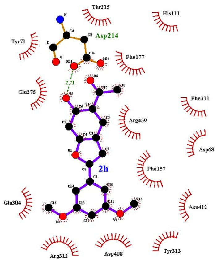
compound 2h with Further analysis of showed
α-glucosidase the interactions
that the of compound
favourable 2h with
binding α-glucosidase
affinity showed
is contributed by that
one
the favourable
hydrogen bondbinding affinity is contributed
and 14 hydrophobic by one6).hydrogen
contacts (Figure bond andPhe177,
The α-glucosidase 14 hydrophobic
Phe311 and contacts
Tyr71
(Figure
formed6). The α-glucosidase
stable π-π interactions Phe177,with Phe311 and Tyr71
compound 2h formed
benzofuranstableregion
π-π interactions
while Tyr313with compound
stabilized
2h benzofuran
compound region while Tyr313region.
2h dimethoxybenzene stabilized compound 2h dimethoxybenzene region.
Figure 6. The details interactions of compound 2h at the binding site of built structure of α-glucosidase.
Figure 6. The details interactions of compound 2h at the binding site of built structure of α-
glucosidase.You can also read