The Bacterial Microbiome in the Small Intestine of Hooded Seals (Cystophora cristata)
←
→
Page content transcription
If your browser does not render page correctly, please read the page content below
microorganisms
Article
The Bacterial Microbiome in the Small Intestine of
Hooded Seals (Cystophora cristata)
Mario Acquarone *, Alejandro Salgado-Flores and Monica Alterskjær Sundset
 Department of Arctic and Marine Biology, UiT—The Arctic University of Norway, 9037 Tromsø, Norway;
 alejandro.f.salgado@uit.no (A.S.-F.); monica.a.sundset@uit.no (M.A.S.)
 * Correspondence: mario.acquarone@gmail.com; Tel.: +47-406-26-266

 Received: 6 August 2020; Accepted: 23 October 2020; Published: 27 October 2020

 Abstract: Arctic hooded seals (Cystophora cristata) are monogastric carnivores that go through extreme
 fasting and re-feeding in early life. They are born isolated on sea ice; suckle high-fat milk for four
 days and may then fast for up to one month before they start hunting and feeding on small prey
 (fish and crustaceans). Previous studies of the gut microbiota in pinnipeds have focused on the
 large intestine, while little data exist on the small intestinal microbiota. In this study, the bacterial
 microbiome in the proximal and distal small intestine of four captive two-year old seals (two males
 and two females) fed herring (Clupea harengus) was sampled post-mortem and characterized using
 16S rRNA metabarcoding from the V1–V3 hypervariable region of the 16S ribosomal RNA (rRNA)
 genes. The seals were originally born in the wild and taken into human care at the end of the suckling
 period. Molecular-based analysis using Illumina Hiseq resulted in 569,910 16S rRNA sequences
 from the four seals (both sampling sites together). Taxonomical classification applying a naive
 Bayesian algorithm gave 412 Operational Taxonomic Units (OTUs). Firmicutes was the major phylum
 across samples (Proximal (P): 90.5% of total sequences, on average; Distal (D): 94.5%), followed by
 Actinobacteria (P: 7%; D: 0.3%) and Proteobacteria (P: 1.7%; D: 1.9%). Bacterial spp. belonging to the
 Clostridium (P: 54.1%; D: 41.6%) and SMB53 (P: 15.3%; D: 21.5%) constituted the major genera in both
 the proximal and distal small intestine. Furthermore, comparison with hindgut and fecal samples
 from geographically diverse marine mammals highlighted similarities in the microbiome between
 our seals and those sharing similar aquatic environments. This study has provided a first reliable
 glimpse of the bacterial microbiota in the small intestine microbiome of hooded seals.

 Keywords: 16S rRNA; metabarcoding; gut; bacteria; arctic; seal

1. Introduction
      Hooded seals (Cystophora cristata) are deep-diving, long-distance swimmers that occupy vast
ocean areas throughout the North Atlantic and adjacent Arctic marine waters [1–3]. They spend most
of the year at sea presumably foraging except during the breeding and molting periods that are spent
in ice-covered waters off the coast of Northeast Canada and Greenland [3–7]. Hooded seal new-born
pups undergo extreme nutritional transitions during their first month of life. In four days they almost
double their body weight [8] suckling milk with up to 60% fat [9] after which the mother abandons
them on the ice. Even though the pups are ingesting large quantities of fat, their digestive organs
(stomach, small and large intestines, and the pancreas) are neither particularly large at birth nor do
these organs gain in weight or length at unusual speed [10]. The 4-week post-weaning fasting period,
during which the pups presumably ingest only snow and seawater [11] ends when the pups begins to
hunt and feed on small prey [12,13]. The fast results in weight loss/nutrient depletion, with liver and
spleen decreasing in weight by about 70% so that they are actually lighter one month postpartum than
at birth [10].

Microorganisms 2020, 8, 1664; doi:10.3390/microorganisms8111664         www.mdpi.com/journal/microorganisms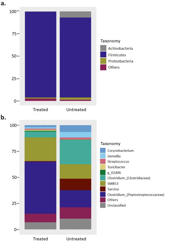
Microorganisms 2020, 8, 1664                                                                          2 of 16

      Seals are monogastric carnivores with simple hindguts (a rudimentary cecum and a short simple
colon) [14,15]. The stomach is cylindroid with a sharp pyloric bend, it weighed between 395 and 2080
g and had a pH ranging between 1.2 and 7.1 in 40 hooded seals examined (body mass 37–2015 kg) [15].
The concentration of pepsin, an endopeptidase key for protein breakdown, was found to be significantly
higher in hooded seals that had been eating a mixed diet of fish and crustaceans (146 µg mL−1 ) compared
to only crustaceans (50 µg mL−1 ) [15].
      The relative length of the pinniped small intestine varies considerably. Some species have
significantly longer small intestines than those in terrestrial carnivores of similar size [14,16]. In hooded
seals, the length of the small intestine is 13× body length, while the small intestines of the northern
elephant seals (Mirounga angustirostris) and southern elephant seals (Mirounga leonina) average 25×
body length and only 4.8× body length in the Ross seal (Ommatophoca rossii) [16–18]. The hypothesis
that the relatively long small intestine of some seal species evolved to compensate for long periods of
reduced or even abolished blood perfusion to the intestine during diving [19] was later rejected, as no
significant correlation was found between relative intestinal length and diving ability [16]. Instead,
the area of the small intestine was significantly related to body length/size [16].
      Little is known about the hooded seal diet; however, stomach content analyses indicate that
adult seals feed on large prey. This diet may vary considerably depending of the location where seals
happened to be sampled and can include large cod, halibut, and redfish [20]. A diet study using
quantitative fatty acid signature analysis of hooded seals sampled along the Northeastern coast of
Newfoundland and Southern Labrador indicated a diet of amphipods, Atlantic argentine, capelin,
euphausiids and redfish [21] highlighting that the hooded seal diet is variable. Transitions from
feeding to fasting, and from fasting to feeding, pose severe challenges to the digestive system of
vertebrates [22]. Symbiosis between mammals and their gut microbiome is important for the extraction
of energy and nutrients from food and influences both immune response and brain development.
The gastrointestinal tract is colonized during birth and then by maternal, social, and environmental
contact. The composition of the adult gut microbiome depends on initial colonization, food chemistry
and intake, and hereditary aspects such as host-genetics. Several studies have described the gut
microbiota in pinnipeds [23,24], which is mostly dominated by a ‘core’ of a few bacterial phyla [25,26].
Nonetheless, these studies were focused on the microbiota from the large intestine of the digestive
tract. There are few data available on the microbiome from the small intestine despite the essential role
of this section of the gut in carbohydrate and lipid metabolism [27]. Here we present the first study on
the bacterial microbiome in the small intestine in the hooded seal—a mammalian model for extreme
fasting and re-feeding.

2. Materials and Methods

2.1. Animals and Sampling
     We sampled and characterized the bacterial microbiome from the proximal and distal small
intestine (contents) of four healthy adult hooded seals (age 2 years), two males (81.5 kg and 86 kg) and
two females (88 kg and 103 kg).
     The seals had originally been born in the wild and taken in human care after weaning between
the 26th and the 28th March 2012 (by permit from the Danish Foreign Ministry and the Greenland
authorities). They were transported fasted, by ship from Greenland to Tromsø, Norway and maintained
in two 40,000 L sea water pools in the approved animal holding facilities at the Department of Arctic
and Marine Biology of UiT—The Arctic University of Norway. During the period in human care the
seals were fed freshly thawed, human food grade frozen herring (Clupea harengus) integrated daily
by marine animal dietary supplement (Sea Tabs ® MA, Pacific Research Labs Inc., PO Box 675890,
Rancho Santa Fe, CA 92067, USA). The animals were euthanized by bleeding in isoflurane anesthesia
and were also sampled extensively as part of other research projects unrelated to this study (permit
Microorganisms 2020, 8, 1664 3 of 16
no. 5399 (reference 2013/87412-119) issued by the National Animal Research Authority of Norway
3rd June 2013).
All seals received routine antiparasitic treatment with ivermectin (10 mg sub cutaneous injection)
upon arrival at the facility on 3rd April. An oral antiparasitic treatment was also administered for
three days starting on 22nd June and consisted on a total dose of 250 mg of Panacur.
One of the females was treated with antibiotics for an infection in the jaw (enrofloxacin 300 mg oral
dose daily between 4th and 12th December 2012 and again between 5th and 14th March 2013). One of
the males also suffered from an infection of the jaw and received a daily intra-muscular (i.m.) injection
with enrofloxacin (400 mg) 31st July–11th August 2013 followed up by a daily oral dose (300 mg)
between 12th and 16th August. The same animal was treated with a daily oral dose of enrofloxacin
(400 mg) between 23rd and 30th December and between 7th and 14th February 2014 for a new infection
of the jaw. This last treatment was administered 12 days before sampling.
For this study, the abdominal cavity of the freshly euthanized animal was immediately opened
and the intestinal system, sectioned at the end of the duodenum and before the colon, was removed
from the carcass. The contents of approximately 2 m at each end of the removed intestinal tract were
manually squeezed out of the gut and into 50 mL centrifuge tubes. The samples were immediately
frozen and kept at −40 ◦ C, until analysis.
2.2. DNA Extraction
DNA extraction was based on the protocol of the Repeated Bead Beating plus Column (RBB+C)
Method developed by Yu and Morrison [28]. DNA quantification was done with NanoDrop 2000c
spectrophotometer and solutions were stored at −20 ◦ C until PCR amplification.
2.3. Sequencing
PCR amplifications for Bacteria were performed with the bacterial primer set 27F
(50 -AGAGTTTGATCCTGGCTCAG -30 and 519R (50 -GWATTACCGCGGCKGCTG -30 ) [29,30], giving
a 500-nt size amplicon product targeting the V1-V3 hypervariable region of the 16S ribosomal RNA
(rRNA) genes. PCR reactions were run as described in [31]. Sample products were then pooled in
equimolar amounts, checked in a 1% agarose gel electrophoresis, and excised and purified from gel
using a NucleoSpin Gel and PCR Clean-up kit (Macherey-Nagel, Düren, Germany). The resulting
DNA was stored at −20 ◦ C until sequencing. PCR amplicons were sequenced with Illumina MiSeq at
the MrDNA company (Shallowater, TX, USA).
2.4. Sequence Processing
Sequences for 16S rRNA genes from both microbial groups were analyzed using the Quantitative
Insights Into Microbial Ecology (QIIME) (v. 1.9.0) pipeline [32]. First, Illumina forward and reverse
fast reads were merged using the join_paired_ends.py script. The resulting merged reads were quality
checked and discarded when: lengthMicroorganisms 2020, 8, 1664 4 of 16
Alpha diversity indicators evaluating intra-group species richness (chao1), evenness (Shannon) and
overall diversity (Simpson) were analyzed using the alpha_diversity.py script in QIIME from randomly
subsampled dataset.
To facilitate direct comparisons across studies, the raw dataset was also processed with the
open-source pipeline DADA2 (v1.16.0; May 2020 release) [36]. DADA2 entails a different approach
compared to traditional OTU-based clustering by processing exact sequences as Amplicon Sequence
Variants (ASVs). The lack of clustering and use of exact sequences allows for higher reproducibility
when compared to ASVs covering a comparable target region. The dataset was processed following
the standard recommended pipeline parameters (https://benjjneb.github.io/dada2/tutorial.html v1.16;
May 2020). The resulting sequence table including all the filtered, trimmed, merged and chimera-free
ASVs was made available (Table S1).
2.6. Statistical Analysis
Considering the limited sample size for the current study (n = 4), it may not be sufficient to
conduct robust statistical comparisons. Consequently, we have made a special emphasis on presenting
a qualitative report of the bacterial communities dwelling the proximal and distal part of the small
intestine in the captive hooded seals as a proxy for potential future, broader-scope assessments.
3. Results
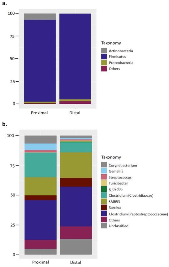
3.1. Bar Chart Plot
A total 1,081,347 bacterial 16S rRNA sequences were produced from small intestine proximal (n = 4)
and distal (n = 4) samples of hooded seals. Quality checked samples resulted in a range between 57,381
and 72,161 sequences per sample, with an average length of 405 bases for downstream analysis. Based
upon a 97% similarity criterion the sequences were clustered in 412 chimera-free OTUs. Taxonomical
classification at phylum level showed a microbiota dominated by the phylum Firmicutes in both
proximal and distal small intestine samples (Proximal: 90.5 ± 12.2% of total sequences, on average;
Distal: 94.5 ± 4%). Actinobacteria (Proximal: 7 ± 12.7%; Distal: 0.4 ± 0.3%) and Proteobacteria
(Proximal: 1.7 ± 1.5%; Distal: 1.9 ± 2.3%) constituted the remaining major phyla (Figure 1a; Figure S1).
The standard deviation in the relative abundance at phylum level ranged from 0.9% to 12.7% for the
proximal sample group, and 0.3% to 4% for the distal sample group.
Classification at genus level showed bacterial phylotypes assigned to Clostridium from the
families Peptostreptococcaceae (Proximal: 33.4 ± 32.7%; Distal: 33.3 ± 26.8%) and Clostridiaceae
(Proximal: 20.7 ± 32.1%; Distal: 8.3 ± 7.2%) as the major genera, followed by genus SMB53 (Proximal:
15.3 ± 14.5%; Distal: 21.5 ± 15.2%), and unclassified genera from the family Peptostreptococcaceae
(Proximal: 4.8 ± 6.96%; Distal: 13.3 ± 11.5%) (Figure 1b). The standard deviation in the relative
abundance at genus level ranged from 0.1% to 32.7% for the proximal sample group, and 1.4% to 26.8%
for the distal sample group.Microorganisms 2020, 8, 1664 5 of 16
Microorganisms 2020, 8, x FOR PEER REVIEW 5 of 16
Figure
Figure 1.1. Major
Major bacterial
bacterialphyla
phylaandand genus
genus in the
in the proximal
proximal and distal
and distal small small intestine
intestine of captive
of captive hooded
hooded
seals. Theseals. The bar
bar charts charts the
represent represent
relativethe relative abundance
abundance of the
of the total 16S rRNA total 16S rRNA
sequence sequence
taxonomically
taxonomically
classified at (a)classified
phylum andat (a)
(b)phylum and (b)
genus level. genus level.was
Classification Classification was performed
performed with with partial
partial sequences of the
sequences of the
bacterial 16S rRNAbacterial 16S rRNA
gene against the gene
RDP-IIagainst the RDP-II
database database
using RDP using
classifier RDP classifier tool.
tool.
Some of the studied
Classification sealslevel
at genus wereshowed
providedbacterial
occasional antibiotic treatment
phylotypes assigned to (enrofloxacin)
Clostridiumthroughout
from the
the captive
families period. Considering(Proximal:
Peptostreptococcaceae the widely33.4 reported impact
± 32.7%; posed
Distal: 33.3by± antibiotics
26.8%) andonClostridiaceae
the intestinal
microbiota20.7
(Proximal: [37],±taxonomical
32.1%; Distal:results were as
8.3 ± 7.2%) also
thepresented separating
major genera, followedthose
byseals
genusreceiving antibiotics
SMB53 (Proximal:
and ±
15.3 not receiving
14.5%; antibiotics
Distal: to assess
21.5 ± 15.2%), andforunclassified
potential differences
genera from (Figure 2). As observed,
the family classification
Peptostreptococcaceae
at phylum 4.8
(Proximal: level
± is also represented
6.96%; Distal: 13.3 by a microbiota
± 11.5%) dominated
(Figure 1b). The by the phylum
standard Firmicutes
deviation in the (Treated:
relative
95.6 ± 4.1%;atUntreated:
abundance genus level ± 11.4%)
89.3ranged (Figure
from 0.1%2a). Classification
to 32.7% for the at genus level
proximal showed
sample sealsand
group, treated
1.4%with
to
antibiotics
26.8% to be
for the dominated
distal sample by member of the genus Clostridium from the family Peptostreptococcaceae
group.
Some of the studied seals were provided occasional antibiotic treatment (enrofloxacin)
throughout the captive period. Considering the widely reported impact posed by antibiotics on theintestinal microbiota [37], taxonomical results were also presented separating those seals receiving
antibiotics and not receiving antibiotics to assess for potential differences (Figure 2). As observed,
classification at phylum level is also represented by a microbiota dominated by the phylum
Microorganisms
Firmicutes 2020, 8, 1664 95.6 ± 4.1%; Untreated: 89.3 ± 11.4%) (Figure 2a). Classification at genus
(Treated: level
6 of 16
showed seals treated with antibiotics to be dominated by member of the genus Clostridium from the
family Peptostreptococcaceae (50.2 ± 22.5%) and family Clostridiaceae (22.9 ± 14%) (Figure 2b).
(50.2 ± 22.5%) and family Clostridiaceae (22.9 ± 14%) (Figure 2b). Likewise, the bacterial microbiota from
Likewise, the bacterial microbiota from untreated seals was also dominated by the genus
untreated seals was also dominated by the genus Clostridium from the family Peptostreptococcaceae
Clostridium from the family Peptostreptococcaceae (16.5 ± 23.1%) and family Clostridiaceae (23.5 ±
(16.5 ± 23.1%) and family Clostridiaceae (23.5 ± 31.1%); however, other genera were also importantly
31.1%); however, other genera were also importantly represented such as SBM53 (13.9 ± 14.7) and
represented such as SBM53 (13.9 ± 14.7) and Sarcina (11 ± 13.6). Interestingly, “unclassified” bacteria
Sarcina (11 ± 13.6). Interestingly, “unclassified” bacteria constituted a comparable relative
constituted a comparable relative proportion of the total microbiota in both treated (7.7 ± 10.5) and
proportion of the total microbiota in both treated (7.7 ± 10.5) and untreated seals (10.4 ± 10.7) (Figure
untreated seals (10.4 ± 10.7) (Figure 2).
2).
Figure 2. Major bacterial phyla and genus in the proximal and distal small intestine of captive hooded
Figure 2. Major bacterial phyla and genus in the proximal and distal small intestine of captive
seals depending on whether they received antibiotic treatment or not. The bar charts represent the
hooded
relative seals depending
abundance on16S
of the total whether they received
rRNA sequence antibiotic
taxonomically treatment
classified at (a)or not. The
phylum bargenus
and (b) charts
level. Classification was performed with partial sequences of the bacterial 16S rRNA gene against the
RDP-II database using RDP classifier tool.Microorganisms 2020, 8, 1664 7 of 16
3.2. Group-Based Diversity Tests
Intra-group (alpha) diversity indicators for samples grouped based on sampling site (i.e., proximal
or distal small intestine) showed comparable values for species richness (chao1), evenness (Shannon),
and overall diversity (Simpson’s) (Figure S1a–d).
Similar parameters were interrogated when samples were grouped based on antibiotic treatment
(Figure S3a–d). Overall, alpha diversity parameters were comparable for every individual metric
between both groups of samples; however, Simpson diversity index was on average lower in samples
from seals undergoing antibiotic treatment (Figure S3c).
3.3. Comparison with the Microbiota from Other Marine Mammals
To assess the findings presented here from a broader perspective, we compared them with the
results described from other marine mammals (Table 1). Due to the lack of information of the microbiota
in the small intestine in general, and in particular for marine mammals, all the additional data were
obtained from studies using cecal or fecal samples. In addition, some of the studies applied different
sequencing platforms to the one used in the current study (Illumina HiSeq) or hybridization-based
techniques, aspects that may potentially pose bias to the results. The majority of the listed species
possessed a gut microbiota dominated by the phylum Firmicutes, similar to that reported in the current
study (Figure 1a). Only a few studies reported animals whose gut microbiota was dominated by other
microbial phyla apart from Firmicutes. It was interesting to observe that the fecal microbiota in wild
hooded seals was dominated by Bacteroidetes (68% relative sequence abundance), in contrast to the
results from our captive seals (no presence of Bacteroidetes). In addition, other phocids such as harbor
seals presented a more balanced microbiota where Firmicutes and Bacteroidetes were the dominant
phyla, both in captive and wild seals. Actinobacteria (5%) and Proteobacteria (2%), which were present
at a very low relative abundance in the current study, constituted an important fraction of the microbiota
in some species such as wild and captive Australian sea lion, wild leopard seals, and porpoises from
China (Table 1). Fusobacterium was also largely present in some marine mammals such as southern
elephant seals and captive leopard seals, but this phylum was not detected in the small intestine of the
captive hooded seals.Microorganisms 2020, 8, 1664 8 of 16
Table 1. Intestinal microbiota composition of carnivore marine mammals from Arctic and Antarctic latitudes. The results were presented per host animal species,
living conditions (wild or captive), and sampling origin. Taxonomy was indicated at phylum level considering the total relative abundance. An indication on the
applied methodology by which the results were obtained was also included.
Species Latin Name Location—Diet Samples Bacterial Community Method Reference
Bacteroidetes (68%)
Wild (Greenland Sea)—Halibut, herring, BigDye—Sanger
Hooded seal Cystophora cristata Colon Firmicutes (22%) [23]
cod, squid, crustaceans sequencing
Proteobacteria (9%)
Wild (Ringvassøy, Troms)—saithe, cod, Firmicutes (50%) BigDye—Sanger
Harbor seal Phoca vitulina Colon [23]
herring, sculpin Bacteroidetes (49%) sequencing
Wild (Ringvassøy, Troms)—Cod, saithe, Firmicutes (76%) BigDye—Sanger
Gray seal Halichoerus grypus Colon [23]
herring, sandeel, catfish Bacteroidetes (24%) sequencing
Firmicutes (33 ± 11%)
Bacteroidetes (27 ± 7%)
Harbor seals Phoca vitulina Captive – herring, sprat, crustaceans Feces 454 pyrosequencing [24]
Fusobacteria (26 ± 4%)
Proteobacteria (13 ± 5%)
Firmicutes (42 ± 10%)
Southern elephant Bacteroidetes (22 ± 5%)
Mirounga leonina Wild (Western Antarctica)—squid, fish Rectal swab 454 pyrosequencing [26]
seals Fusobacteria (20 ± 8)
Proteobacteria (16 ± 8%)
Firmicutes (45 ± 13%)
Proteobacteria (33 ± 12%)
Wild (Western Antarctica)—seals, krill,
Leopard seals Hydrurga leptonyx Rectal swab Fusobacteria (14 ± 8%) 454 pyrosequencing [26]
penguins, fish/captive (Taronga zoo)—fish
Firmicutes (60 ± 32%)
Fusobacteria (34 ± 13%)
Firmicutes (67%)
Australian fur Arctocephalus Wild (Kanowna island, Australia)—fish
Feces Bacteroidetes (15%) FISH [38]
seals pusillus doriferus and cephalops
Actinobacteria (3%)
Bacteroidetes (40 ± 21%)
Zalophus
California sea lion Captive—fed fish and squid Rectal swab Firmicutes (29 ± 20%) Sanger sequencing [39]
californianus
Fusobacteria (25 ± 9)Microorganisms 2020, 8, 1664 9 of 16
Table 1. Cont.
Species Latin Name Location—Diet Samples Bacterial Community Method Reference
Firmicutes (74 ± 28%)
Proteobacteria (10 ± 20%)
Wild—benthic fish, squid, lobster, small Bacteroidetes (9 ± 16%) Illumina MiSeq
Australian sea lion Neophoca cinerea Feces [40]
crustacean)/captive—frozen fish Firmicutes (58 ± 33%) sequencing
Proteobacteria (30 ± 35.9%)
Bacteroidetes (5 ± 8.6%)
Firmicutes (89 ± 6%)
South American Arctocephalus Wild dead animal (Southern coast of
Feces Proteobacteria (6 ± 6%) Ion Torrent [41]
fur seals australis Brazil)—fish, cephalopods, crustaceans
Actinobacteria (3 ± 2%)
Wild dead animal (Southern coast of
Subantarctic fur Arctocephalus Firmicutes (84 ± 6%)
Brazil)—fish, cephalopods, crustaceans, Feces Ion Torrent [41]
seals tropicalis Actinobacteria (11 ± 3%)
rock hoper penguins
Firmicutes (51.3%)
Neophocaena
Wild (Jianxi, China)—fish, crustaceans, Tenericutes (17.9%)
Yangtze porpoise phocaenoides Feces Sanger sequencing [42]
cephalopods Proteobacteria (15.4%)
asiaeorientalis
Actinobacteria (7.7%)
Firmicutes (93 ± 9%)
Illumina HiSeq
Hooded seals Cystophora cristata Captive—herring and diet supplements Small intestine Actinobacteria (4 ± 9%) Current study
sequencing
Proteobacteria (2 ± 3%)Microorganisms 2020, 8, 1664 10 of 16
4. Discussion
4.1. Homogenous Bacterial Microbiome Along the Small Intestine of the Hooded Seals
The microbiome of the small and the large intestine in mammals differs fundamentally, but only
limited information is available about the microbiota of the small intestine, despite its relevance to many
physiological mechanisms and also pathological states [27]. The small intestine is characterized by a
relatively short transit time, and also influx of bile and digestive enzymes that creates harsh conditions
not favorable for a bacterial growth. The small intestinal microbiome adapts rapidly to changes
in availability of nutrients and metabolizes simple carbohydrates for community maintenance [43].
Studies from human samples reported differences in the bacterial communities throughout sections of
the small intestine, with a higher overall bacterial density and increased presence of anaerobic bacteria
toward the distal ileum where transit is slower [27]. However, a major concept shown across studies
is that the microbiota of the small intestine is less diverse than that of the large intestine although
more dynamic. In the current study diversity indicators assessing species richness and evenness gave
similar results for samples from the distal and proximal small intestine of hooded seals (Figure S1a–d).
A cross-sectional analysis of the bacterial microbiota in the digestive tract of a Brazilian ruminant
showed dissimilar communities between the duodenum and ileum, but in multi-dimensional analysis
of those samples clustered together when compared with samples from other gastrointestinal tract (GIT)
sections [44]. Dissimilar bacterial profiles were also found between samples from the small intestine
and cecum in swine with different fatness [45]. Such results indicated the presence of a comparable
microbiota along the small intestine, but different compared to other GIT compartments. Furthermore,
the same microbial groups were found to dominate the proximal and distal part of the small intestine
in the studied captive hooded seals at phyla and genus level (Figure 1a,b), which suggests the existence
of a homogeneous microbiota across this organ. The nature of the ingested food, consisting mostly
of fats and proteins that are mostly digested and absorbed in the small intestine, together with a
short retention time, would leave little substrate for fermentation in these carnivorous animals. Thus,
these aspects would lead to the growth of a similar microbiota between the proximal and distal parts.
4.2. Comparing the Small Intestine Microbiome of the Hooded Seal to That of Other Marine Mammals
A previous study describing the colonic microbiota in wild hooded seals showed dominance by the
phylum Bacteroidetes followed by Firmicutes [23]. In contrast, the current study presented Firmicutes
as the dominant bacterial taxa, and the absence of Bacteroidetes-related phylotypes (Figure 1a).
One factor that may potentially influence bacterial diversity is the living conditions, i.e., captivity
versus free-ranging state. For instance, the samples used in the current study were collected from
captive seals, and captivity has been discussed to largely influence the gut microbiota composition in
other marine mammals such as leopard seals (Hydrurga leptonix), where increased relative abundance
of Firmicutes-related bacteria was described in the captive animals [26]. A presumably less varied
diet given to the captive leopard seals was accounted for the main cause driving to such differences
(see discussion in [26]). Likewise, wild hooded seals combine a diet including several fish species,
squid, and some invertebrates (crustaceans) [21], which overall constitutes a richer diet than that fed to
our captive seals (herring and dietary supplements; see Methods). In addition, other factors directly
associated with a captive lifestyle, such as the administration of antibiotics, and physiological alterations
(e.g., hormonal production), may also trigger such differences [25,46]. Nonetheless, as previously
discussed, the part of the digestive tract chosen for sampling (i.e., colon vs small intestine) would pose
a stronger effect on the composition of the bacterial microbiota and would account for most of the
differences observed between wild and captive hooded seals.
Clostridium constituted the dominant genus in the small intestine of the studied captive hooded
seals (Figure 1b). The ubiquity of this genus in the digestive tract of several other marine mammals,
from Arctic and Antarctic latitudes, has made it to be considered to be part of a putative ‘phocid
seals core microbiota’, together with several other bacterial genera [24,25]. In principle, such a sharedMicroorganisms 2020, 8, 1664 11 of 16
microbiota would be passed from mother to pups, and it would be involved in several physiological
aspects, from host-immunity, maturation of the gut tissue, and, the breakdown of milk components
during the 2-month period after birth [25]. The lack of data on the small intestine microbiota across
marine mammals gives us no possibility to associate the presence of Clostridium spp. with a putative
core microbiota. Nonetheless, a potential link between an increase in the relative abundance of
bacterial members belonging to the class Clostridia and the consumption of diets high in fat have been
reported [27], which may hint a role played by these bacteria in fat metabolism in this intestinal section.
The genus SMB53 also constituted a substantial fraction of the microbiota in the small intestine of
our captive hooded seals (Figure 1b). SMB53 is a poorly studied genus belonging to the class Clostridia,
which has mostly been reported in captive hosts such as birds, pigs, and obese laboratory mice [45,47,48].
SMB53 was also present in free-ranging carnivores consuming a high-fat, high-protein diet, but it
constituted a small fraction of the microbiota [49]. In addition to the current study, the only record of
this genus in the small intestine was found in farmed pigs with high body fatness, whose microbiome
was enriched in inflammation-related genes speculated to trigger increased fat adiposity [45]. Whether
the presence of this genus in our captive seals is directly linked to the consumption of a high-fat,
high-protein diet or altered by antibiotics (which captive animals are normally provided) remains to be
elucidated until more information on its physiological features is reported.
4.3. Potential Effect of Antimicrobial and Antiparasitic Treatment on the Gut Microbiota
The ingestion of antibiotics has been reported to pose an impact on the gut microbiota [37].
For instance, broad-spectrum antibiotics may drop the overall community diversity, an effect that may
last months or even years [37]. In the current study, two of the four captive seals received occasional
antibiotic treatment (enrofloxacin) due to an infection in the jaw (see Methods). Enrofloxacin is a
fluoroquinolone antibiotic commonly used for animals; some studies described a decrease in overall
diversity in the gut microbiota of laboratory mice, with a particular emphasis in members from the
family Bacteroidaceae [50]. Likewise, in this study, a reduction in microbial richness (Simpson index)
was observed in samples from captive hooded seals that were administered this antibiotic (Figure S3c),
which showed dominance by one particular group belonging to the genus Clostridium (family
Peptostreptococcaceae) (Figure 2b). Although the use of antibiotics has been commonly associated with
an increase in members of the phylum Bacteroidetes over Firmicutes [37], some pathogenic bacteria
such as Clostridium difficile (Firmicutes) may also be favored after antibiotic treatment [37]. As indicated
in the Methods section, statistical analysis was not addressed in the current study due to limitation in
sample size, which makes it impossible to test the differences in bacterial taxonomy between treated
and untreated hooded seals. Nonetheless, in light of the result for both groups of seals (treated vs.
untreated with antibiotics), a direct link between the use of antibiotics and dominance by Firmicutes
may be discarded.
Knowledge of the potential effects exerted by the use of antiparasitic drugs on the gut microbiome
has been investigated less than for antibiotics, with some studies performed in human subjects indicating
a drop in overall diversity as well as alterations in some individual microbial groups [51]. In the
current study, all captive hooded seals underwent an intitial treatment with ivermectin (a macrocyclic
lactone) and Panacur (fenbendazole), broad-spectrum antiparatic drugs categorized as anthelmintics
(see Methods). Research on the effect posed by each specific drug on the gut microbiota is limited and
mostly shown a lack of effect in community-wide diversity [52,53]. Instead, certain positive effect was
observed at individual level to bacteria within the families Enterobacteriaceae and Lachnospiraceae [52].
Based on this information and considering that antiparasitic treatment was administered exclusively
upon arrival to the animal facilities at UiT, almost two years prior to sampling, we may suggest a
negligible effect posed by the administration of either drug on the small intestine microbiota in the
captive hooded seals in this study.Microorganisms 2020, 8, 1664 12 of 16
4.4. Importance of the Gut Microbiota in Food Digestion in Carnivores
Although the small intestine is the main site for the complex physiological processes of protein
and lipid digestion and absorption, its associated microbiota has been substantially neglected
compared to other GIT regions, e.g., the large intestine. Still, some studies have described its
composition and reported the potential role assigned to this microbial consortium on lipid metabolism,
mainly by increasing lipid absorption via enteroendocrine signaling in the proximal small intestine
(duodenum-jejunum) [54,55]. For instance, when germ-free mice were transplanted with the small
intestinal microbiota of mice conditioned on a high-fat diet, lipid absorption was significantly increased
regardless the fat content in the diet [56]. Some microbial phyla (e.g., Firmicutes) have been associated
with increased fat absorption and metabolic disorders such as obesity [49], although the extent of the
effect is a matter of debate [57]. All the marine mammals used for the interspecies comparison were
carnivores ingesting generally a diet rich in fat and protein (Table 1), with most of them presenting
a gut microbiota dominated by Firmicutes bacteria. Our captive hooded seals were mostly fed
herring, a foodstuff rich in protein and polyunsaturated fatty acids (PUFAs) such as omega-3 [58].
It has been reported that ingestion of omega-3 dietary supplements is linked with an increase in
butyrate-producing bacteria belonging to the phylum Firmicutes [59]. Altogether, it may be speculated
that dominance by Firmicutes-related bacteria in the small intestine of captive hooded seals may be
related to the ingestion of a fatty acid-rich carnivore diet, which in turn, may play a role in easing
lipid metabolism and energy uptake. New experiments targeting to unveil the genetic information
(metagenomics) must be conducted to elucidate the functional potential in fat metabolism exerted by
the small intestine microbiota in hooded seals or other carnivore marine mammals.
5. Conclusions
Despite the increasing body of knowledge regarding the gut microbiota of marine mammals,
there is still little information available on their small intestine microbes for both captive and wild
individuals. To date, the current study is the first endeavor to unveil the bacterial constituents found
in the small intestine of captive hooded seals originally captured in the pack ice off the coast of East
Greenland. No significant differences were observed between the bacteria found in the proximal and
distal parts of the small intestine, indicating a seemingly homogenous microbiota throughout the
small intestine. We suggest that the diet of fatty fish, which is mostly digested in the small intestine,
would leave little material for subsequent bacterial degradation at distal sections and, therefore,
rendered a similar microbiota at both ends of the small intestine. Dominance by bacterial groups
previously associated with individuals feeding on high-energy diets, in some instances also related to
metabolic disorders (e.g., obesity), may indicate a small intestine populated by microbes adapted to
metabolize foodstuffs rich in fat and protein. Such microbiota would help maximize the degradation
and energy retrieval from the diet. In addition, a broad comparison with the microbiota reported
from several other marine mammals allowed us to identify the presence of microbial groups in our
captive seals that have been hypothesized to belong to a putative phocid core microbiota. Nonetheless,
any potential conclusion drawn by direct comparisons should be considered with care until more
information on the small intestine microbiome can be obtained from other marine mammals. Further
analyses of the small intestine microbiome at a functional level (i.e., metagenomics/metatranscriptomics)
would help understand the role played by such microbes in physiological functions related to lipid
absorption and energy metabolism, relevant to comprehend the digestive physiology in hooded seals.
Supplementary Materials: The following are available online at http://www.mdpi.com/2076-2607/8/11/1664/s1,
Figure S1: Major bacterial groups in the small intestine of treated and untreated with antibiotics captive hooded
seals. The bar charts represent the relative abundance of the total 16S rRNA sequence taxonomically classified
at (a) class, (b) order, and (c) family level. Classification was performed with partial sequences of the bacterial
16S rRNA gene against the RDP-II database using RDP classifier tool. Figure S2: Boxplot intra-group microbial
diversity indicators from proximal and distal small intestine samples. Mean alpha diversity values for: (a) species
richness (Chao1); (b) evenness (Shannon index); (c) overall diversity (Simpson); and (d) total observed species.
Figure S3: Boxplot intra-group microbial diversity indicators from the small intestine of treated and untreatedMicroorganisms 2020, 8, 1664 13 of 16
with antibiotics captive hooded seals. Mean alpha diversity values for: (a) species richness (Chao1); (b) evenness
(Shannon index); (c) overall diversity (Simpson); and (d) total observed species. Table S1: Amplicon Sequence
Variants (ASVs) from the proximal and distal parts of the small intestine of Hooded seals. ASVs were obtained
from raw sequencing dataset and processed as indicated in Materials and Methods (2.5. Sequence Analysis).
Author Contributions: Conceptualization, M.A., A.S.-F., and M.A.S.; methodology, M.A., A.S.-F., and M.A.S.;
software, A.S.-F..; validation, A.S.-F., M.A., and M.A.S.; formal analysis, A.S.-F.; investigation, M.A., A.S.-F., and
M.A.S.; resources, M.A., A.S.-F., and M.A.S.; data curation, M.A., A.S.-F., and M.A.S.; writing—original draft
preparation, M.A.S., A.S.-F., and M.A.; writing—review and editing, A.S.-F., M.A., and M.A.S.; visualization,
A.S.-F.; project administration, M.A., A.S.-F., and M.A.S.; funding acquisition, M.A., and M.A.S. All authors have
read and agreed to the published version of the manuscript.
Funding: This project was financed by internal funding from the Department of Arctic and Marine Biology and
received no external funding. The publication charges for this article have been funded by a grant from the
publication fund of UiT The Arctic University of Norway.
Acknowledgments: We are grateful to Erling S. Nordøy who led the cruise and the field sampling as well as to
Lars P. Folkow who assisted with euthanasia procedures and provided laboratory space.
Conflicts of Interest: The authors declare no conflict of interest.
Data Availability: The sequence reads obtained from 16S amplicon sequencing and shotgun metagenomics
are available at the Sequence Read Archive (SRA) database under the BioProject identifier PRJNA665267
(SAMN16249586-SAMN16249593).
References
1. Folkow, L.P.; Mårtensson, P.-E.; Blix, A.S. Annual distribution of hooded seals (Cystophora cristata) in the
Greenland and Norwegian seas. Polar Biol. 1996, 16, 179–189. [CrossRef]
2. Andersen, J.M.; Wiersma, Y.F.; Stenson, G.B.; Hammill, M.O.; Rosing-Asvid, A.; Skern-Maurizen, M. Habitat
selection by hooded seals (Cystophora cristata) in the Northwest Atlantic Ocean. ICES J. Mar. Sci. 2012, 70,
173–185. [CrossRef]
3. Vacquie-Garcia, J.; Lydersen, C.; Biuw, M.; Haug, T.; Fedak, M.A.; Kovacs, K.M. Hooded seal Cystophora
cristata foraging areas in the Northeast Atlantic Ocean—Investigated using three complementary methods.
PLoS ONE 2017, 12, e0187889. [CrossRef] [PubMed]
4. Sergeant, D.E. A rediscovered whelping population of hooded seals, Cystophora cristata Erxleben, and its
possible relationship to other populations. Polarforschung 1974, 44, 1–7.
5. Folkow, L.; Blix, A.S. Distribution and Diving Behaviour of Hooded Seals; Elsevier BV: Amsterdam,
The Netherlands, 1995; Volume 4, pp. 193–202.
6. Folkow, L.P.; Blix, A.S. Diving behavior of hooded seals (Cystophora cristata) in the Greenland and Norwegian
Seas. Polar Biol. 1999, 22, 61–74. [CrossRef]
7. Hammill, M.O.; Stenson, G. Abundance of Northwest Atlantic hooded seals (1960–2005). DFO Canada.
Canadian Science Advisory Secretariat Research Document 2006. p. 19. Available online: http://www.dfo-
mpo.gc.ca/csas/ (accessed on 30 May 2020).
8. Bowen, W.D.; Oftedal, O.T.; Boness, D.J. Birth to weaning in 4 days: Remarkable growth in the hooded seal,
Cystophora cristata. Can. J. Zool. 1985, 63, 2841–2846. [CrossRef]
9. Lydersen, C.; Kovacs, K.M.; Hammill, M.O. Energetics during nursing and early postweaning fasting in
hooded seal (Cystophora cristata) pups from the Gulf of St Lawrence, Canada. J. Comp. Physiol. B 1997, 167,
81–88. [CrossRef]
10. Oftedal, O.T.; Bowen, D.; Widdowson, E.M.; Boness, D.J. Effects of Suckling and the Postsuckling Fast on
Weights of the Body and Internal Organs of Harp and Hooded Seal Pups. Neonatology 1989, 56, 283–300.
[CrossRef]
11. Schots, P.C.; Bue, M.E.; Nordøy, E.S. Hooded seal (Cystophora cristata) pups ingest snow and seawater during
their post-weaning fast. J. Comp. Physiol. B 2016, 187, 493–502. [CrossRef]
12. Bowen, W.D.; Boness, D.J.; Oftedal, O.T. Mass transfer from mother to pup and subsequent mass loss by the
weaned pup in the hooded seal, Cystophora cristata. Can. J. Zool. 1987, 65, 1–8. [CrossRef]
13. Haug, T.; Nilssen, K.T.; Lindblom, L. First independent feeding of harp seal (Phoca groenlandica) and hooded
seal (Cystophora cristata) pups in the Greenland Sea. NAMMCO Sci. Publ. 2000, 2, 29–39. [CrossRef]
14. Olsen, M.A.; Nilssen, K.T.; Mathiesen, S.D. Gross anatomy of the gastrointestinal system of harp seals
(Phoca groenlandica). J. Zool. 1996, 238, 581–589. [CrossRef]Microorganisms 2020, 8, 1664 14 of 16
15. Christiansen, J.S.; Gildberg, A.; Nilssen, K.T.; Lindblom, C.; Haug, T. The gastric properties of free-ranging
harp (Pagophilus groenlandicus (Erxleben, 1777)) and hooded (Cystophora cristata (Erxleben, 1777)) seals.
ICES J. Mar. Sci. 2004, 61, 287–292. [CrossRef]
16. Mårtensson, P.-E.; Nordøy, E.S.; Messelt, E.B.; Blix, A.S. Gut length, food transit time and diving habit in
phocid seals. Polar Biol. 1998, 20, 213–217. [CrossRef]
17. Bryden, M. Size and growth of viscer in the southern elephant seal, Mirounga leonina (L.). Aust. J. Zool. 1971,
19, 103. [CrossRef]
18. Helm, R.C. Intestinal length of three California pinniped species. J. Zool. 2009, 199, 297–304. [CrossRef]
19. Krockenberger, M.B.; Bryden, M.M. Rate of passage of digesta through the alimentary tract of southern
elephan seals (Mirounga leonina) (Carnivora: Phocidae). J. Zool. Lond. 1994, 234, 229–237. [CrossRef]
20. Kapel, F. Feeding habits of harp and hooded seals in Greenland waters. NAMMCO Sci. Publ. 2000, 2, 50–64.
[CrossRef]
21. Tucker, S.; Bowen, W.; Iverson, S.; Blanchard, W.; Stenson, G. Sources of variation in diets of harp and hooded
seals estimated from quantitative fatty acid signature analysis (QFASA). Mar. Ecol. Prog. Ser. 2009, 384,
287–302. [CrossRef]
22. Wang, T.; Hung, C.C.Y.; Randall, D.J. The comparative physiology of food deprivation: From Feast to Famine.
Annu. Rev. Physiol. 2006, 68, 223–251. [CrossRef]
23. Glad, T.; Kristiansen, V.F.; Nielsen, K.M.; Brusetti, L.; Wright, A.-D.G.; Sundset, M.A. Ecological
characterisation of the colonic microbiota in arctic and sub-arctic seals. Microb. Ecol. 2010, 60, 320–330.
[CrossRef] [PubMed]
24. Numberger, D.; Herlemann, D.P.R.; Jürgens, K.; Dehnhardt, G.; Schulz-Vogt, H. Comparative analysis of
the fecal bacterial community of five harbor seals (Phoca vitulina). Microbiology 2016, 5, 782–792. [CrossRef]
[PubMed]
25. Nelson, T.M. Factor Influencing the gut microbiota of antartic seals. Ph.D. Thesis, University of New South
Wales, Sydney, Australia, 2012.
26. Nelson, T.M.; Rogers, T.L.; Carlini, A.R.; Brown, M.V. Diet and phylogeny shape the gut microbiota of
Antarctic seals: A comparison of wild and captive animals. Environ. Microbiol. 2012, 15, 1132–1145.
[CrossRef] [PubMed]
27. Kastl, A.J.; Terry, N.A.; Wu, G.D.; Albenberg, L.G. The structure and function of the human small intestinal
microbiota: Current understanding and future directions. Cell. Mol. Gastroenterol. Hepatol. 2020, 9, 33–45.
[CrossRef] [PubMed]
28. Yu, Z.; Morrison, M. Improved extraction of PCR-quality community DNA from digesta and fecal samples.
Biotechnology 2004, 36, 808–812. [CrossRef]
29. Ishaq, L.S.; Wright, A. High-throughput DNA sequencing of the ruminal bacteria from moose (Alces alces)
in Vermont, Alaska, and Norway. Microb. Ecol. 2014, 68, 185–195. [CrossRef]
30. Ovreås, L.; Forney, L.; Daae, F.L.; Torsvik, V. Distribution of bacterioplankton in meromictic Lake Saelenvannet,
as determined by denaturing gradient gel electrophoresis of PCR-amplified gene fragments coding for 16S
rRNA. Appl. Environ. Microbiol. 1997, 63, 3367–3373. [CrossRef]
31. Salgado-Flores, A.; Hagen, L.H.; Ishaq, S.L.; Zamanzadeh, M.; Wright, A.-D.G.; Pope, P.B.; Sundset, M.A.
Rumen and Cecum Microbiomes in Reindeer (Rangifer tarandus tarandus) Are Changed in Response to a
Lichen Diet and May Affect Enteric Methane Emissions. PLoS ONE 2016, 11, e0155213. [CrossRef]
32. Caporaso, J.G.; Kuczynski, J.; Stombaugh, J.; Bittinger, K.; Bushman, F.D.; Costello, E.K.; Fierer, N.; Peña, A.G.;
Goodrich, J.K.; Gordon, J.I.; et al. QIIME allows analysis of high-throughput community sequencing data.
Nat. Methods 2010, 7, 335–336. [CrossRef]
33. Edgar, R.C.; Haas, B.J.; Clemente, J.C.; Quince, C.; Knight, R. UCHIME improves sensitivity and speed of
chimera detection. Bioinformatics 2011, 27, 2194–2200. [CrossRef]
34. Caporaso, J.G.; Bittinger, K.; Bushman, F.D.; DeSantis, T.Z.; Andersen, G.L.; Knight, R. PyNAST: A flexible
tool for aligning sequences to a template alignment. Bioinformatics 2009, 26, 266–267. [CrossRef]
35. Cole, J.R. The Ribosomal Database Project (RDP-II): Previewing a new autoaligner that allows regular
updates and the new prokaryotic taxonomy. Nucleic Acids Res. 2003, 31, 442–443. [CrossRef] [PubMed]
36. Callahan, B.J.; McMurdie, P.J.; Rosen, M.J.; Han, A.W.; A Johnson, A.J.; Holmes, S.P. DADA2: High-resolution
sample inference from Illumina amplicon data. Nat. Methods 2016, 13, 581–583. [CrossRef] [PubMed]Microorganisms 2020, 8, 1664 15 of 16
37. Efrancino, M.P. Antibiotics and the Human Gut Microbiome: Dysbioses and Accumulation of Resistances.
Front. Microbiol. 2016, 6, 1543. [CrossRef]
38. Smith, S.C.; Chalker, A.; Dewar, M.L.; Arnould, J.P.Y. Age-related differences revealed in Australian fur seal
Arctocephalus pusillus doriferus gut microbiota. FEMS Microbiol. Ecol. 2013, 86, 246–255. [CrossRef]
39. Bik, E.M.; Costello, E.K.; Switzer, A.D.; Callahan, B.J.; Holmes, S.P.; Wells, R.S.; Carlin, K.P.; Jensen, E.D.;
Venn-Watson, S.; Relman, D.A. Marine mammals harbour unique microbiotas shaped by and yet distinct
from the sea. Nature Comm. 2016, 7. [CrossRef]
40. Delport, T.C.; Power, M.L.; Harcourt, R.G.; Webster, K.N.; Tetu, S.G. Colony location and captivity
influence the gut microbial community composition of the Australian sea lion (Neophoc cinerea). AEM Appl.
Environ. Microbiol. 2016. [CrossRef]
41. Medeiros, A.W.; Giongo, A.; Valdez, F.P.; de Amorin, D.B.; Tavares, M.; d’Awevedo, P.A.; Franco, A.C.;
Frazzon, J.; Frazzon, A.P.G. Characterization of the faecal bacterial community of wild young South American
(Arctocephalus australis) and Subantarctic fur seals (Arctocephalus tropicalis). FEMS Microbiol. Ecol. 2016, 92.
[CrossRef]
42. McLaughlin, R.W.; Chen, M.; Zheng, J.; Zhao, Q.; Wang, D. Analysis of the bacterial diversity in the fecal
material of the endangered Yangtze finless porpoise, Neophocaena phocaenoides asiaeorientalis. Mol. Biol. Rep.
2012, 39, 5669–5676. [CrossRef]
43. Zoetendal, E.G.; Raes, J.; Bogert, B.V.D.; Arumugam, M.; Booijink, C.C.G.M.; Troost, F.J.; Bork, P.; Wels, M.; De
Vos, W.M.; Kleerebezem, M. The human small intestinal microbiota is driven by rapid uptake and conversion
of simple carbohydrates. ISME J. 2012, 6, 1415–1426. [CrossRef]
44. De Oliveira, M.N.V.; Jewell, K.A.; Freitas, F.S.; Benjamin, L.A.; Tótola, M.R.; Borges, A.C.; Moraes, C.A.;
Suen, G. Characterizing the microbiota across the gastrointestinal tract of a Brazilian Nelore steer. Vet. Microbiol.
2013, 164, 307–314. [CrossRef] [PubMed]
45. Yang, H.; Huang, X.; Fang, S.; Xin, W.; Huang, L.; Chen, C. Uncovering the composition of microbial
community structure and metagenomics among three gut locations in pigs with distinct fatness. Sci. Rep.
2016, 6, 27427. [CrossRef] [PubMed]
46. McKenzie, V.J.; Song, S.J.; Delsuc, F.; Prest, T.L.; Oliverio, A.M.; Korpita, T.M.; Alexiev, A.; Amato, K.R.;
Metcalf, J.L.; Kowalewski, M.; et al. The Effects of Captivity on the Mammalian Gut Microbiome.
Integr. Comp. Biol. 2017, 57, 690–704. [CrossRef] [PubMed]
47. Wang, W.; Cao, J.; Li, J.-R.; Yang, F.; Li, Z.; Li, L. Comparative analysis of the gastrointestinal microbial
communities of bar-headed goose (Anser indicus) in different breeding patterns by high-throughput
sequencing. Microbiol. Res. 2016, 182, 59–67. [CrossRef]
48. Horie, M.; Miura, T.; Hirakata, S.; Hosoyama, A.; Sugino, S.; Umeno, A.; Murotomi, K.; Yoshida, Y.; Koike, T.
Comparative analysis of the intestinal flora in type 2 diabetes and nondiabetic mice. Exp. Anim. 2017, 66,
405–416. [CrossRef]
49. Menke, S.; Wasimuddin; Meier, M.; Melzheimer, J.; Mfune, J.K.E.; Heinrich, S.; Thalwitzer, S.; Wachter, B.;
Sommer, S. Oligotyping reveals differences between gut microbiomes of free-ranging sympatric Namibian
carnivores (Acinonyx jubatus, Canis mesomelas) on a bacterial species-like level. Front. Microbiol. 2014, 5, 526.
[CrossRef]
50. Sun, L.; Zhang, X.; Zhang, Y.; Zheng, K.; Xiang, Q.; Chen, N.; Chen, Z.; Zhang, N.; Zhu, J.; He, Q.
Antibiotic-Induced Disruption of Gut Microbiota Alters Local Metabolomes and Immune Responses.
Front. Cell. Infect. Microbiol. 2019, 9, 99. [CrossRef]
51. Easton, A.V.; Quiñones, M.; Vujkovic-Cvijin, I.; Oliveira, R.G.; Kepha, S.; Odiere, M.R.; Anderson, R.M.;
Belkaid, Y.; Nutman, T.B. The impact of anthelmintic treatment on human gut microbiota based on
cross-sectional and pre- and postdeworming comparisons in Western Kenya. mBio 2019, 10, e005191-9.
[CrossRef]
52. Jenkins, T.P.; Formenti, F.; Castro, C.; Piubelli, C.; Perandin, F.; Buonfrate, D.; Otranto, D.; Griffin, J.L.;
Krause, L.; Bisoffi, Z.; et al. A comprehensive analysis of the fecal microbiome and metabolome of Strongyloides
stercoralis infected volunteers from a non-endemic area. Sci. Rep. 2018, 8, 15651. [CrossRef]
53. Crotch-Harvey, L.; Thomas, L.-A.; Worgan, H.J.; Douglas, J.-L.; Gilby, D.E.; McEwan, N.R. The effect of
administration of fenbendazole on the microbial hindgut population of the horse. J. Equine Sci. 2018, 29,
47–51. [CrossRef]Microorganisms 2020, 8, 1664 16 of 16
54. Semova, I.; Carten, J.D.; Stombaugh, J.; Mackey, L.C.; Knight, R.; Farber, S.A.; Rawls, J.F. Microbiota regulate
intestinal absorption and metabolism of fatty acids in the zebrafish. Cell Host Microbe 2012, 12, 277–288.
[CrossRef] [PubMed]
55. Martinez-Guryn, K.; Hubert, N.; Frazier, K.; Urlass, S.; Musch, M.W.; Ojeda, P.; Pierre, J.F.; Miyoshi, J.;
Sontag, T.J.; Cham, C.M.; et al. Small intestine microbiota regulate host digestive and absorptive adaptive
responses to dietary lipids. Cell Host Microbe 2018, 23, 458–469. [CrossRef] [PubMed]
56. Turnbaugh, P.J.; Ley, R.E.; Mahowald, M.A.; Magrini, V.; Mardis, E.R.; Gordon, J.I. An obesity-associated gut
microbiome with increased capacity for energy harvest. Nat. Cell Biol. 2006, 444, 1027–1031. [CrossRef]
57. Schwiertz, A.; Taras, D.; Schaefer, K.; Beijer, S.; Bos, N.A.; Donus, C.; Hardt, P.D. Microbiota and SCFA in lean
and overweight healthy subjects. Obesity 2010, 18, 190–195. [CrossRef]
58. Jensen, K.N.; Jacobsen, C.; Nielsen, H.H. Fatty acid composition of herring (Clupea harengus L.): Influence of
time and place of catch on n-3 PUFA content. J. Sci. Food Agric. 2007, 87, 710–718. [CrossRef]
59. Costantini, L.; Molinari, R.; Farinon, B.; Merendino, N. Impact of omega-3 fatty acids on the gut microbiota.
Int. J. Mol. Sci. 2017, 18, 2645. [CrossRef]
Publisher’s Note: MDPI stays neutral with regard to jurisdictional claims in published maps and institutional
affiliations.
© 2020 by the authors. Licensee MDPI, Basel, Switzerland. This article is an open access
article distributed under the terms and conditions of the Creative Commons Attribution
(CC BY) license (http://creativecommons.org/licenses/by/4.0/).You can also read