Mast Cell Activation Disorders - Review - MDPI
←
→
Page content transcription
If your browser does not render page correctly, please read the page content below
medicina
Review
Mast Cell Activation Disorders
Arianna Giannetti 1 , Emanuele Filice 1 , Carlo Caffarelli 2 , Giampaolo Ricci 1 and Andrea Pession 1, *

                                          1   Pediatric Unit, IRCCS Azienda Ospedaliero-Univerisitaria di Bologna, 40138 Bologna, Italy;
                                              ariannagiannetti25@gmail.com (A.G.); eman.filice@gmail.com (E.F.); giampaolo.ricci@unibo.it (G.R.)
                                          2   Clinica Pediatrica, Department of Medicine and Surgery, University of Parma, 43126 Parma, Italy;
                                              carlo.caffarelli@unipr.it
                                          *   Correspondence: andrea.pession@unibo.it; Tel.: +39-051-2144443; Fax: +39-051-346044

                                          Abstract: Background and Objectives: Mast cell disorders comprise a wide spectrum of syndromes
                                          caused by mast cells’ degranulation with acute or chronic clinical manifestations. Materials and Methods:
                                          In this review article we reviewed the latest findings in scientific papers about mast cell disorders with
                                          a particular focus on mast cell activation syndrome and mastocytosis in pediatric age. Results: Patients
                                          with mast cell activation syndrome have a normal number of mast cells that are hyperreactive upon
                                          stimulation of various triggers. We tried to emphasize the diagnostic criteria, differential diagnosis,
                                          and therapeutic strategies. Another primary mast cell disorder is mastocytosis, a condition with a long-
                                          known disease, in which patients have an increased number of mast cells that accumulate in different
                                          regions of the body with different clinical evolution in pediatric age. Conclusions: Mast cell activation
                                          syndrome overlaps with different clinical entities. No consensus was found on biomarkers and no
                                          clearly resolutive treatment is available. Therefore, a more detailed knowledge of this syndrome is of
                                          fundamental importance for a correct diagnosis and effective therapy.

                                          Keywords: mast cell; tryptase; mastocytosis; hereditary alpha-tryptasemia; C-Kit; N-methylhistamine;
                                          1,4-methylhistamine; urinary leukotriene E4; 9 alpha,11 beta- prostaglandin F2α


Citation: Giannetti, A.; Filice, E.;
Caffarelli, C.; Ricci, G.; Pession, A.
                                          1. Introduction
Mast Cell Activation Disorders.
Medicina 2021, 57, 124. https://doi.           Primary mast cell disorders include monoclonal mast cell activation syndrome (MCAS)
org/10.3390/medicina57020124              and mastocytosis. MCAS is a recently recognized entity, belonging to the wide spectrum of
                                          syndromes provoked by mast cells’ (MCs) degranulation.
Academic Editor: Patrick Geraghty              Because of the great heterogeneity of its clinical pattern, it has been underdiagnosed
Received: 31 December 2020                for a long time, requiring a huge effort in standardizing its definition and management.
Accepted: 27 January 2021                 Furthermore, misdiagnosis is common since it is often not recognized as such and the
Published: 30 January 2021                number of reported cases is like the tip of an iceberg [1].
                                               Mastocytosis, on the contrary, defines a long-known group of disorders whose main
Publisher’s Note: MDPI stays neutral      feature is represented by the accumulation of MCs in different body regions.
with regard to jurisdictional claims in
published maps and institutional affil-   2. Background on Pathophysiology of Mast Cell Degranulation
iations.                                        Much has been written about MC function. MCs represent the effector cells of allergy.
                                          Their activation is classically induced by the bounding of allergens to the high-affinity Fc
                                          epsilon receptor 1 (FCeRI), but also by a wide range of other mediators such as complement
                                          anaphylotoxins, physical triggers, drugs, physical stimuli, cytokines, and more [2].
Copyright: © 2021 by the authors.               MCs are located inside the connective tissue of vascularized organs, in mucosal tissues
Licensee MDPI, Basel, Switzerland.        (respiratory and intestinal, exerting a “sentinel” function), and mostly in the skin. They con-
This article is an open access article    tain many cytoplasmic granules containing prestored mediators, including histamine and
distributed under the terms and           tryptase, that are massively released in the blood stream after MCs are activated, leading
conditions of the Creative Commons        to clinical manifestations.
Attribution (CC BY) license (https://
                                                These cells produce not only pro-inflammatory mediators and cytokines, including
creativecommons.org/licenses/by/
                                          histamine, cysteinyl leukotrienes, and prostaglandins, but also certain proteoglycans such
4.0/).

Medicina 2021, 57, 124. https://doi.org/10.3390/medicina57020124                                           https://www.mdpi.com/journal/medicina
Medicina 2021, 57, 124 2 of 13
as heparin and various proteases [2–4]. Tryptases are serine proteases that are preferentially
produced and stored in MCs [5,6] and account for >20% of the total protein content in
tissue MCs [7]. While mature tryptases are mostly stored in the metachromatic granules,
the precursor forms (pro-tryptases) are released spontaneously and continuously from rest-
ing MCs [8,9]. Based on a stable release rate of tryptases, the resulting basal serum tryptase
level is extremely constant in healthy individuals. Only during and soon after a severe
anaphylactic event where MCs release large amounts of tryptase, the serum tryptase level
increases significantly over the individual’s baseline [10–15]. Subsequently, the tryptase
level returns to baseline, in several hours (up to 24 h), providing a consistent diagnostic
window of about 2–4 h for laboratory tests [16] and if other chemical mediators, like his-
tamine, prostaglandin D2, and heparin, may also provide as indicators. In daily practice,
tryptase is considered the reliable biomarker of severe systemic MC activation [10–16].
Tryptase is also released by activated basophils, but MCs contain 500-fold higher levels of
basophils, so that tryptase results in being somewhat specific for their function.
More than 90% of patients with mastocytosis have a somatic gain-of-function muta-
tion in the KIT receptor tyrosine kinase, primarily an aspartic acid-to-valine substitution
(D816V), which results in enhanced survival and cell autonomous growth of neoplastic
mast cells (MCs). Under normal physiological conditions, MCs are strongly regulated
by the availability of KIT ligand Stem Cell Factor (SCF). Otherwise, under pathological
conditions, uncontrolled proliferation and enhanced survival of MCs can contribute to the
disease pathogenesis [17].
3. Definition of MCAS
According to 2012 diagnostic criteria by Valent et al. [18] and the subsequent 2019
update [19], MCAS can be diagnosed by the simultaneous presence of:
• typical clinical signs and symptoms of severe recurrent acute systemic (involving at
least two organ systems) MC activation.
• laboratory-confirmed MCs’ involvement, defined by increase in serum total tryptase,
histamine or its metabolites, or prostaglandin D2 or its urinary metabolites compared
to a baseline level.
• favorable response to drugs with MC-stabilizing agents or acting against MC-derived
mediators.
The typical signs and symptoms associated with MC activation are flushing and
hypotension (for a 95% of consensus level among the authors), pruritus, nasal congestion,
nasal pruritus, headache and diarrhea (90% consensus level), urticaria and throat swelling
(85% consensus level), angioedema (75%), and wheezing (70%). None of them is specific to
MCAS, even if rash, urticaria, and wheezing [20] are more typical of mast cell activation.
MCAS should be suspected when complaints are episodic, reiterated, and unexplained.
In order to define a systemic involvement, at least two organ systems must be affected.
Serum total tryptase is considered the preferred biomarker for diagnosing MCAS.
It requires to be analyzed through a fluoroimmune enzyme assay and, ideally, the blood
sample should be promptly collected after onset of symptoms because of the short half-life
of the enzyme. In order to define a significative increase in tryptase levels above the
baseline, the level of the enzyme should be higher than 20% plus 2 ng/mL [21]. When a
baseline level prior to the onset of acute phase is not available, it can be detected at least 24
h following acute phase’s complete resolution.
No consensus has been reached on the diagnostic role of different biomarkers, though the
urinary histamine metabolites N-methylhistamine and 1,4-methylhistamine, urinary leukotriene
E4 (LTE4), and 9 alpha,11 beta-prostaglandin F2α (9a-11b-PGF2) appear to be promising.
Urinary level of 9a-11b-PGF2 24-h is higher within 3 h from acute episode when
compared to controls and appears to be suggestive of idiopathic MCAS. Though more
difficult to collect, 9a-11b-PGF2 concentration was found to be more sensitive than tryptase
for MCAS and more specific for MC activation since it derives from prostaglandin D2
(PGD2), a molecule that is not produced by basophils [22].Medicina 2021, 57, 124 3 of 13
MCAS can be classified into three different subtypes (Table 1).
Table 1. MCAS (Mastcell activation syndrome) recognizes three different subtypes.
Definition Main Features
Primary/Clonal MCAS D816V detectable in C-Kit gene with/without CD25 expression in clonal MCs
Detectable conditions such as atopy, autoimmune diseases, bacterial infections,
Secondary MCAS
adverse drug reactions without mutations in CD25 or C-Kit
Idiopathic MCAS Diagnosis of exclusion
The presence of a clonal line of MCs, which often express CD25, carrying a mutation
in c-kit protooncogene defines “primary MCAS” or “clonal MCAS”. C-Kit gene encodes
KIT, a type III transmembrane tyrosine kinase with an extracellular domain activated by
SCF [23].
When c-kit gene is mutated, the activation of its product KIT becomes SCF-independent
and constitutive.
When an underlying disease such as allergy or autoimmune disorders is diagnosed,
MCAS is defined as “secondary”.
Finally, patients who do not qualify for either primary or secondary MCAS are diag-
nosed with “idiopathic MCAS”.
MCAS overlaps with different clinical entities. According to a multicenter European
research from 2019 on 2083 patients suffering from functional gastrointestinal disorders
(FGID), 85% of patients presented a clinical overlap between the two entities. MCAs was
most likely overdiagnosed in this study because of the adopted diagnostic definition,
which did not fulfill the 2012 consensus. Consequently, the conclusions drawn from
the authors are to be carefully analyzed. Nevertheless, interestingly, FGID appear to be
characterized by mast cell involvement, testified by both biopsy findings and urinalysis [24].
Weiler [25] recently stressed that probably too many medical conditions are associated
to MCAS, often leading to misdiagnosis and possibly disproportionate therapies with no
benefit for either the physician or the patient.
4. Definition of Mastocytosis
Mastocytosis is defined by an abnormal clonal MCs’ expansion, leading to their
accumulation in many body regions, mainly skin and bone marrow [26].
Its prevalence rate is estimated at 9 cases per 100,000 persons in the USA [27] and 13
cases per 100,000 persons in the Dutch region, aged over 15 years [28]. About 35 out of 100
cases of cutaneous mastocytosis and 1.8 out of 100 cases of systemic mastocytosis occur in
children [29].
Despite a long history, started in 1869 with the first description of Cutaneous Mastoci-
tosys [30], it was only in 2016 that World Health Organization (WHO) finally recognized
mastocytosis as a distinct disease category no longer belonging to myeloproliferative
neoplasms [31].
Under the category of Mastocytosis, a wide range of clinical forms is included, ranging
from the most benign and, luckily, most common “Indolent Systemic Mastocytosis” to the
rarer but potentially lethal “Mast-Cell Leukemia” and “Mast Cell Sarcoma” [2].
WHO puts a huge effort to organize the heterogeneity of mastocytosis into different
nosological categories (Table 2).
Cutaneous mastocytosis (CM) is the most common form in the pediatric population,
consisting of a local accumulation of MCs limited to the skin and, in its turn, has three
different forms:
• Maculopapular CM (MPCM), also known as urticaria pigmentosa, with its two vari-
ants, monomorphic and polymorphic [32]. Clinically, it is recognizable for its small,
monomorphic, red-brown macules/papules ,typically on the trunk, sparing face and
extremities [33].Medicina 2021, 57, 124 4 of 13
• Diffuse CM is uncommon, accounting for no more than 2% of all forms of CM,
and consists of an infiltration of MCs involving the entire skin, often with a neonatal
onset [34].
• Solitary/localized cutaneous mastocytoma, accounting for up to 15% of all pediatric
forms of CM, is characterized by a limited accumulation in a specific skin site, typically
post-traumatic (e.g., in the sites of vaccination) probably following the releas of SCF,
nerve growth factor, and interleukin-5 [27].
CM is to be suspected when Darier sign is present, namely, the induction of localized
urticaria and erythema after rubbing, scratching, or stroking affected skin because of the
release of MC mediators [35].
Definite diagnosis requires a skin biopsy with immunohistochemical staining for
tryptase and KIT.
In most cases, CM develops with a benign course and often regresses by puberty.
Recent data showed that localized mastocytomas tend to regress at a rate of 10% per year
(Gurnee), while MPCM presents a slower rate of 1.9% per year [32,36,37].
Systemic Mastocytosis (SM) is, on the contrary, characterized by a multi-organ in-
volvement that requires a differential diagnosis from MCAS and usually involves the bone
marrow (BM), spleen, liver, and gastrointestinal tract. Table 3 depicts the diagnostic criteria
for SM.
Table 2. World Health Organization (WHO) classification of mastocytosis (adapted from Pardanani [31]).
1. Cutaneous mastocytosis (CM)
2. Indolent systemic mastocytosis (ISM)
3. Smoldering systemic mastocytosis (SSM)
4. Systemic mastocytosis with an associated hematological neoplasm (SM-AHN)
5. Aggressive systemic mastocytosis (ASM)
6. Mast cell leukemia (MCL)
7. Mast cell sarcoma (MCS)
Table 3. SM (Systemic Mastocytosis) diagnostic criteria (Adapted from Valent et al. [37]).
Multifocal Dense Infiltrates of MCs (≥15 MCs in Aggregates) in Bone Marrow
Major Criterion
Biopsies and/or in Extracutaneous Organs
Minor criteria a. >25% of all MCs are atypical cells (type I or type II) on BM or are spindle-shaped in
MC infiltrates detected on sections of visceral organs
b. D816V C-Kit mutation in cells from bone marrow, blood or extracutaneous organs
c. CD2 and/or CD25 expressed by cells from bone marrow, blood or
extracutaneous organs
d. Baseline serum tryptase level > 20 ng/mL (Warning: this criterion must not be
counted if a myeloid neoplasm is present)
Diagnosis can be made if:
• 1 Major criteria + at least 1 minor criteria are present;
• No major criteria but at least 3 minor criteria are present.
Among the different forms of SM, indolent systemic mastocytosis (ISM) is, luckily,
the most common. It is characterized by a stable or slowly progressing clinical course,
often with a good prognosis [38].Medicina 2021, 57, 124 5 of 13
Smoldering systemic mastocytosis (SSM) is a more invasive and rarer form, over-
lapping with myelodysplasia, caused by a huge involvement of bone marrow by MCs
(>30%).
Systemic mastocytosis with an associated hematological neoplasm (SM-AHN) is, as its
name suggests, a form of SM possibly accompanying all forms of hematological neoplasms.
It accounts for up to 40% of all SM cases (second most frequent subvariant following ISM) [38].
Aggressive systemic mastocytosis (ASM) is a fearsome, relatively rare (12%) form of
SM where the term “aggressive” refers to the entity of involvement of tissue infiltration,
potentially leading to multi-organ dysfunction.
A special form of ASM is transforming ASM, a transitional state from ASM to Mast
cell leukemia (MCL).
MCL is a rare, aggressive form with a poor prognosis, where immature forms of MCs
are found both in peripheral blood and bone marrow.
Finally, mast cell sarcoma (MCS) is a poorly known (23 cases in the literature), destruc-
tive, neoplastic form in which neoplastic MCs are found often in bone tissue [39].
5. MCAS vs. Mastocytosis
Primary MCAS and Systemic Mastocytosis (SM) share a common genetic mutation
since in both entities c-Kit shows a missense mutation (D816V). The mutation occurs in the
vast majority of adult SM, regardless of the nosological category [31].
In pediatric mastocytosis, c-Kit activating mutations have been suggested to have a
germinal origin with a wide number of different mutations, rather than just the D816V one,
being found [31].
When in doubt between primary MCAS and SM, the clinical features need to be
thoroughly evaluated: If criteria for diagnosing SM (Table 3) are not met, primary MCAS
can be diagnosed.
Since both MCAS and mastocytosis share a massive activation of mast cells and
consequent laboratory anomalies, Theoharides et al. recommended to perform a bone
marrow biopsy in order to rule out a SM before safely diagnosing a MCAS [2].
The authors’ recommendation applies mainly to adult patients with typical skin
lesions or unexplained hypotensive episodes and/or syncope and, secondly, to patients
who also present unexplained osteoporosis, splenomegaly, or lymphadenopathy.
Pediatric patients, on the contrary, should not undergo bone marrow biopsy unless
anomalies in spleen, liver, lymph nodes, and peripheral blood are present, because of the
rarity of SM and of the lower frequency of V816F mutation in this age group [40].
6. Differential Diagnosis
To make things even more complicated, researchers recently found that up to 4% of
general populations presents an autosomal dominant-inherited genetic feature, described
as Hereditary Alpha-Tryptasemia (HAT) and consisting of an increase in the numbers of
copies for one of the genes encoding for serum tryptase that results in increased baseline
serum tryptase levels. Clinical manifestations are very variable, including atopy (94% of
patients), gastroesophageal reflux disease (up to 100%), abdominal pain with diarrhea
(87%), and cutaneous manifestations such as flushing, pruritus, and urticaria (up to 80%),
often mimicking those of MC disorders [25]. Faced with these findings, Wieler [25] recom-
mends to test for alpha-tryptase gene copy number in patients fulfilling all three MCAS
criteria before diagnosing MCAS, in order to properly rule out the chance of a HAT.
Differential diagnoses include a huge number of clinical conditions potentially mimicking
MCAS or mastocytosis, some sharing a pathogenesis involving MCs (such as allergic reactions,
scombroid syndrome) and most characterized by completely different pathogenesis.
Table 4 (adapted from Weiler and Caimmi [25,41]) shows the most frequent.
Cardiovascular conditions, including acute myocardial infarction, myocarditis (rela-
tively frequent in pediatric population), and hypovolemic shock, are easier to be ruled out
because of the typical accompanying symptoms usually with a suggesting clinical history.Medicina 2021, 57, 124 6 of 13
Table 4. Differential diagnoses of primary MC activation conditions (adapted from Weiler and
Caimmi [25,41]).
Acquired angioedema
Hereditary angioedema
Idiopathic flushing
Cutaneous
Erythema nodosum
Atopic dermatitis
Allergy contact dermatitis
Vasculitis
Autoimmune disorders
Systemic lupus erythematosus (SLE)
Thyroid dysfunction
Endocrine
Adrenal gland dysfunction
Myocardial Infarction
Cardiovascular Myocarditis
Hypovolemic shock
Inflammatory bowel disease
Gastrointestinal
Irritable bowel syndrome
VIPoma
Carcinoid tumours
Neoplasms
Pheochromocytoma
Medullary thyroid carcinoma
Stroke
Neurological
Multiple Sclerosis
Serotonin syndrome
Scombroid syndrome
Miscellanea
Panic attacks
Toxic or allergic reactions
Skin disease such as atopic dermatitis or allergy contact dermatitis are excluded by
clinical criteria.
A variety of endocrine diseases, including adrenal gland and thyroid dysfunction
(both hypofunction and hyperfunction), can be ruled out by typical signs (goiter, excessive
hair growth, striae rubrae, bronzing of skin) followed by laboratory findings.
Singular conditions, especially in pediatric patients (such as neurological or gastroin-
testinal diseases), may need specialist consultations.
7. Proposed Diagnostic Algorithm
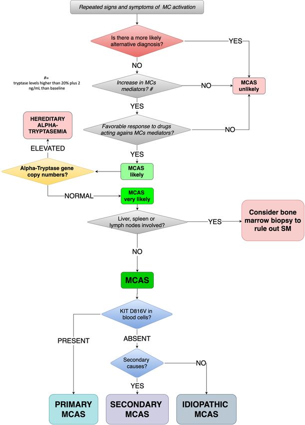
In Figure 1 we have proposed a diagnostic algorithm of MCAS for children.
In patients suffering from repeated signs and symptoms of MC activation, we suggest
testing for laboratory levels of mediators released by MCs, according to the availability
of local laboratory, tryptase being the patient shows a significant most feasible. If both
the previous conditions are satisfied and the favorable response to drugs acting against
MC mediators (such as H1/H2-histamine receptor blockers, Leukotriene receptor blockers,
or Mast cell stabilizers), MCAS is to be taken into account.
As previously stated, all three features need to be fulfilled at the same time.cina 2021, 57, x FOR2021,
Medicina PEER57,
REVIEW
124 8 of 14 7 of 13
Figurediagnostic
Figure 1. Proposed 1. Proposed diagnostic
algorithm algorithm
for MCAS for MCAS
in children. in children.
MC(s): MC(s):MCAS:
mast cell(s); mast cell(s); MCAS:
mast cell mast syndrome;
activation
cell activation
SM: systemic mastocytosis. syndrome; SM: systemic mastocytosis.
8. Treatment Options
Before proceeding with diagnosing MCAS (always keeping in mind that when hearing
Management hoof beats,
of MCAS anda horse should be suspected before zebras), clinicians should carefully run a
Mastocytosis
No resolutive treatment is availabledifferential
re-evaluation of all possible diagnoses
yet for MCAS, so its (Table 4).
management, such as that
If MCAS still remains the more likely diagnosis, Hereditary
of other MC disorders, needs to be multifactorial and ideally coordinated by Alpha-Tryptasemia
comprehen- should
be ruled out by testing for alpha-tryptase gene copy number: Increased numbers of copies
sive care centers with adequate experience.
are diagnostic for HAT while a normal number of copies should lead to diagnosis of MCAS.
The purpose of management is to minimize, as much as possible, the risk for recur-
At this stage, MCAS needs to be further characterized. The characterizing c-Kit muta-
rence of symptoms and, when symptoms are present, to efficiently treat them.
tion at D816V is to be searched in peripheral blood cells: When present, primary/clonal
MCAS can be diagnosed. If D816V is absent, MCAS can be secondary or idiopathic. If a con-Medicina 2021, 57, 124 8 of 13
dition such as atopy, autoimmune diseases, bacterial infections, or adverse drug reaction is
found, MCAS can be classified as secondary, otherwise as idiopathic.
8. Treatment Options
Management of MCAS and Mastocytosis
No resolutive treatment is available yet for MCAS, so its management, such as that of
other MC disorders, needs to be multifactorial and ideally coordinated by comprehensive
care centers with adequate experience.
The purpose of management is to minimize, as much as possible, the risk for recurrence
of symptoms and, when symptoms are present, to efficiently treat them.
Firstly, Castells and Butterfield [42] recommend to adequately educate patients to
avoid any kind of factor that may trigger degranulation of MCs (Table 5) and to use
premedication regimens when about to undergo surgery, invasive procedures, or radiol-
ogy procedures.
Table 5. Factors that trigger degranulation of MCs and recommended premedication regimens (Adapted from Castells and
Butterfield [42]).
A. Avoidance of Triggers: Patient Specific
Specific foods, medications (NSAIDs, vancomycin, quinolones), environmental allergens, and general triggers
(stress, lack of sleep, emotions)
Physical triggers (exercise, rubbing, pressure)
Changes in temperature (heat, cold)
Extreme temperatures
Dryness of skin
B. Premedications Recommended for Surgery, Invasive Procedures (Endoscopy, Colonoscopy, Others), radiological
Procedures with Contrast Dyes, Dental Procedures, and Vaccinations: 12 and 1 h
Antihistamine receptors H1 and H2
Leukotriene blocker
Steroid
NSAID, nonsteroidal anti-inflammatory drug
In avoidance of triggers, anesthesia requires a specific mention. A huge Spanish study
from 2015 retrospectively evaluated the data of 726 anesthetic procedures performed in
501 patients (both pediatric and adults) suffering from mastocytosis. Authors found a
higher frequency of perioperative anaphylaxis than in the general population but still
recommended not to avoid anesthesia in patients with mastocytosis though managing
them as high-risk patients, premedicating them with histamine receptor blockers and
benzodiazepines 1 h before procedure to relieve stress [43].
Coltoff et al. recommend to prescribe epinephrine autoinjectors to all patients with
known SM. This recommendation is explained by the tendency of patients with mas-
tocytosis to develop more severe forms of anaphylactic reactions potentially leading to
death [38].
Especially hymenoptera stings represent a very dangerous situation for patients.
Studies have been demonstrating that they induce more severe systemic reactions than
in patients who do not suffer from MC disorders. Hymenoptera stings are considered to
be the most frequent risk factor for anaphylaxis in patients with MCAS or mastocytosis
who are, consequently, eligible for specific venom immunotherapy (VIT) who appear to
significantly reduce the risk for anaphylactic recurrence. VIT safety in patients suffering
from MCs’ disorders appears to be safe and life-saving. To reduce both symptoms and
adverse reactions, Omalizumab was tested and showed satisfactory results [44,45].
All patients suffering from MC disorders with anaphylactic reaction should be pro-
vided with two intramuscular epinephrine autoinjectors. The jab can be repeated no more
than twice (1 to 5 min after the first administration) in case of massive MC activation.
Vaccination is a major concern in the pediatric population suffering from MCs’ disorders.Medicina 2021, 57, 124 9 of 13
Immunization schedule in Europe and US begins very early in life and accompanies
children for the first years of life.
Case reports have been describing a huge range of reactions to vaccination in this
particular population, ranging from the development of mastocytomas at injections sites to
severe anaphylaxis [46–50].
Reactions appear to be transient without recurrence on booster administrations [45,50].
Combination vaccines seem to increase risk for reactions more than single shots,
since most reactions were described following hexavalent formulations. Consequently, it is
safer to consider single vaccine injections in substitution of the combined ones [45].
Physicians should adequately inform parents and children, reassuring and educating
them about the management of adverse reactions [51]. Vaccinations are still recommended
in children with mastocytosis since benefits outweigh potential disadvantages. Never-
theless, it is reasonable to be cautious and administer vaccinations in a clinical setting
provided with adequate drugs, including epinephrine, putting children on observation for
a prolonged time (30 min according to Zanoni et al. [52], 2 h according to Parente et al. [50])
following the administration.
9. Pharmacological Treatment
The treatment of mild clinical manifestations is largely symptomatic and consists of:
(1) H1-histamine receptor blockers, for general symptoms (such as Cetirizine, Levoceti-
rizine, Loratadine, Desloratadine, Fexofenadine);
(2) H2-histamine receptor blockers, mainly for gastrointestinal symptoms (Ranitidine,
Cimetidine);
(3) Leukotriene receptor blockers (such as Montelukast, Zafirlukast and Zileuton); and
(4) Mast cell stabilizers (Cromolyn, Nedocromil).
9.1. H1/H2-Histamine Receptor Blockers
Though widely used, a recent systematic review found that little evidence supports
this habitude. Authors found only one well-designed randomized controlled trial (RCT),
which demonstrated rupatadine to be superior to placebo with a globally significant
improvement of clinical conditions of adults suffering from MCAS symptoms [53].
9.2. Leukotriene Receptor Blockers
The rational for their use in MCs’ disorders lays on the massive release of leukotrienes
by MCs’ degranulation. Nevertheless, no strong evidence has been found yet about their
effectiveness [38].
9.3. Mast Cell Stabilizers
Instead, mast cell stabilizers, especially cromolyn sodium, have shown efficacy in
different RCTs [38].
10. Other Therapeutic Options
In a previously cited retrospective study from Ravi et al. [22], the use of acetylsalicylic
acid for MCAS was mentioned. The rational lays on the production of PGD2 by MCs,
directly depending on prostaglandin–endoperoxide synthase, more commonly known as
cyclooxygenase (COX), whose function is blocked by acetylsalicylic acid. Most patients
treated with acetylsalicylic acid and with elevated 9a-11b-PGF2 urinary levels responded
with both an improvement of symptoms and normalization of 9a-11b-PGF2 levels [22].
Anti-Immunoglobulin (-Ig)E recombinant humanized monoclonal antibody Oma-
lizumab has been tested and proven to be effective in reducing symptoms in primary,
secondary, and idiopathic MCAS other than in pediatric diffuse CM [41].
Patients only suffering from CM may benefit from topical emollients, topical cal-
cineurin inhibitors, or phototherapy [38].Medicina 2021, 57, 124 10 of 13
10.1. Tyrosine Kinase Inhibitors
The theoretical possibility that a tyrosine kinase inhibitor, such as Imatinib, could block
the pathological reaction has been proposed. Unfortunately, trials conducted on patients
affected by SM with c-Kit D816V mutation with tyrosine kinase inhibitors showed that the
mutation itself confers resistance to Imatinib, such as to other tyrosine kinase inhibitors
(Dasatinib, Masitinib, Nilotinib). This is likely due to a conformational change of the
activation loop, interfering with the binding of imatinib to the receptor [54].
A multicenter experiment about the role of Imatinib in the treatment of “advanced
MC disease” reported a 29% efficacy only in patients without D816V c-kit mutation [55].
Nevertheless, Imatinib has been found to be useful in SM with different c-Kit muta-
tions involving transmembrane receptors, mainly F522C and K509I [31,37]. To the best of
our knowledge, a trial with Imatinibin MCAS has never been published.
Ustun et al. recommended to avoid the use of Imatinib for SM in pediatric patients
because of the self-limiting feature of some of the childhood-onset mastocytosis and because
of the lack of knowledge about long-term adverse effects of imatinib in children [54].
10.2. Midostaurin
A promising therapeutic agent is represented by midostaurin, an N-benzoyl derivative
of staurosporine, initially developed as a protein kinase C (PKC)-targeting drug that, later,
proved to be a multitargeted kinase inhibitor. In particular, midostaurin appears to be very
promising since it exhibits an ability to suppress D816V c-Kit mutated clonal cells and,
at the same time, it showed the ability to inhibit IgE-dependent mediator release in MCs
and basophils [56].
The cornerstone of midostaurin’s use in MC disorders was the huge multicenter,
open-label, phase II (single-arm) trial by Gotlib et al. [57] conducted on patients with
advanced SM. Patients showed a 60% response rate even though the drug was not globally
well tolerated by patients since 52% were forced to temporarily discontinue therapy [57].
Data of its use in children are lacking.
10.3. Cytoreductive Therapies
Interferon alpha-2b was studied on patients suffering from D816V c-Kit mutated SM
not responding to less invasive therapies, combined with prednisolone by Hauswirth
et al. [58]. Despite the small number of patients included in the study (five, all suffering
from ASM), the authors obtained a good response in 3/5 (60%) patients with complete or
partial resolution of symptoms and stabilized the disease in 1/5 (20%). One of the included
patients (20%), unluckily, decided to interrupt the therapy because of side effects [58].
The 2-chloro-20 -deoxyadenosine, or Cladribine, is an antimetabolite, currently ap-
proved to treat Hairy Cell Leukemia and Multiple Sclerosis [59].
Ever since 2001, it has been experimented in SM patients, leading to response rates > 50%
in SM [42].
11. Conclusions
MCAS is a highly complex disease with associated disorders ranging from relatively
common IgE-mediated disease to less frequent conditions such as mastocytosis or mon-
oclonal MC activation disorder. The diagnostic criteria established by an international
consensus should always be applied [1]. Serum total tryptase is considered the preferred
enzyme for diagnosing MCAS, but no consensus was found on different biomarkers.
The level of the tryptase should be higher than 20% plus 2 ng/mL.
Based on the underlying condition, patients with MCAS should then be further
classified into primary/clonal MCAS, secondary MCAS, or idiopathic MCAS.
No definitive treatment is yet available for MCAS, so its management must be multidisci-
plinary, integrated, and aimed primarily at avoiding triggers and the risk of symptom recurrence.Medicina 2021, 57, 124 11 of 13
Clinical practitioners’ training and well-designed and thought-out prospective clinical
research protocols are needed for improved recognition and knowledge of this syndrome
and treatments.
Author Contributions: Conceptualization, A.G., E.F., and A.P.; resources A.G., E.F., and C.C.; method-
ology, A.G., E.F., and C.C.; writing—original draft preparation A.G., E.F., C.C., G.R., and A.P.;
writing—review and editing A.G., C.C., G.R., and A.P. All authors have read and agreed to the
published version of the manuscript.
Funding: This research received no external funding.
Institutional Review Board Statement: Not applicable.
Informed Consent Statement: Not applicable.
Data Availability Statement: The study not report any data.
Conflicts of Interest: The authors declare no conflict of interest.
References
1. Afrin, L.B.; Ackerley, M.B.; Bluestein, L.S.; Brewer, J.; Brook, J.B.; Buchanan, A.D.; Cuni, J.R.; Davey, W.P.; Dempsey, T.T.;
Dorff, S.R.; et al. Diagnosis of mast cell activation syndrome: A global “consensus-2”. Diagnosis 2020. [CrossRef] [PubMed]
2. Theoharides, T.C.; Valent, P.; Akin, C. Mast Cells, Mastocytosis, and Related Disorders. N. Engl. J. Med. 2015, 373, 163–172.
[CrossRef] [PubMed]
3. Metcalfe, D.D. Mast cells and mastocytosis. Blood 2008, 112, 946–956. [CrossRef] [PubMed]
4. Galli, S.J.; Tsai, M. IgE and mast cells in allergic disease. Nat. Med. 2012, 18, 693–704. [CrossRef]
5. Schwartz, L.B. Tryptase from human mast cells: Biochemistry, biology and clinical utility. Monogr. Allergy 1990, 27, 90–113.
6. Schwartz, L.B. Tryptase, a mediator of human mast cells. J. Allergy Clin. Immunol. 1990, 86, 594–598. [CrossRef]
7. Schwartz, L.B.; Lewis, R.A.; Austen, K.F. Tryptase from human pulmonary mast cells. Purification and characterization.
J. Biol. Chem. 1981, 256, 11939–11943. [CrossRef]
8. Schwartz, L.B.; Min, H.-K.; Ren, S.; Xia, H.-Z.; Hu, J.; Zhao, W.; Moxley, G.; Fukuoka, Y. Tryptase precursors are preferentially and
spontaneously released, whereas mature tryptase is retained by HMC-1 cells, mono-mac-6 cells, and human skin-derived mast
cells. J. Immunol. 2003, 170, 5667–5673. [CrossRef]
9. Fukuoka, Y.; Schwartz, L.B. The B12 anti-tryptase monoclonal antibody disrupts the tetrameric structure of heparin-stabilized
betatryptase to form monomers that are inactive at neutral pH and active at acidic pH. J. Immunol. 2006, 176, 3165–3172. [CrossRef]
10. Schwartz, L.B.; Metcalfe, D.D.; Miller, J.S.; Earl, H.; Sullivan, T. Tryptase levels as an indicator of mast-cell activation in systemic
anaphylaxis and mastocytosis. N. Engl. J. Med. 1987, 316, 1622–1626. [CrossRef]
11. Bosso, J.V.; Schwartz, L.B.; Stevenson, D.D. Tryptase and histamine release during aspirin-induced respiratory reactions. J. Allergy
Clin. Immunol. 1991, 88, 830–837. [CrossRef]
12. Schwartz, L.B.; Sakai, K.; Bradford, T.R.; Ren, S.; Zweiman, B.; Worobec, A.S.; Metcalfe, D.D. The alpha form of human tryptase
is the predominant type present in blood at baseline in normal subjects and is elevated in those with systemic mastocytosis.
J. Clin. Investig. 1995, 96, 2702–2710. [CrossRef] [PubMed]
13. Schwartz, H.J. Elevated serum tryptase in exercise-induced anaphylaxis. J. Allergy Clin. Immunol. 1995, 95, 917–919. [CrossRef]
14. Schwartz, L.B. Diagnostic value of tryptase in anaphylaxis and mastocytosis. Immunol. Allergy Clin. N. Am. 2006, 26, 451–463.
[CrossRef]
15. Lin, R.Y.; Schwartz, L.B.; Curry, A.; Pesola, G.R.; Knight, R.J.; Lee, H.-S.; Bakalchuk, L.; Tenenbaum, C.; Westfal, R.E. Histamine
and tryptase levels in patients with acute allergic reactions: An emergency department–based study. J. Allergy Clin. Immunol.
2000, 106, 65–71. [CrossRef]
16. Schwartz, L.B.; Yunginger, J.W.; Miller, J.; Bokhari, R.; Dull, D. Time course of appearance and disappearance of human mast cell
tryptase in the circulation after anaphylaxis. J. Clin. Investig. 1989, 83, 1551–1555. [CrossRef]
17. Chatterjee, A.; Ghosh, J.; Kapur, R. Mastocytosis: A mutated KIT receptor induced myeloproliferative disorder. Oncotarget 2015, 6,
18250–18264. [CrossRef]
18. Valent, P.; Akin, C.; Arock, M.; Brockow, K.; Butterfield, J.H.; Carter, M.C.; Castells, M.; Escribano, L.; Hartmann, K.; Lieberman, P.; et al.
Definitions, criteria and global classification of mast cell disorders with special reference to mast cell activation syndromes: A consensus
proposal. Int. Arch. Allergy Immunol. 2012, 157, 215–225. [CrossRef]
19. Valent, P.; Akin, C.; Bonadonna, P.; Hartmann, K.; Brockow, K.; Niedoszytko, M.; Nedoszytko, B.; Siebenhaar, F.; Sperr, W.R.;
Elberink, J.N.O.; et al. Proposed diagnostic algorithm for patients with suspected mast cell activation syndrome. J. Allergy Clin.
Immunol. Pr. 2019, 7, 1125–1133.e1. [CrossRef]
20. Mastrorilli, C.; Posa, D.; Cipriani, F.; Caffarelli, C. Asthma and allergic rhinitis in childhood: What’s new. Pediatr. Allergy Immunol.
2016, 27, 795–803. [CrossRef]Medicina 2021, 57, 124 12 of 13
21. Valent, P.; Bonadonna, P.; Hartmann, K.; Broesby-Olsen, S.; Brockow, K.; Butterfield, J.H.; Triggiani, M.; Lyons, J.J.; Elberink,
J.N.O.; Arock, M.; et al. Why the 20% + 2 tryptase formula is a diagnostic gold standard for severe systemic mast cell activation
and mast cell activation syndrome. Int. Arch. Allergy Immunol. 2019, 180, 44–51. [CrossRef] [PubMed]
22. Ravi, A.; Butterfield, J.; Weiler, C.R. Mast cell activation syndrome: Improved identification by combined determinations of serum
tryptase and 24-hour urine 11β-Prostaglandin2α. J. Allergy Clin. Immunol. Pr. 2014, 2, 775–778. [CrossRef] [PubMed]
23. Longley, B.J.; Metcalfe, D.D.; Tharp, M.; Wang, X.; Tyrrell, L.; Lu, S.-Z.; Heitjan, D.; Ma, Y. Activating and dominant inactivating
c-KIT catalytic domain mutations in distinct clinical forms of human mastocytosis. Proc. Natl. Acad. Sci. USA 1999, 96, 1609–1614.
[CrossRef] [PubMed]
24. Wilder-Smith, C.H.; Drewes, A.M.; Materna, A.; Olesen, S.S. Symptoms of mast cell activation syndrome in functional gastroin-
testinal disorders. Scand. J. Gastroenterol. 2019, 54, 1322–1325. [CrossRef]
25. Weiler, C.R. Mast cell activation syndrome: Tools for diagnosis and differential diagnosis. J. Allergy Clin. Immunol. Pr. 2020, 8,
498–506. [CrossRef]
26. Valent, P.; Horny, H.-P.; Escribano, L.; Longley, B.; Li, C.Y.; Schwartz, L.B.; Marone, G.; Nuñez, R.; Akin, C.; Sotlar, K.; et al.
Diagnostic criteria and classification of mastocytosis: A consensus proposal. Leuk. Res. 2001, 25, 603–625. [CrossRef]
27. Lam, J.M.; Leong, K.F. Childhood solitary cutaneous mastocytoma: Clinical manifestations, diagnosis, evaluation, and manage-
ment. Curr. Pediatr. Rev. 2019, 15, 42–46. [CrossRef]
28. Van Doormaal, J.J.; Arends, S.; Brunekreeft, K.L.; Van Der Wal, V.B.; Sietsma, J.; van Voorst Vader, P.C.; Elberink, J.N.O.; Kluin-
Nelemans, H.C.; van der Veer, E.; de Monchy, J.G. Prevalence of indolent systemic mastocytosis in a Dutch region. J. Allergy
Clin. Immunol. 2013, 131, 1429–1431.e1. [CrossRef]
29. Jennings, S.; Russell, N.; Jennings, B.; Slee, V.; Sterling, L.; Castells, M.; Valent, P.; Akin, C. The Mastocytosis Society survey on
mast cell disorders: Patient experiences and perceptions. J Allergy Clin Immunol Pract. 2014, 2, 70–76. [CrossRef]
30. Nettleship, E.; Tay, W. Rare forms of urticaria. Br. Med. J. 1869, 2, 323–324.
31. Pardanani, A.D. Systemic mastocytosis in adults: 2019 update on diagnosis, risk stratification and management. Am. J. Hematol.
2018, 94, 363–377. [CrossRef] [PubMed]
32. Gurnee, E.A.; Johansen, M.L.; Phung, T.L.; Guo, E.L.; Fong, A.; Tollefson, M.; Nguyen, H.; Brandling-Bennett, H.; Moriarty, N.;
Paller, A.S.; et al. Pediatric maculopapular cutaneous mastocytosis: Retrospective review of signs, symptoms, and associated
conditions. Pediatr. Dermatol. 2020. [CrossRef] [PubMed]
33. Stevenson, R.L. Urticaria Pigmentosa. In Treasure Island (FL); Macri, A., Cook, C., Eds.; StatPearls Publishing: Treasure Island, FL,
USA, 2020.
34. Hosking, A.-M.; Makdisi, J.; Ortenzio, F.; De Feraudy, S.; Smith, J.; Linden, K. Diffuse cutaneous mastocytosis: Case report and
literature review. Pediatr. Dermatol. 2018, 35, e348–e352. [CrossRef] [PubMed]
35. Soter, N.A. Mastocytosis and the skin. Hematol. Oncol. Clin. N. Am. 2000, 14, 537–555. [CrossRef]
36. Hartmann, K.; Escribano, L.; Grattan, C.; Brockow, K.; Carter, M.C.; Alvarez-Twose, I.; Matito, A.; Broesby-Olsen, S.; Siebenhaar,
F.; Lange, M.; et al. Cutaneous manifestations in patients with mastocytosis: Consensus report of the European Competence
Network on Mastocytosis; the American Academy of Allergy, Asthma & Immunology; and the European Academy of Allergology
and Clinical Immunology. J. Allergy Clin. Immunol. 2016, 137, 35–45. [CrossRef]
37. Valent, P.; Akin, C.; Metcalfe, D.D. Mastocytosis: 2016 updated WHO classification and novel emerging treatment concepts. Blood
2017, 129, 1420–1427. [CrossRef]
38. Coltoff, A.; Mascarenhas, J. Relevant updates in systemic mastocytosis. Leuk. Res. 2019, 81, 10–18. [CrossRef]
39. Raymond, L.; Funk, T.; Braziel, R.M.; Fan, G.; Gatter, K.; Loriaux, M.; Traer, E.; Raess, P.W. Mast cell sarcoma with concurrent
mast cell leukaemia. Br. J. Haematol. 2020, 189, e160–e164. [CrossRef]
40. Broesby-Olsen, S.; Carter, M.; Kjaer, H.F.; Mortz, C.G.; Moller, M.B.; Kristensen, T.K.; Bindslev-Jensen, C.; Agertoft, L. Pediatric
expression of mast cell activation disorders. Immunol. Allergy Clin. N. Am. 2018, 38, 365–377. [CrossRef]
41. Caimmi, D.; Barni, S.; Mastrorilli, C. Le sindromi da attivazione mastocitaria. Riv. Immunol. Allergol. Pediatr. 2020, 34, I.
42. Castells, M.; Butterfield, J. Mast cell activation syndrome and mastocytosis: Initial treatment options and long-term management.
J. Allergy Clin. Immunol. Pr. 2019, 7, 1097–1106. [CrossRef] [PubMed]
43. Matito, A.; Morgado, J.M.; Sánchez-López, P.; Álvarez-Twose, I.; Sánchez-Muñoz, L.; Orfao, A.; Escribano, L. Management of
Anesthesia in adult and pediatric mastocytosis: A Study of the Spanish Network on Mastocytosis (REMA) based on 726 anesthetic
procedures. Int. Arch. Allergy Immunol. 2015, 167, 47–56. [CrossRef] [PubMed]
44. Niedoszytko, M.; De Monchy, J.; Van Doormaal, J.J.; Jassem, E.; Elberink, J.N.G. Mastocytosis and insect venom allergy: Diagnosis,
safety and efficacy of venom immunotherapy. Allergy 2009, 64, 1237–1245. [CrossRef] [PubMed]
45. Carter, M.C.; Metcalfe, D.D.; Matito, A.; Escribano, L.; Butterfield, J.H.; Schwartz, L.B.; Bonadonna, P.; Zanotti, R.; Triggiani, M.;
Castells, M.; et al. Adverse reactions to drugs and biologics in patients with clonal mast cell disorders: A Work Group Report of
the Mast Cells Disorder Committee, American Academy of Allergy, Asthma & Immunology. J. Allergy Clin. Immunol. 2019, 143,
880–893. [CrossRef]
46. Brockow, K.; Jofer, C.; Behrendt, H.; Ring, J. Anaphylaxis in patients with mastocytosis: A study on history, clinical features and
risk factors in 120 patients. Allergy 2008, 63, 226–232. [CrossRef]
47. Bankova, L.G.; Walter, J.E.; Iyengar, S.R.; Lorenzo, M.E.; Hornick, J.L.; Castells, M. Generalized bullous eruption after routine
vaccination in a child with diffuse cutaneous Mastocytosis. J. Allergy Clin. Immunol. Pr. 2013, 1, 94–96. [CrossRef]Medicina 2021, 57, 124 13 of 13
48. Koh, M.J.A.; Chong, W.S. Red plaque after hepatitis B vaccination. Pediatr. Dermatol. 2008, 25, 381–382. [CrossRef]
49. Poulton, J.K.; Kauffman, C.L.; Lutz, L.L.; Sina, B. Solitary mastocytoma arising at a hepatitis B vaccination site. Cutis 1999, 63,
37–40.
50. Parente, R.; Pucino, V.; Magliacane, D.; Petraroli, A.; Loffredo, S.; Marone, G.; Triggiani, M. Evaluation of vaccination safety in
children with mastocytosis. Pediatr. Allergy Immunol. 2016, 28, 93–95. [CrossRef]
51. Franceschini, F.; Bottau, P.; Caimmi, S.; Cardinale, F.; Crisafulli, G.; Liotti, L.; Pellegrini, G.; Peroni, D.; Saretta, F.; Mastror-
illi, C.; et al. Evaluating children with suspected allergic reactions to vaccines for infectious diseases. Allergy Asthma Proc. 2018,
39, 177–183. [CrossRef]
52. Zanoni, G.; Zanotti, R.; Schena, D.; Sabbadini, C.; Opri, R.; Bonadonna, P. Vaccination management in children and adults with
mastocytosis. Clin. Exp. Allergy 2017, 47, 593–596. [CrossRef] [PubMed]
53. Nurmatov, U.; Rhatigan, E.; Simons, F.E.R.; Sheikh, A. H1-antihistamines for primary mast cell activation syndromes: A systematic
review. Allergy 2015, 70, 1052–1061. [CrossRef] [PubMed]
54. Ustun, C.; DeRemer, D.L.; Akin, C. Tyrosine kinase inhibitors in the treatment of systemic mastocytosis. Leuk. Res. 2011, 35,
1143–1152. [CrossRef] [PubMed]
55. Pagano, L.; Valentini, C.G.; Caira, M.; Rondoni, M.; Van Lint, M.T.; Candoni, A.; Allione, B.; Cattaneo, C.; Marbello, L.;
Caramatti, C.; et al. Advanced mast cell disease: An Italian Hematological Multicenter experience. Int. J. Hematol. 2008, 88,
483–488. [CrossRef]
56. Valent, P.; Akin, C.; Hartmann, K.; George, T.I.; Sotlar, K.; Peter, B.; Gleixner, K.V.; Blatt, K.; Sperr, W.R.; Manley, P.W.; et al.
Midostaurin: A magic bullet that blocks mast cell expansion and activation. Ann. Oncol. 2017, 28, 2367–2376. [CrossRef]
57. Gotlib, J.; Kluin-Nelemans, H.C.; George, T.I.; Akin, C.; Sotlar, K.; Hermine, O.; Awan, F.T.; Hexner, E.; Mauro, M.J.; Stern-
berg, D.W.; et al. Efficacy and safety of midostaurin in advanced systemic mastocytosis. N. Engl. J. Med. 2016, 374, 2530–2541.
[CrossRef]
58. Hauswirth, A.W.; Simonitsch-Klupp, I.; Uffmann, M.; Koller, E.; Sperr, W.R.; Lechner, K.; Valent, P. Response to therapy with
interferon alpha-2b and prednisolone in aggressive systemic mastocytosis: Report of five cases and review of the literature.
Leuk. Res. 2004, 28, 249–257. [CrossRef]
59. Cladribine. Available online: https://www.drugs.com/international/cladribine.html (accessed on 10 November 2020).You can also read