Identification and Characterization of Avihepadnaviruses Isolated from Exotic Anseriformes Maintained in Captivity
←
→
Page content transcription
If your browser does not render page correctly, please read the page content below
JOURNAL OF VIROLOGY, Mar. 2005, p. 2729–2742                                                                                    Vol. 79, No. 5
0022-538X/05/$08.00⫹0 doi:10.1128/JVI.79.5.2729–2742.2005
Copyright © 2005, American Society for Microbiology. All Rights Reserved.

Identification and Characterization of Avihepadnaviruses Isolated from
              Exotic Anseriformes Maintained in Captivity
      Haitao Guo,1 William S. Mason,1* Carol E. Aldrich,1 Jeffry R. Saputelli,1 Darren S. Miller,2,3
                              Allison R. Jilbert,2,3 and John E. Newbold4
     Fox Chase Cancer Center, Philadelphia, Pennsylvania1; Department of Microbiology and Immunology, University of North
         Carolina, Chapel Hill, North Carolina4; and Infectious Diseases Laboratories, Institute of Medical and Veterinary
             Science,2 and School of Molecular and Biomedical Science, University of Adelaide,3 Adelaide, Australia
                                            Received 14 June 2004/Accepted 19 October 2004

            Five new hepadnaviruses were cloned from exotic ducks and geese, including the Chiloe wigeon, mandarin

                                                                                                                                                 Downloaded from http://jvi.asm.org/ on March 1, 2021 by guest
         duck, puna teal, Orinoco sheldgoose, and ashy-headed sheldgoose. Sequence comparisons revealed that all but
         the mandarin duck viruses were closely related to existing isolates of duck hepatitis B virus (DHBV), while
         mandarin duck virus clones were closely related to Ross goose hepatitis B virus. Nonetheless, the S protein,
         core protein, and functional domains of the Pol protein were highly conserved in all of the new isolates. The
         Chiloe wigeon and puna teal hepatitis B viruses, the two new isolates most closely related to DHBV, also lacked
         an AUG start codon at the beginning of their X open reading frame (ORF). But as previously reported for the
         heron, Ross goose, and stork hepatitis B viruses, an AUG codon was found near the beginning of the X ORF
         of the mandarin duck, Orinoco, and ashy-headed sheldgoose viruses. In all of the new isolates, the X ORF
         ended with a stop codon at the same position. All of the cloned viruses replicated when transfected into the
         LMH line of chicken hepatoma cells. Significant differences between the new isolates and between these and
         previously reported isolates were detected in the pre-S domain of the viral envelope protein, which is believed
         to determine viral host range. Despite this, all of the new isolates were infectious for primary cultures of Pekin
         duck hepatocytes, and infectivity in young Pekin ducks was demonstrated for all but the ashy-headed sheld-
         goose isolate.

   Hepadnaviruses are small, enveloped DNA viruses that rep-                transcription factors, and has a role in virus replication, though
licate via reverse transcription of an RNA pregenome. The                   the relationship between these observations and the role of X
family Hepadnaviridae is divided into two genera, Orthohepad-               in vivo remains elusive (3, 5–7). Recently, expression of an
navirus and Avihepadnavirus, each with restricted host speci-               X-like protein from an open reading frame (ORF) on the
ficity. Ortho (mammalian) hepadnaviruses have been found in                 DHBV genome lacking a conventional start codon was re-
humans (hepatitis B virus [HBV]) and great apes, woolly mon-                ported (12, 27). An ORF is also present in the same location
keys (woolly monkey HBV), woodchucks (woodchuck hepatitis                   on other avihepadnavirus genomes; however, whether X-like
virus), ground squirrels (ground squirrel hepatitis virus), arctic          proteins are expressed during natural infections by all of these
squirrels (arctic squirrel hepatitis virus), and Richardson                 avihepadnaviruses is unknown. Expression of the DHBV X-
ground squirrels (Richardson ground squirrel hepatitis virus).              like protein in cell culture led, as with the HBV X protein, to
The avian hepadnaviruses include duck HBV (DHBV) (40, 43,                   transcriptional activation of several heterologous promoters
58, 66), heron HBV (HHBV) (52), Ross goose HBV                              (12). However, a knockout mutation in the X ORF did not
(RGHBV; GenBank accession no. M95589), snow goose HBV                       alter the ability of DHBV either to replicate in culture or to
(SGHBV) (11), stork HBV (STHBV) (50), and crane HBV                         induce transient and persistent infections in ducks (12, 44), and
(CHBV) (48).                                                                the relevance of the transcriptional activation data remains
   Sequence similarities between the ortho- and avihepadnavi-               unclear.
ruses are minimal except in highly conserved functional do-                    All known hepadnaviruses are hepatotropic and cause tran-
mains, though the overall genome organization is similar. Pro-              sient and persistent infections with variable degrees of patho-
teins encoded by both groups include the nucleocapsid (core)                genesis (51). Infection is limited to the species from which a
and envelope polypeptides (L and S), the nonstructural                      virus has been isolated or to closely related species. It is gen-
HBeAg (a secretory protein of unknown function), and the                    erally believed that host range and tissue restriction are regu-
polymerase/reverse transcriptase protein (Pol protein). Ortho-              lated at the level of virus entry, specificity being determined by
hepadnaviruses also encode a third envelope protein, M, and a               the pre-S1 domain of the L envelope protein of the orthohe-
regulatory protein, termed X, that is required for efficient                padnaviruses and the homologous pre-S domain of the avihe-
replication in vivo (13, 65, 67). Studies with liver cell lines             padnaviruses. Between hepadnaviruses, this domain is the
suggest that X stimulates signal transduction, regulates several            most divergent region of the viral envelope, consistent with a
                                                                            role in species-specific receptor recognition. This idea is sup-
  * Corresponding author. Mailing address: Fox Chase Cancer Cen-
                                                                            ported by a report that replacement of a small region (69
ter, 333 Cottman Ave., Philadelphia, PA 19111. Phone: (215) 728-            amino acids) of the HHBV-specific pre-S domain by the cor-
2462. Fax: (215) 728-3105. E-mail: ws_mason@fccc.edu.                       responding DHBV domain facilitated infection of duck hepa-

                                                                     2729
2730 GUO ET AL. J. VIROL.
tocytes, which are not otherwise susceptible to HHBV (20). supernatant was precipitated with 2 volumes of ethanol with addition of 20 g of
Similarly, exchange of as little as 9 amino acids of the pre-S1 dextran as carrier. The pellet was washed with 70% ethanol, dried, and resus-
pended in 20 l of TE (10 mM Tris-HCl [pH 7.4], 1 mM EDTA). The virus DNA
domain of HBV with corresponding sequences from woolly was cleaved with XbaI or, for MDHBV, EcoRI, and ligated into the respective
monkey HBV reduced infectivity for human hepatocytes (14). site of the lambda ZAP II vector (Stratagene). The bacteriophage DNA was
Carboxypeptidase D (gp180) has been identified as a possible packaged in vitro, and viral DNA-containing plaques were identified by hybrid-
receptor for DHBV (8, 28, 29, 57), mediating virus attachment ization (2). The entire insert from bacteriophage lambda DNA was released with
and internalization. Unfortunately, transfection of nonpermis- XbaI or EcoRI, as appropriate, and cloned into the corresponding site of plasmid
pGEM-3Z (Promega). For MDHBVb and OSHBV, due to poor growth of the
sive cells with gp180 does not confer susceptibility to DHBV, lambda clones of these viruses, the cloned viral genomes were amplified from the
indicating that additional, or other, cellular factors are in- bacteriophage by using the Advantage-HF 2 PCR kit (Clontech) with the
volved in host range determination during or subsequent to M13fwd and M13rev primers and then cloned into pGEM-3Z. Head-to-tail
virus attachment and entry (32, 48, 51). dimer clones of the complete viral genomes were used for transfection studies.
As an approach to determining the significance of pre-S Initial analyses revealed that the clones of mandarin duck, Chiloe wigeon,
puna teal, and Orinoco sheldgoose were full length (⬃3 kbp). The ashy-headed
variation in viral host range determination and evaluating the sheldgoose clone was only 2,451 nucleotides in length, due to the loss of an XbaI
possibility that the X ORF is conserved throughout the avihe- fragment (⬃600 bp) present within the core gene region. This XbaI fragment was
padnavirus genus, which may argue for a functional role, we
Downloaded from http://jvi.asm.org/ on March 1, 2021 by guest
amplified by using the Advantage-HF 2 PCR kit (Clontech) from serum-derived
cloned, sequenced, and evaluated the infectivity in Pekin ducks viral DNA and ligated to the 2,451-bp XbaI fragment in the pGEM-3Z plasmid
(Anas domesticus) of a number of new viruses identified in to produce a full-length genome.
DNA sequencing of cloned avihepadnavirus genomes. Both strands of each
anseriforme birds maintained in aviculture collections. Serum cloned virus genome were sequenced by using an ABI PRISM 377 automated
samples taken from exotic ducks and geese were screened by sequencing system. Plasmid DNA was extracted and purified by using a Midiprep
DNA hybridization for DHBV-related genomes. New hepad- kit (QIAGEN). The nucleotide sequence data for the six distinct avihepadnavi-
navirus isolates were detected in the mandarin duck, Chiloe ruses (MDHBVa, MDHBVb, CWHBV, PTHBV, OSHBV, and ASHBV) were
wigeon, puna teal, Orinoco sheldgoose, and ashy-headed deposited in GenBank (see “Nucleotide sequence accession number” below).
The isolates from the two Chiloe wigeons were identical in sequence.
sheldgoose. These have been designated mandarin duck HBVa Comparative sequence analysis of avihepadnaviruses. The relationship of the
and b (MDHBVa and MDHBVb, differing slightly in sequence new avihepadnaviruses to the known avian hepadnaviruses was investigated by
and isolated from a male and female mandarin duck from phylogenetic analysis by using the ClustalX, version 1.81, and TreeView, version
different aviculture collections, respectively), Chiloe wigeon 1.6.6, programs as described previously (47, 55). Other avihepadnavirus se-
HBV (CWHBV), puna teal HBV (PTHBV), Orinoco sheld- quences were obtained from GenBank as follows (accession numbers are indi-
cated in parentheses): Western country DHBV isolates, DHBV1 (X58567),
goose HBV (OSHBV), and ashy-headed sheldgoose HBV DHBVF16 (X12798), DHBVCG (X74623), HPUCGE (M60677), HPUCGD
(ASHBV). All isolates differed significantly from DHBV in (K01834), DHBV493986.1 (AF493986), DHBV47045 (AF047045), DHBV26
pre-S. Nonetheless, all primary isolates were able to infect the (X58569), DHBV22 (X58568); Chinese DHBV isolates, HPUGA (M21953),
mallard (J. E. Newbold, unpublished data), the species from DHBV404406 (AF404406), HPUS5CG (M32990), DHVBCG (X60213),
which most domesticated ducks are derived, and all of the HPUS31CG (M32991); Australian DHBV isolate, DHV6350 (AJ006350);
SGHBV isolates, SGHBV1-7 (AF110999), SGHBV1-9 (AF111000),
cloned isolates but ASHBV were shown to infect the Pekin SGHBV1-13 (AF110996), SGHBV1-15 (AF110997), SGHBV1-19 (AF110998);
duck. Sequence comparisons provided evidence of an X-like RGHBV isolate, HPUGENM (M95589); STHBV isolates, SHE251934
ORF, beginning with an AUG start codon, in some but not all (AJ251934), SHE251935 (AJ251935), SHE251936 (AJ251936), SHE251937
of these virus isolates, and ending at the same stop codon. Full (AJ251937); HHBV, HPUCG (M22056); CHBV isolates, CHBV1 (AJ441111),
genome sequence alignments and comparisons suggested that CHBV2 (AJ441112), CHBV3 (AJ441113).
Cell culture and transfection. The chicken hepatoma cell line LMH (25) was
the Mandarin duck and Ross goose viruses are closely related grown at 37°C on 60-mm-diameter tissue culture dishes in 1:1 Dulbecco’s min-
and possibly define a new species within the avihepadnavirus imal essential medium–Ham’s nutrient mixture F-12 supplemented with 10 mM
genus, a decision that will ultimately require characterization NaH2CO3 and 10% calf serum. Transfection of purified plasmid DNA (5 g) was
of these viruses, in comparison to DHBV, in their natural performed by using Lipofectamine 2000 reagent (Invitrogen) when cultures were
hosts. 70 to 80% confluent. The medium was changed daily until harvest, at which time
the medium was clarified to remove floating cells and cell debris and stored at
⫺80°C. The monolayers were rinsed twice with chilled phosphate-buffered saline
MATERIALS AND METHODS (PBS) and used for intracellular core particle DNA isolation.
Serum samples. Sera containing DHBV-related viruses were collected from Isolation and analysis of viral DNA from transfected cells and culture super-
two mandarin ducks (genus Aix, male and female) from two different aviculture natants. Viral DNA replication intermediates were isolated from cytoplasmic
collections in North Carolina, two Chiloe wigeons, probably siblings (genus Anas, nucleocapsids 5 days posttransfection as described previously (10). Viral DNA in
male and female), from an aviculture collection in Ohio, a puna teal (genus secreted, enveloped virus particles was extracted and purified from 400 l of
Anas) from an aviculture collection in Pennsylvania (original serum sample clarified culture fluids by using a published method (31). Viral DNAs were
passed once in a domestic mallard to expand the stock), an Orinoco sheldgoose subjected to electrophoresis through a 1.5% agarose gel and analyzed by South-
(genus Neochen) from an aviculture collection in North Carolina (original serum ern blotting (15) with [32P]TTP-labeled DNA probes representing the entire
passed once in a paradise shelduck to expand the stock), and an ashy-headed DHBV genome. The relative efficiency of hybridization of this probe to the other
sheldgoose (genus Chloephaga) from an aviculture collection in North Carolina. viruses was 30 to 50% when tested against the full-length cloned DNAs.
Screening for DHBV-related viruses via hybridization spot test. Hybridization PDH infection and viral DNA analysis. Primary duck hepatocyte (PDH) cul-
spot tests for DHBV-like virus in serum samples was carried out as previously tures were prepared from 1-week-old DHBV-negative Pekin ducklings (49).
described (42). PDH were plated onto 60-mm-diameter tissue culture dishes in L15 medium
Cloning of viral genomic DNA. Serum (300 l) from each bird containing a supplemented with 5% fetal calf serum and maintained at 37°C. The next day,
DHBV-related virus was layered on a 10 to 20% (wt/vol) sucrose step gradient the cultures were shifted to serum-free medium L15. The cells were infected on
in 0.15 M NaCl–0.02 M Tris-HCl (pH 7.4) and centrifuged at 45,000 rpm in a day 2 after seeding with approximately 1.5 ⫻ 107 enveloped virus particles
Beckman SW60 rotor for 3 h at 4°C. The virus pellet was suspended in 100 l of purified from the supernatants of transfected LMH cells. The concentration and
0.2% (wt/vol) sodium dodecyl sulfate (SDS)–1 mg of pronase/ml, 0.015 M Tris- titration of enveloped virus particles produced following transfection of LMH
HCl (pH 7.5), and 0.01 M EDTA and incubated at 37°C for 1 h. The nucleic acids cells was performed as described previously (31). After 16 h, medium was re-
were extracted twice with an equal volume of phenol-chloroform (1:1), and the placed and changed daily thereafter. Cultures were harvested at 10 days postin-VOL. 79, 2005 NEW AVIHEPADNAVIRUS ISOLATES 2731
fection. The plates were washed once with PBS, and viral core DNA was ex- mined. Sequences were the same for each clone of an isolate
tracted and analyzed by Southern blotting (49). with the exception of a PTHBV clone (data not shown) that
Duck infection experiments. One hundred microliters of virus stock purified
from the supernatants of transfected LMH cells and containing 6 ⫻ 107 (MDHBVa,
had a 3-nucleotide deletion that removed a histidine residue
MDHBVb, CWHBV, and PTHBV) or 1.5 ⫻ 107 (OSHBV and ASHBV) genome (amino acid [aa] 107) from the avian insertion domain of the
equivalents of enveloped viral particles, as measured by the procedure of Lenhoff core protein. Because this DNA was cloned and expanded
and Summers (31), were inoculated into groups of 3-day-old DHBV-negative from the original serum without employing PCR, this PTHBV
ducks by intraperitoneal injection. Two groups of ducklings were injected with
clone may be a natural variant of the virus. This variant was
the two different amounts of DHBV to serve as positive controls. Ducks were
monitored by a weekly spot test for viremia. Tissue samples (liver, pancreas, replication defective (H. Guo, unpublished data). Differences
spleen, and kidney) were dissected and divided into pieces that were either between MDHBVa and MDHBVb were found throughout the
snap-frozen in liquid N2 for total DNA and covalently closed circular DNA viral genome. The two Chiloe wigeon isolates, from a male and
(cccDNA) extraction or fixed in ethanol-acetic acid (EAA) (3:1) for in situ a female from the same aviculture collection, had the same
hybridization (23). EAA fixation was performed at room temperature for 30 min,
followed by treatment in chilled (⫺20°C) 70% ethanol overnight. The tissues
sequence.
were then dehydrated, embedded in paraffin wax, and sectioned onto gelatin- Phylogenetic relationships. The six genomes were aligned
coated slides. Viral nucleic acids were detected in sections of EAA-fixed liver, with prototypes of the known avihepadnavirus genomes:
spleen, kidney, and pancreas tissues by in situ hybridization with a genome- DHBV, HHBV, RGHBV, SGHBV, STHBV, and CHBV (Fig.
Downloaded from http://jvi.asm.org/ on March 1, 2021 by guest
length DHBV DNA probe labeled with digoxigenin-UTP by using a nick trans-
1). The new avihepadnavirus isolates had a high percentage of
lation kit (Roche Applied Science). The vector backbone was used as a negative
control. Section preparation and detection was as previously described (21, 23). sequence identity with previously described duck and goose
Sections were counterstained with hematoxylin and mounted in 25% glycerol in hepadnaviruses (Table 1). The two mandarin duck isolates
PBS. MDHBVa and MDHBVb were highly homologous to
One-day-old ducklings were purchased from Metzer Farms, Gonzales, Calif. RGHBV (45), with percent identities of 99.1 and 93.3, respec-
All experiments were reviewed and approved by the Institutional Animal Care
and Use Committee of the Fox Chase Cancer Center.
tively. CWHBV and PTHBV and OSHBV and ASHBV had
Total and cccDNA extraction from duck tissues. One hundred milligrams of high homology to each other and were more closely related to
duck liver and spleen tissue was homogenized in 1.5 ml of ice-cold TE (10:10) DHBV. As expected from published studies of other avihep-
buffer (10 mM Tris-HCl [pH 7.5], 10 mM EDTA) with 25 to 30 strokes of a adnavirus isolates (11, 45, 48, 50), cis sequence elements with
Dounce homogenizer and divided into two aliquots for cccDNA and total DNA
roles in viral RNA and DNA synthesis were conserved. These
extractions, respectively, as described previously (53), with some modifications.
Nuclear counts in the homogenates were determined, after staining with included the sequence elements involved in viral DNA synthe-
ethidium bromide, by using a hemocytometer under fluorescent illumination. sis, such as epsilon, a stem-loop structure on pregenomic RNA
Total DNA was extracted from 0.7 ml of homogenate, initially diluted to 3 ml involved in pregenomic RNA encapsidation and initiation of
with TE (10:10), and digested with an equal volume of pronase-SDS solution reverse transcription, and the identical 12-nucleotide se-
(final concentrations, 4 mg of pronase per ml, 0.5% SDS, 0.1 M NaCl, 50 mM
Tris-HCl [pH 7.5], 20 mM EDTA) at 37°C for at least 2 h. The total DNA was
quences, DR1 and DR2, which regulate early steps in minus-
phenol-chloroform extracted, ethanol precipitated at ⫺20°C overnight, washed and plus-strand synthesis (24, 33, 34, 39, 54, 61). Other con-
with 70% ethanol, and redissolved in 400 l of TE (10:1). The cccDNA was served elements included sequences involved in transcriptional
extracted from the remaining 0.7 ml of liver homogenate by increasing the regulation, including a TATA box motif in the pre-S and core
volume to 3 ml with TE (10:10) and then mixing with 0.2 ml of 10% SDS,
promoters, transcription factor binding sites for C/EBP,
followed by incubation at room temperature (RT) for 5 min. One milliliter of 2.5
M KCl was then added, and the mixture was incubated at RT for 30 min, HNF1, and HNF3 in the DHBV enhancer (35, 36), the poly-
followed by centrifugation at 10,000 rpm for 20 min at 4°C in a small SA600 adenylation signal for all viral transcripts that is located within
rotor. The cccDNA in the supernatant was then phenol-chloroform extracted, the core gene (9), and mRNA splice donor and acceptor sites
ethanol precipitated overnight at RT, washed with 70% ethanol, and redissolved (46).
in 100 l of TE (10:10). Samples of total DNA isolated from 1.6 ⫻ 106 liver cells
and 1.2 ⫻ 106 spleen cells and cccDNA isolated from 3.2 ⫻ 106 liver cells or 4.8
Evolutionary relationships among the isolates were exam-
⫻ 106 spleen cells were subjected to agarose gel electrophoresis followed by ined with ClustalX and Treeview. The results are presented in
Southern blot hybridization (23). Table 1 and Fig. 2. Analysis of previously reported isolates
Nucleotide sequence accession number. The nucleotide sequence data for divides the tree into seven major branches, with all isolates of
MDHBVa, MDHBVb, CWHBV, PTHBV, OSHBV, and ASHBV were depos-
DHBV, as well as those of SGHBV, falling into three closely
ited in GenBank under accession numbers AY494848, AY494849, AY494850,
AY494851, AY494852, and AY494853, respectively. related branches. The other branches include RGHBV (45),
STHBV (50), HHBV (45), and CHBV (11). The avihepadna-
viruses isolates reported here could be segregated into three
RESULTS
clades on the phylogenetic tree. The CWHBV and PTHBV
Detection of DHBV-related avihepadnaviruses in serum form a sister clade to the Western country DHBV isolates (58)
samples. Serum samples were collected from ducks and geese from domesticated ducks (Anas platyrhynchos), while OSHBV
of the anseriforme family Anatidae. DHBV-related viruses and ASHBV identify a clade of sequences less closely related
were detected in the serum of 2 mandarin ducks (genus Aix, to DHBV. MDHBVa and MDHBVb were closely related to
male and female), 2 Chiloe wigeons (genus Anas, male and RGHBV and localized with it to a third clade. The similarity is,
female), a puna teal (genus Anas), an Orinoco sheldgoose in fact, so striking that it appears that transmission of RGHBV
(genus Neochen), and an ashy-headed sheldgoose (genus Chlo- and MDHBV occurred between birds of the Aix and Anser
ephaga). These DHBV-related viruses are here designated genera. In contrast, although snow geese and Ross geese nest
MDHBVa (male mandarin duck), MDHBVb (female manda- in mixed colonies, where young may be in very close contact
rin duck), CWHBV, PTHBV, OSHBV, and ASHBV, respec- and interbreeding is thought to occur (56), the viruses isolated
tively. from these apparently closely related birds are very different.
To further characterize the isolates, the complete genome Protein sequence comparisons. The relationship of the new
sequences of two full-length clones of each isolate were deter- isolates to other avian hepadnaviruses was next examined atDownloaded from http://jvi.asm.org/ on March 1, 2021 by guest
FIG. 1. Nucleotide sequence alignment with prototypic members of the avihepadnavirus family (DHBV, HHBV, RGHBV, SGHBV, STHBV,
and CHBV). Genomes are numbered from an EcoRI sequence in the viral core gene. Dots and dashes represent identical and deleted nucleotides,
respectively. Start and stop codons for the viral genes (Pol, polymerase; pre-S/S, envelope proteins; pre-C/C, precore and core; X, X-like protein)
are indicated with arrows and asterisks, respectively. Additionally, both features are boxed by rectangles. Selected transcription factor binding sites
(HNF, hepatocyte nuclear factor; C/EBP, CCAAT/enhancer-binding protein), the TATA box of the pre-S and core promoter, and other regulatory
sequence elements (DR, direct repeats; epsilon; PAS, polyadenylation signal) are also boxed.
2732VOL. 79, 2005 NEW AVIHEPADNAVIRUS ISOLATES 2733
Downloaded from http://jvi.asm.org/ on March 1, 2021 by guest
FIG. 1—Continued.2734 GUO ET AL. J. VIROL.
Downloaded from http://jvi.asm.org/ on March 1, 2021 by guest
FIG. 1—Continued.
the protein sequence level (Fig. 3). As expected, the pre-S a serine- or threonine-proline kinase recognition site that has
region was quite variable (Fig. 3A), especially in the host- been speculated to have a role in nucleocapsid assembly and
range-determining region and in the minimal binding domains disintegration (1).
for the duck proteins gp180/p120, which are thought to define The Pol protein was also conserved in all three functional
part of the cell surface receptor for DHBV (8, 20, 32, 57, 59, domains, including terminal protein (TP), reverse transcrip-
60). There was an 11-amino-acid insertion in ASHBV at the tase, and RNase H (Fig. 3C). These conserved motifs included
position corresponding to the C terminus of the gp180-binding the tyrosine residue at position 96 within the TP that functions
site in DHBV pre-S. All of the new avihepadnavirus isolates as the primer for reverse transcription (68) and the YMDD
had a potential myristylation site at the amino terminus of motif, known to be essential for the DNA polymerase activity
pre-S (38). One or more potential phosphorylation sites were of the reverse transcriptase. The nonfunctional spacer domain
found in the pre-S domain of all the isolates, though their role of Pol that overlaps the variable pre-S region was itself highly
in the DHBV life cycle remains elusive (4, 17, 18). The S variable between isolates.
protein domain was highly conserved in all of the avihepadna- It has been proposed that avihepadnaviruses have an X-like
viruses. protein expressed in vivo that, like the HBV X protein, has mul-
The pre-C/C proteins of the new isolates were also con- tiple regulatory activities (12). MDHBVa, MDHBVb, OSHBV,
served (Fig. 3B), including a hydrophobic heptad repeat (hhr), and ASHBV, like RGHBV, have sequences that could encode
important for nucleocapsid assembly (62), SPXX phosphory- a protein from this region of the viral genome that begins with
lation sites that appear to play a regulatory role in virus mor- an AUG translation start codon (Fig. 3D) and is followed by a
phogenesis (26, 63, 64), a nuclear localization signal (37), and G in position ⫹4, which is believed to favor initiation at AUG
TABLE 1. Sequence identity among new avihepadnaviruses and prototypes of other avihepadnavirusesa
% Identity with:
Virus
HHBV RGHBV SGHBV STHBV CHBV MDHBVa MDHBVb CWHBV PTHBV OSHBV ASHBV
DHBV 78.4 83.0 88.7 78.3 84.5 82.9 85.9 91.0 91.1 87.7 87.6
HHBV 78.9 78.7 85.9 78.9 78.9 79.3 78.8 78.7 78.3 77.9
RGHBV 81.5 79.0 84.5 99.1 93.3 82.7 82.6 81.7 82.0
SGHBV 78.4 84.2 81.5 83.2 88.4 88.8 86.7 85.8
STHBV 80.3 79.0 79.5 78.4 78.2 78.5 78.2
CHBV 84.5 85.1 84.5 84.6 84.3 84.9
MDHBVa 93.6 82.8 82.7 82.0 82.1
MDHBVb 84.6 84.3 83.4 83.2
CWHBV 94.8 89.0 88.0
PTHBV 89.1 87.7
OSHBV 89.5
a
Sequence comparisons were made by using the following sequences (identified by GenBank accession number): DHBV, K01834; HHBV, M22056; RGHBV,
M95589; SGHBV, AF110999; STHBV, AJ251934; CHBV, AJ441111; MDHBVa, AY494848; MDHBVb, AY494849; CWHBV, AY494850; PTHBV, AY494851;
OSHBV, AY494852; ASHBV, AY494853.VOL. 79, 2005 NEW AVIHEPADNAVIRUS ISOLATES 2735
Downloaded from http://jvi.asm.org/ on March 1, 2021 by guest
FIG. 2. Phylogenetic relationships of avian hepadnaviruses based on full-length sequences. A dendrogram file was constructed by using
ClustalX and then displayed by using Treeview. The clades of previously described (58) and new avihepadnavirus isolates are identified by ovals
and rectangles, respectively. The number 0.1 above the scale bar indicates 0.1 nucleotide substitutions per site.
as well as at alternative initiator codons (27). CWHBV and stranded DNA, were revealed by Southern blotting of nucleic
PTHBV show conservation in the X-like protein region with acids extracted from viral nucleocapsids (Fig. 4A). Enveloped
DHBV, especially at the C termini of the proposed proteins, viral DNA containing particles (Fig. 4B) were also present in
but like DHBV, these two lacked a conventional start codon at the culture medium. Thus, all six clones could support the
the beginning of the ORF (12). Not inconsistent with the idea synthesis of viral DNA replicative intermediates and the pro-
that this X-like ORF encodes a viral protein, the ORF ends duction of extracellular virions.
with a stop codon located at the same position on each viral Infection of primary cultures of Pekin duck hepatocytes. To
genome sequence shown in Fig. 1 as well as those sequences test for production of infectious virus from the viral clones,
analyzed in Fig. 2. PDH cultures were inoculated with 1.5 ⫻ 107 viral genome
Replication of cloned viruses in the LMH line of chicken equivalents of particles harvested from the culture fluids of
hepatoma cells. To determine whether the cloned avian hep- DHBV-, MDHBVa-, MDHBVb-, CWHBV-, PTHBV-,
adnavirus DNAs were replication competent, plasmids con- OSHBV-, and ASHBV-transfected LMH cells. Viral core
taining a head-to-tail dimer of viral DNA were transfected into DNA was extracted at day 10 postinfection and analyzed by
LMH cells (15). pSPDHBV5.2galx2, a DHBV tandem EcoRI Southern blotting. Viral DNA replication intermediates were
dimer clone (15), was used as a positive control. Viral DNA observed in MDHBVa-, MDHBVb-, CWHBV-, PTHBV-, and
replication in the cytoplasm and release of virus particles into DHBV-infected PDH and, at lower levels, in PDH infected
the culture fluids was analyzed by Southern blotting. Typical with OSHBV and ASHBV (Fig. 5). While entry and/or repli-
hepadnavirus DNA replication intermediates, including re- cation of OSHBV and ASHBV in duck hepatocytes appeared
laxed circle DNA, double-stranded linear DNA, and single- to be restricted, it was not excluded that these isolates pro-2736 GUO ET AL. J. VIROL.
Downloaded from http://jvi.asm.org/ on March 1, 2021 by guest
FIG. 3. Amino acid sequence alignments. Dots and dashes represent identical and deleted amino acids, respectively; asterisks in the sequences
represent stop codons. Translation initiation codons are indicated by arrows. (A) pre-S and S proteins. Putative myristylation sites are boxed. The WTP
motif of DHBV involved in virus neutralization, transmembrane domains, the host-range-determining region (aa 22 to 90), and the gp180 (aa 30 to
115)/p120 (aa 98 to 102) binding site are indicated. Potential phosphorylation sites on pre-S are marked by asterisks. (B) pre-C and C proteins. The avian
insertion domain, hydrophobic heptad repeat (hhr), and nuclear localization signal (NLS) are marked. A cysteine residue in hhr, which forms a disulfide
bridge in the core protein dimer is boxed. Asterisks above the sequences indicate predicted phosphorylation sites. (C) Pol protein. The TP, the spacer,
the reverse transcriptase, and the RNase H domains are indicated, and conserved motifs within these domains are boxed. A tyrosine residue that primes
initiation of reverse transcription is also boxed. (D) X-like protein. Possible translation start sites (27) are boxed.
duced viruses in LMH cultures with a low infectivity-to-virus with each of the isolates, as described in Materials and Meth-
particle ratio. ods. As summarized in Table 2, a high-titer viremia developed
Analysis of infectivity in ducklings. To test infectivity in vivo, in at least some inoculated ducklings for every isolate but
3-day-old Pekin ducklings were inoculated intraperitoneally ASHBV. To assay for replication in the liver, DNA was ex-VOL. 79, 2005 NEW AVIHEPADNAVIRUS ISOLATES 2737
Downloaded from http://jvi.asm.org/ on March 1, 2021 by guest
FIG. 3—Continued.2738 GUO ET AL. J. VIROL.
TABLE 2. Viremia in inoculated ducklingsa
No. of ducklings
No. of ducklings No. of ducklings
developing high-
with transient with persistent
Virus titer viremia/
viremia/total no. viremia/total no.
total no. of
of ducklings of ducklings
ducklings
DHBV (6 ⫻ 107) 6/6 NTb 4/4
DHBV (1.5 ⫻ 107)c 5/6 NT NT
MDHBVa 5/5 2/5 3/5
MDHBVbd 3/6 NT NT
CWHBV 5/5 NT 4/4
PTHBV 4/6 1/6 3/6
OSHBV 2/4 0/4 2/4
ASHBV 0/6
a
Three-day-old ducklings were inoculated intraperitoneally with 100 l of
virus stock containing 6 ⫻ 107 (for DHBV, MDHBVa, MDHBVb, CWHBV, and
Downloaded from http://jvi.asm.org/ on March 1, 2021 by guest
PTHBV) or 1.5 ⫻ 107 (for DHBV, OSHBV; and ASHBV) genome equivalents
of virus particles, as described in Materials and Methods, and bled weekly,
beginning at 6 days postinfection. Viremia was detected by a hybridization spot
test (42). High-titer viremia represents ⬎2 ⫻ 107 virus particles per ml of duck
serum. Ducks that remained viremic at the end of the experiment, at 6 to 10
weeks postinfection, were scored as persistently infected. Viremia was not de-
tected in ducks inoculated with ASHBV during this same interval.
b
NT, not tested.
c
Ducklings in this group were euthanized 1 week postinfection.
d
Ducklings in this group were euthanized at 5 week postinfection.
tracted from selected birds, except those inoculated with
ASHBV, and analyzed by Southern blotting. Evidence of in-
FIG. 4. Viral DNA replication in LMH cells. LMH cells were
transfected with 5 g of the indicated plasmids. Monolayers and cul- fection of the spleen and pancreas was also assessed by South-
ture fluids were harvested 5 days later for analyses of virus expression. ern blotting, and liver, spleen, pancreas, and kidney tissues
(A) Southern blot analysis of viral DNA from cytoplasmic nucleocap- were also examined by in situ hybridization for viral nucleic
sids isolated from transfected LMH cells. Each lane contained one- acids.
fourth of the sample isolated from the cells on a 60-mm-diameter
tissue culture dish at 5 days posttransfection. (B) Southern blot anal-
As shown in Fig. 6 to 8 and summarized in Table 3, DHBV,
ysis of viral DNA prepared from viral particles shed into the culture
media of transfected LMH cells. Each lane contained viral DNA iso-
lated from particles from 400 l of tissue culture medium (of 4 ml
total). The positions of the relaxed circle (RC), double-stranded linear
(DL), and single-stranded (SS) DNAs are indicated. The marker con-
tains 30 pg of cloned unit-length DHBV DNA migrating at 3 kbp.
FIG. 6. Southern blot analysis of liver samples from infected ducks.
FIG. 5. Southern blot analysis of cytoplasmic replicative interme- Total DNA and cccDNA were prepared from liver samples collected at
diates isolated from PDH after infection with the viral particles from autopsy at 5 weeks (MDHBVb) or 10 weeks postinfection (DHBV,
culture fluids of transfected LMH cells. PDH were infected with 1.5 ⫻ OSHBV, PTHBV, CWHBV, and MDHBVa). Total and cccDNA from
107 virus particles purified from the supernatants of transfected LMH 1.6 ⫻ 106 and 3.2 ⫻ 106 liver cells, respectively, were subjected to
cells. Each lane contains one-fourth of the viral DNA recovered from Southern blot analysis following electrophoresis on 1.5% agarose gels
PDH cultures maintained on 60-mm-diameter tissue culture dishes. as described in Materials and Methods. The positions of the relaxed
The positions of the relaxed circle (RC), double-stranded linear (DL), circle (RC), double-stranded linear (DL), and single-stranded (SS)
and single-stranded (SS) DNAs are indicated. The marker contains 30 DNAs are indicated. The marker contains 30 pg of cloned unit-length
pg of cloned unit-length DHBV DNA migrating at 3 kbp. DHBV DNA migrating at 3 kbp.VOL. 79, 2005 NEW AVIHEPADNAVIRUS ISOLATES 2739
Downloaded from http://jvi.asm.org/ on March 1, 2021 by guest
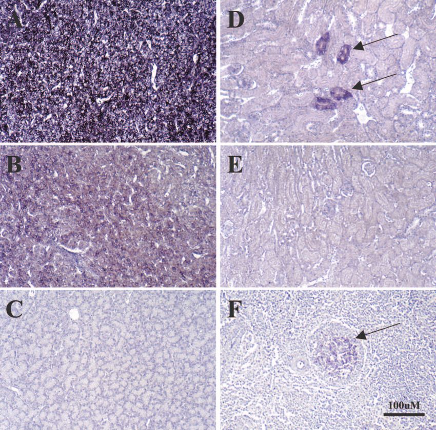
FIG. 7. In situ hybridization of tissues from infected ducks for detection of viral nucleic acids. EAA-fixed sections of liver (A, B, C), kidney (D,
E) and spleen (F) collected at autopsy from ducks (see legend to Fig. 6) infected with OSHBV (A), PTHBV (B, C, D, E), or DHBV (F) were used
for in situ hybridization to detect viral nucleic acids with a digoxigenin-labeled full-length DHBV DNA (A, B, D, F) or pUC19 DNA (C, E) probe,
as described in Materials and Methods. Viral nucleic acid detection in kidney tubular cells (D) and splenic germinal centers (F) is indicated by
arrows. All panels were counterstained with hematoxylin. Magnification, ⫻160. Bar, 100 M.
MDHBVa, MDHBVb, CWHBV, PTHBV, and OSHBV all Fig. 7), and in addition, cccDNA was detected in the spleens of
replicated in the liver. In contrast, none, including DHBV, ducks infected with DHBV, MDHBVa, and CWHBV (Fig. 8).
appeared to infect the exocrine pancreas (19) under these Comparison of Table 3 and Fig. 7 reveals that cccDNA detec-
conditions (Table 3 and data not shown). Accumulation of tion in the spleen did not completely correlate with detection
virus in germinal centers in the spleen, thought to be present in of viral DNA in germinal centers, suggesting that cccDNA
follicular dendritic cells, has been observed before, but it re- accumulated elsewhere in the spleen. No single-stranded, mi-
mains unclear whether DHBV replicates in the spleen (22). nus-strand DNA, diagnostic of virus replication (41), was de-
Similar observations were made in the present study (Table 3; tected in the spleen. Thus, the data suggest that some cells in2740 GUO ET AL. J. VIROL.
and variability of the pre-S domain between isolates is believed
to reflect adaptation to host receptors. If this were strictly true,
we would expect that divergence in pre-S would have an impact
on cross-species infection. Surprisingly, all of the isolates re-
ported here, despite considerable variation in pre-S, were able
to infect Pekin duck hepatocytes.
A question unresolved by this study is whether the new
isolates are present in wild birds of the same species, as most
isolates described to date have been found in aviculture, zoos,
farms, and wild mallards (16, 30). For instance, the Ross goose
is found mostly along the west coast of North America, where
snow geese and the Ross goose share nesting space and also
interbreed, yet the Ross goose virus is completely distinct from
SGHBV. Both viruses were, however, isolated from captive
FIG. 8. Southern blot analysis of total and cccDNA from the
birds (11; Newbold, unpublished). In contrast, the Ross goose
Downloaded from http://jvi.asm.org/ on March 1, 2021 by guest
spleens of infected ducks. Total and cccDNA from 1.2 ⫻ 106 and 4.8
⫻ 106 spleen cells, respectively, from the ducks described in the legend and the mandarin duck have virtually identical viruses. These
to Fig. 6, were subjected to electrophoresis in 1.5% agarose gels and birds would not be expected to have any contact in the wild,
Southern blot analysis, as described in Materials and Methods. DHBV and it seems surprising that a virus with 99.1% sequence con-
DNA, including cccDNA, was detected in the spleens of ducks infected servation would be found in each if these viruses were native to
with DHBV, while spleen tissue from ducks infected with the new
isolates, although containing cccDNA, did not contain detectable lev- the feral birds. In this regard, we note that the Ross goose and
els of other viral DNAs. The marker contains 30 pg of cloned unit- mandarin duck virus were isolated from aviculture collections
length DHBV DNA migrating at 3 kbp. in different states with no obvious contact. Thus, while it is
convenient to identify these virus isolates with the species from
which each was first isolated, it is possible that neither
the spleen may be latently infected, but their identity remains RGHBV nor MDHBV are native to these birds in the wild.
unknown. Evidence was also obtained for infection in the kid- The situation with the Chiloe wigeon and puna teal is sim-
ney tubular epithelium of some of the ducks (Fig. 7; Table 3), ilar. Both are found in South America in a range that does not
probably reflecting virus replication in these cells (19). overlap the mallard, found in the northern hemisphere and
reported to be infected with DHBV (16, 30). In addition, these
ducks are presumably a distinct species from the mallard, yet
DISCUSSION
they carry a virus almost identical to DHBV. To a lesser extent,
We report on the isolation of five new strains of avihepad- this holds true for the Orinoco and ashy-headed sheldgeese,
naviruses. Two, from the puna teal and Chiloe wigeon, are not which are also found in South America. In summary, while the
obviously distinct from previous isolates of DHBV (Fig. 2 and isolates from these species are not identical to previously re-
3; Table 1). Another two, from the Orinoco and ashy-headed ported isolates of DHBV, they are similar enough, especially
sheldgeese, were also closely related to DHBV isolates but considering the divergence of DHBV isolates (Fig. 2), to raise
somewhat more distantly than those from the puna teal and the possibility that these different birds were cross-infected
Chiloe wigeon. The most distantly related, from the mandarin with DHBV in captivity, an issue that can only be resolved by
duck, were closely related to RGHBV (93.3 to 99.1% homol- study of these birds in the wild.
ogy). Hepadnaviruses are generally considered to be highly The second issue we hoped to resolve in these experiments
species specific, but this seems less true of those found in ducks was the prevalence of X-like ORFs in avihepadnavirus isolates.
and geese. Host range is believed to be determined by pre-S, Again, the picture is mixed. The mandarin duck and sheld-
goose isolates contain an ORF beginning with an AUG in the
appropriate location for X, whereas the puna teal and Chiloe
TABLE 3. In situ hybridization detection of viral nucleic acids in wigeon isolates, like DHBV, lack an AUG. In view of the
tissues collected from infected ducksa recent report that the X ORF of DHBV does not express a
protein needed for either persistent or chronic infection of
Resultc for:
ducklings, the significance of this ORF, if any, remains unclear.
Virus Spleen Kidney
Liverb (germinal Pancreas (tubular
centers) epithelium) ACKNOWLEDGMENTS
DHBV (6 ⫻ 10 ) 7
⬎95% ⫹ ⫹ ⫺ ⫺ We are grateful to Maxine Linial (Fred Hutchinson Cancer Center),
MDHBVa ⬎95% ⫹ ⫺ ⫺ ⫺ Christoph Seeger, and Jesse Summers (University of New Mexico) for
MDHBVb ⬎95% ⫹ ⫹ ⫺ ⫹⫹ helpful suggestions.
CWHBV ⬎95% ⫹ ⫹ ⫺ ⫺ This work was supported by USPHS grant AI-18641 (to W.S.M.), a
PTHBV ⬎95% ⫹ ⫹ ⫺ ⫹⫹ project grant from the National Health and Medical Research Council
OSHBV ⬎95% ⫹ ⫹ ⫺ ⫺ of Australia (to A.R.J.), and an appropriation from the Common-
wealth of Pennsylvania.
a
In situ hybridization was carried out as described in Materials and Methods
and as illustrated in Fig. 7.
b REFERENCES
Infection of both bile ductular epithelium and hepatocytes was detected.
c
⫹, infected cells detected by in situ hybridization; ⫺, infected cells not 1. Barrasa, M. I., J. T. Guo, J. Saputelli, W. S. Mason, and C. Seeger. 2001.
detected. Does a cdc2 kinase-like recognition motif on the core protein of hepadna-VOL. 79, 2005 NEW AVIHEPADNAVIRUS ISOLATES 2741
viruses regulate assembly and disintegration of capsids? J. Virol. 75:2024– Central role of a serine phosphorylation site within duck hepatitis B virus
2028. core protein for capsid trafficking and genome release. J. Biol. Chem. 278:
2. Benton, W. D., and R. W. Davis. 1977. Screening lambda gt recombinant 28123–28129.
clones by hybridization to single plaques in situ. Science 196:180–182. 27. Kozak, M. 1997. Recognition of AUG and alternative initiator codons is
3. Biermer, M., R. Puro, and R. J. Schneider. 2003. Tumor necrosis factor augmented by G in position ⫹4 but is not generally affected by the nucleo-
alpha inhibition of hepatitis B virus replication involves disruption of capsid tides in positions ⫹5 and ⫹6. EMBO J. 16:2482–2492.
integrity through activation of NF-B. J. Virol. 77:4033–4042. 28. Kuroki, K., R. Cheung, P. L. Marion, and D. Ganem. 1994. A cell surface
4. Borel, C., C. Sunyach, O. Hantz, C. Trepo, and A. Kay. 1998. Phosphoryla- protein that binds avian hepatitis B virus particles. J. Virol. 68:2091–2096.
tion of DHBV pre-S: identification of the major site of phosphorylation and 29. Kuroki, K., F. Eng, T. Ishikawa, C. Turck, F. Harada, and D. Ganem. 1995.
effects of mutations on the virus life cycle. Virology 242:90–98. gp180, a host cell glycoprotein that binds duck hepatitis B virus particles, is
5. Bouchard, M., S. Giannakopoulos, E. H. Wang, N. Tanese, and R. J. Schnei- encoded by a member of the carboxypeptidase gene family. J. Biol. Chem.
der. 2001. Hepatitis B virus HBx protein activation of cyclin A–cyclin-de- 270:15022–15028.
pendent kinase 2 complexes and G1 transit via a Src kinase pathway. J. Virol. 30. Lambert, V., L. Cova, P. Chevallier, R. Mehrotra, and C. Trepo. 1991.
75:4247–4257. Natural and experimental infection of wild mallard ducks with duck hepatitis
6. Bouchard, M. J., R. J. Puro, L. Wang, and R. J. Schneider. 2003. Activation B virus. J. Gen. Virol. 72:417–420.
and inhibition of cellular calcium and tyrosine kinase signaling pathways 31. Lenhoff, R. J., and J. Summers. 1994. Construction of avian hepadnavirus
identify targets of the HBx protein involved in hepatitis B virus replication. variants with enhanced replication and cytopathicity in primary hepatocytes.
J. Virol. 77:7713–7729. J. Virol. 68:5706–5713.
7. Bouchard, M. J., L. H. Wang, and R. J. Schneider. 2001. Calcium signaling 32. Li, J., S. Tong, and J. R. Wands. 1999. Identification and expression of
by HBx protein in hepatitis B virus DNA replication. Science 294:2376–2378. glycine decarboxylase (p120) as a duck hepatitis B virus pre-S envelope-
Downloaded from http://jvi.asm.org/ on March 1, 2021 by guest
8. Breiner, K. M., S. Urban, and H. Schaller. 1998. Carboxypeptidase D binding protein. J. Biol. Chem. 274:27658–27665.
(gp180), a Golgi-resident protein, functions in the attachment and entry of 33. Lien, J. M., C. E. Aldrich, and W. S. Mason. 1986. Evidence that a capped
avian hepatitis B viruses. J. Virol. 72:8098–8104. oligoribonucleotide is the primer for duck hepatitis B virus plus-strand DNA
9. Buscher, M., W. Reiser, H. Will, and H. Schaller. 1985. Transcripts and the synthesis. J. Virol. 57:229–236.
putative RNA pregenome of duck hepatitis B virus: implications for reverse 34. Lien, J. M., D. J. Petcu, C. E. Aldrich, and W. S. Mason. 1987. Initiation and
transcription. Cell 40:717–724. termination of duck hepatitis B virus DNA synthesis during virus maturation.
10. Calvert, J., and J. Summers. 1994. Two regions of an avian hepadnavirus J. Virol. 61:3832–3840.
RNA pregenome are required in cis for encapsidation. J. Virol. 68:2084– 35. Lilienbaum, A., C. B. Crescenzo, A. A. Sall, J. Pillot, and E. Elfassi. 1993.
2090. Binding of nuclear factors to functional domains of the duck hepatitis B virus
11. Chang, S. F., H. J. Netter, M. Bruns, R. Schneider, K. Frolich, and H. Will. enhancer. J. Virol. 67:6192–6200.
1999. A new avian hepadnavirus infecting snow geese (Anser caerulescens) 36. Liu, C., W. S. Mason, and J. Burch. 1994. Identification of factor binding
produces a significant fraction of virions containing single-stranded DNA. sites in the DHBV enhancer and in vivo effects of enhancer mutations.
Virology 262:39–54. J. Virol. 68:2286–2296.
12. Chang, S. F., H. J. Netter, E. Hildt, R. Schuster, S. Schaefer, Y. C. Hsu, A. 37. Mabit, H., K. M. Breiner, A. Knaust, B. Zachmann-Brand, and H. Schaller.
Rang, and H. Will. 2001. Duck hepatitis B virus expresses a regulatory 2001. Signals for bidirectional nucleocytoplasmic transport in the duck hep-
HBx-like protein from a hidden open reading frame. J. Virol. 75:161–170. atitis B virus capsid protein. J. Virol. 75:1968–1977.
13. Chen, H. S., S. Kaneko, R. Girones, R. W. Anderson, W. E. Hornbuckle, B. C. 38. Macrae, D. R., V. Bruss, and D. Ganem. 1991. Myristylation of a duck
Tennant, P. J. Cote, J. L. Gerin, R. H. Purcell, and R. H. Miller. 1993. The hepatitis B virus envelope protein is essential for infectivity but not for virus
woodchuck hepatitis virus X gene is important for establishment of virus assembly. Virology 181:359–363.
infection in woodchucks. J. Virol. 67:1218–1226. 39. Mandart, E., A. Kay, and F. Galibert. 1984. Nucleotide sequence of a cloned
14. Chouteau, P., J. Le Seyec, I. Cannie, M. Nassal, C. Guguen-Guillouzo, and duck hepatitis B virus genome: comparison with woodchuck and human
P. Gripon. 2001. A short N-proximal region in the large envelope protein hepatitis B virus sequences. J. Virol. 49:782–792.
harbors a determinant that contributes to the species specificity of human 40. Mangisa, N. P., H. E. Smuts, A. Kramvis, C. W. Linley, M. Skelton, T. J.
hepatitis B virus. J. Virol. 75:11565–11572. Tucker, P. M. De La Hall, D. Kahn, A. R. Jilbert, and M. C. Kew. 2004.
15. Condreay, L. D., C. E. Aldrich, L. Coates, W. S. Mason, and T. T. Wu. 1990. Molecular characterization of duck hepatitis B virus isolates from South
Efficient duck hepatitis B virus production by an avian liver tumor cell line. African ducks. Virus Genes 28:179–186.
J. Virol. 64:3249–3258. 41. Mason, W. S., C. Aldrich, J. Summers, and J. M. Taylor. 1982. Asymmetric
16. Cova, L., V. Lambert, A. Chevallier, O. Hantz, I. Fourel, C. Jacquet, C. replication of duck hepatitis B virus in liver cells: free minus-strand DNA.
Pichoud, J. Boulay, B. Chomel, L. Vitvitski, et al. 1986. Evidence for the Proc. Natl. Acad. Sci. USA 79:3997–4001.
presence of duck hepatitis B virus in wild migrating ducks. J. Gen. Virol. 42. Mason, W. S., M. S. Halpern, J. M. England, G. Seal, J. Egan, L. Coates, C.
67:537–547. Aldrich, and J. Summers. 1983. Experimental transmission of duck hepatitis
17. Grgacic, E. V., and D. A. Anderson. 1994. The large surface protein of duck B virus. Virology 131:375–384.
hepatitis B virus is phosphorylated in the pre-S domain. J. Virol. 68:7344– 43. Mason, W. S., G. Seal, and J. Summers. 1980. Virus of Pekin ducks with
7350. structural and biological relatedness to human hepatitis B virus. J. Virol.
18. Grgacic, E. V., B. Lin, E. V. Gazina, M. J. Snooks, and D. A. Anderson. 1998. 36:829–836.
Normal phosphorylation of duck hepatitis B virus L protein is dispensable 44. Meier, P., C. A. Scougall, H. Will, C. J. Burrell, and A. R. Jilbert. 2003. A
for infectivity. J. Gen. Virol. 79:2743–2751. duck hepatitis B virus strain with a knockout mutation in the putative X ORF
19. Halpern, M. S., J. M. England, D. T. Deery, D. J. Petcu, W. S. Mason, and shows similar infectivity and in vivo growth characteristics to wild-type virus.
K. L. Molnar-Kimber. 1983. Viral nucleic acid synthesis and antigen accu- Virology 317:291–298.
mulation in pancreas and kidney of Pekin ducks infected with duck hepatitis 45. Netter, H. J., S. Chassot, S. F. Chang, L. Cova, and H. Will. 1997. Sequence
B virus. Proc. Natl. Acad. Sci. USA 80:4865–4869. heterogeneity of heron hepatitis B virus genomes determined by full-length
20. Ishikawa, T., and D. Ganem. 1995. The pre-S domain of the large viral DNA amplification and direct sequencing reveals novel and unique features.
envelope protein determines host range in avian hepatitis B viruses. Proc. J. Gen. Virol. 78:1707–1718.
Natl. Acad. Sci. USA 92:6259–6263. 46. Obert, S., B. Zachmann-Brand, E. Deindl, W. Tucker, R. Bartenschlager,
21. Jilbert, A. R. 2000. In situ hybridization protocols for detection of viral DNA and H. Schaller. 1996. A spliced hepadnavirus RNA is essential for virus
using radioactive and nonradioactive DNA probes. Methods Mol. Biol. 123: replication. EMBO J. 15:2565–2574.
177–193. 47. Page, R. D. M. 1996. TREEVIEW: an application to display phylogenetic
22. Jilbert, A. R., J. S. Freiman, E. J. Gowans, M. Holmes, Y. E. Cossart, and trees on personal computer. Comput. Appl. Biosci. 22:357–358.
C. J. Burrell. 1987. Duck hepatitis B virus DNA in liver, spleen, and pan- 48. Prassolov, A., H. Hohenberg, T. Kalinina, C. Schneider, L. Cova, O. Krone,
creas: analysis by in situ and Southern blot hybridization. Virology 158:330– K. Frolich, H. Will, and H. Sirma. 2003. New hepatitis B virus of cranes that
338. has an unexpected broad host range. J. Virol. 77:1964–1976.
23. Jilbert, A. R., T. T. Wu, J. M. England, P. M. Hall, N. Z. Carp, A. P. 49. Pugh, J. C., and J. W. Summers. 1989. Infection and uptake of duck hepatitis
O’Connell, and W. S. Mason. 1992. Rapid resolution of duck hepatitis B B virus by duck hepatocytes maintained in the presence of dimethyl sulfox-
virus infections occurs after massive hepatocellular involvement. J. Virol. ide. Virology 172:564–572.
66:1377–1388. 50. Pult, I., H. J. Netter, M. Bruns, A. Prassolov, H. Sirma, H. Hohenberg, S. F.
24. Junker-Niepmann, M., R. Bartenschlager, and H. Schaller. 1990. A short Chang, K. Frolich, O. Krone, E. F. Kaleta, and H. Will. 2001. Identification
cis-acting sequence is required for hepatitis B virus pregenome encapsida- and analysis of a new hepadnavirus in white storks. Virology 289:114–128.
tion and sufficient for packaging of foreign RNA. EMBO J. 9:3389–3396. 51. Seeger, C., and W. S. Mason. 2000. Hepatitis B virus biology. Microbiol. Mol.
25. Kawaguchi, T., K. Nomura, Y. Hirayama, and T. Kitagawa. 1987. Establish- Biol. Rev. 54:51–68.
ment and characterization of a chicken hepatocellular carcinoma cell line, 52. Sprengel, R., E. F. Kaleta, and H. Will. 1988. Isolation and characterization
LMH. Cancer Res. 47:4460–4464. of a hepatitis B virus endemic in herons. J. Virol. 62:932–937.
26. Kock, J., M. Kann, G. Putz, H. E. Blum, and F. Von Weizsacker. 2003. 53. Summers, J., P. M. Smith, and A. L. Horwich. 1990. Hepadnaviral envelope2742 GUO ET AL. J. VIROL.
proteins regulate covalently closed circular DNA amplification. J. Virol. 61. Wang, G. H., F. Zoulim, E. H. Leber, J. Kitson, and C. Seeger. 1994. Role of
64:2819–2824. RNA in enzymatic activity of the reverse transcriptase of hepatitis B viruses.
54. Tavis, J. E., S. Perri, and D. Ganem. 1994. Hepadnavirus reverse transcrip- J. Virol. 68:8437–8442.
tion initiates within the stem-loop of the RNA packaging signal and employs 62. Yu, M., R. H. Miller, S. Emerson, and R. H. Purcell. 1996. A hydrophobic
a novel strand transfer. J. Virol. 68:3536–3543. heptad repeat of the core protein of woodchuck hepatitis virus is required for
55. Thompson, J. D., T. J. Gibson, F. Plewniak, F. Jeanmougin, and D. G. capsid assembly. J. Virol. 70:7085–7091.
Higgins. 1997. The ClustalX windows interface: flexible strategies for mul- 63. Yu, M., and J. Summers. 1994. Multiple functions of capsid protein
tiple sequence alignments aided by quality analysis tools. Nucleic Acids Res. phosphorylation in duck hepatitis B virus replication. J. Virol. 68:4341–
24:4876–4882. 4348.
56. Todd, F. S. 1996. Natural history of the waterfowl. Ibis Publishing Company,
64. Yu, M., and J. Summers. 1994. Phosphorylation of the duck hepatitis B virus
Vista, Calif.
capsid protein associated with conformational changes in the C terminus.
57. Tong, S., J. Li, and J. R. Wands. 1999. Carboxypeptidase D is an avian
J. Virol. 68:2965–2969.
hepatitis B virus receptor. J. Virol. 73:8696–8702.
58. Triyatni, M., P. Ey, T. Tran, M. Le Mire, M. Qiao, C. Burrell, and A. Jilbert. 65. Zhang, Z., N. Torii, Z. Hu, J. Jacob, and T. J. Liang. 2001. X-deficient
2001. Sequence comparison of an Australian duck hepatitis B virus strain woodchuck hepatitis virus mutants behave like attenuated viruses and induce
with other avian hepadnaviruses. J. Gen. Virol. 82:373–378. protective immunity in vivo. J. Clin. Investig. 108:1523–1531.
59. Urban, S., K. M. Breiner, F. Fehler, U. Klingmuller, and H. Schaller. 1998. 66. Zhou, Y. Z. 1980. A virus possibly associated with hepatitis and hepatoma in
Avian hepatitis B virus infection is initiated by the interaction of a distinct ducks. Shanghai Med. J. 3:641–644.
pre-S subdomain with the cellular receptor gp180. J. Virol. 72:8089–8097. 67. Zoulim, F., J. Saputelli, and C. Seeger. 1994. Woodchuck hepatitis virus X
60. Urban, S., C. Schwarz, U. C. Marx, H. Zentgraf, H. Schaller, and G. Mul- protein is required for viral infection in vivo. J. Virol. 68:2026–2030.
Downloaded from http://jvi.asm.org/ on March 1, 2021 by guest
thaup. 2000. Receptor recognition by a hepatitis B virus reveals a novel 68. Zoulim, F., and C. Seeger. 1994. Reverse transcription in hepatitis B viruses
mode of high affinity virus-receptor interaction. EMBO J. 19:1217–1227. is primed by a tyrosine residue of the polymerase. J. Virol. 68:6–13.You can also read