Displays conserved function in ovule integument development - Nature
←
→
Page content transcription
If your browser does not render page correctly, please read the page content below
www.nature.com/scientificreports
OPEN Orchid B
sister gene PeMADS28
displays conserved function
in ovule integument development
Ching‑Yu Shen1,10, You‑Yi Chen2,10, Ke‑Wei Liu3,4, Hsiang‑Chia Lu1,5,6, Song‑Bin Chang2,
Yu‑Yun Hsiao7, Fengxi Yang8, Genfa Zhu8, Shuang‑quan Zou5,9, Lai‑Qiang Huang3,4,
Zhong‑Jian Liu5* & Wen‑Chieh Tsai1,2,7*
The ovules and egg cells are well developed to be fertilized at anthesis in many flowering plants.
However, ovule development is triggered by pollination in most orchids. In this study, we
characterized the function of a Bsister gene, named PeMADS28, isolated from Phalaenopsis equestris,
the genome-sequenced orchid. Spatial and temporal expression analysis showed PeMADS28
predominantly expressed in ovules between 32 and 48 days after pollination, which synchronizes with
integument development. Subcellular localization and protein–protein interaction analyses revealed
that PeMADS28 could form a homodimer as well as heterodimers with D-class and E-class MADS-box
proteins. In addition, ectopic expression of PeMADS28 in Arabidopsis thaliana induced small curled
rosette leaves, short silique length and few seeds, similar to that with overexpression of other species’
Bsister genes in Arabidopsis. Furthermore, complementation test revealed that PeMADS28 could rescue
the phenotype of the ABS/TT16 mutant. Together, these results indicate the conserved function of
Bsister PeMADS28 associated with ovule integument development in orchid.
In plants, MADS-box genes control the development of distinct organs, such as flower, ovule, fruit, leaf, and
root1–3. Plant MADS-box genes can be classified into types I and II genes on the basis of phylogenetic a nalysis4.
The best studied plant type II MADS-box transcription factors are those involved in floral organ identity deter-
mination. The determination of floral organ primordia by genes of the A, B, C, D and E classes led to the ABCDE
model5–10. Furthermore, most of plant type II MADS-box proteins share a conserved structure consisting of
four domains: MADS (M), intervening (I), keratin-like (K), and C-terminal (C)11,12. The DNA binding partner
specificity is mediated to a large extent by the I domain, and the K domain likely promotes protein dimerization
as well as tetramerization13–15. Proteins of floral MADS-box genes participating in floral organ identity interact
with each other to control downstream genes16–19. Most of the protein–protein interactions necessary for the con-
stitution of quaternary complexes, as recommended by the “quartet model”, are conserved20. Proliferating ovule
primordia is specified by specific ovule identity factors, such as the MADS-box family members SEEDSTICK
(STK), SHATTERPROOF1 (SHP1), SHP2, SEPALLATA (SEP) and AGAMOUS21–23. Moreover, Bsister genes are
“marker genes” for the development of (inner) integument structures, phylogenetically the “oldest” structures
surrounding the female gametophyte of seed p lants24.
1
Institute of Tropical Plant Sciences and Microbiology, National Cheng Kung University, Tainan 701,
Taiwan. 2Department of Life Sciences, National Cheng Kung University, Tainan 701, Taiwan. 3Center for
Biotechnology and BioMedicine, Shenzhen Key Laboratory of Gene & Antibody Therapy, State Key Laboratory of
Health Science & Technology (prep) and Division of Life & Health Sciences, Graduate School at Shenzhen, Tsinghua
University, Shenzhen, China. 4School of Life Science, Tsinghua University, Beijing 100084, China. 5Key Laboratory
of National Forestry and Grassland Administration for Orchid Conservation and Utilization at College of Landscape
Architecture, College of Landscape Architecture, Fujian Agriculture and Forestry University, Fuzhou, China. 6Fujian
Colleges and Universities Engineering Research Institute of Conservation and Utilization of Natural Bioresources,
College of Forestry, Fujian Agriculture and Forestry University, Fuzhou 350002, China. 7Orchid Research and
Development Center, National Cheng Kung University, Tainan 701, Taiwan. 8Guangdong Key Laboratory of
Ornamental Plant Germplasm Innovation and Utilization, Environmental Horticulture Research Institute,
Guangdong Academy of Agricultural Sciences, Guangzhou 510640, China. 9Fujian Colleges and Universities
Engineering Research Institute of Conservation and Utilization of Natural Biosciences, College of Forestry, Fujian
Agriculture and Forestry University, Fuzhou 350002, China. 10These authors contributed equally: Ching-Yu Shen
and You-Yi Chen. *email: zjliu@fafu.edu.cn; tsaiwc@mail.ncku.edu.tw
Scientific Reports | (2021) 11:1205 | https://doi.org/10.1038/s41598-020-79877-9 1
Vol.:(0123456789)
www.nature.com/scientificreports/
sister gene was isolated from Arabidopsis thaliana and was named ARABIDOPSIS
In dicotyledons, the first B
BSISTER (ABS/TT16)25,26. ABS/TT16 is expressed mainly in the innermost integument layer, the endothelium; a
closely related paralog is GORDITA (GOA, formerly known as AGL63). Besides study of eudicots, B sister MADS-
box genes have been investigated in the monocot Oryza sativa. ABS/TT16 and GOA were found not functionally
redundant in ovule d evelopment24,26–28. ABS/TT16 is required for the proper differentiation of the inner integu-
ment, and GOA is necessary for at least the early development of the outer integument28. The single mutants abs/
tt16 and goa still produce seeds, which germinate p roperly24,26,27. In addition, silencing of OsMADS29, a B sister
gene in rice, led to severe phenotypes with degeneration of the pericarp, ovular vascular trace, integuments,
nucellar epidermis and nucellar p rojection29.
Orchids, constituting approximately 10% of all seed plant species, have enormous value for commercial
horticulture and are of specific scientific interest because of their extraordinary diversity of floral morphology,
ecological adaptations, and unique reproductive s trategies30. The unique reproductive strategies include mature
pollen grains packaged as pollinia, pollination-regulated ovary/ovule development, synchronized timing of
micro- and mega-gametogenesis for effective fertilization, and release of thousands or millions of immature
embryos (seeds without endosperm) in mature pods31.
In most flowering plants, the ovules are mature, and the egg cells are ready for fertilization at anthesis. In
contrast, in orchids, ovule development is triggered by pollination. In most orchids such as Cattleya, Sophronitis,
Epidendron, Laelia, Phalaenopsis, Dendrobium and Doritis, ovules are completely absent in unpollinated ovaries,
and the development of ovule is triggered only after pollination32. The long-term progressive process of ovule
development in orchids as compared with other flowering plants is an attractive system for investigating ovule
initiation and subsequent development.
With high economic value, Phalaenopsis orchids are beautiful ornamental plants and very popular world-
wide. The genome of P. equestris was recently sequenced33 and the information provides a great opportunity to
identify and characterize the genes involved in regulating orchid ovule d evelopment34. In this study, we identified
and functionally characterized PeMADS28, a B sister MADS-box gene, in P. equestris. Our results indicate that
the function of PeMADS28 plays an important role in ovule integument development in orchid and reveals the
functional conservation of B sister genes between monocots and dicots.
Results
Identification of PeMADS28 MADS‑box gene in P. equestris. Only one Bsister MADS-box gene,
PeMADS28 (predicted proteome gene ID Peq004141), exists in the P. equestris genome33. The sequence of
PeMADS28 was retrieved from OrchidBase35,36. The ORF including 723 bp encodes a protein of 240 amino
acids. Multiple sequence alignment with other Bsister proteins from gymnosperm, dicots and monocots demon-
strated that PeMADS28 has a typical MIKC-type domain structure (Supplementary Fig. S1). B sister proteins also
contain a conserved PI Motif-Derived sequence in their C-terminal regions that are also representative of B-class
MADS-box proteins (Supplementary Fig. S1)25.
Phylogenetic relationship of PeMADS28 and other MADS‑box genes. To determine the phy-
logenetic relationships of PeMADS28 and other Bsister genes, we constructed a phylogenetic tree by using the
amino acid sequences of PeMADS28 with other known gymnosperm and angiosperm B sister sequences and the
AGL63-like sequence from Brassicaceae. Amino acid sequences of B sister proteins and AGL63-like proteins were
retrieved from the National Center for Biotechnology Information (NCBI). This phylogeny has two supported
major clades, one containing monocot Bsister proteins and the other dicot Bsister proteins (Fig. 1). Moreover, the
Bsister and AGL63-like proteins were divided into two groups in dicots clade. PeMADS28 is close to the orchid
Erycina pusilla Bsister protein EpMADS24 (Fig. 1). These results strongly suggest that PeMADS28 belongs to the
Bsister gene family.
Spatial and temporal expression of PeMADS28 in P. equestris. RT-PCR and quantitative real-time
RT-PCR were used to survey the spatial and temporal expression patterns of PeMADS28. Because B sister MADS-
box genes are involved in ovule development and pollination is a key regulatory event in orchid ovule initia-
tion, we determined the temporal mRNA expression patterns of PeMADS28 in developing ovules triggered by
pollination. During ovule development, PeMADS28 transcript level was highest from 32 to 48 days after pol-
lination (DAP) (Fig. 2a,b), then decreased from 56 to 100 DAP (Fig. 2a,b). However, PeMADS28 expression
was barely observed in flower buds and was absent from vegetative tissues (Supplementary Fig. S2). Previous
research showed that ovule development between 32 and 48 DAP is associated with inner and outer integument
development37. These results suggest PeMADS28 has functions in ovule integument development.
In situ hybridization of PeMADS28 transcripts. We further examined the detailed spatial and tempo-
ral expression patterns of PeMADS28 during ovule development by in situ hybridization with antisense RNA
probes. During the early stage of ovule development, when the final branches of placental protuberances differ-
entiate ovular primordia, PeMADS28 transcript expression was detected in all ovule primordia at their initiation
(Fig. 3a,c,d). In the later stage, the expression was more concentrated in developing ovules (Fig. 3e). At 48 DAP,
PeMADS28 mRNA was detected in the whole ovule including nucellus and integument (Fig. 3f). PeMADS28
transcript expression was not detected in 56-DAP ovules (Fig. 3h). The negative control was sense RNA used as
a probe (Fig. 3b,g,i). These results supported that PeMADS28 might be involved in orchid ovule initiation and
integument development.
Scientific Reports | (2021) 11:1205 | https://doi.org/10.1038/s41598-020-79877-9 2
Vol:.(1234567890)
www.nature.com/scientificreports/
Figure 1. Phylogenetic analysis of B
sister proteins. GGM13 from Gnetum gnemon and GbMADS10 from Ginkgo
biloba were outgroup representatives. Bootstrap values from 1000 replicates are indicated on most major nodes.
PeMADS28 is highlighted by the asterisks.
Subcellular localization of PeMADS28‑GFP fusion protein. As a member of MADS-box tran-
scription factors, PeMADS28 was expected to localize in the nucleus. To test the subcellular localization of
PeMADS28, a PeMADS28-GFP fusion protein was generated with a GFP reporter gene fused in-frame to the
PeMADS28 coding region under control of the 35S promoter. Transient expression of PeMADS28-GFP fusion
protein was analyzed in Phalaenopsis petal protoplasts. PeMADS28-GFP fusion protein signals were observed in
both the nucleus and cytoplasm along with GFP signals (Fig. 4). Thus, PeMADS28 might need to interact with
other proteins to exclusively localize in the nucleus.
Interaction behavior of PeMADS28 analyzed by bimolecular fluorescence complementation
(BiFC) assay. A number of previous studies demonstrated that MADS-box transcription factors form
dimers or higher-order complexes for their functions in flower and ovule d evelopment17,20,21,24,38–40. To investi-
gate the ability of homodimer formation of PeMADS28 and nuclear localization of this self-association, we used
BiFC assay. A BiFC vector pair with PeMADS28 fused to N- or C-terminal halves of YFP (PeMADS28:YFPn
and PeMADS28:YFPc) was prepared and used to co-transfect Phalaenopsis petal protoplasts. Fluorescence
YFP signal clearly indicated an interaction between the two PeMADS28 monomers. The formed homodimer
was exclusively localized in the nucleus (Fig. 5a). These results demonstrate that dimerization of PeMADS28
monomers plays an important role in retaining PeMADS28 in the nucleus. Previous reports indicated that
PeMADS1 (C-class), PeMADS7 (D-class), and PeSEP3 (E-class) MADS-box genes are involved in orchid ovule
development37,41. To gain more insight into the interaction of MADS-box proteins involved in orchid ovule
development, interaction behaviors among Bs and C-class, D-class, and E-class proteins were further inves-
tigated by BiFC assay. Interaction fluorescence signals were observed in the combination of PeMADS28 and
PeSEP3 (Fig. 5b,c) as well as PeMADS28 and PeMADS7 (Fig. 5d,e), which suggests that the orchid B sister protein
can interact with E- and D-class MADS-box proteins. In addition, the signals were localized in the nucleus,
as indicated by use of the nuclear dye propidium iodide (PI). However, interaction was not observed with the
combination of PeMADS28 and PeMADS1 (Fig. 5f,g). Therefore, PeMADS28 may not form heterodimers with
C-class MADS-box proteins. No fluorescence was detected with the empty vector control (Fig. 5h).
Functional analysis of PeMADS28 gene by ectopic expression and complementation in Arabi-
dopsis thaliana. For functional characterization of PeMADS28, we constructed transgenic Arabidopsis
plants expressing PeMADS28 under control of the cauliflower mosaic virus (CaMV) 35S promoter via Agro-
bacterium-mediated transformation. A total of 20 independent overexpressed PeMADS28 transgenic lines
were obtained based on kanamycin selection and a similar phenotype. Among twenty transgenic lines, nine
showed a 3:1 segregating kanamycin resistance phenotype. As compared with wild-type plants, six independent
PeMADS28 overexpressed lines shows the early flowering phenotype (Fig. 6a,c [wild-type plant]; 6b,d [trans-
Scientific Reports | (2021) 11:1205 | https://doi.org/10.1038/s41598-020-79877-9 3
Vol.:(0123456789)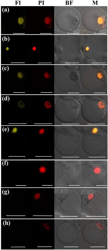
www.nature.com/scientificreports/
Figure 2. Expression patterns of PeMADS28 at various developing ovule stages in Phalaenopsis equestris (a)
RT-PCR analysis of PeMADS28. Expression of Phalaenopsis actin was an internal control. (b) Quantitative real-
time RT-PCR analysis of PeMADS28. DAP: days after pollination.
genic plant]) and fewer flower bud production (Fig. 6i [wild-type plant]; 6j [transgenic plant]). The rosette and
cauline leaves of transgenic plants had upwardly curled profiles and were smaller than those of wild-type plants
(Fig. 6a,k [wild type plant]; 6b,l [transgenic plant]). No homeotic conversion of floral organs was observed in
transgenic plants. Moreover, transgenic plants had smaller flowers with cracked sepals than wild-type plants
(Fig. 6e,g [wild-type plant]; 6f,h [transgenic plant]). The length of siliques was shorter in transgenic plants
(Fig. 6n and Table 1). Transgenic plants also showed more undeveloped seeds in siliques than did wild-type
plants (Fig. 6o and Table 1). In addition, transgenic seeds were larger and heavier (Fig. 6m and Table 1). Previ-
ously, GORDITA and ABS/TT16 are the paralogs in Arabidopsis. Consistently, over-expressed the GORDITA or
ABS/TT16 in Arabidopsis caused that the plant size shorter, and all organs are smaller than those in the wild-
type24,26,28. Both two over-expressed plants were affected the fruit development, and ABS/TT16 led to the rosette
leaves curled24,26,28. It is similar to 35::PeMADS28 phenotype. These results appeared that the PeMADS28 may
play a role in fruit development.
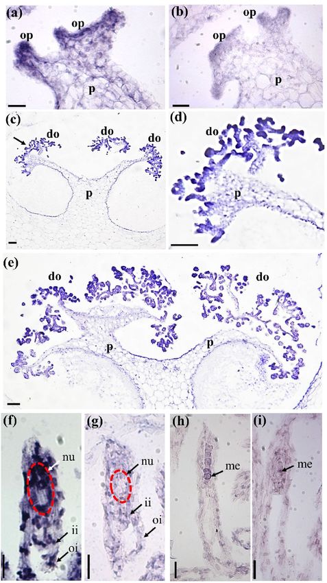
To further validate the function of PeMADS28, we used complementation testing with the tt16-1 mutant and
examined the seed pigmentation and development of the endothelium. A total of 8 transgenic lines were obtained
and 4 showed a 3:1 segregating kanamycin-resistance phenotype. All of the T2 line seeds showed restoration of
pigmentation to a brown color from the straw color of the tt16-1 mutant (Fig. 7a–c). In addition, PeMADS28
could rescue the development of endothelium in tt16-1 plants. Endothelial cells in immature wild-type seeds were
small, almost rectangular in shape and regularly spaced (Fig. 7d–f). In abs/tt16 immature seeds, endothelium
cells seemed to be flatter and more irregularly shaped than wild-type cells, resembled parenchymatic cells, and
often seemed to collapse (Fig. 7f)26. All of the T2 line (35S::PeMADS28 transgenic tt16-1) seed coats showed the
normal endothelium of the wild-type seed coat (Fig. 7e), so PeMADS28 was sufficient to complete the function
of Arabidopsis ABS/TT16.
Discussion
In this work, we identified a B sister-like gene, PeMADS28, from the P. equestris genome and characterized its func-
tion by sequence comparison, expression profile analysis, protein–protein interaction behavior, ectopic expres-
sion and complementation experiments in Arabidopsis. Protein sequence alignment showed that PeMADS28
is a typical B
sister protein with respect to its protein sequence because it contains a conserved sub-terminal “PI
motif-derived sequence,” which is also representative of B-class MADS-box proteins25. Phylogenetic analysis with
Scientific Reports | (2021) 11:1205 | https://doi.org/10.1038/s41598-020-79877-9 4
Vol:.(1234567890)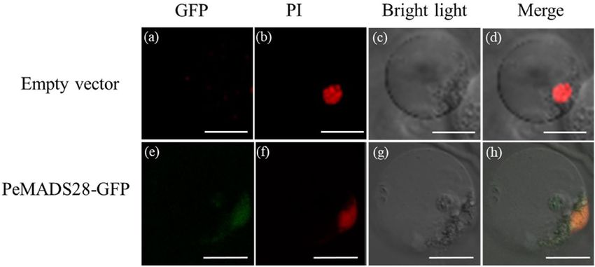
www.nature.com/scientificreports/

                                  Figure 3.  In situ hybridization of PeMADS28 in developing ovules of P. equestris. (a,b) Placenta with ovule primordium at 4
                                  DAP; (c) placenta with developing ovule at 32 DAP; (d) enlarged region of the dark arrow in (c); (e) placenta with developing
                                  ovule at 40 DAP; (f,g) developing ovules at 48 DAP; (h,i) developing ovule at 56 DAP. In (a), (c), (d), (e), (f) and (h), antisense
                                  probes were used to detect PeMADS28 transcripts. In (b), (g) and (i), hybridization involved sense probes (negative controls).
                                  Bars, 0.1 mm. p, placenta; op, ovule primordium; do, developing ovule; ii, inner integument; oi, outer integument; nu,
                                  nucellus; me, megaspores; DAP: days after pollination. The nucellus is highlighted by the red dash line.

Scientific Reports |   (2021) 11:1205 |                   https://doi.org/10.1038/s41598-020-79877-9                                                                5

                                                                                                                                                              Vol.:(0123456789)
www.nature.com/scientificreports/
Figure 4. Localization patterns of PeMADS28-GFP fusions in Phalaenopsis protoplasts. Images show
fluorescence and bright-field confocal microscopy images and merged images of flower protoplast. (a) Empty
vector was no green fluorescence in the cytoplasm and nucleus. (b) Cell in (a) stained with propidium iodide
(PI) represented in red to confirm the nucleus. (c) Cell in (a) and (b) by bright-field confocal microscopy. (d)
Merged image of (a), (b) and (c) to confirm green fluorescence in the cytoplasm of a flower cell. (e) PeMADS28-
GFP green fluorescence in nucleus and cytoplasm. (f) Cell in (e) stained with PI represented in red to confirm
the nucleus. (g) Cell in (e) and (f) by bright-field confocal microscopy. (h) Merged image of (e), (f) and (g) to
confirm green fluorescence in cytoplasm. Bars: 20 µm.
use of a deduced amino acid sequence revealed that PeMADS28 belongs to the monocot Bsister subclade. Both
analyses suggested that PeMADS28 is a putative orchid ortholog of Bsister genes like ABS/TT16 from Arabidopsis.
Bsister genes are closely related to B-class genes but express predominantly in the female reproductive organ.
Previous studies showed that B sister genes are expressed in the ovule and envelope in gymnosperms and in the
ovule and integuments of angiosperms. In the gymnosperm Gnetum gnemon, expression of B sister GGM13 is
specifically strong at the adaxial base of the cupules, where ovules subsequently develop42. When ovules appear,
GGM13 expression is limited to the developing nucellus and inner e nvelopes42. In dicots, the Arabidopsis Bsister
gene ABS/TT16 is expressed mainly in endothelium43. In petunia, FBP24 is expressed in young ovule primordia,
nucellus and integument. Later, the expression is confined to the endothelium in mature o vules38. In snapdragon,
DEFH21 expression was found in only a few inner cell layers of the inner integuments of the ovules25. In mono-
cots, wheat WBsis mRNA was detected in the developing inner integument at the late floral organ developmental
stage44. In rice, OsMADS29 transcripts are localized in the ovule, including integuments and nucellus throughout
ovule development29.
These results reveal a similarity of expression of Bsister genes suggesting conservation of the gene expres-
sion pattern over at least 300 million years27. In our study, temporal expression analysis revealed significant
PeMADS28 transcript expression between 32 and 48 DAP (Fig. 2a,b). In addition, in situ hybridization signals
of PeMADS28 transcripts were concentrated in the developing ovules (Fig. 3c–f). Hence, B sister genes may have
conserved expression patterns in seed plants. Interestingly, although Arabidopsis genome contains two and rice
has three B sister genes, these homologous genes have been occurred diversified expression and functional dif-
ferentiation. ABS/TT16 is involvement in endothelial cell specification and control of flavonoid biosynthesis in
Arabidopsis seed c oat26. The GOA is a young paralog of ABS/TT16 and play a role in fruit longitudinal g rowth27.
The rice OsMADS29 was identified as a key regulator of early rice seed development by regulating the pro-
grammed cell death of maternal t issues29. OsMADS30 does not have a canonical ‘Bsister function’, and revealed
neo-function in shoot size and architecture45. In fact, the development of orchid ovule is the typical monosporic
Polygonum type in which the functional megaspore passes through three mitotic divisions producing a seven
celled embryo sac consisting of three antipodal cells, one central cell formed by two polar nuclei, two synergid
cells, and the egg cell. In addition, the embryo sac is enclosed by inner and outer integuments. The orchid ovule
structure and development is highly similar to that of Arabidopsis and cereal except that the inner integument
gradually degenerated during the early stages of embryo proper formation and ovule initiation and development
is precisely triggered by pollination. Our data considered that the B sister gene PeMADS28 might involve in the
typical ovule development including integument morphogenesis.
Previously, it has been shown that the antagonistic development of nucellus and endosperm in Arabidopsis46.
The endosperm delivers the signal for the differentiation of seed coat and then both of tissues orchestrates seed
growth. However, the endosperm could also initiate nucellus degeneration via vacuolar cell death and n ecrosis46.
It also has been demonstrated that TT16/ABS can regulate proanthocyanidins synthesis in the seed coat and con-
versely TT16/ABS expression in the seed coat is sufficient to activate the nucellus degeneration46. In Phalaenopsis
orchids, double fertilization could be observed. However, the triple fusion nucleus of the endosperm initial is
amorphous in shape and apparently begins to degenerate immediately, consequently forming no e ndosperm47,48.
We speculated that the signal generated by fertilization of the central cell triggers its degeneration through
Scientific Reports | (2021) 11:1205 | https://doi.org/10.1038/s41598-020-79877-9 6
Vol:.(1234567890)www.nature.com/scientificreports/
Figure 5. Analysis of protein–protein interactions among Bsister PeMADS28, C-class PeMADS1, D-class
PeMADS7 and E-class PeSEP3 proteins by BiFC method. Fusion proteins were expressed in Phlaenopsis
petal protoplasts. (a) PeMADS28:YFPc + PeMADS28:YFPn. (b) PeMADS28:YFPc + PeSEP3:YFPn.
(c) PeSEP3:YFPc + PeMADS28:YFPn. (d) PeMADS28:YFPc + PeMADS7:YFPn. (e)
PeMADS7:YFPc + PeMADS28:YFPn. (f) PeMADS28:YFPc + PeMADS1:YFPn. (g)
PeMADS1:YFPc + PeMADS28:YFPn. (h) YFPc + YFPn as a negative control. BF, bright field; Fl, fluorescence
image; M, Merged image; PI, propidium iodide. Bars, 20 µm.
activation of the PeMADS28 expression. Because endosperm initial lives shortly, the signal might not spread
to the seed coat. In fact, the Phalaenopsis seed coat do not accumulate p roanthocyanidins49. However, whether
signal generated by fertilization of the central cell could reach to the nucellus and initiates the nucellus cell death
should be necessary for further study.
As transcriptional regulatory proteins, a number of MADS-box proteins have been shown to localize in
the nucleus. However, some MADS-box proteins are unable to translocate into the nucleus by themselves, but
their dimers deposit in the nucleus; examples are AP3-PI50 and UNSHAVEN-FLORAL BINDING PROTEIN
9 (FBP9)51. In this study, we detected PeMADS28-GFP fusion proteins in the nucleus and cytoplasm (Fig. 4h).
However, BiFC results showed the PeMADS28 homodimer specifically retained in the nucleus. The results sug-
gest that homodimerization of PeMADS28 drives a conformational change to bring it into a nuclear-retaining
structure. This kind of behavior of orchid Bsister PeMADS28 is similar to that of rice OsMADS2952. Our data
Scientific Reports | (2021) 11:1205 | https://doi.org/10.1038/s41598-020-79877-9 7
Vol.:(0123456789)www.nature.com/scientificreports/
Figure 6. Phenotype analysis of transgenic Arabidopsis overexpressing PeMADS28. (a) 20-day-old wild-
type plant; (b) 20-day-old 35S::PeMADS28 transgenic plant; (c) 31-day-old wild-type plant; (d) 31-day-old
35S::PeMADS28 transgenic plant. Bars (a–d), 5 mm. (e) Side-view of wild-type flower; (f) side-view of
35S::PeMADS28 transgenic flower; (g) top-view of wild-type flower; (h) top-view of 35S::PeMADS28 transgenic
flower; (i) Wild-type floral inflorescence; (j) 35S::PeMADS28 transgenic floral inflorescence; (k) cauline leaf of
wild-type plant; (l) cauline leaf of 35S::PeMADS28 transgenic plant. Bars (e–l) 1 mm. (m) Wild-type seed (left);
35S::PeMADS28 transgenic seed (right); (n) silique of wild-type (upper); silique of 35S::PeMADS28 transgenic
plant (lower); (o) silique of wild-type without one valve (upper); silique of 35S::PeMADS28 transgenic plant
without one valve (lower). Bars (m–o) 1 mm.
Plant WT OXPeMADS28
Silique length (cm) 1.29 ± 0.13 (n = 15) 0.85 ± 0.16 (n = 15), decrease*
Seeds/silique 45.33 ± 3.81 (n = 15) 24.26 ± 4.04 (n = 15), decrease*
100 seeds (mg) 3.04 ± 0.4 (n = 3) 5.05 ± 0.2( n = 3), increase*
Table 1. Silique length and seeds in OXPeMADS28 transgenic plants and wild-type (WT) plants. Asterisks
indicate statistically significant differences (*P < 0.05 compared with WT by Student’s t-test); The ± standard
deviation (SD) of the three biological repeats.
suggest the probability of PeMADS28 being regulated at the post-translational level via its interactions, which
may affect its function by regulating entry into the nucleus and regulation of its targets.
In Arabidopsis, previous study suggested a specific interaction of ABS with STK, SEP3, SHP1 and (much
weaker) SHP2 but not A G24,38. OsMADS29 could interact with OsMADS3 (C-class proteins) and all five E-class
proteins of r ice52. Our results indicate that PeMADS28 can form a homodimer in the nucleus. In addition, it
could interact with D-class (PeMADS7) and E-class (PeMADS8) MADS-box proteins. However, PeMADS28 and
PeMADS1 may not form heterodimers directly. These results suggest that protein interaction behaviors among
Bsister, D- and E-class proteins are conserved in angiosperms. Furthermore, in previous study, protein–pro-
tein interaction analyses revealed that PeSEP3 could bridge the interaction between PeMADS1 and PeMADS7
involved in Phalaenopsis gynostemium and ovule development37. Thus, a higher-order protein complex formed
by C-E-D-Bsister genes (PeMADS1-PeMADS8-PeMADS-PeMADS28) might have an important role in regulation
of orchid ovule development.
Functional analysis of ABS has shown abnormal characteristics in vegetative and reproductive organs of ABS-
overexpressing Arabidopsis, including curled rosette leaves, late flowering, small flowers and shrunken siliques
with few developed seeds26. Overexpression of GOA, the paralog of ABS, showed similar phenotypes as ABS-
overexpressed plants, except that GOA-overexpressing plants displayed early fl owering27. Overexpression of the
Ginkgo Bsister gene GBM10 in tobacco resulted in reduced size of transgenic seedlings, small and curled leaves,
small flowers, small fruit with wrinkled surface and massive abortion of undeveloped ovules42. Similar to these
phenotypes, our PeMADS28-overexpression Arabidopsis showed curled and small rosette leaves, early flowering,
Scientific Reports | (2021) 11:1205 | https://doi.org/10.1038/s41598-020-79877-9 8
Vol:.(1234567890)www.nature.com/scientificreports/
Figure 7. Phenotypes of seed pigmentation and structure of the seed coat. Seed pigmentation of mature
seeds from wild-type (Col) (a), a tt16-1 mutant (b), and transgenic 35S::PeMADS28 in tt16-1 mutant plant
(c). The development of seed coat from cleaned seed in wild-type (Col) (a), tt16-1 mutant (b), and transgenic
35S::PeMADS28 in tt16-1 mutant plant (c). The dark arrows are indicated the endothelium. cb, chalazal bulb; ii,
inner integument; mi, micropyle; oi, outer integument.
small flowers, short siliques and few developed seeds. Overexpressing PeMADS28 in wild-type Arabidopsis
demonstrated that PeMADS28 has functions similar to those of B sister genes in regulating ovule development.
Moreover, overexpression of PeMADS28 could restore the development of endothelial cells in the tt16 mutant.
Conserved functions of orchid Bsister genes for specifying integument development could occur in developing
seeds of Arabidopsis, which indicates that a competent endothelium is needed for PeMADS28 function to specify
integument development.
In most orchids, ovary and ovule development is precisely triggered by pollination32. Previous studies showed
that pollination inhibits PeMADS6 (B-PI MADS-box gene) expression in the ovary via the auxin signaling
pathway to promote Phalaenopsis ovary/ovule development32,53. In addition, expression of C-class PeMADS1
and D-class PeMADS7 was significantly induced by p ollination37. Furthermore, the TCP gene PeCIN8 showed
a parallel expression pattern in the developing ovules of Phalaenopsis to that of PeMADS2834. Understanding
the interaction as well as regulation networks of these genes, then stimulating pollination will help in further
exploring the molecular mechanism of orchid ovule development. Moreover, the availability of several whole-
genome sequences of orchids, including P. equestris, Dendrobium catenatum, and Apostasia shenzhenica33,54–56,
can lead to promising exploration of more genes involved in the orchid ovule development.
Materials and methods
Plant materials and growth conditions. The plants of wild-type P. equestris (S82–159) were grown in
greenhouses under natural light and controlled temperature from 23 to 27 °C48. A. thaliana ecotype Columbia
was used in transformation experiments. Seeds were surface-sterilized in 10% (v/v) bleach for 15 min, then
rinsed 3–4 times with sterile water. Sterilized seeds were grown on half-strength Murashige and Skoog medium
(INVITROGEN, CARLSBAD CA, USA) in the presence of 1% (w/v) sucrose and 0.8% (w/v) agar. Plated seeds
were incubated at 4 °C for 48 h, then maintained in a fully automated growth chamber (CHIN HSIN, Taiwan)
under a 16-h light/8-h dark photoperiod at 22 °C for 10 days before being transplanted to s oil37.
Sequence alignment and phylogenetic analysis. Sequence alignment involved use of CLUSTALW
and phylogenetic analysis MEGA 6 by the neighbor-joining method. Bootstrap analysis was with 1000 repli-
cates. The GeneBank accession numbers for amino acid sequences are AtsMADS29 (XP_020188803), TaWM25
(CAM59071), HvBsister (BAK06913), BdMADS29 (NP_001288325), OsMADS29 (XP_015624837), SbB-
sister (XP_002453370), ZMM17 (NP_001105130), OsMADS30 (Q655V4), OsMADS31 (Q84NC2), MaM-
ADS29 (XP_018678849), PdAP3-like (XP_00880798), AcAP3-like (XP_020109780), PeMADS28 (KT865880),
EpMADS24 (AHM92100), VvFBP24 (RVW42148), VrFBP24-like (XP_034698718), ABS (Q8RYD9), RsTT16-
like (XP_018481949), AmDEFH21 (CAC85225), FBP24 (AAK21255), SlFBP24-like (XP_019066630),
AmtGGM13 (XP_006829168.2), CcBsister1 (ADD25185), GGM13 (CAB44459), GbMADS10 (BAD93174),
GORDITA (NP_174399.2), CrAGL63 (XP_006306362.2), LcAGL63 (APB93359).
RNA extraction. We collected unpollinated ovaries; ovaries and ovules at 1, 2, 4, 8 day after pollination
(DAP); ovules at 16, 32, 40, 48, 56, 64 DAP; and developing seeds at 80 and 100 DAP from P. equestris37. Samples
were immersed in liquid nitrogen, and stored at – 80 °C until the RNA was extracted. Total RNA was isolated
with use of TRIZOL reagent (SIGMA-ALDRICH). Briefly, frozen tissue (0.5–1 g) was ground with liquid nitro-
gen with a pestle and mortar and homogenized in TRIZOL reagent. Then the dissolved RNA was extracted with
chloroform. After centrifugation in 13,000 rpm to remove insoluble material, total RNA was precipitated with
isopropanol and 0.8 M sodium citrate was added to dissolve polysaccharides at − 20 °C overnight; then samples
were precipitated again with 4 M LiCl, pelleted, washed, and the final RNA precipitate was dissolved in a suitable
Scientific Reports | (2021) 11:1205 | https://doi.org/10.1038/s41598-020-79877-9 9
Vol.:(0123456789)www.nature.com/scientificreports/
volume of sterilized DEPC-treated water. Before cDNA synthesis, RNA was treated with RNase-free DNase I
(INVITROGEN) to remove DNA contamination.
RT‑PCR and quantitative real‑time PCR. RNA was used as a template for cDNA synthesis with reverse
transcriptase and the SuperScript II kit (INVITROGEN). Transcripts of PeMADS28 were detected by RT-PCR
with gene-specific primers (Supplementary Table S1) for 25–30 cycles. The RT-PCR program was 95 °C for
7 min for denaturation of DNA and activation of polymerase, then amplification at 95 °C for 30 s, 55 °C for
30 s, 72 °C for 30 s and extension at 72 °C for 10 min as described previously37. The amplified products were
analyzed on 1% agarose gels. Quantitative real-time PCR involved using the ABI Prism 7000 sequence detection
system (APPLIED BIOSYSTEMS) with 2X SYBR green PCR master mix (APPLIED BIOSYSTEMS)34. Reaction
involved incubation at 50 °C for 2 min, then 95 °C for 10 min, and thermal cycling for 40 cycles (95 °C for 15 s
and 60 °C for 1 min). The relative quantification was calculated according to the manufacturer’s instructions
(APPLIED BIOSYSTEMS)34. The expression of PeActin4 (PACT4, AY134752) was used for n ormalization34.
Primers used for amplification are in Supplementary Table S1.
In situ hybridization. Developing ovules and developing seeds of P. equestris were fixed in 4% (v/v) para-
formaldehyde and 0.5% (v/v) glutaraldehyde for 24 h at 4 °C, dehydrated through an ethanol series, embed-
ded in Histoplast and longitudinal sectioned at 6–8 μm with use of a rotary microtome. Tissue sections were
deparaffinized with xylene, rehydrated through an ethanol series, pre-treated with proteinase K (2 μg ml−1) in
1 × phosphate-buffered saline (PBS) at 37 °C for 60 min, acetylated with 0.5% acetic anhydride for 10 min, and
dehydrated with an ethanol series. The resulting PCR fragments were used as templates for synthesis of both
antisense and sense riboprobes with digoxigenin-labeled UTP-DIG (ROCHE APPLIED SCIENCE) and the T7/
SP6 Riboprobe in vitro Transcription System (PROMEGA) following the manufacturer’s instructions. For qual-
ity control, hybridization probes were tested by using dot blot to analyze the sensitivity before in situ hybridi-
zation. Hybridization and immunological detection of signals with alkaline phosphatase were performed as
described48.
Subcellular localization of PeMADS28‑GFP fusion protein. Template-specific primers were
designed by the addition of an attB1 adapter primer (5′-GGG GAC AAG TTT GTA CAA AAA AGC AGG CTG
G-3′) to the 5′ end of the first 18–25 nt of the open reading frame (ORF) and attB2 adapter primer (5′- GGG
GAC CAC TTT GTA CAA GAA AGC TGG GTT-3′) to the 3′ end of the first 18–25 nt of the ORF, which gen-
erated the full-length attB1 and attB2 sites flanking the ORF (Supplementary Table S1). Gateway -compatible
amplified ORFs were recombined into the pDONR 221 vector (INVITROGEN) by BP cloning: 1 µl (15–150 ng)
PCR products, 2 µl BP clonase II Enzyme Mix (INVITROGEN), 150 ng pDONR vector plasmid and TE buffer
(pH 8.0) were incubated at 25 °C for 1 h. Entry clones were used directly for transformation of E. coli DH5α
cells, and bacteria were plated on LB medium containing 50 µg/ml of kanamycin. These entry clones were for
recombination of target genes into the destination vector p2GWF7, C-terminal fusions57 in a reaction mixture
containing 2 µl LR clonase II Enzyme Mix (INVITROGEN), 150 ng p2GWF7 vector, and TE buffer (pH 8.0),
and incubation at 25 °C for 1 h. The LR reactions were used for transformation, then transformants were selected
in plates containing 50 µg/ml ampicillin. The plasmids were transfected into Phalaenopsis protoplasts by PEG
transformation. After culturing for 16 h, signals were visualized under a confocal laser microscope (CARL ZEISS
LSM780, Instrument Development Center, NCKU). Separate bright field and fluorescence images were overlaid
by using Axio Vision 4 Rel.4.8.
Bimolecular fluorescence complementation assay (BiFC). To construct the interaction vectors, we
used gene-specific primers with an additional attB1 adapter primer (5′-GGG GAC AAG TTT GTA CAA AAA
AGC AGG CTG G-3′) added to the 5′ end of the first 18–25 nt of the ORF and attB2 adapter primer (5′- GGG
GAC CAC TTT GTA CAA GAA AGC TGG GTT-3′) to the 3′ end of the first 18–25 nt of the ORF by using
Pfu DNA polymerase. Primers for amplification are in Supplementary Table S1. Gateway compatible amplified
ORFs were recombined into the pDONR 221 vector (INVITROGEN) by BP cloning described p reviously34.
Gateway LR clonase enzyme mix was used for cloning the entry clones into the BiFC destination vectors pSAT4-
DEST-nEYFP-C1 (pE3136) and pSAT5(A)-DEST-cEYFP-N1 (pE3132). After the LR reactions, plasmids were
transformed into DH5α cells and transfected into Phalaenopsis protoplasts by PEG transformation34. Signals
were visualized by confocal laser microscopy (CARL ZEISS LSM780, Instrument Development Center, NCKU).
Arabidopsis transformation. cDNA fragments containing the coding regions of PeMADS28 were cloned
into the pBI121 vector (primers are in Supplementary Table S1). Constructs were then introduced into Agro-
bacterium tumefaciens (strain GV3101). GV3101 was inoculated drop-by-drop into closed floral buds by using
a micropipette. Arabidopsis transformation was modified by the addition of 0.05% (v/v) Silwet L-77 (LEHLE
SEEDS, ROUND ROCK, TX, USA) in the transformation media. To select transformed Arabidopsis, seeds
(T0) were screened on media supplemented with 50 μg/ml kanamycin (SIGMA- ALDRICH). After 2 weeks
of selection, the kanamycin-resistant seedlings (T1) were transferred to soil and grown under the conditions
described above. Kanamycin segregation in the T1 generation was analyzed by chi-square test. The homozygous,
kanamycin-resistant T2 generation was used to confirm the integration fragment by PCR for each construct.
Transformed lines with segregation ratio 3:1 were collected for further analysis. The seeds of 35S::PeMADS28
transgenic Arabidopsis plants were grown in the same environment as described previously37.
Scientific Reports | (2021) 11:1205 | https://doi.org/10.1038/s41598-020-79877-9 10
Vol:.(1234567890)www.nature.com/scientificreports/
Complementation assay. The tt16-1 mutant was obtained from Dr. L. Lepiniec (Institut Jean-Pierre
Bourgin, France26). The pBI121-PeMADS28 construct was transformed into the tt16-1 mutant and screened
on media supplemented with 50 μg/ml kanamycin. The seeds of 35S::PeMADS28 tt16-1 transgenic Arabidopsis
plants were used.
Differential interference contrast (DIC) microscopy. Immature seeds were removed from different
developmental stages of siliques and soaked overnight in clear solution (chloral hydrate:water:glycerol, 8:2:1
[w/v/v]). The Cleared seeds were examined by using a microscope equipped with Nomarski optics.
Code availability
Accession numbers for sequence data PeMADS1 (AF234617), PeMADS7 (JN983500), PeSEP3 (KF673859),
PeMADS28 (KT865880), AtsMADS29 (XP_020188803), TaWM25 (CAM59071), HvBsister (BAK06913),
BdMADS29 (NP_001288325), OsMADS29 (XP_015624837), SbBsister (XP_002453370), ZMM17
(NP_001105130), OsMADS30 (Q655V4), OsMADS31 (Q84NC2), MaMADS29 (XP_018678849), PdAP3-like
(XP_00880798), AcAP3-like (XP_020109780), EpMADS24 (AHM92100), VvFBP24 (RVW42148), VrFBP24-
like (XP_034698718), ABS (Q8RYD9), RsTT16-like (XP_018481949), AmDEFH21 (CAC85225), FBP24
(AAK21255), SlFBP24-like (XP_019066630), AmtGGM13 (XP_006829168.2), CcBsister1 (ADD25185), GGM13
(CAB44459), GbMADS10 (BAD93174), GORDITA (NP_174399.2), CrAGL63 (XP_006306362.2), LcAGL63
(APB93359).
Received: 25 February 2020; Accepted: 14 December 2020
References
1. Riechmann, J. L. et al. Arabidopsis transcription factors: Genome-wide comparative analysis among eukaryotes. Science 290,
2105–2110 (2000).
2. Smyth, D. A reverse trend–MADS functions revealed. Trends Plant Sci. 5, 315–317 (2000).
3. Ng, M. & Yanofsky, M. F. Function and evolution of the plant MADS-box gene family. Nat. Rev. Genet. 2, 186–195 (2001).
4. Nam, J. et al. Type I MADS-box genes have experienced faster birth-and-death evolution than type II MADS-box genes in angio-
sperms. Proc. Natl. Acad. Sci. 101, 1910–1915 (2004).
5. Coen, E. S. & Meyerowitz, E. M. The war of the whorls: Genetic interactions controlling flower development. Nature 353, 31–37
(1991).
6. Weigel, D. & Meyerowitz, E. M. The ABCs of floral homeotic genes. Cell 78, 203–209 (1994).
7. Colombo, L. et al. The petunia MADS box gene FBP11 determines ovule identity. Plant Cell 7, 1859–1868 (1995).
8. Pelaz, S., Ditta, G. S., Baumann, E., Wisman, E. & Yanofsky, M. F. B and C floral organ identity functions require SEPALLATA
MADS-box genes. Nature 405, 200–203 (2000).
9. Kramer, E. M. & Hall, J. C. Evolutionary dynamics of genes controlling floral development. Curr. Opin. Plant Biol. 8, 13–18 (2005).
10. Zahn, L. M., Leebens-Mack, J., Depamphilis, C. W., Ma, H. & Theissen, G. To B or not to B a flower: The role of DEFICIENS and
GLOBOSA orthologs in the evolution of the angiosperms. J. Hered. 96, 225–240 (2005).
11. Theissen, G., Kim, J. T. & Saedler, H. Classification and phylogeny of the MADS-box multigene family suggest defined roles of
MADS-box gene subfamilies in the morphological evolution of eukaryotes. J. Mol. Evol. 43, 484–516 (1996).
12. Münster, T. et al. Floral homeotic genes were recruited from homologous MADS-box genes preexisting in the common ancestor
of ferns and seed plants. Proc. Natl. Acad. Sci. 94, 2415–2420 (1997).
13. Riechmann, J. L., Krizek, B. A. & Meyerowitz, E. M. Dimerization specificity of Arabidopsis MADS domain homeotic proteins
APETALA1, APETALA3, PISTILLATA, and AGAMOUS. Proc. Natl. Acad. Sci. 93, 4793–4798 (1996).
14. Riechmann, J. L. & Meyerowitz, E. M. MADS domain proteins in plant development. Biol. Chem. 378, 1079–1102 (1997).
15. Puranik, S. et al. Structural basis for the oligomerization of the MADS domain transcription factor SEPALLATA3 in Arabidopsis.
Plant Cell 26, 3603–3615 (2014).
16. Egea-Cortines, M., Saedler, H. & Sommer, H. Ternary complex formation between the MADS-box proteins SQUAMOSA, DEFI-
CIENS and GLOBOSA is involved in the control of floral architecture in Antirrhinum majus. EMBO J. 18, 5370–5379 (1999).
17. Honma, T. & Goto, K. Complexes of MADS-box proteins are sufficient to convert leaves into floral organs. Nature 409, 525 (2001).
18. Liu, C. et al. Interactions among proteins of floral MADS-box genes in basal eudicots: Implications for evolution of the regulatory
network for flower development. Mol. Biol. Evol. 27, 1598–1611 (2010).
19. Tsai, W.-C., Pan, Z.-J., Su, Y.-Y. & Liu, Z.-J. New insight into the regulation of floral morphogenesis. Int. Rev. Cell Mol. Biol. 311,
157–182 (2014).
20. Theissen, G. & Saedler, H. Floral quartets. Nature 409, 469–471 (2001).
21. Favaro, R. et al. MADS-box protein complexes control carpel and ovule development in Arabidopsis. Plant Cell 15, 2603–2611
(2003).
22. Pinyopich, A. et al. Assessing the redundancy of MADS-box genes during carpel and ovule development. Nature 424, 85–88 (2003).
23. Brambilla, V. et al. Genetic and molecular interactions between BELL1 and MADS box factors support ovule development in
Arabidopsis. Plant Cell 19, 2544–2556 (2007).
24. Kaufmann, K., Anfang, N., Saedler, H. & Theissen, G. Mutant analysis, protein–protein interactions and subcellular localization
of the Arabidopsis B sister (ABS) protein. Mol. Genet. Genomics 274, 103–118 (2005).
25. Becker, A. et al. A novel MADS-box gene subfamily with a sister-group relationship to class B floral homeotic genes. Mol. Genet.
Genomics 266, 942–950 (2002).
26. Nesi, N. et al. The TRANSPARENT TESTA16 locus encodes the ARABIDOPSIS BSISTER MADS domain protein and is required
for proper development and pigmentation of the seed coat. Plant Cell 14, 2463–2479 (2002).
27. Erdmann, R., Gramzow, L., Melzer, R., Theissen, G. & Becker, A. GORDITA (AGL63) is a young paralog of the Arabidopsis thaliana
B(sister) MADS box gene ABS (TT16) that has undergone neofunctionalization. Plant J. 63, 914–924 (2010).
28. Prasad, K., Zhang, X., Tobón, E. & Ambrose, B. A. The Arabidopsis B-sister MADS-box protein, GORDITA, represses fruit growth
and contributes to integument development. Plant J. 62, 203–214 (2010).
29. Yang, X. et al. Live and let die-the Bsister MADS-box gene OsMADS29 controls the degeneration of cells in maternal tissues during
seed development of rice (Oryza sativa). PLoS ONE 7, e51435 (2012).
30. Hsiao, Y.-Y. et al. Research on orchid biology and biotechnology. Plant Cell Physiol. 52, 1467–1486 (2011).
31. Yu, H. & Goh, C. J. Molecular genetics of reproductive biology in orchids. Plant Physiol. 127, 1390–1393 (2001).
32. Tsai, W.-C. et al. The role of ethylene in orchid ovule development. Plant Sci. 175, 98–105 (2008).
Scientific Reports | (2021) 11:1205 | https://doi.org/10.1038/s41598-020-79877-9 11
Vol.:(0123456789)www.nature.com/scientificreports/
33. Cai, J. et al. The genome sequence of the orchid Phalaenopsis equestris. Nat. Genet. 47, 65–72 (2015).
34. Lin, Y. F. et al. Genome-wide identification and characterization of TCP genes involved in ovule development of Phalaenopsis
equestris. J. Exp. Bot. 67, 5051–5066 (2016).
35. Fu, C.-H. et al. OrchidBase: A collection of sequences of the transcriptome derived from orchids. Plant Cell Physiol. 52, 238–243
(2011).
36. Tsai, W.-C. et al. OrchidBase 2.0: Comprehensive collection of Orchidaceae floral transcriptomes. Plant Cell Physiol. 54, e7 (2013).
37. Chen, Y. Y. et al. C- and D-class MADS-box genes from Phalaenopsis equestris (Orchidaceae) display functions in gynostemium
and ovule development. Plant Cell Physiol. 53, 1053–1067 (2012).
38. de Folter, S. et al. A Bsister MADS-box gene involved in ovule and seed development in petunia and Arabidopsis. Plant J. 47,
934–946 (2006).
39. Kater, M. M., Dreni, L. & Colombo, L. Functional conservation of MADS-box factors controlling floral organ identity in rice and
Arabidopsis. J. Exp. Bot. 57, 3433–3444 (2006).
40. Arora, R. et al. MADS-box gene family in rice: Genome-wide identification, organization and expression profiling during repro-
ductive development and stress. BMC Genomics 8, 242 (2007).
41. Pan, Z. J. et al. Flower development of Phalaenopsis orchid involves functionally divergent SEPALLATA-like genes. New Phytol.
202, 1024–1042 (2014).
42. Lovisetto, A., Guzzo, F., Busatto, N. & Casadoro, G. Gymnosperm B-sister genes may be involved in ovule/seed development and
in some species, in the growth of fleshy fruit-like structures. Ann. Bot. 112, 535–544 (2013).
43. Mizzotti, C. et al. The MADS box genes SEEDSTICK and ARABIDOPSIS Bsister play a maternal role in fertilization and seed
development. Plant J. 70, 409–420 (2012).
44. Yamada, K. et al. Class D and B sister MADS-box genes are associated with ectopic ovule formation in the pistil-like stamens of
alloplasmic wheat (Triticum aestivum L). Plant Mol. Biol. 71, 1–14 (2009).
45. Schilling, S. et al. Non-canonical structure, function and phylogeny of the Bsister MADS-box gene OsMADS30 of rice (Oryza
sativa). Plant J. 84, 1059–1072 (2015).
46. Xu, W. et al. Endosperm and nucellus develop antagonistically in Arabidopsis seeds. Plant Cell 28, 1343–1360 (2016).
47. Niimoto, D. H. & Sagawa, Y. Ovule development in Phalaenopsis. Caryologia 15, 89–97 (1962).
48. Yeung, E. C. A perspective on orchid seed and protocorm deveopment. Bot. Stud. 58, 33 (2017).
49. Lee, Y. I., Yeung, E. C., Lee, N. & Chung, M. C. Embryology of Phalaenopsis amabilis var. formosa: Embryo development. Bot. Stud.
49, 139–146 (2008).
50. McGonigle, B., Bouhidel, K. & Irish, V. F. Nuclear localization of the Arabidopsis APETALA3 and PISTILLATA homeotic gene
products depends on their simultaneous expression. Genes Dev. 10, 1812–1821 (1996).
51. Ferrario, S. et al. Ectopic expression of the petunia MADS box gene UNSHAVEN accelerates flowering and confers leaf-like char-
acteristics to floral organs in a dominant-negative manner. Plant Cell 16, 1490–1505 (2004).
52. Nayar, S., Kapoor, M. & Kapoor, S. Post-translational regulation of rice MADS29 function: Homodimerization or binary interac-
tions with other seed-expressed MADS proteins modulate its translocation into the nucleus. J. Exp. Bot. 65, 5339–5350 (2014).
53. Tsai, W.-C. et al. PeMADS6, a GLOBOSA/PISTILLATA-like gene in Phalaenopsis equestris involved in petaloid formation, and
correlated with flower longevity and ovary development. Plant Cell Physiol. 46, 1125–1139 (2005).
54. Zhang, G. Q. et al. The Dendrobium catenatum Lindl. genome sequence provides insights into polysaccharide synthase, floral
development and adaptive evolution. Sci. Rep. 6, 19029 (2016).
55. Zhang, G. Q. et al. The Apostasia genome and the evolution of orchids. Nature 549, 379–383 (2017).
56. Yeh, C.-M., Liu, Z.-J. & Tsai, W.-C. Advanced applications of next-generation sequencing technologies to orchid biology. Curr.
Issues Mol. Biol. 27, 51–70 (2018).
57. Karimi, M., Bleys, A., Vanderhaeghen, R. & Hilson, P. Building blocks for plant gene assembly. Plant Physiol. 145, 1183–1191
(2007).
Acknowledgements
We thank Dr. Loïc Lepiniec (Institut Jean-Pierre Bourgin, UMR1318 INRA-AgroParisTech, France) for the gift
of tt16-1 seeds. This work was supported by the Ministry of Science and Technology, Taiwan [grants MOST
103-2313-B-006-001-MY3, MOST 103-2321-B-006-016-, MOST 104-2321-B-006-025- and MOST 105-2321-B-
006-026-], and the teamwork projects funded by Guangdong Natural Science Foundation (no. 2017A030312004).
Author contributions
W.-C.T. and Z.-J.L. planned and coordinated the project and wrote the manuscript; C.-Y.S. and Y.-Y.C. conducted
all experimental works; K.-W.L. prepared mRNA extracted from the collected samples; H.-C.L. conducted com-
plementation assay; Y.-Y.H., S.-B.C., F.Y., G.Z., S.-Q.Z. and L.-Q.H. analyzed the data.
Competing interests
The authors declare no competing interests.
Additional information
Supplementary Information The online version contains supplementary material available at https://doi.
org/10.1038/s41598-020-79877-9.
Correspondence and requests for materials should be addressed to Z.-J.L. or W.-C.T.
Reprints and permissions information is available at www.nature.com/reprints.
Publisher’s note Springer Nature remains neutral with regard to jurisdictional claims in published maps and
institutional affiliations.
Scientific Reports | (2021) 11:1205 | https://doi.org/10.1038/s41598-020-79877-9 12
Vol:.(1234567890)www.nature.com/scientificreports/
Open Access This article is licensed under a Creative Commons Attribution 4.0 International
License, which permits use, sharing, adaptation, distribution and reproduction in any medium or
format, as long as you give appropriate credit to the original author(s) and the source, provide a link to the
Creative Commons licence, and indicate if changes were made. The images or other third party material in this
article are included in the article’s Creative Commons licence, unless indicated otherwise in a credit line to the
material. If material is not included in the article’s Creative Commons licence and your intended use is not
permitted by statutory regulation or exceeds the permitted use, you will need to obtain permission directly from
the copyright holder. To view a copy of this licence, visit http://creativecommons.org/licenses/by/4.0/.
© The Author(s) 2021
Scientific Reports | (2021) 11:1205 | https://doi.org/10.1038/s41598-020-79877-9 13
Vol.:(0123456789)You can also read