Testis-expressed protein 33 is not essential for spermiogenesis and fertility in mice
←
→
Page content transcription
If your browser does not render page correctly, please read the page content below
Molecular Medicine REPORTS 23: 317, 2021

                          Testis-expressed protein 33 is not essential for
                               spermiogenesis and fertility in mice
                          MENGMENG XIA1,2*, JING XIA1,2*, CHANGMIN NIU1,2, YANAN ZHONG1,2,
                                 TINGTING GE1,2, YUE DING1,2 and YING ZHENG1,2

         1
             Department of Histology and Embryology, School of Medicine, Yangzhou University; 2Jiangsu Key Laboratory of
                   Experimental and Translational Noncoding RNA Research, Yangzhou, Jiangsu 225001, P.R. China

                                        Received May 20, 2020; Accepted November 11, 2020

                                                   DOI: 10.3892/mmr.2021.11956

Abstract. Gene expression analyses have revealed that there           esis. However, Tex33 knockout mice presented no detectable
are >2,300 testis-enriched genes in mice, and gene knockout           difference in testis-to-body weight ratios when compared
models have shown that a number of them are responsible for           with wild-type mice. PAS staining and CASA revealed that
male fertility. However, the functions of numerous genes have         spermatogenesis and sperm quality were normal in mice
yet to be clarified. The aim of the present study was to identify     lacking Tex33. In addition, fertility testing suggested that
the expression pattern of testis-expressed protein 33 (TEX33)         the Tex33 knockout mice had normal reproductive functions.
in mice and explore the role of TEX33 in male reproduc-               In summary, the findings of the present study indicate that
tion. Reverse transcription-polymerase chain reaction and             TEX33 is associated with spermiogenesis but is not essential
western blot assays were used to investigate the mRNA and             for sperm development and male fertility.
protein levels of TEX33 in mouse testes during the first wave
of spermatogenesis. Immunofluorescence analysis was also              Introduction
performed to identify the cellular and structural localiza-
tion of TEX33 protein in the testes. Tex33 knockout mice              Male fertility relies on the continuous production of sperm
were generated by CRISPR/Cas9 gene-editing. Histological              through a complex developmental process termed spermatogen-
analysis with hematoxylin and eosin or periodic acid-Schiff           esis. This process involves the differentiation of spermatogonia
(PAS) staining, computer-assisted sperm analysis (CASA)               into spermatocytes, the development of spermatocytes into
and fertility testing, were also carried out to evaluate the effect   round spermatids through two successive meiotic divisions
of TEX33 on mouse spermiogenesis and male reproduction.               and the final elongation of spermatids (1,2). The process of
The results showed that Tex33 mRNA and protein were exclu-            spermatid elongation is also known as spermiogenesis and
sively expressed in mouse testes and were first detected on           involves the differentiation of round spermatids into sperm
postnatal days 21-28 (spermiogenesis phase); their expression         via marked morphological changes. Spermiogenesis includes
then remained into adulthood. Immunofluorescence analysis             chromatin condensation, elongation of the nucleus, biogenesis
revealed that TEX33 protein was located in the spermatids             of the acrosome, extension of the flagellum and removal of
and sperm within the seminiferous tubules of the mouse                excess cytoplasm (3-5).
testes, and exhibited specific localization to the acrosome,              The acrosome is a unique cap-like organelle that is derived
flagellum and manchette during spermiogenesis. These results          from the Golgi apparatus and gradually expands to cover
suggested that TEX33 may play a role in mouse spermiogen-             the apical region of the sperm nucleus (6). Formation of the
                                                                      acrosome is one of the most readily detectable changes in
                                                                      the seminiferous tubules (7), and the process responsible for
                                                                      acrosomal development has been well characterized. First,
                                                                      proacrosomal granules originating from the trans-Golgi
Correspondence to: Professor Ying Zheng, Department of                stacks assemble in the concave region of the nuclear surface
Histology and Embryology, School of Medicine, Yangzhou                and fuse with each other to form a single acrosomal granule.
University, 136 Jiangyang Middle Road, Yangzhou, Jiangsu 225001,      This subsequently enlarges and spreads to form a cap over the
P.R. China
                                                                      nucleus. Finally, the acrosome migrates over the surface of the
E-mail: yzzkl@163.com
                                                                      sperm nucleus and forms its characteristic shape (8-10).
*
    Contributed equally                                                   Microtubule structure plays an important role in spermio-
                                                                      genesis. For example, the manchette, a temporary microtubular
Key words: TEX33, acrosome, sperm flagellum, manchette,               structure, first appears in step-8 spermatids but subsequently
spermiogenesis, male reproduction                                     disappears as spermiogenesis enters steps 13 and 14 (2).
                                                                      During the elongation of spermatids, the manchette shapes the
                                                                      distal half of the head and facilitates the intracellular transport
                                                                      of proteins to the flagellum (11,12).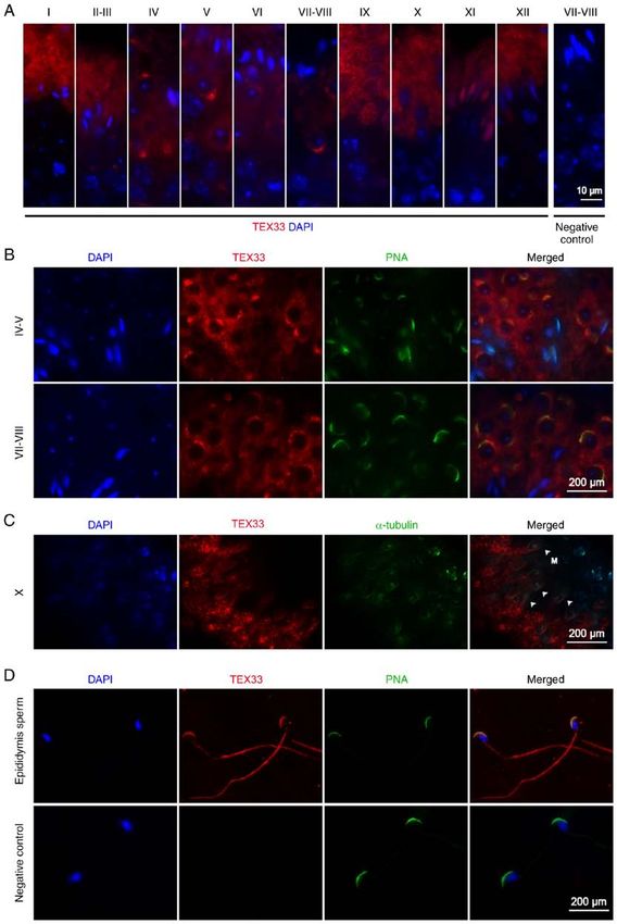
2                                            XIA et al: ROLES OF TEX33 IN SPERMIOGENESIS

    A previous study reported that >2,300 genes are expressed              All mice were maintained under specific pathogen-free
predominantly in mouse testes (13), and a number of them are           conditions with free access to standard mouse lab pellet food
identified as serving important roles in numerous events during        and water, and housed at 20-22˚C and 50-70% humidity with
spermiogenesis through gene-editing technology, such as the            a 12-h light/dark cycle. All animal experiments were approved
biogenesis of the acrosome, the morphology of the manchette            by the Animal Ethics Committee of Yangzhou University.
and the assembly of the flagellum. For example, mice lacking
autophagy related 7, sirtuin 1, serine-rich single-pass membrane       Reverse transcription-polymerase chain reaction (RT-PCR).
protein 1, vacuolar protein sorting (Vps)13b, Vps54 and                For RNA extraction, heart, brain, spleen, liver, lung, kidney
sperm acrosome associated 1 exhibit abnormal round-headed              and trachea were collected from adult wild-type mice (n=3)
sperm as a result of aberrant acrosome formation (8,9,14-16).          and testes were harvested from wild-type mice at different
Leucine-rich repeats and guanylate kinase domain containing            spermatogenesis stages (postnatal days 7, 14, 21, 28, 35 and 56;
isoform 1, serine/threonine kinase 22, katanin-like 2, sperm           n=3 each). Total RNAs were isolated on ice using TRIzol®
flagellar 2, hook microtubule tethering protein 1 and calcium          reagent (Invitrogen; Thermo Fisher Scientific, Inc.) and then
and integrin binding family member 4 are essential for the             dissolved in RNase-free distilled water. The purity of the total
correct formation of the manchette; deficiency in any of these         RNAs was determined using ultraviolet spectrophotometry
genes can lead to abnormal elongation of the manchette and             (Eppendorf) and agarose gel electrophoresis. Only RNA
thus, a malformed head or flagellum (11,12,17-20). In addi-            samples with a ratio of absorbance at 260 and 280 nm of ≥1.8
tion, the deletion of sperm associated antigen 16, kinesin             were used for RT-PCR. For reverse transcription, 500 ng total
light chain 3, kinase suppressor of ras 2, testis-specific serine      RNA in a final volume of 10 µl was mixed with 5X PrimeScript
kinase 4 or genes of the septin (Sep) family (Sept2, Sept4 and         RT Master Mix (Takara Bio, Inc.) and RNase-free distilled
Sept12) causes alterations in the intra-flagellum structure and        water. For cDNA synthesis, the mixture was incubated at 37˚C
reduces sperm motility (21-25). Despite the crucial function of        for 15 min, and then at 85˚C for 15 sec. Tex33 transcript vari-
these genes in spermiogenesis, the localization and functional         ants were then amplified by PCR using the following primers:
requirement of other genes requires further characterization.          Tex33 transcript variant V1: 5'-GGTTCATTTGCCTTCTTC
    Testis-expressed protein 33 (TEX33), also known as Ean57,          CC-3' (forward), 5'-TCCTTGTCGTTCGGGTGTG-3'
1700061J05Rik, cE81G9.2 and C22orf33, is evolutionarily                (reverse); Tex33 transcript variant V2: 5'-ACCTGGGGAGA
conserved. Orthologs of this protein have been identified in a         GCTGCTGCTCG-3' (forward), 5'-GTTGGTGGTAGAGGT
number of different species, including humans, mice, Norwegian         GGAG-3' (reverse); Tex33 transcript variant V3: 5'-TGGGTCA
rats, zebrafish and Rhesus monkeys. A previous bioinformatics          GGAAGGAGTGTG-3' (forward), 5'-GTTGGTGGTAG
analysis revealed that TEX33 was expressed abundantly in the           AGGTGGAG-3' (reverse). Glyceraldehyde 3-phosphate dehy-
testis and associated with the formation of cilia (26). Therefore,     drogenase (GAPDH) was used as a positive control using the
we hypothesized that TEX33 may be a testis-enriched or specific        following primer sequences: 5'-GACGGCCGCATCTTCT
protein that localizes to the sperm flagellum and is potentially       TGT-3' (forward) and 5'-ACACCGACCTTCACCATTTT
associated with spermiogenesis.                                        GT-3' (reverse). PCR was performed using a Taq DNA poly-
    The aim of the present study was to investigate the function       merase kit (cat. no. M1661S; Promega Corporation).
of TEX33 in mouse spermiogenesis and fertility using CRISPR/           Amplification conditions for Tex33 transcript variant V1, V2
Cas9 gene-editing. It is hoped that the results will enrich our        and V3 were as follows: V1, initial denaturation step at 95˚C
understanding of the molecular networks underlying spermio-            for 5 min followed by 35 cycles of at 95˚C for 30 sec, an
genesis and provide new insight into the genetics of male fertility.   annealing step at 60˚C for 45 sec, an extension step at 72˚C for
                                                                       45 sec, and a final extension at 72˚C for 10 min; V2, initial
Materials and methods                                                  denaturation at 95˚C for 5 min, followed by 35 cycles at 95˚C
                                                                       for 30 sec, 58˚C for 30 sec, 72˚C for 45 sec, and elongation at
Animals. A total of three male C57BL/6N mice aged                      72˚C for 10 min; V3, an initial denaturation step of 5 min at
10 weeks and two female C57BL/6N mice aged 8 weeks                     95˚C, followed by 35 cycles 95˚C for 30 sec, 52˚C for 30 sec,
were purchased from Cyagen Biosciences, Inc. The                       72˚C for 45 sec, ended by a final elongation at 72˚C for 10 min.
CRISPR/Cas9 system was used to generate Tex33 knockout                 PCR protocol for GAPDH included a 5 min denaturation step
mice. Two guide RNAs (gRNAs) were designed using the                   at 95˚C followed by 35 cycles of 50 sec at 95˚C, 1 min annealing
E-CRISPR database (http://www.e-crisp.org/E-CRISP/                     step at 55˚C and 1 min extension at 72˚C and a final elongation
designcrispr.html) to target exons 2-4 of Tex33: gRNA1,                at 72˚C for 10 min. PCR products were electrophoresed on
TACCAGAATCATCTAGTCCCTG G (match i ng t he                              1.2% agarose gels using 1X Tris/Borate/EDTA buffer (TBE).
forward strand of the gene) and gRNA2, GCTAGCCAAGG                     Gels were stained with SYBR Safe™ (Thermo Fisher
CCAACACCTGGG (matching the reverse strand of the                       Scientific, Inc.) and the bands visualized using a gel imaging
gene). The two gRNAs and Cas9 NLS endonuclease                         system (Tanon Science and Technology Co., Ltd.).
(cat. no. M0646M; New England BioLabs, Inc.) were micro-
injected into the cytoplasm of fertilized eggs from C57BL/6N           Synthesis of an anti-TEX33 antibody. PCR was used to
mice. These eggs were then implanted into the oviducts                 amplify the open reading fragment (ORF) of Tex33 transcript
of pseudo-pregnant female mice. Founder mice were then                 variant V2 (encoding 266 amino acids), which represents the
crossed with C57BL/6N mice to generate Tex33+/- offspring.             predominant transcript according to the GenBank database and
Finally, Tex33 -/- mice were obtained by breeding from the             shares the same ORF as V1. The following primers were used:
Tex33+/- mice.                                                         5'-GGGAATTCATGGAGCTGAGCCACCGAC-3' (forward)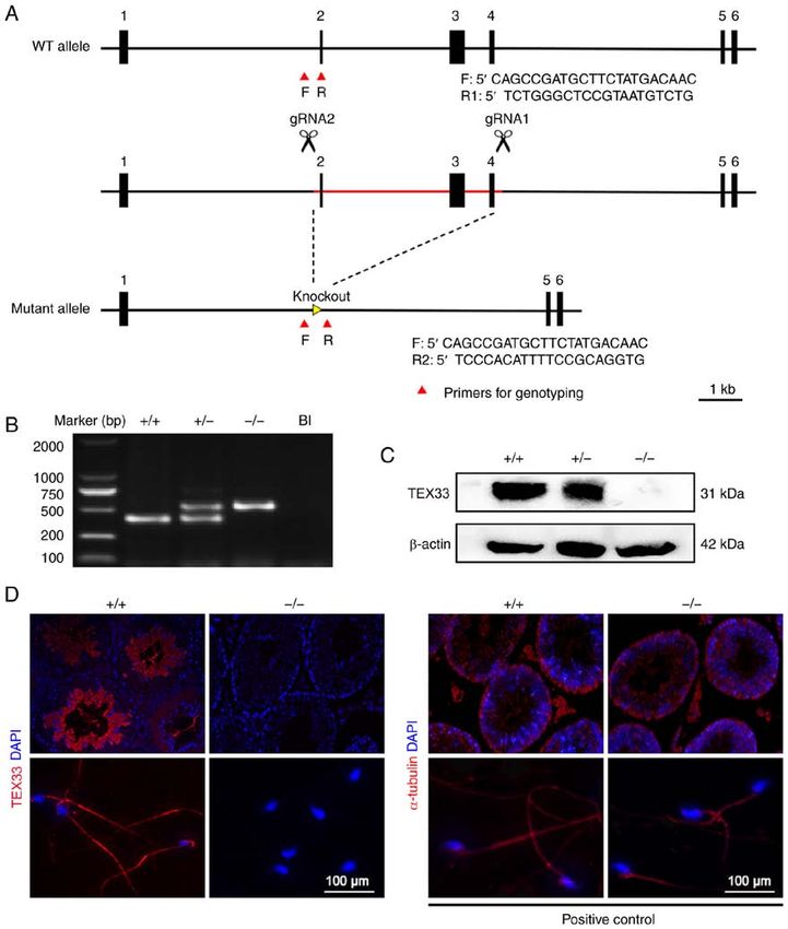
Molecular Medicine REPORTS 23: 317, 2021                                              3

and 5'-GGAAGCTTGCTTGGCCTTGGATCTGCTC-3'                            10% SDS-polyacrylamide gels and transferred onto poly-
(reverse). The PCR product was purified with a DNA gel            vinylidene fluoride membranes (Millipore; Merck KGaA).
extraction kit (Corning, Inc.), sequenced by next generation      Non-specific antibody binding was blocked using 5% non-fat
sequencing (BGI Group) and finally subcloned into a pET-30a       milk in Tris-buffered saline (TBS) for 1 h at room tempera-
(+) vector (Novagen; Merck KGaA). After induction with            ture. Membranes were then incubated at 4˚C overnight with
isopropyl β-D-thiogalactoside for 12-14 h at 37˚C, the prokary-   the following primary antibodies: TEX33 antibody (1:1,000
otic protein samples were analyzed by electrophoresis on          dilution; generated in the present study), GAPDH antibody
10% sodium dodecyl sulfate (SDS) polyacrylamide gels using        (cat. no. CW0100M; 1:2,500 dilution; CWBio) and β -actin
Coomassie Brilliant Blue solution. The TEX33 prokaryotic          antibody (cat. no. ET1701-80; 1:2,500 dilution; HuaBio).
protein was successfully expressed in Escherichia coli BL21       After washing with 0.05% TBS-T, membranes were incu-
(DE3) plysS (Novagen; Merck KGaA). The resulting His-tag          bated with goat-anti-rabbit IgG-HRP secondary antibody
fusion protein was purified using Ni-NTA affinity resin           (cat. no. ZB-2301; 1:2,500 dilution; OriGene Technologies,
(RuiXiang Bio Inc.) in accordance with the manufacturer's         Inc.) for 1 h at room temperature. After washing three times
instructions. The concentration of the purified protein was       with TBS-T, protein bands were visualized with enhanced
determined using a bicinchoninic acid (BCA) kit (CWBio).          chemiluminescence reagent (New Cell and Molecular Biotech
     A total of 5 male New Zealand white rabbits (~2 kg body      Co., Ltd.) using an automatic chemiluminescence image
weight) aged 10 weeks were used for animal immuniza-              analysis system (Tanon Science and Technology Co., Ltd.).
tion. They were purchased from Laboratory Animal Center
of Shanghai Branch of Chinese Academy of Science and              Immunofluorescence analysis. In order to analyze tissue
maintained in individual cages at 22˚C, 42% humidity, with        sections by immunofluorescence, testicular samples were
a 12-h light/dark cycle and free access to food and water.        collected from the mice and fixed in 4% paraformaldehyde
Purified protein (0.75 mg/ml) was mixed with an equal             solution (pH 7.0) for 24-48 h at 4˚C. The testicular samples
volume of Freund's adjuvant (Sigma-Aldrich; Merck KGaA)           were dehydrated in an ascending alcohol gradient, then
and 500 µl purified protein/adjuvant mixture was subsequently     cleared in chloroform overnight. The cleared samples were
injected into the backs of adult male rabbits at 0, 2, 4, 6 and   infiltrated in xylene, embedded in paraffin and cut into 4-µm
8 weeks. After the last immunization, blood was harvested         sections. The sections were rehydrated in a descending alcohol
from each rabbit and the serum was separated. The antibody        gradient followed by antigen retrieval in 10 mM sodium citrate
titer from each rabbit was determined using an enzyme-linked      (pH 6.0) at 100˚C for 10 min. After blocking with 1% BSA
immunosorbent assay (ELISA; Fig. S1A).                            for 1 h at room temperature, the sections were incubated
     For ELISA, 5 µg/ml purified TEX33 prokaryotic protein        with primary antibodies at 4˚C overnight. The following
was added to each well of a 96-well plate (Corning Inc.)          primary antibodies were used: TEX33 antibody (1:200 dilu-
and incubated at 4˚C overnight. After washing with 0.05%          tion; generated in this study) and mouse α-tubulin antibody
Tween-20 solution in phosphate-buffered saline (PBS-T),           (cat. no. AC012; 1:100 dilution; ABclonal Biotech Co., Ltd.).
each well was blocked with 1% bovine serum albumin (BSA);         After washing with PBS, sections were further incubated with
Beijing Solarbio Science & Technology Co., Ltd.) for 1 h          Alexa Fluor 488-conjugated donkey-anti-mouse secondary
at room temperature. After washing, 1:10, 1:100, 1:1,000,         antibody (cat. no. A32766TR; 1:1,000 dilution; Thermo Fisher
1:10,000, 1:100,000, and 1:1,000,000 dilutions of serum were      Scientific, Inc.) and Alexa Fluor 555-conjugated donkey-anti-
added and the plate was incubated at 37˚C for 1 h. The wells      rabbit secondary antibody (cat. no. A-31572; 1:1,000 dilution;
were then washed thrice with PBS-T and incubated with goat-       Thermo Fisher Scientific, Inc.) for 1 h at room temperature,
anti-rabbit IgG-HRP secondary antibody (cat. no. ZB-2301;         and then counterstained with 4',6-diamidino-2-phenylindole
1:2,500 dilution; OriGene Technologies, Inc.) at 37˚C for 1 h.    (DAPI) dye (cat. no. C0060; 1:1,000 dilution; Beijing Solarbio
TMB Two-Component Substrate solution (Beijing Solarbio            Science & Technology Co., Ltd.) for 10 min at room tempera-
Science & Technology Co., Ltd.) was then added and allowed        ture. Fluorescence intensity was finally detected by confocal
to react for 10 min at room temperature in the dark. The          laser-scanning microscopy (Carl Zeiss AG).
reaction was stopped with 1 mM hydrochloric acid and the              In order to investigate sperm using immunocytochem-
absorbance was measured at 450 nm using a Multiskan FC            istry, the cauda epididymis of each mouse was cut into small
microplate reader (Thermo Fisher Scientific, Inc.). The speci-    pieces to allow motile sperm to swim out. After filtration and
ficity of the antibody was confirmed by western blot (Fig. S1B)   centrifugation at 800 x g for 15 min at room temperature,
and immunofluorescence analyses (Fig. S1C-F).                     the sperm sample was rinsed with PBS, incubated in PBS at
                                                                  37˚C for 15 min, smeared on slides and fixed with 4% para-
Western blot analysis. For protein extraction, heart, brain,      formaldehyde solution for 30 min at room temperature. The
spleen, liver, lung, kidney and trachea were collected from       slides were blocked with donkey serum (cat. no. SL050;
multiple tissues of adult wild-type mice (n=3) and testes         1:100 dilution; Beijing Solarbio Science & Technology Co.,
were harvested from wild-type mice at different spermato-         Ltd.) at room temperature for 1 h, and then incubated with
genesis stages (postnatal days 7, 14, 21, 28, 35 and 56; n=3      anti-TEX33 primary antibody (1:100 dilution; generated in the
each). Tissues were lysed in RIPA buffer (CWBio) on ice,          present study) at 4˚C overnight. This was followed by incuba-
followed by sonication on ice for 10 min. After centrifuga-       tion with FITC-conjugated peanut agglutinin (PNA-FITC;
tion at 14,000 x g for 10 min at 4˚C, the supernatants were       cat. no. L7381; 1:50 dilution; Sigma-Aldrich; Merck KGaA)
collected. The protein concentration was determined using         and Alexa Fluor 555 donkey-anti-rabbit secondary antibody
a BCA kit. Protein samples (40 µg) were then separated on         (cat. no. A-31572; 1:1,000 dilution). After washing three times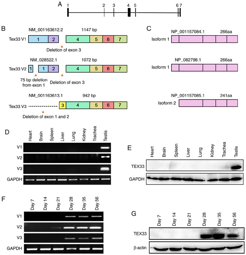
4                                         XIA et al: ROLES OF TEX33 IN SPERMIOGENESIS

with PBS, the nuclei were stained with DAPI for 10 min at          warm human tubal fluid (Wisent Biotechnology) at 37˚C. The
room temperature. Images were captured by confocal laser-          total sperm count was determined using a hemocytometer
scanning microscopy.                                               and sperm motility was determined with a computer-assisted
                                                                   sperm analysis system (CASA; Sperm Class Analyzer v. 4.0.0;
Mouse genotyping. Mouse tail samples (0.2-0.3 cm) were             Microptic S.L., Barcelona, Spain). The sperm deformity rate
obtained and lysed at 55˚C overnight in lysis buffer containing    was studied using sperm smears and microscopy counting
1 mM Tris-HCl (pH 8.0), 1 mM NaCl, 1 mM EDTA (pH 8.0),             methods. Sperm smears were stained with H&E and images
0.5% SDS and 10 µg/µl proteinase K (Sigma-Aldrich; Merck           were captured by light microscopy (Nikon-70i). More than
KGaA). Genomic DNA was extracted from the lysate using             1,000 sperm were observed and counted from Tex33+/+ and
phenol and chloroform (Beijing Solarbio Science & Technology       Tex33 -/- males (n=3 mice/group). The numbers of sperm
Co., Ltd.), precipitated with isopropanol (Sinopharm Chemical      showing normal and abnormal morphology were counted and
Reagent Co., Ltd.) and dissolved in distilled water at 60˚C.       analyzed.
Subsequent PCR was performed using a Taq DNA poly-
merase kit (cat. no. M1661S; Promega Corporation) according        Fertility testing. Tex33+/+ and Tex33-/- male mice (10-12 weeks
to the manufacturer's instruction. Primers for genotyping          old) were continuously mated with Tex33+/+ female mice
were designed: 5'-CAGCCGATGCTTCTATGACAAC-3'                        (8-10 weeks old) with demonstrable fertilizing ability (at
(forward), 5'-TCCCACATTTTCCGCAGGTG-3' (reverse 1),                 a ratio of 1:2) for 1 month. The Tex33+/+ female mice were
5'-TCTGGGCTCCGTAATGTCTG-3' (reverse 2). The PCR                    changed once a week. Plugs and the numbers of pups born
conditions were as follows: Denaturation at 94˚C for 5 min,        were recorded.
followed by 35 cycles at 94˚C for 50 sec, 55˚C for 1 min, 65˚C
for 50 sec, and a final extension at 65˚C for 10 min. PCR prod-    Statistical analysis. Sperm quality and average litter sizes are
ucts were separated on 1.2% agarose gels by electrophoreses        expressed as mean ± standard error of mean and the Student’s
in 1X TBE buffer. The products were visualized using a gel         t-test was used to identify significant differences. Plugs and
imaging system (Tanon Science and Technology Co., Ltd.)            pregnancies were compared between Tex33+/+ and Tex33 -/-
after staining with SYBR Safe™ (Thermo Fisher Scientific,          mice using the Chi-squared test. P
Molecular Medicine REPORTS 23: 317, 2021 5
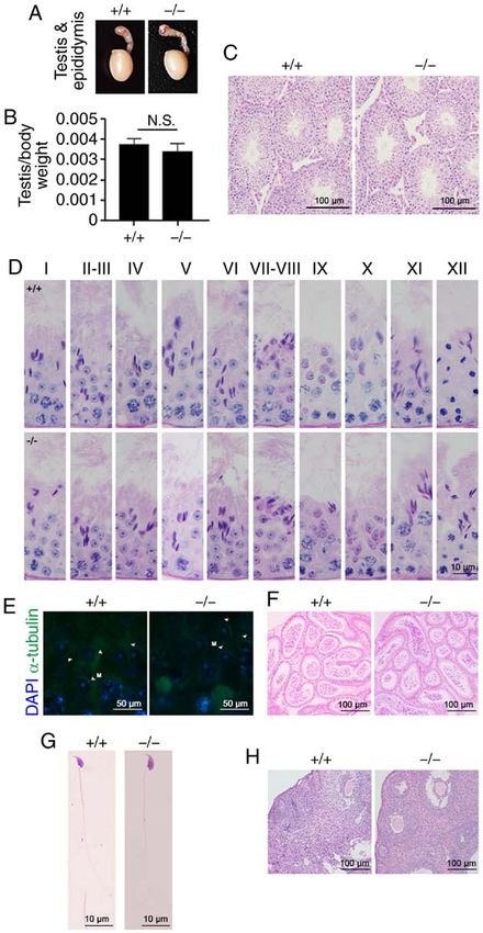
Figure 1. Characterization of mouse Tex33. (A) Analysis of the Tex33 gene in mice. The Tex33 gene consists of seven exons. (B) Tex33 transcript variants in
mice. Deleted regions reveal alternate splicing events and different combinations of exons. Transcript variant V1 contains six exons, transcript variant V2 has
seven exons and transcript variant V3 has five exons. (C) Two different isoforms are encoded by transcript variants V1, V2 and V3. (D) mRNA levels of Tex33
transcript variants in various mouse tissues were analyzed by RT-PCR. Transcript variant V1, 220 bp; transcript variant V2, 348 bp; transcript variant V3,
261 bp. (E) Expression of TEX33 protein in various mouse tissues, as determined by western blot analysis. (F) Expression of Tex33 transcript variants during
the first wave of spermatogenesis, as determined by RT-PCR. (G) Expression of TEX33 protein during the first wave of spermatogenesis, as determined by
western blot analysis. TEX33, testis-expressed protein 33; RT-PCR, reverse transcription-polymerase chain reaction.
consistent with the expression profiles of the transcript vari- hood, while V1 and V3 were expressed from day 28 and
ants (Fig. 1E). maintained peak expression levels into adulthood (Fig. 1F).
To determine the expression profile of Tex33 during the Western blotting showed that TEX33 protein first presented
first wave of spermatogenesis, mouse testes were obtained on day 28 and maintained high expression levels into adult-
at postnatal day 7 (containing only spermatogonia), day 14 hood (Fig. 1G). The appearance of Tex33 transcript variants
(containing spermatogonia and spermatocytes), day 21 and proteins coincides with the spermiogenesis phase,
(in which round spermatids begin to appear), day 28 (in during which time the appearance of round spermatids
which spermatids begin to elongate), day 35 (around which begins and spermiogenesis is initiated (28).
time the first wave of spermatogenesis is completed) and Immunofluorescence analysis demonstrated that TEX33
day 56 (3,28). The results of RT-PCR analysis suggested protein was localized in spermatids and testicular sperm
that V2 was expressed weakly from day 21 in the mouse within the seminiferous tubules (Fig. 2A). In spermatids,
testes, peaked at day 28 and was expressed into adult- TEX33 was detected in Golgi-derived pro-acrosomal granules,6 XIA et al: ROLES OF TEX33 IN SPERMIOGENESIS Figure 2. Localization of TEX33 during spermatogenesis. (A) Immunofluorescence staining of TEX33 in the seminiferous tubules during different stages of spermatogenic development (stages I-XII). (B) Co-immunofluorescence staining of TEX33 with PNA-FITC in the seminiferous tubules during different stages of spermatogenic development. (C) Co-immunofluorescence staining of TEX33 and α-tubulin in elongating spermatids during spermatogenic stage X. (D) Co-immunofluorescence staining of TEX33 with PNA-FITC in sperm. TEX33, testis-expressed protein 33; PNA-FITC, FITC-conjugated peanut agglu- tinin; DAPI, 4',6-diamidino-2-phenylindole; M, manchette (indicated by arrowheads). the cap-like acrosome and the fully formed acrosome from mammalian species (29,30), such as peanut agglutinin (PNA), spermatogenic stages I to XII (Fig. 2A). It has been reported which reacts specifically with acrosomal sugar components that the acrosome could bind many lectins in a variety of and is widely used to visualize acrosomal formation (31-35).
Molecular Medicine REPORTS 23: 317, 2021                                                               7

Figure 3. Generation of Tex33 knockout mice. (A) Schematic representation of the deletion of Tex33 exons 2-4 via Cas9 microinjection. Primers were designed
for genotyping. F targets intron 1, R1 targets exon 2 and R2 targets intron 4. (B) Genotyping of Tex33 offspring by PCR assay. (C) Comparison of TEX33
knockout efficiency between Tex33+/+, Tex33+/- and Tex33-/- mice by western blot analysis. (D) Detection of TEX33 expression in testes (upper panels) and
sperm (lower panels) by immunofluorescence analysis. WT, wild-type; F, forward; R, reverse; gRNA, guide RNA; TEX33, testis-expressed protein 33; +/+,
Tex33+/+ (wild-type) mice; +/-, Tex33+/- (heterozygous) mice; -/-, Tex33-/- (homozygote) mice; Bl, blank; DAPI, 4',6-diamidino-2-phenylindole.

In the present study, TEX33 was co-localized with PNA on                       this, the biological function of TEX33 during spermatogenesis
the acrosomes during different stages of spermatid develop-                    was investigated. CRISPR/Cas9-mediated genome engi-
ment (Fig. 2B) and on mature acrosomes (Fig. 2D). TEX33                        neering was used to create Tex33 knockout mice. Exons 2, 3
was also detected on the manchette of elongating spermatids                    and 4 were selected as the target sites. Cas9 and gRNA were
(spermatogenic stage X) via α-tubulin co-labeling (Fig. 2C), as                co-injected into fertilized eggs in order to delete exons 2-4
well as the sperm flagellum (Fig. 2D). These findings indicate                 (Fig. 3A). Mice were then genotyped by PCR using specific
that TEX33 may be involved in the formation of the acrosome,                   primers (Fig. 3A and B). Western blot analysis was performed
manchette and flagellum during spermiogenesis.                                 on testis samples to validate the efficiency of Tex33 removal.
                                                                               As shown in Fig.3C, TEX33 protein was present in the
Deletion of Tex33 in mice. The initial data indicate that TEX33                Tex33+/+ and Tex33+/- mice, but absent from the Tex33-/- mice.
is expressed at high levels in mouse testes. On the basis of                   Furthermore, immunofluorescence analysis of the testis and
8 XIA et al: ROLES OF TEX33 IN SPERMIOGENESIS Figure 4. Phenotypic analysis of Tex33 knockout mice. (A) Testis and epididymis from Tex33+/+ and Tex33 -/- males. Tissue samples were collected from 10-week-old male mice (n=5 per genotype). (B) Testis/body weight ratio of Tex33+/+ and Tex33-/- males. Values are expressed as mean ± standard error of mean. (C) H&E staining showing seminiferous tubules with normal histology in Tex33+/+ and Tex33-/- males. (D) Periodic acid-Schiff staining showing normal acro- some development and spermatogenesis in Tex33+/+ and Tex33-/- males. (E) Co-immunofluorescence staining of TEX33 and α-tubulin in elongating spermatids revealed normal manchette morphology in both Tex33+/+ and Tex33-/- males. H&E staining of Tex33+/+ and Tex33-/- males showing normal (F) cauda epididymis and (G) sperm. (H) H&E staining revealed normal ovarian morphology in Tex33+/+ and Tex33-/- females. H&E, hematoxylin and eosin; Tex33, testis-expressed protein 33; +/+, Tex33+/+ (wild-type) mice; -/-, Tex33-/- (homozygote) mice; M, manchette (indicated by arrowheads); DAPI, 4',6-diamidino-2-phenylindole; N.S., not significant. sperm confirmed that TEX33 expression was absent from the Tex33 deletion does not affect spermatogenesis and fertility. Tex33-/- mice (Fig. 3D). To determine whether the deletion of Tex33 has an effect on
Molecular Medicine REPORTS 23: 317, 2021                                                        9

Table I. Analysis of sperm quality in Tex33+/+ and Tex33-/- male mice.

Genotype         Sperm count (x106/ml)          Total sperm motility (%)          Progressive motility (%)          Sperm deformation (%)

+/+ (n=3)               9.02±0.46                       80.82±3.69                        57.59±5.06                       16.31±3.76
-/- (n=3)               8.98±0.35                       77.96±4.51                        58.26±3.47                       18.12±4.08
P-value                    0.91                            0.47                              0.86                             0.60

Data are expressed as mean ± standard error of the mean and were analyzed using Student's t-test. Tex33, testis-expressed protein 33; +/+,
Tex33+/+ (wild-type) males; -/-, Tex33-/- (homozygous) males.

Table II. Fertility analysis of Tex33+/+ and Tex33-/- mice.

Test                                 Males                        Females	Average litter size                                         P-value

Male fertility                      +/+ (n=8)                   +/+ (n=64)                         8.69±0.37                           0.13
                                    -/- (n=8)                   +/+ (n=64)                         8.41±0.34
Female fertility                    +/+ (n=5)                   +/+ (n=12)                         8.67±0.86                           0.61
                                    +/+ (n=5)                   -/- (n=12)                         8.92±0.80

Data are expressed as mean ± standard error of the mean and were analyzed using the Student's t-test. Tex33, testis-expressed protein 33; +/+,
Tex33+/+ (wild-type) mice; -/-, Tex33-/- (homozygous) mice.

Table III. Comparison of pregnancies in Tex33+/+ and Tex33-/- mice.

Test                                  Males                        Females                      Pregnancies (%)                       P-value

Male fertility                      +/+ (n=8)                    +/+ (n=64)                           90.00                            0.23
                                    -/- (n=8)                    +/+ (n=64)                           94.64
Female fertility                    +/+ (n=5)                    +/+ (n=12)                           81.81                            0.59
                                    +/+ (n=5)                    -/- (n=12)                           90.00

Data were analyzed using Chi-square tests. Tex33, testis-expressed protein 33; +/+, Tex33+/+ (wild-type) mice; -/-, Tex33-/- (homozygous) mice.

fertility, functional analyses were then carried out on Tex33            Tex33-/- mice (Fig. 4F). In addition, the morphology of sperm
knockout mice. As shown in Fig. 4A, the sizes of the testes              exhibited no clear differences between the Tex33+/+ and
and epididymis were comparable between adult Tex33+/+                    Tex33-/- mice (Fig. 4G and Table I). Sperm were counted and
and Tex33 -/- males. The testis-to-body weight ratios were               their motility assessed to reveal the effect of Tex33 knockout
not significantly different between the Tex33+/+ and Tex33-/-            on sperm quality. Sperm counts were determined using a
male mice (Fig. 4B). The process of sperm development                    counting board while motility was determined by a CASA
in the testis was examined by H&E staining. The results                  analysis that provided total motility and progressive motility
revealed that Tex33+/+ and Tex33 -/- mice exhibited seminif-             data. These investigations revealed no detectable differences
erous tubules with normal morphology and spermatogenic                   in either sperm count or motility between the Tex33+/+ and
cells (Fig. 4C).                                                         Tex33-/- mice (Table I).
    PAS staining was then performed to analyze the process of                A breeding assay was performed to evaluate the fertility
acrosome development during spermatogenesis. No apparent                 of the Tex33 knockout mice. Tex33+/+ and Tex33-/- male mice
morphological differences were observed between the testes               (10-12 weeks old) were mated separately with female mice
of Tex33+/+ and Tex33-/- mice in spermatogenic stages I-XII              (8-10 weeks old) for 1 month. The Tex33+/+ and Tex33-/- male
(Fig. 4D). The morphology of the manchette was evaluated by              mice exhibited normal fertility (Tables II-IV), and the litter
α-tubulin immunofluorescence staining and immunostaining                 sizes sired by Tex33 -/- mice were similar to those sired by
was identified in elongating spermatids in the seminiferous              Tex33+/+ mice (Table II). These results indicate that the
tubules of Tex33+/+ and Tex33-/- male mice at spermatogenic              fertilizing ability of Tex33-/- male mice was not significantly
stage IX (Fig. 4E). H&E staining revealed the presence of                different from that of Tex33+/+ mice. No marked differences
mature sperm in the cauda epididymides of Tex33+/+ and                   in female fertility were observed between the two groups of
10 XIA et al: ROLES OF TEX33 IN SPERMIOGENESIS
Table IV. Comparison of plugs in Tex33+/+ and Tex33-/- mice.
Test Males Females Plugs (%) P-value
Male fertility +/+ (n=8) +/+ (n=64) 93.75 0.35
-/- (n=8) +/+ (n=64) 87.50
Female fertility +/+ (n=5) +/+ (n=12) 91.67 0.54
+/+ (n=5) -/- (n=12) 83.33
Data were analyzed using Chi-square tests. Tex33, testis-expressed protein 33; +/+, Tex33+/+ (wild-type) mice; -/-, Tex33-/- (homozygous) mice.
mice, as the Tex33-/- female mice exhibited normal ovarian was first detected at day 28, and localized on the acrosome
structures (Fig. 4H), underwent normal pregnancies and at different developmental stages, including at maturity.
produced equivalent litter sizes to the Tex33+/+ female mice Furthermore, it was also detected on the manchette of elon-
(Tables II and III). gating spermatids during spermatogenesis stages IX and X,
as well as on the sperm flagellum. These results confirm that
Discussion Tex33 is not only associated with cilia, as previously reported,
but is also testis-specific and associated with spermiogenesis.
Mature sperm is the end product of spermiogenesis and Thus, Tex33 may serve a crucial role in mouse spermiogen-
exhibits highly specialized morphology. Spermiogenesis is esis, particularly in terms of acrosome biogenesis, manchette
facilitated by an important microtubule structure referred morphology and flagellum formation.
to as the manchette, which appears in elongating spermatids Considering the enrichment and cellular localization of
and helps to create the shape of the sperm head and transport TEX33 in the testis, Tex33 knockout mice were generated
proteins to the flagellum (2,11). Failure in spermiogenesis, by application of the CRISPR/Cas9 system, as reported by
commonly results in subfertility or infertility due to sperm Miyata et al (44). The association between TEX33 and sper-
malformation and/or dysfunction (36,37). A number of matid elongation was then explored. Exons 2-4 were selected
genes have been reported to be responsible for spermiogen- as targets to ensure the non-functionality of Tex33 after
esis (38-42); however, the expression profiles of other genes knockout. The deletion was confirmed by genotyping, western
and their respective roles in spermiogenesis remain unknown blotting and immunofluorescence analysis.
and require further investigation. In the present study, Tex33, Recent studies have identified that numerous testis-
a gene previously shown to be associated with cilia forma- enriched/specific genes are necessary for male fertility,
tion (26), was demonstrated for the first time, to the best of our including CFAP97 domain containing 1, kinesin family
knowledge, to be testis-specific and associated with spermio- member 9, intraflagellar transporter protein 140, protease
genesis. Its role in spermatogenesis was investigated further serine 55 and RIKEN cDNA 1700102P08 (39,40,45-47). Mice
using a Tex33 knockout mouse model. lacking any of these genes have been shown to be sterile due
In mammals, primary RNA transcripts give rise to to a failure to complete spermatogenesis. However, the Tex33
different transcript variants through alternative splicing, knockout mice were found to be fertile. Further analyses of
thereby resulting in different mRNA and protein isoforms that testis and cauda epididymis histology, acrosome biogenesis,
may have distinct functions and localization patterns (43). manchette morphology and sperm quality also indicated that
Analysis of GenBank data revealed that Tex33 pre-mRNA there were no significant differences between Tex33-/- mice
undergoes alternative splicing to produce three transcripts. and the controls. The possible reason for these knockout mice
Exon 3 is present in V3, but lacking from V1 and V2; this exhibiting normal spermatogenesis and fertility may be func-
may be due to exon skipping. Similarly, exon skipping may tional redundancy (48-50). Paralogous genes may compensate
have contributed to the deletion of exons 1 and 2 from V3. for the loss of function of knockout genes when they contribute
In addition, unlike exon 1 of V1, exon 1 of V2 has a 75-bp to single function (50). Likewise, it is plausible that Tex33
deletion. The results of RT-PCR analysis revealed that all paralogs, such as Tex11 and Tex14, have compensational func-
three transcript variants are testis specific. Furthermore, tions in the absence of Tex33.
TEX33 protein was also detected only in the testis, indicating The function of Tex33 during mouse spermatogenesis
that the Tex33 gene is testis specific and conserved from has been preliminarily clarified using CRISPR/Cas9 system
transcription to RNA translation. However, the distribution in the present study. However, considering the cellular and
of this gene in the testis has not previously been elucidated. subcellular localization of Tex33 on sperm and the acrosome,
It has been reported that Tex33 is associated with cilia forma- further investigations using transmission electron microscopy
tion; therefore, we hypothesized that the TEX33 protein may or in vitro fertilization experiments are recommended.
localize on the sperm flagellum. RT-PCR analysis revealed In summary, the present study preliminarily describes the
that the expression of Tex33 transcript variants began during expression profile and role of TEX33 during spermatogenesis
spermiogenesis, between days 21 and 28, when round sper- in mice. Although Tex33 was testis-specific and associated
matids begin to appear and undergo morphological changes. with spermiogenesis, deletion of this gene had no effect on
Consistent with this finding, the expression of TEX33 protein sperm development and reproduction in mice. This indicatesMolecular Medicine REPORTS 23: 317, 2021 11
that Tex33 may regulate spermiogenesis in association with 7. Kistler WS, Baas D, Lemeille S, Paschaki M, Seguin-Estevez Q,
Barras E, Ma W, Duteyrat JL, Morlé L, Durand B, et al: RFX2
cooperating factors; however, it is not required for spermio- is a major transcriptional regulator of spermiogenesis. PLoS
genesis and male fertility. Genet 11: e1005368, 2015.
8. Wang H, Wan H, Li X, Liu W, Chen Q, Wang Y, Yang L, Tang H,
Acknowledgements Zhang X, Duan E, et al: Atg7 is required for acrosome biogenesis
during spermatogenesis in mice. Cell Res 24: 852-869, 2014.
9. Liu C, Song Z, Wang L, Yu H, Liu W, Shang Y, Xu Z, Zhao H,
Not applicable. Gao F, Wen J, et al: Sirt1 regulates acrosome biogenesis by
modulating autophagic flux during spermiogenesis in mice.
Development 144: 441-451, 2017.
Funding 10. Khawar MB, Gao H and Li W: Mechanism of acrosome
biogenesis in mammals. Front Cell Dev Biol 7: 195, 2019.
The present study was supported by the National Natural 11. Okuda H, DeBoer K, O'Connor AE, Merriner DJ, Jamsai D
Science Foundation of China (grant no. 81871205), the and O'Bryan MK: LRGUK1 is part of a multiprotein complex
required for manchette function and male fertility. FASEB J 31:
Postgraduate Research & Practice Innovation Program of 1141-1152, 2017.
Yangzhou University (grant no. XKYCX18_129) and the 12. Martins LR, Bung RK, Koch S, Richter K, Schwarzmüller L,
Undergraduate Science & Technology Innovation Program of Terhardt D, Kurtulmus B, Niehrs C, Rouhi A, Lohmann I, et al:
Stk33 is required for spermatid differentiation and male fertility
Yangzhou University (grant no. 20180744). in mice. Dev Biol 433: 84-93, 2018.
13. Schultz N, Hamra FK and Garbers DL: A multitude of genes
Availability of data and materials expressed solely in meiotic or postmeiotic spermatogenic cells
offers a myriad of contraceptive targets. Proc Natl Acad Sci USA
100: 12201-12206, 2003.
The datasets used and/or analyzed during the current study are 14. Nozawa K, Zhang Q, Miyata H, Devlin DJ, Yu Z, Oura S,
available from the corresponding author on reasonable request. Koyano T, Matsuyama M, Ikawa M and Matzuk MM: Knockout
of serine-rich single-pass membrane protein 1 (Ssmem1) causes
globozoospermia and sterility in male mice. Biol Reprod 103:
Authors' contributions 244-253, 2020.
15. Da Costa R, Bordessoules M, Guilleman M, Carmignac V,
MX, JX and CN designed and performed the experiments. Lhussiez V, Courot H, Bataille A, Chlémaire A, Bruno C,
Fauque P, et al: Vps13b is required for acrosome biogenesis
MX wrote the manuscript. YaZ collected and analyzed the through functions in Golgi dynamic and membrane trafficking.
data. MX, TG and YD generated knockout mice and the Cell Mol Life Sci 77: 511-529, 2020.
TEX33 antibody. YiZ designed the study. All authors read and 16. Paiardi C, Pasini ME, Gioria M and Berruti G: Failure of
approved the final manuscript. acrosome formation and globozoospermia in the wobbler mouse,
a Vps54 spontaneous recessive mutant. Spermatogenesis 1:
52-62, 2011.
Ethics approval and consent to participate 17. Dunleavy JEM, Okuda H, O'Connor AE, Merriner DJ,
O'Donnell L, Jamsai D, Bergmann M and O'Bryan MK:
Katanin-like 2 (KATNAL2) functions in multiple aspects
All animal experiments were approved by the Animal Ethics of haploid male germ cell development in the mouse. PLoS
Committee of Yangzhou University. Genet 13: e1007078, 2017.
18. Lehti MS, Zhang FP, Kotaja N and Sironen A: SPEF2 functions
Patient consent for publication in microtubule-mediated transport in elongating spermatids to
ensure proper male germ cell differentiation. Development 144:
2683-2693, 2017.
Not applicable. 19. Schwarz T, Prieler B, Schmid JA, Grzmil P and Neesen J:
Ccdc181 is a microtubule-binding protein that interacts with
Competing interests Hook1 in haploid male germ cells and localizes to the sperm tail
and motile cilia. Eur J Cell Biol 96: 276-288, 2017.
20. Xu Z, Miyata H, Kaneda Y, Castaneda JM, Lu Y, Morohoshi A,
The authors declare that they have no competing interests. Yu Z, Matzuk MM and Ikawa M: CIB4 is essential for the
haploid phase of spermatogenesis in mice. Biol Reprod 103:
235-243, 2020.
References 21. Zhang Z, Kostetskii I, Tang W, Haig-Ladewig L, Sapiro R, Wei Z,
Patel AM, Bennett J, Gerton GL, Moss SB, et al: Deficiency of
1. Bao J, Tang C, Li J, Zhang Y, Bhetwal BP, Zheng H and Yan W: SPAG16L causes male infertility associated with impaired sperm
RAN-binding protein 9 is involved in alternative splicing and is motility. Biol Reprod 74: 751-759, 2006.
critical for male germ cell development and male fertility. PLoS 22. Zhang Y, Ou Y, Cheng M, Saadi HS, Thundathil JC and van der
Genet 10: e1004825, 2014. Hoorn FA: KLC3 is involved in sperm tail midpiece formation
2. Lehti MS and Sironen A: Formation and function of the and sperm function. Dev Biol 366: 101-110, 2012.
manchette and flagellum during spermatogenesis. Reproduction 23. Kuo YC, Shen YR, Chen HI, Lin YH, Wang YY, Chen YR,
151: R43-R54, 2016. Wang CY and Kuo PL: SEPT12 orchestrates the formation
3. Li RK, Tan JL, Chen LT, Feng JS, Liang WX, Guo XJ, Liu P, of mammalian sperm annulus by organizing core octameric
Chen Z, Sha JH, Wang YF, et al: Iqcg is essential for sperm complexes with other SEPT proteins. J Cell Sci 128: 923-934,
flagellum formation in mice. PLoS One 9: e98053, 2014. 2015.
4. Nishimura H and L'Hernault SW: Spermatogenesis. Curr Biol 27: 24. Moretti E, Collodel G, Mazzi L, Russo I and Giurisato E:
R988-R994, 2017. Ultrastructural study of spermatogenesis in KSR2 deficient mice.
5. Kazarian E, Son H, Sapao P, Li W, Zhang Z, Strauss JF III and Transgenic Res 24: 741-751, 2015.
Teves ME: SPAG17 is required for male germ cell differentiation 25. Wang X, Wei Y, Fu G, Li H, Saiyin H, Lin G, Wang Z, Chen S and
and fertility. Int J Mol Sci 19: 1252, 2018. Yu L: Tssk4 is essential for maintaining the structural integrity of
6. Bryant JM, Donahue G, Wang X, Meyer-Ficca M, Luense LJ, sperm flagellum. Mol Hum Reprod 21: 136-145, 2015.
Weller AH, Bartolomei MS, Blobel GA, Meyer RG, Garcia BA, 26. McClintock TS, Glasser CE, Bose SC and Bergman DA:
et al: Characterization of BRD4 during mammalian postmeiotic Tissue expression patterns identify mouse cilia genes. Physiol
sperm development. Mol Cell Biol 35: 1433-1448, 2015. Genomics 32: 198-206, 2008.12 XIA et al: ROLES OF TEX33 IN SPERMIOGENESIS
27. Xia M, Xia J, Yang D, Liu M, Niu C, Shen X, Sun H and Zheng Y: 40. Miyata H, Shimada K, Morohoshi A, Oura S, Matsumura T,
Preparation and application of rabbit polyclonal antibody against Xu Z, Oyama Y and Ikawa M: Testis-enriched kinesin KIF9
mouse Tex33. Xi Bao Yu Fen Zi Mian Yi Xue Za Zhi 34: 643-649, is important for progressive motility in mouse spermatozoa.
2018 (In Chinese). FASEB J 34: 5389-5400, 2020.
28. Busada JT, Velte EK, Serra N, Cook K, Niedenberger BA, 41. Zhang L, Zhen J, Huang Q, Liu H, Li W, Zhang S, Min J, Li Y,
Willis WD, Goulding EH, Eddy EM and Geyer CB: Rhox13 is Shi L, Woods J, et al: Mouse spermatogenesis-associated
required for a quantitatively normal first wave of spermatogenesis protein 1 (SPATA1), an IFT20 binding partner, is an acrosomal
in mice. Reproduction 152: 379-388, 2016. protein. Dev Dyn 249: 543-555, 2020.
29. Lee MC and Damjanov I: Lectin binding sites on human sperm 42. Lehti MS and Sironen A: Formation and function of sperm
and spermatogenic cells. Anat Rec 212: 282-287, 1985. tail structures in association with sperm motility defects. Biol
30. Arya M and Vanha-Perttula T: Comparison of lectin-staining Reprod 97: 522-536, 2017.
pattern in testis and epididymis of gerbil, guinea pig, mouse, and 43. Wang ET, Sandberg R, Luo S, Khrebtukova I, Zhang L, Mayr C,
nutria. Am J Anat 175: 449-469, 1986. Kingsmore SF, Schroth GP and Burge CB: Alternative isoform
31. Wakayama T, Nakata H, Kumchantuek T, Gewaily MS and regulation in human tissue transcriptomes. Nature 456: 470-476,
Iseki S: Identification of 5-bromo-2'-deoxyuridine-labeled cells 2008.
during mouse spermatogenesis by heat-induced antigen retrieval 44. Miyata H, Castaneda JM, Fujihara Y, Yu Z, Archambeault DR,
in lectin staining and immunohistochemistry. J Histochem Isotani A, Kiyozumi D, Kriseman ML, Mashiko D, Matsumura T,
Cytochem 63: 190-205, 2015. et al: Genome engineering uncovers 54 evolutionarily conserved
32. Fàbrega A, Puigmulé M, Dacheux JL, Bonet S and Pinart E: and testis-enriched genes that are not required for male fertility
Glycocalyx characterisation and glycoprotein expression of Sus in mice. Proc Natl Acad Sci USA 113: 7704-7710, 2016.
domesticus epididymal sperm surface samples. Reprod Fertil 45. Zhang Y, Liu H, Li W, Zhang Z, Zhang S, Teves ME, Stevens C,
Dev 24: 619-630, 2012. Foster JA, Campbell GE, Windle JJ, et al: Intraflagellar trans-
33. Wu Y, Zhong A, Zheng H, Jiang M, Xia Z, Yu J, Chen L and porter protein 140 (IFT140), a component of IFT-A complex,
is essential for male fertility and spermiogenesis in mice.
Huang X: Expression of flotilin-2 and acrosome biogenesis are Cytoskeleton (Hoboken) 75: 70-84, 2018.
regulated by MiR-124 during spermatogenesis. PLoS One 10: 46. Kobayashi K, Endo T, Matsumura T, Lu Y, Yu Z, Matzuk MM
e0136671, 2015. and Ikawa M: Prss55 but not Prss51 is required for male fertility
34. Zhong L, Yang M, Zou X, Du T, Xu H and Sun J: Human in mice. Biol Reprod 103: 223-234, 2020.
umbilical cord multipotent mesenchymal stromal cells alleviate 47. Wu XL, Yun DM, Gao S, Liang AJ, Duan ZZ, Wang HS,
acute ischemia-reperfusion injury of spermatogenic cells via Wang GS and Sun F: The testis-specific gene 1700102P08Rik is
reducing inflammatory response and oxidative stress. Stem Cell essential for male fertility. Mol Reprod Dev 87: 231-240, 2020.
Res Ther 11: 294, 2020. 48. Krakauer DC and Plotkin JB: Redundancy, antiredundancy,
35. Huang Q, Liu H, Zeng J, Li W, Zhang S, Zhang L, Song S, Zhou T, and the robustness of genomes. Proc Natl Acad Sci USA 99:
Sutovsky M, Sutovsky P, et al: COP9 signalosome complex 1405-1409, 2002.
subunit 5, an IFT20 binding partner, is essential to maintain male 49. Khan M, Jabeen N, Khan T, Hussain HMJ, Ali A, Khan R, Jiang L,
germ cell survival and acrosome biogenesis. Biol Reprod 102: Li T, Tao Q, Zhang X, et al: The evolutionarily conserved genes:
233-247, 2020. Tex37, Ccdc73, Prss55 and Nxt2 are dispensable for fertility in
36. Anawalt BD: Approach to male infertility and induction of sper- mice. Sci Rep 8: 4975, 2018.
matogenesis. J Clin Endocrinol Metab 98: 3532-3542, 2013. 50. Holcomb RJ, Oura S, Nozawa K, Kent K, Yu Z, Robertson MJ,
37. García-Vázquez FA, Gadea J, Matás C and Holt WV: Importance Coarfa C, Matzuk MM, Ikawa M and Garcia TX: The testis-
of sperm morphology during sperm transport and fertilization in specific serine proteases PRSS44, PRSS46, and PRSS54 are
mammals. Asian J Androl 18: 844-850, 2016. dispensable for male mouse fertility. Biol Reprod 102: 84-91, 2020.
38. Morohoshi A, Miyata H, Shimada K, Nozawa K, Matsumura T,
Yanase R, Shiba K, Inaba K and Ikawa M: Nexin-Dynein regu-
latory complex component DRC7 but not FBXL13 is required This work is licensed under a Creative Commons
for sperm flagellum formation and male fertility in mice. PLoS Attribution-NonCommercial-NoDerivatives 4.0
Genet 16: e1008585, 2020. International (CC BY-NC-ND 4.0) License.
39. Oura S, Kazi S, Savolainen A, Nozawa K, Castañeda J, Yu Z,
Miyata H, Matzuk RM, Hansen JN, Wachten D, et al: Cfap97d1
is important for flagellar axoneme maintenance and male mouse
fertility. PLoS Genet 16: e1008954, 2020.You can also read