Meiotic failure in male mice lacking an X-linked factor
←
→
Page content transcription
If your browser does not render page correctly, please read the page content below
Downloaded from genesdev.cshlp.org on March 20, 2015 - Published by Cold Spring Harbor Laboratory Press

Meiotic failure in male mice lacking
an X-linked factor
Fang Yang,1 Katarina Gell,2 Godfried W. van der Heijden,3,7 Sigrid Eckardt,4 N. Adrian Leu,4
David C. Page,3 Ricardo Benavente,5 Chengtao Her,6 Christer Höög,2 K. John McLaughlin,4 and
Peijing Jeremy Wang1,8
1
 Department of Animal Biology, School of Veterinary Medicine, University of Pennsylvania, Philadelphia, Pennsylvania
19104, USA; 2Department of Cell and Molecular Biology, Karolinska Institutet, SE-171 77 Stockholm, Sweden; 3Howard
Hughes Medical Institute, Whitehead Institute, and Department of Biology, Massachusetts Institute of Technology,
Cambridge, Massachusetts 02142, USA; 4Center for Animal Transgenesis and Germ Cell Research, New Bolton Center,
University of Pennsylvania, Kennett Square, Pennsylvania 19348, USA; 5Department of Cell and Developmental Biology,
Biocenter of the University of Würzburg, Am Hubland, Würzburg 97074, Germany; 6School of Molecular Biosciences and
Center for Reproductive Biology, Washington State University, Pullman, Washington 99164, USA

Meiotic silencing of sex chromosomes may cause their depletion of meiosis-specific genes during evolution.
Here, we challenge this hypothesis by reporting the identification of TEX11 as the first X-encoded
meiosis-specific factor in mice. TEX11 forms discrete foci on synapsed regions of meiotic chromosomes and
appears to be a novel constituent of meiotic nodules involved in recombination. Loss of TEX11 function
causes chromosomal asynapsis and reduced crossover formation, leading to elimination of spermatocytes,
respectively, at the pachytene and anaphase I stages. Specifically, TEX11-deficient spermatocytes with
asynapsed autosomes undergo apoptosis at the pachytene stage, while those with only asynapsed sex
chromosomes progress. However, cells that survive the pachytene stage display chromosome nondisjunction
at the first meiotic division, resulting in cell death and male infertility. TEX11 interacts with SYCP2, which
is an integral component of the synaptonemal complex lateral elements. Thus, TEX11 promotes initiation
and/or maintenance of synapsis and formation of crossovers, and may provide a physical link between these
two meiotic processes.
[Keywords: TEX11; male infertility; meiosis; synapsis; meiotic recombination; X chromosome]
Supplemental material is available at http://www.genesdev.org.
Received September 7, 2007; revised version accepted January 2, 2008.

In sexually reproducing organisms, meiosis reduces the                      defects (trisomy and monosomy) in humans (Hassold
chromosome complement by half to generate haploid                           and Hunt 2001).
gametes to allow for genome doubling at fertilization.                         In principle, meiosis-specific genes could be located
During meiosis, homologous chromosomes undergo pair-                        anywhere in the genome. However, no mouse sex chro-
ing, synapsis, recombination, and faithful segregation                      mosome-linked mutants with meiosis-specific defects
(Zickler and Kleckner 1999; Page and Hawley 2004). Syn-                     have been reported, leading to the perception that meio-
apsis is mediated by the assembly of the synaptonemal                       sis-specific factors are rarely if ever encoded by the sex
complex (SC), a multiprotein structure, and facilitates                     chromosomes (Matzuk and Lamb 2002). This could re-
completion of homologous recombination that in turn                         sult from an evolutionary situation set up by the process
allows exchange of genetic material between the pater-                      of meiotic sex chromosome inactivation (MSCI) that si-
nal and maternal genomes. Homologous recombination,                         lences the transcriptional activity of the X and Y chro-
on the other hand, facilitates chromosome synapsis and                      mosomes during meiosis (Solari 1974; Handel et al.
ensures proper chromosome segregation. Defects in                           1994). Our previous study that systematically identified
meiosis are a leading cause of both infertility and birth                   germ cell-specific genes from mouse spermatogonia re-
                                                                            vealed that nearly one-third of these genes are located on
                                                                            the X chromosome (Wang et al. 2001). Enrichment of
7
 Present address: Carnegie Institution of Washington, Department of         early spermatogenesis genes on the X chromosome was
Embryology, Baltimore, MD 21218, USA.                                       also substantiated by a separate genomic study (Khil et
8
 Corresponding author.
E-MAIL pwang@vet.upenn.edu; FAX (215) 573-5188.                             al. 2004). Although these X-linked germ cell-specific
Article is online at http://www.genesdev.org/cgi/doi/10.1101/gad.1613608.   genes undergo MSCI during the pachytene stage of male

682       GENES & DEVELOPMENT 22:682–691 © 2008 by Cold Spring Harbor Laboratory Press ISSN 0890-9369/08; www.genesdev.org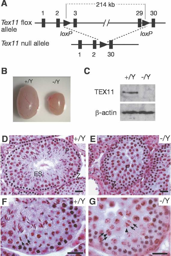
Downloaded from genesdev.cshlp.org on March 20, 2015 - Published by Cold Spring Harbor Laboratory Press

                                                                                                 TEX11 and X-linked male infertility

meiosis, they are expressed in early meiosis, implicating         null pachytene cells (Fig. 1N,O; de Vries et al. 2005).
a possible role in meiosis (Wang et al. 2005). To test this       Consistent with the immunofluorescence results, elec-
possibility, we characterized the function of one of the          tron microscopy showed that immunogold labeling of
X-linked germ cell-specific genes, Tex11, and now dem-            TEX11 was present as foci between lateral elements (Fig.
onstrate that TEX11 is a novel X-encoded meiosis factor           1P). Double immunoelectron microscopy revealed that
required for male fertility.                                      TEX11 and RPA colocalize to electron-dense nodules on
                                                                  SCs (Supplemental Fig. S2). These data suggest that
                                                                  TEX11 might be a novel constituent of synapsis-inde-
Results                                                           pendent transition meiotic recombination nodules and
                                                                  suggest that TEX11 might take part in the homologous
TEX11 forms discrete foci on meiotic chromosomes
                                                                  recombination process.
Tex11 is a germ cell-specific gene encoding a 947-amino-
acid protein of unknown function (Wang et al. 2001).
Protein sequence analysis revealed that TEX11 contains            TEX11 is required for meiosis and fertility in males
a tetratricopeptide repeat (TPR) protein–protein interac-
                                                                  To uncover the functional role of Tex11 in meiosis,
tion domain but no other known protein motifs (Blatch
                                                                  we generated a Tex11 conditional null mutant allele
and Lassle 1999). TEX11 protein was detected in testis
                                                                  (Tex11fl) in mice using the Cre-loxP strategy (Fig. 2A).
but not in somatic tissues (data not shown). By immu-
                                                                  Sequence analysis revealed that the mouse Tex11 gene
nostaining of spread nuclei, we found that TEX11 forms
                                                                  consists of 30 exons spanning a 224-kb genomic region
arrays of discrete foci along SCs in spermatocytes and
                                                                  on the X chromosome. We performed two consecutive
fetal oocytes. In spermatocytes, TEX11 foci were not ob-
                                                                  targeting steps in embryonic stem (ES) cells to obtain a
served on asynapsed chromosomes in leptotene (Fig. 1A)
                                                                  Tex11 conditional allele with loxP sites inserted in in-
but became apparent on synapsed regions in zygotene
                                                                  tron 2 and intron 29, which are 214 kb apart (Fig. 2A;
(Fig. 1B) and on the fully synapsed chromosomes in early
                                                                  Supplemental Fig. S3). Both Tex11fl/Y males and Tex11fl/fl
pachytene (∼110 foci per cell) (Fig. 1C), suggesting that
                                                                  females were fertile. To delete the Tex11 gene, Tex11fl
TEX11 may play a role in chromosomal synapsis. The
                                                                  mice were crossed with ACTB-Cre mice, in which Cre
TEX11 foci then disappeared in late pachytene (Fig. 1D)
                                                                  recombinase is expressed in all cells of the early embryo
and were not observed in diplotene spermatocytes (data
                                                                  (Lewandoski et al. 1997). Tex11−/Y males and Tex11−/−
not shown). A similar spatiotemporal localization pat-
                                                                  females were viable and appeared to be healthy.
tern of TEX11 was detected on female meiotic chromo-
                                                                    While Tex11−/− females were fertile with reduced litter
somes (Supplemental Fig. S1), suggesting that TEX11
                                                                  size, Tex11−/Y males were sterile. Testes from 12-wk-old
carries out a similar meiotic function in both sexes. The
                                                                  Tex11−/Y mice (84.5 ± 21.3 mg, n = 5 pairs) weighed 50%
TEX11 foci were independently observed in wild-type
                                                                  less than those from wild-type mice (180.5 ± 17.6 mg,
meiocytes using two different antibodies, but they were
                                                                  n = 5 pairs) (Fig. 2B). In mutant mice, 98% of the Tex11
absent in Tex11-deficient spermatocytes/oocytes (data
                                                                  coding region was deleted, and absence of TEX11 protein
not shown), validating the specificity of the TEX11 an-
                                                                  in Tex11−/Y testes (Fig. 2C) confirmed that the mutant
tibodies.
                                                                  Tex11 allele was null. Sequence analysis did not reveal
   We sought to address whether TEX11 colocalizes with
                                                                  the presence of other transcripts or known noncoding
recombination-related proteins that also form foci on
                                                                  RNAs in the Tex11 gene of mice, humans, and rats,
meiotic chromosomes. Mouse RAD51/DMC1, compo-
                                                                  strongly suggesting that the fertility defects observed in
nents of early meiotic nodules, form foci earlier during
                                                                  mutant mice were caused by loss of Tex11 function.
meiosis than RPA (replication protein A), a ssDNA-bind-
                                                                     Histological analysis of testis from adult mice showed
ing protein, which localizes to transition meiotic nod-
                                                                  that disruption of Tex11 causes arrest of male meiosis
ules (Plug et al. 1998; Moens et al. 2002). The protein
                                                                  and aberrant chromosome segregation in anaphase sper-
composition of transition meiotic nodules changes dur-
                                                                  matocytes (Fig. 2D–G). Tubules of wild-type testis con-
ing the course of meiotic recombination, and their other
                                                                  tained a full spectrum of spermatogenic cells (Fig. 2D,F).
components include MSH4, MSH5, and BLM (Marcon
                                                                  However, tubules of Tex11−/Y testis exhibited an arrest
and Moens 2005). A subset of transition nodules trans-
                                                                  in late meiosis (Fig. 2E). We observed that anaphase sper-
forms into late meiotic recombination nodules (sites of
                                                                  matocytes in Tex11−/Y testis were defective (Fig. 2G). In
crossovers) marked by MLH1.
                                                                  wild-type anaphase cells, two sets of chromosomes seg-
   Interestingly, a majority of TEX11 foci colocalized
                                                                  regated synchronously toward opposite spindle poles
with RPA foci in pachynema (Fig. 1E–H,H⬘), whereas no
                                                                  (Fig. 2F). In contrast, in Tex11-deficient anaphase cells, a
overlap between TEX11 and DMC1 or MLH1 foci were
                                                                  few chromatin masses either migrated ahead or lagged
observed (Fig. 1M; data not shown), suggesting that
                                                                  behind the two major chromatin masses (Fig. 2G).
TEX11 might be a component of transition meiotic nod-
ules. In further support of this notion, more than half of
TEX11 foci overlapped with MSH4 foci in spermatocytes
                                                                  TEX11 promotes chromosomal synapsis in males
(Fig. 1I–L,L⬘). In addition, we found that TEX11 foci, like
RPA foci, were located between the two aligned but                We next investigated whether Tex11−/Y pachytene sper-
separated lateral elements in synapsis-deficient Sycp1-           matocytes exhibit defects in chromosomal synapsis by

                                                                                               GENES & DEVELOPMENT              683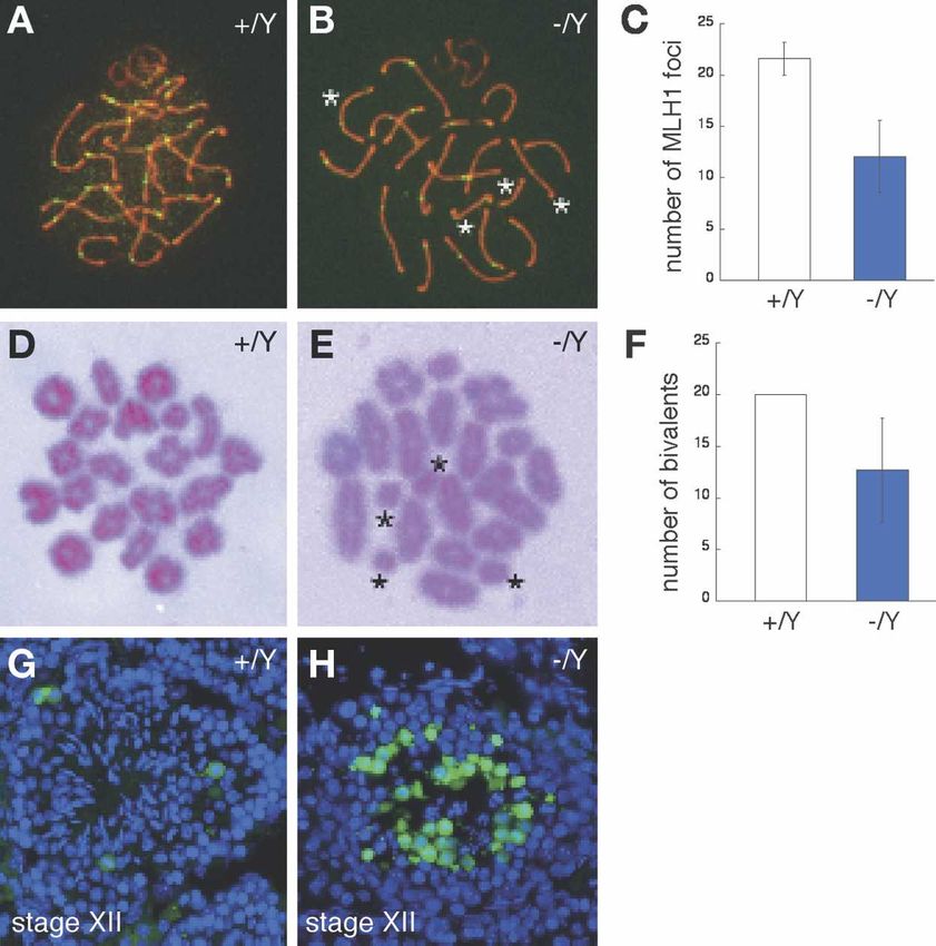
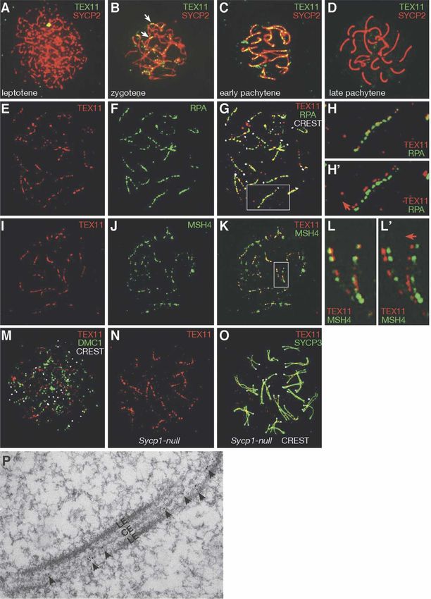
Downloaded from genesdev.cshlp.org on March 20, 2015 - Published by Cold Spring Harbor Laboratory Press

Yang et al.

Figure 1. TEX11 forms distinct foci on meiotic chromosomes. (A–D) Distribution of TEX11 foci in spermatocytes. TEX11 foci are
absent in leptonema (A), present in zygonema (B) and early pachynema (C), but absent in late pachynema (D). (B) Note that TEX11 foci
are present on synapsed regions (arrows) in zygonema. (E–G) TEX11 foci mostly (74%) colocalize with RPA foci in spermatocytes. The
same spermatocyte is shown for TEX11 alone (E), RPA alone (F), and a composite image (G). CREST antiserum stains centromeres.
Expanded view of the chromosome in the square in G is shown in H and H⬘ (with offset channels). (I–K) A majority of TEX11 foci (60%)
overlap with MSH4 foci in spermatocytes. The same spermatocyte is shown for TEX11 alone (I), MSH4 alone (J), and a composite image
(K). Expanded view of the two chromosomes in the square in K is shown in L and L⬘ (with offset channels). (H⬘,L⬘) The direction of
deliberate shift of the red channel is indicated by an arrow. (M) TEX11 foci rarely overlap with DMC1 foci in spermatocytes. (N) TEX11
forms arrays of foci in Sycp1−/− pachynema. (O) TEX11 foci are located between the two aligned but separated lateral elements in
Sycp1−/− pachynema. The same cell is shown in N. (P) Electron microscopy shows immunogold labeling of TEX11 between lateral
elements (LEs) in spermatocytes. (CE) Central element.

684       GENES & DEVELOPMENT
Downloaded from genesdev.cshlp.org on March 20, 2015 - Published by Cold Spring Harbor Laboratory Press

                                                                                                   TEX11 and X-linked male infertility

                                                                    pachytene nuclei scored). In contrast, only one of 150
                                                                    wild-type pachytene nuclei analyzed had asynapsed sex
                                                                    chromosomes (900                  3I; Roeder and Bailis 2000; Eaker et al. 2002; Baarends et

                                                                                                 GENES & DEVELOPMENT              685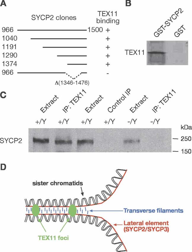
Downloaded from genesdev.cshlp.org on March 20, 2015 - Published by Cold Spring Harbor Laboratory Press
Yang et al.
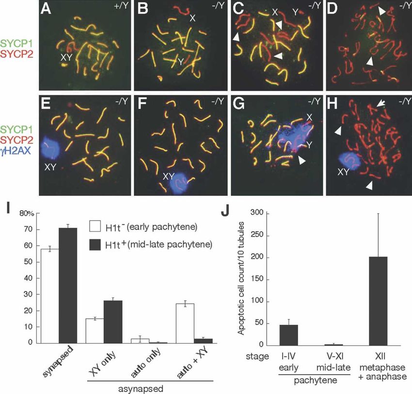
Figure 3. TEX11 promotes chromosome
synapsis. (A) Wild-type pachynema with
19 pairs of fully synapsed autosomes (yel-
low) and partially synapsed X–Y chromo-
somes (red). (B) Tex11−/Y pachynema with
asynapsed X–Y. (C) Tex11−/Y pachynema
with eight asynapsed chromosomes: the
X–Y and three pairs of autosomes (arrow-
heads). Notably, each pair of asynapsed ho-
mologs (as judged by equal SC length ex-
cept for XY) is in close proximity and align-
ment, indicating a possible interaction.
(D) Tex11−/Y diplonema with univalents
(arrowheads). (E) An apparently normal
Tex11−/Y pachynema with partially syn-
apsed X–Y in the ␥H2AX-positive sex
body. (F) Tex11−/Y pachynema. Asynapsed
X–Y chromosomes are confined to a sin-
gle ␥H2AX-positive domain. (G) Tex1−/Y
pachynema with one pair of asynapsed au-
tosomes (arrowhead) and asynapsed X–Y,
all of which are ␥H2AX-positive. (H)
Tex11−/Y diplonema with a large number of
univalents (arrowheads). Some autosomes
are bivalents (arrow). X–Y univalents are
confined to a single ␥H2AX-positive do-
main. In contrast, all autosomal univalents
are ␥H2AX-negative. (I) Analysis of asyn-
apsed chromosomes in early (H1t-negative)
and mid-to-late (H1t-positive) Tex11−/Y
pachynema. Testes from four 7.5-wk-old
Tex11−/Y mice were analyzed by triple im-
munostaining of spread nuclei with anti-SYCP1, anti-SYCP3, and anti-histone H1t antibodies. All pachynema in view were first
divided into H1t− and H1t+ groups, and asynapsed chromosomes were then examined. More than 200 pachynema from each mouse
were analyzed. (J) Two distinct rounds of apoptosis in adult Tex11−/Y testis. Seminiferous tubules were divided into three groups:
H1t-negative (Stages I–IV), H1t-positive (Stages V–XI), and Stage XII.
al. 2005; Turner et al. 2005). The question arises as to ence of asynapsed X–Y chromosomes. Analysis of meta-
what could account for the second round of apoptosis phase I chromosomes by Giemsa staining revealed a re-
(Stage XII) in Tex11−/Y testis (Fig. 3J). The majority (70%) duced number of bivalents (12.7 ± 5.0 bivalents per
of mid–late pachytene mutant spermatocytes had nor- nucleus) in Tex11−/Y metaphase I spermatocytes com-
mal synapsis (Fig. 3I), but nearly all (96%) Tex11−/Y dip- pared with wild-type (20 bivalents per nucleus) (Fig. 4D–
lotene spermatocytes contained univalent chromosomes F). The presence of univalents could lead to chromosom-
(Fig. 3D,H). Additionally, in these diplotene cells, sex al segregation defects at the subsequent anaphase I stage,
chromosomal univalents remained ␥H2AX- and ATR- and aberrant chromosome attachment to the spindle or
positive, whereas autosomal univalents were ␥H2AX- chromosome nondisjunction could trigger the spindle
and ATR-negative (Fig. 3H; Supplemental Fig. S4). These checkpoint and induce apoptosis (Ashley et al. 1994; Li
results suggest de novo formation of autosomal univa- and Nicklas 1995; Odorisio et al. 1998; Eaker et al. 2002).
lents by desynapsis at the diplotene stage, possibly due We detected both defective chromosome segregation in
to defective crossover formation at pachytene. Tex11−/Y anaphase spermatocytes (Fig. 2G) and massive
apoptosis of germ cells in Stage XII tubules (containing
the metaphase I and anaphase I spermatocytes) in
TEX11 promotes formation of meiotic crossovers Tex11−/Y testis (Figs. 3J, 4H), indicating that TEX11 pro-
in males motes the formation of crossovers and thus is required
We monitored the formation of crossovers at pachytene. for proper chromosome segregation during male meiosis.
MLH1 is essential for crossover formation and marks
sites of crossover (Baker et al. 1996; Edelmann et al.
Chromosomal asynapsis and reduced crossover
1996). We detected a sharply reduced number of MLH1
formation in Tex11-deficient females
foci (12.1 ± 3.5, n = 42 nuclei) in Tex11−/Y pachynema
compared with wild-type (21.6 ± 1.6, n = 24 nuclei) (Fig. In contrast to Tex11−/Y males, Tex11−/− females were
4A–C), mainly due to a loss of foci from synapsed chro- fertile and produced offspring. However, Tex11−/− fe-
mosomes, and only to a lesser degree caused by the pres- males displayed a reduction in litter size, generating on
686 GENES & DEVELOPMENT
Downloaded from genesdev.cshlp.org on March 20, 2015 - Published by Cold Spring Harbor Laboratory Press

                                                                                                 TEX11 and X-linked male infertility

                                                                                    Figure 4. TEX11 modulates the formation of
                                                                                    meiotic crossovers. (A–C) A dramatic reduc-
                                                                                    tion in the number of MLH1 foci (green) in
                                                                                    Tex11−/Y pachynema (∼15 foci) relative to
                                                                                    wild type (22 foci). (B) Note the absence of
                                                                                    MLH1 foci on four synapsed chromosomes
                                                                                    (asterisks) in the mutant. (D) Twenty biva-
                                                                                    lents (pairs of homologs) are connected via
                                                                                    chiasmata in wild-type metaphase I sper-
                                                                                    matocytes. (E) Presence of univalents in
                                                                                    Tex11-deficient metaphase I spermatocytes.
                                                                                    Asterisks indicate four univalents. (F) A sharp
                                                                                    decrease in the number of bivalents in Tex11-
                                                                                    deficient metaphase I spermatocytes. (G)
                                                                                    TUNEL analysis of wild-type tubules. A few
                                                                                    apoptotic cells (green) are occasionally ob-
                                                                                    served in Stage XII tubules. (H) Massive apo-
                                                                                    ptosis in Tex11−/Y Stage XII tubules. Apopto-
                                                                                    tic cells presumably corresponded to ana-
                                                                                    phase I spermatocytes.

average 5.2 offspring per litter (5.2 ± 2.2). In compari-         elements of the SC (Offenberg et al. 1998; Schalk et al.
son, heterozygous (Tex11+/−) littermates produced 9.6             1998). The SYCP2 C-terminal region (1374–1500 amino
offspring per litter (9.6 ± 2.0; P < 0.0001). Analysis of         acids) was sufficient for interaction with TEX11 in both
pachytene asynapsis and formation of MLH1 foci in fetal           two-hybrid and GST pulldown assays (Fig. 5A,B). Fur-
oocytes revealed a higher percentage (31.3%) of Tex11-            thermore, an internal deletion of SYCP2 (1346–1476
deficient pachytene oocytes containing asynapsed chro-            amino acids) abolished its interaction with TEX11 (Fig.
mosomes compared with wild-type/Tex11+/− oocytes                  5A). This internal deletion (1346–1476 amino acids) was
(5.5%). The number of MLH1 foci (18.6 ± 2.6) in Tex11-            previously generated in Sycp2 mutant mice (Yang et al.
deficient oocytes was slightly lower than that (23.5 ± 2.4)       2006). We found that TEX11 foci were abundant on mei-
in wild-type/Tex11+/− oocytes. These data, together with          otic chromosomes in Sycp2 mutant pachytene oocytes
studies of Tex11-deficient males, demonstrate that loss           (data not shown), suggesting that formation of TEX11
of Tex11 function causes the same effect on synapsis and          foci is independent of SYCP2. Coimmunoprecipitation
crossover formation in both sexes, even though these              from testicular protein extracts further demonstrated
defects in females are milder. The fertility in Tex11-de-         that TEX11 associates with SYCP2 in vivo (Fig. 5C).
ficient females could be explained by the fact that the           Thus, through its association with SYCP2, an SC com-
number of MLH1 foci is close to 20 per cell in some               ponent, TEX11 could provide a link between chromo-
oocytes. The reduced fertility in Tex11-deficient females         somal synapsis and meiotic recombination (Fig. 5D).
could be caused by aneuploidy due to asynapsis and/or a
reduced number of MLH1 foci in oocytes.
                                                                  Discussion
                                                                  Our studies implicate TEX11 in initiation of chromo-
TEX11 interacts with SYCP2
                                                                  some synapsis. In support of such a function, loss of
To elucidate a potential molecular mechanism for the              TEX11 results in an “all-or-none” synapsis defect on a
function of TEX11 in meiosis—i.e., to identify protein            per chromosome basis in early pachytene spermatocytes:
partners of TEX11—we performed a yeast two-hybrid                 A subset of chromosomes is completely asynapsed (pre-
screen of a mouse testis cDNA library using the full-             sumably due to a failure in initiation), but other chro-
length TEX11 as bait. The most frequently identified              mosomes are synapsed. The role in synapsis is further
protein was SYCP2, an integral component of the lateral           supported by colocalization of TEX11 with RPA foci,

                                                                                                GENES & DEVELOPMENT             687
Downloaded from genesdev.cshlp.org on March 20, 2015 - Published by Cold Spring Harbor Laboratory Press
Yang et al.
verse filaments, localizes only in foci, indicating a failure
of full synapsis (Tsubouchi et al. 2006). Furthermore, de-
spite absence of sequence homology between Tex11 and
Zip3, the yeast Zip3 mutant resembles the Tex11 mouse
null mutant in one key aspect of synapsis. As in Tex11-
deficient pachynema, Zip3-deficient pachytene cells ex-
hibit an “all-or-none” synaptic defect on a per chromo-
some basis (Agarwal and Roeder 2000). These studies
suggest that the TEX11-mediated synapsis pathways are
functionally conserved among diverse organisms.
Several lines of evidence support the conclusion that
TEX11 also plays a distinct role in meiotic recombina-
tion. Firstly, a majority of TEX11 foci overlap with RPA
foci and MSH4 foci, which correspond to transition mei-
otic nodules. The formation of TEX11 foci between lat-
eral elements in Sycp1-null pachytene spermatocytes
further demonstrates that the synapsed structure is not
essential for holding TEX11 to chromosomes; instead, it
is likely that TEX11 is part of a recombination complex.
Secondly, loss of TEX11 caused a subtle but distinct ef-
fect on initiation and/or maintenance of synapsis (a
small subset of chromosomes are affected) but a more
severe effect on crossover formation (almost a 50% loss
of MLH1 foci). In particular, the number of MLH1 foci in
Tex11−/Y pachytene spermatocytes with normal synapsis
was greatly reduced, suggesting that the role of TEX11 in
crossover formation is separate from its role in synapsis.
Thirdly, yeast Zip4 and Arabidopsis Zip4 mutants ex-
hibit defects in crossover formation, demonstrating the
Figure 5. TEX11 interacts with SYCP2. (A) Five unique over-
lapping SYCP2 fragments identified in the two-hybrid screen evolutionary conservation of the TEX11/ZIP4 functions
using the full-length TEX11 protein as bait. Numbers indicate in meiotic recombination (Tsubouchi et al. 2006; Chely-
the position of terminal residues. The bottom clone with an sheva et al. 2007). Lastly, we observed two rounds of
internal deletion of SYCP2 (1346–1476 amino acids) does not apoptosis in Tex11-deficient testis: in early pachytene
interact with TEX11. (B) Binding of in vitro translated TEX11 to spermatocytes and in anaphase I spermatocytes. These
GST-SYCP2 (amino acids 1374–1500). (C) Coimmunoprecipita- two rounds of apoptosis were distinct from each other,
tion of SYCP2 with TEX11 from testis. Immunoprecipitation because increased apoptosis (the first round) was ob-
was performed with rabbit anti-TEX11 antibodies or preim- served in H1t-negative tubules from juvenile Tex11-de-
mune control serum and probed with guinea pig anti-SYCP2
ficient testis, in which meiosis has yet to reach the ana-
antibodies on Western blot. (D) Model illustrating the associa-
phase of prophase I (Supplemental Fig. S6). Thus, in
tion of TEX11 foci and the lateral elements at the zygotene
stage. A pair of homologous chromosomes and transverse fila- Tex11-deficient testis, asynapsis (if affecting autosomes)
ments of the SC are also shown. would result in cell elimination by the pachytene check-
point and/or MSUC, whereas those cells that succeed in
synapsis would fail to complete crossover formation and
which are known to be present on synapsed regions (Plug would be eliminated by the spindle checkpoint, resulting
et al. 1998; Moens et al. 2002). Importantly, TEX11 in- in male sterility (Roeder and Bailis 2000; Eaker et al.
teracts with SYCP2, which is required for chromosomal 2002; de Rooij and de Boer 2003; Turner et al. 2005).
synapsis (Yang et al. 2006). Additionally or alternatively, We propose that the dual function of TEX11 in synap-
TEX11 could contribute to the elongation or stabiliza- sis and crossover fixation might be linked by the TEX11–
tion of the SC. Thus, in the absence of TEX11, chromo- SYCP2 interaction and could be explained by its focal
some synapsis may be initiated but not propagated or localization on synapsed but not asynapsed chromo-
maintained. Although little is known about the molecu- somes (Fig. 5D). Consistent with this model, the short
lar underpinnings of synapsis initiation in mammals, length of the X–Y synapsed region might explain why
this process has been extensively studied in yeast. A set asynapsis preferentially affects X–Y chromosomes.
of yeast proteins (Zip2, Zip3, Zip4, Msh4, Msh5, etc.), TEX11 forms distinct foci on the meiotic chromosomes,
referred to as SIC (synapsis initiation complex) or ZMM and these foci are located between the lateral elements of
proteins, are required for both chromosomal synapsis the SC. In contrast, SYCP2, an integral component of the
initiation and meiotic recombination (Borner et al. 2004; lateral element, localizes continuously on the lateral el-
Tsubouchi et al. 2006). TEX11 exhibits some sequence ements. How could these two proteins interact while
homology with yeast Zip4 and Arabidopsis ZIP4. In Zip4 they appear to be spatially separate? Interestingly, a pre-
mutant yeast cells, Zip1, a component of yeast trans- vious ultrastructural study reported that while both
688 GENES & DEVELOPMENTDownloaded from genesdev.cshlp.org on March 20, 2015 - Published by Cold Spring Harbor Laboratory Press
TEX11 and X-linked male infertility
SYCP2 and SYCP3 localize continuously on the lateral into intron 2 (Supplemental Fig. S3). HyTK encodes a double
elements, SYCP2 but not SYCP3 also localizes to “fuzzy selection marker for positive selection with hygromycin and
connections” (presumably recombination nodules) be- negative selection with ganciclovir. Construct 1 consists of two
tween the lateral elements (Schalk et al. 1998). Thus, we homologous arms (2.1 kb and 2.0 kb). In targeting construct 2,
the floxed HyTK selection cassette was inserted into intron 29
postulate that TEX11 might interact with the pool of
with two flanking homologous arms (2.3 kb and 2.8 kb). Ho-
SYCP2 that is present in these “fuzzy connections.”
mologous arms in both constructs were fully sequenced, and no
Many mutants of meiotic recombination nodule com- mutations were found.
ponents (Dmc1, Msh4, Mlh1, etc.) abolish chromosomal Four consecutive steps were executed to generate ES cells
synapsis and/or crossover formation in mice (Baker et al. harboring a Tex11fl allele (Supplemental Fig. S3). Step 1, target-
1996; Edelmann et al. 1996; Pittman et al. 1998; Yoshida ing ES cells with construct 1: V6.5 hybrid ES cells were electro-
et al. 1998; Kneitz et al. 2000). Remarkably, unlike these porated with linearized targeting construct 1 and selected for
known meiosis factors, TEX11 promotes synapsis and integration with hygromycin B (120 µg/mL; Invitrogen). Hygro-
crossover formation but is not essential for either pro- mycin-resistant ES cell colonies (192) were screened by long-
cess. Partial elimination of synapsis and crossover is not distance PCR. Twenty percent of clones were resistant due to
homologous recombination, referred to as ES line A. Step 2,
caused by any residual function of Tex11 in this mouse
Cre-mediated recombination: Two independent line A clones
mutant, because the TEX11 protein is absent. One ex-
were expanded, electroporated with the pOG231 plasmid that
planation could be that TEX11 is a regulatory but non- transiently expresses Cre recombinase, and subjected to nega-
essential component of a multiprotein complex that tive selection with ganciclovir (2 µM; Sigma) for removal of the
modulates synapsis and crossover. Such a protein com- HyTK cassette. Viable colonies were screened by PCR. Cre-
plex could be partially functional in the absence of mediated recombination resulted in ES line B, which bears only
TEX11. one loxP site in intron 2. Step 3, targeting with construct 2: Two
Meiotic silencing of unpaired DNA or chromatin has independent line B ES clones were electroporated with targeting
emerged as a common process in diverse organisms such construct 2 and screened as described in Step 1. The homolo-
as filamentous fungi, nematodes, and mammals (Shiu et gous targeting frequency was 17%. The resulting ES cells were
referred to as ES line C. Step 4, second Cre-mediated recombi-
al. 2001; Fong et al. 2002; Baarends et al. 2005; Turner et
nation: Two independent line C ES cell clones were electropor-
al. 2005). Gene expression (microarray) studies show that
ated with the pOG231 plasmid and screened as described in Step
meiotic silencing of sex chromosomes leads to depletion 2. Recombination between the adjacent HyTK-flanking loxP
of male germline-intrinsic genes on the X chromosome sites resulted in the final Tex11fl allele, harboring two loxP sites
in nematodes and mammals during evolution (Reinke et 214 kb apart (Fig. 2A).
al. 2000; Khil et al. 2004). However, here we demonstrate
that TEX11, encoded by the X chromosome, is a meiosis- Generation of Tex11fl and Tex11-deficient mice
specific protein, and that its loss causes meiotic failure
in males. This study counters the hypothesis that the X Four Tex11fl/Y ES cell clones were injected into B6C3F1 blasto-
chromosome may be depleted of meiosis-specific genes cysts that were subsequently transferred to the uteri of pseudo-
pregnant ICR females. One ES clone (C10-3) gave rise to seven
in mammals.
chimeric mice. Two of the chimeric males transmitted the
Our findings have important implications for male in- Tex11fl allele through the germline. Tex11fl mice were bred
fertility in humans, which accounts for about half of the with ACTB-CreTg/Tg mice to delete Tex11 (Lewandoski et al.
cases of infertility among couples. An estimated 15% of 1997).
couples are affected by infertility worldwide (Matzuk Because Tex11−/Y males were sterile, generation of homozy-
and Lamb 2002). Given that disruption of Tex11 causes gous knockout (Tex11−/−) females required a different breeding
azoospermia in mice, mutations in the human TEX11 strategy. We bred Tex11fl/Y males with Tex11+/− ACTB-CreTg/+
gene could cause infertility in men. females. Twenty-five percent of the resulting female offspring
were expected to be Tex11flox/− ACTB-CreTg/+. Because ACTB-
Cre was strongly and ubiquitously expressed since the zygote
Materials and methods stage, Tex11fl/− ACTB-CreTg/+ mice were equivalent to Tex11−/−
ACTB-CreTg/+. The Tex11fl allele was not detected in genomic
Generation of anti-TEX11 polyclonal antibodies DNA prepared from multiple tissues (including ovary) from
The mouse Tex11 cDNA fragment encoding the C-terminal 100 Tex11−/− ACTB-CreTg/+ mice by PCR. In addition, genotyping of
amino acids (residues 848–947) was cloned into the pQE-30 ex- >100 offspring sired by Tex11−/− ACTB-CreTg/+ females did not
pression vector (Qiagen). The 6xHis-TEX11 fusion protein was identify any pup that harbors the Tex11fl allele. Mice used were
expressed in Escherichia coli, affinity-purified with Ni-NTA on a mixed genetic background (FVB, 129, and C57BL/6J). Ani-
beads, and used to immunize two rabbits (Cocalico Biologicals, mals were maintained and used for experimentation according
Inc.). The anti-TEX11 antiserum (serum 1966) was used for to the guidelines of the Institutional Animal Care and Use
Western blot analysis (1:500). In addition, guinea pig polyclonal Committee of the University of Pennsylvania.
antibodies were raised against the C-terminal peptide of mouse
TEX11 (EQLRALIVPPEDQGSVSSTNVAAQNHL) and affinity- TUNEL analysis and H1t immunofluorescence
purified.
Testes were fixed in 4% paraformaldehyde (PFA) for 3 h at 4°C,
dehydrated in 30% sucrose overnight, prepared, and sectioned.
Consecutive homologous gene targeting in ES cells
TUNEL assays were performed with the ApopTag Fluorescein
Two targeting constructs were generated. In the Tex11 targeting In Situ Apoptosis Detection Kit (catalog no. S7110, Serologicals
construct 1, we subcloned the floxed HyTK selection cassette Corporation/Chemicon) with or without concurrent immuno-
GENES & DEVELOPMENT 689Downloaded from genesdev.cshlp.org on March 20, 2015 - Published by Cold Spring Harbor Laboratory Press
Yang et al.
staining with anti-H1t antibodies. Cross-sections of tubules the manuscript. This work was supported by an NIH grant
were divided into three categories: H1t-negative, H1t-positive, GM076327 (to P.J.W.); by grants from the Swedish Cancer So-
and Stage XII. TUNEL-positive cells were counted for each ciety, the Swedish Research Council and the Karolinska Insti-
cross-section (Fig. 3J). More than 150 tubules were analyzed for tutet (to C.H); by support from the Howard Hughes Medical
each mouse. Institute (to D.C.P); and by a grant from the Deutsche For-
schungsgemeinschaft (to R.B.).
Two-hybrid screen and immunoprecipitation
The full-length TEX11 coding region was cloned into the References
pAS2-1 vector through gap repair in yeast. A total of 2 × 10⁶
clones from a mouse testis cDNA library (BD Biosciences) were Agarwal, S. and Roeder, G.S. 2000. Zip3 provides a link between
screened according to standard protocols. After specificity tests, recombination enzymes and synaptonemal complex pro-
53 clones were positive and sequenced. Sequence analysis re- teins. Cell 102: 245–255.
vealed that eight clones encode SYCP2. For immunoprecipita- Ashley, T., Ried, T., and Ward, D.C. 1994. Detection of nondis-
tion experiments, protein extracts were prepared by homogeniz- junction and recombination in meiotic and postmeiotic cells
ing testes from five postnatal day 20 mice per genotype. After from XYSxr [XY,tp(Y)1Ct] mice using multicolor fluores-
sonication and centrifugation, extracts were incubated with cence in situ hybridization. Proc. Natl. Acad. Sci. 91: 524–
protein A Sepharose and centrifuged prior to use for immuno- 528.
precipitation. Baarends, W.M., Wassenaar, E., van der Laan, R., Hoogerbrugge,
J., Sleddens-Linkels, E., Hoeijmakers, J.H., de Boer, P., and
Grootegoed, J.A. 2005. Silencing of unpaired chromatin and
Histological, surface nuclei spread, immunofluorescent, histone H2A ubiquitination in mammalian meiosis. Mol.
and electron microscopy analyses Cell. Biol. 25: 1041–1053.
For histological analysis, testes were fixed in Bouin’s solution, Baker, S.M., Plug, A.W., Prolla, T.A., Bronner, C.E., Harris,
embedded in paraffin, sectioned, and stained with hematoxylin A.C., Yao, X., Christie, D.M., Monell, C., Arnheim, N.,
and eosin. Metaphase spread cells were stained with 4% Gurr Bradley, A., et al. 1996. Involvement of mouse Mlh1 in DNA
Giemsa (Invitrogen). The number of bivalents was derived using mismatch repair and meiotic crossing over. Nat. Genet. 13:
the following formula: the number of bivalents = 40 − n (40 is 336–342.
the theoretical maximum number of univalents; n is the total Blatch, G.L. and Lassle, M. 1999. The tetratricopeptide repeat: A
number of chromatin masses in each metaphase I nucleus). For structural motif mediating protein–protein interactions.
immunofluorescent analysis of spread nuclei, testicular cell Bioessays 21: 932–939.
suspensions were prepared, immunostained, and analyzed. To Borner, G.V., Kleckner, N., and Hunter, N. 2004. Crossover/
obtain fetal oocytes, Tex11−/− ACTB-CreTg/+ females were caged noncrossover differentiation, synaptonemal complex forma-
with Tex11fl/Y males at 4 pm. Vaginal copulatory plugs were tion, and regulatory surveillance at the leptotene/zygotene
checked the next morning and recorded as 0.5 d post-coitum transition of meiosis. Cell 117: 29–45.
(dpc). Fetal oocytes were collected at 17.5 or 18.5 dpc. Primary Chelysheva, L., Gendrot, G., Vezon, D., Doutriaux, M.P., Mer-
antibodies used for immunofluorescence were as follows: rabbit cier, R., and Grelon, M. 2007. Zip4/Spo22 is required for
anti-TEX11, guinea pig anti-TEX11 (1:200), rabbit anti-SYCP1 class I CO formation but not for synapsis completion in
(1:100), rabbit and guinea pig anti-SYCP2 (sera 1918 and GP21, Arabidopsis thaliana. PLoS Genet. 3: e83. doi: 10.1371/
1:100), rabbit anti-SYCP3 (1:500; a gift from S. Chuma) and our journal.pgen.0030083.
own (1:100), guinea pig anti-H1t (1:1000; a gift from M.A. Han- Cobb, J., Cargile, B., and Handel, M.A. 1999. Acquisition of
del), rabbit anti-ATR (1:80; catalog no. PC538, EMD Biosci- competence to condense metaphase I chromosomes during
ences), mouse anti-␥H2AX (1:500; Upstate Biotechnology), hu- spermatogenesis. Dev. Biol. 205: 49–64.
man CREST antiserum (1:3000; a gift from N. Ringertz), rabbit de Rooij, D.G. and de Boer, P. 2003. Specific arrests of spermato-
anti-DMC1 (1:100; a gift from P. Moens), anti-RPA sera (1:400; genesis in genetically modified and mutant mice. Cytogenet.
a gift from P. Moens), anti-RPA monoclonal antibody (EMD Genome Res. 103: 267–276.
Biosciences), and rabbit anti-hMSH4 antibodies (1:100) (Her et de Vries, F.A., de Boer, E., van den Bosch, M., Baarends, W.M.,
al. 2003). Various FITC-, Texas red-, AMCA-, Alexa fluor 488-, Ooms, M., Yuan, L., Liu, J.G., van Zeeland, A.A., Heyting,
Cy5-, or TRITC-conjugated secondary antibodies were used. C., and Pastink, A. 2005. Mouse Sycp1 functions in synap-
Slides were visualized under an Axioskop 40 fluorescence mi- tonemal complex assembly, meiotic recombination, and XY
croscope. Images were captured with a digital camera (Evolution body formation. Genes & Dev. 19: 1376–1389.
QEi, MediaCybernetics) and processed with ImagePro software Eaker, S., Cobb, J., Pyle, A., and Handel, M.A. 2002. Meiotic
(Phase 3 Imaging systems) and Photoshop (Adobe). Immunogold prophase abnormalities and metaphase cell death in MLH1-
labeling was performed on 6-µm cryosections of shock-frozen deficient mouse spermatocytes: Insights into regulation of
mouse testis according to standard protocols with rabbit anti- spermatogenic progress. Dev. Biol. 249: 85–95.
TEX11 antibodies. Secondary antibodies were conjugated to Edelmann, W., Cohen, P.E., Kane, M., Lau, K., Morrow, B., Ben-
6-nm or 12-nm gold particles. nett, S., Umar, A., Kunkel, T., Cattoretti, G., Chaganti, R., et
al. 1996. Meiotic pachytene arrest in MLH1-deficient mice.
Cell 85: 1125–1134.
Fong, Y., Bender, L., Wang, W., and Strome, S. 2002. Regulation
Acknowledgments
of the different chromatin states of autosomes and X chro-
We thank E. Gleason for technical assistance, M.A. Handel for mosomes in the germ line of C. elegans. Science 296: 2235–
advice on the project and anti-H1t antibodies, P.E. Cohen for 2238.
advice on phenotypic analysis, P. Moens for anti-RPA and anti- Handel, M.A., Park, C., and Kot, M. 1994. Genetic control of
DMC1 antibodies, and S. Chuma for anti-SYCP3 antibodies. We sex-chromosome inactivation during male meiosis. Cyto-
thank M.A. Handel, S. Rozen, and P. de Boer for comments on genet. Cell Genet. 66: 83–88.
690 GENES & DEVELOPMENTDownloaded from genesdev.cshlp.org on March 20, 2015 - Published by Cold Spring Harbor Laboratory Press
TEX11 and X-linked male infertility
Hassold, T. and Hunt, P. 2001. To err (meiotically) is human: Meiotic silencing by unpaired DNA. Cell 107: 905–916.
The genesis of human aneuploidy. Nat. Rev. Genet. 2: 280– Solari, A.J. 1974. The behavior of the XY pair in mammals. Int.
291. Rev. Cytol. 38: 273–317.
Her, C., Wu, X., Griswold, M.D., and Zhou, F. 2003. Human Tsubouchi, T., Zhao, H., and Roeder, G.S. 2006. The meiosis-
MutS homologue MSH4 physically interacts with von Hip- specific zip4 protein regulates crossover distribution by pro-
pel-Lindau tumor suppressor-binding protein 1. Cancer Res. moting synaptonemal complex formation together with
63: 865–872. zip2. Dev. Cell 10: 809–819.
Khil, P.P., Smirnova, N.A., Romanienko, P.J., and Camerini- Turner, J.M., Mahadevaiah, S.K., Fernandez-Capetillo, O., Nus-
Otero, R.D. 2004. The mouse X chromosome is enriched for senzweig, A., Xu, X., Deng, C.X., and Burgoyne, P.S. 2005.
sex-biased genes not subject to selection by meiotic sex chro- Silencing of unsynapsed meiotic chromosomes in the
mosome inactivation. Nat. Genet. 36: 642–646. mouse. Nat. Genet. 37: 41–47.
Kneitz, B., Cohen, P.E., Avdievich, E., Zhu, L., Kane, M.F., Hou Wang, P.J., McCarrey, J.R., Yang, F., and Page, D.C. 2001. An
Jr., H., Kolodner, R.D., Kucherlapati, R., Pollard, J.W., and abundance of X-linked genes expressed in spermatogonia.
Edelmann, W. 2000. MutS homolog 4 localization to meiotic Nat. Genet. 27: 422–426.
chromosomes is required for chromosome pairing during Wang, P.J., Page, D.C., and McCarrey, J.R. 2005. Differential
meiosis in male and female mice. Genes & Dev. 14: 1085– expression of sex-linked and autosomal germ-cell-specific
1097. genes during spermatogenesis in the mouse. Hum. Mol.
Lewandoski, M., Meyers, E.N., and Martin, G.R. 1997. Analysis Genet. 14: 2911–2918.
of Fgf8 gene function in vertebrate development. Cold Yang, F., De La Fuente, R., Leu, N.A., Baumann, C., McLaugh-
Spring Harb. Symp. Quant. Biol. 62: 159–168. lin, K.J., and Wang, P.J. 2006. Mouse SYCP2 is required for
Li, X. and Nicklas, R.B. 1995. Mitotic forces control a cell-cycle synaptonemal complex assembly and chromosomal synapsis
checkpoint. Nature 373: 630–632. during male meiosis. J. Cell Biol. 173: 497–507.
Marcon, E. and Moens, P.B. 2005. The evolution of meiosis: Yoshida, K., Kondoh, G., Matsuda, Y., Habu, T., Nishimune, Y.,
Recruitment and modification of somatic DNA-repair pro- and Morita, T. 1998. The mouse RecA-like gene Dmc1 is
teins. Bioessays 27: 795–808. required for homologous chromosome synapsis during meio-
Matzuk, M.M. and Lamb, D.J. 2002. Genetic dissection of mam- sis. Mol. Cell 1: 707–718.
malian fertility pathways. Nat. Cell Biol. 4 (Suppl.): s41–s49. Zickler, D. and Kleckner, N. 1999. Meiotic chromosomes: In-
doi: 10.1038/ncb-nm-fertilitys41. tegrating structure and function. Annu. Rev. Genet. 33: 603–
Moens, P.B., Kolas, N.K., Tarsounas, M., Marcon, E., Cohen, 754.
P.E., and Spyropoulos, B. 2002. The time course and chro-
mosomal localization of recombination-related proteins at
meiosis in the mouse are compatible with models that can
resolve the early DNA–DNA interactions without reciprocal
recombination. J. Cell Sci. 115: 1611–1622.
Odorisio, T., Rodriguez, T.A., Evans, E.P., Clarke, A.R., and
Burgoyne, P.S. 1998. The meiotic checkpoint monitoring
synapsis eliminates spermatocytes via p53-independent ap-
optosis. Nat. Genet. 18: 257–261.
Offenberg, H.H., Schalk, J.A., Meuwissen, R.L., van Aalderen,
M., Kester, H.A., Dietrich, A.J., and Heyting, C. 1998. SCP2:
A major protein component of the axial elements of synap-
tonemal complexes of the rat. Nucleic Acids Res. 26: 2572–
2579.
Page, S.L. and Hawley, R.S. 2004. The genetics and molecular
biology of the synaptonemal complex. Annu. Rev. Cell Dev.
Biol. 20: 525–558.
Pittman, D.L., Cobb, J., Schimenti, K.J., Wilson, L.A., Cooper,
D.M., Brignull, E., Handel, M.A., and Schimenti, J.C. 1998.
Meiotic prophase arrest with failure of chromosome synap-
sis in mice deficient for Dmc1, a germline-specific RecA
homolog. Mol. Cell 1: 697–705.
Plug, A.W., Peters, A.H., Keegan, K.S., Hoekstra, M.F., de Boer,
P., and Ashley, T. 1998. Changes in protein composition of
meiotic nodules during mammalian meiosis. J. Cell Sci. 111:
413–423.
Reinke, V., Smith, H.E., Nance, J., Wang, J., Van Doren, C.,
Begley, R., Jones, S.J., Davis, E.B., Scherer, S., Ward, S., et al.
2000. A global profile of germline gene expression in C. el-
egans. Mol. Cell 6: 605–616.
Roeder, G.S. and Bailis, J.M. 2000. The pachytene checkpoint.
Trends Genet. 16: 395–403.
Schalk, J.A., Dietrich, A.J., Vink, A.C., Offenberg, H.H., van
Aalderen, M., and Heyting, C. 1998. Localization of SCP2
and SCP3 protein molecules within synaptonemal com-
plexes of the rat. Chromosoma 107: 540–548.
Shiu, P.K., Raju, N.B., Zickler, D., and Metzenberg, R.L. 2001.
GENES & DEVELOPMENT 691Downloaded from genesdev.cshlp.org on March 20, 2015 - Published by Cold Spring Harbor Laboratory Press
Meiotic failure in male mice lacking an X-linked factor
Fang Yang, Katarina Gell, Godfried W. van der Heijden, et al.
Genes Dev. 2008 22: 682-691
Access the most recent version at doi:10.1101/gad.1613608
Supplemental http://genesdev.cshlp.org/content/suppl/2008/02/19/22.5.682.DC1.html
Material
References This article cites 40 articles, 12 of which can be accessed free at:
http://genesdev.cshlp.org/content/22/5/682.full.html#ref-list-1
Email Alerting Receive free email alerts when new articles cite this article - sign up in the box at the top
Service right corner of the article or click here.
To subscribe to Genes & Development go to:
http://genesdev.cshlp.org/subscriptions
Copyright © 2008, Cold Spring Harbor Laboratory PressYou can also read