CORRELATED WITH DIFFERENTIAL METHYLATION OF AN INTERNAL CPG ISLAND
←
→
Page content transcription
If your browser does not render page correctly, please read the page content below
MOLECULAR AND CELLULAR BIOLOGY, July 2005, p. 5514–5522 Vol. 25, No. 13
0270-7306/05/$08.00⫹0 doi:10.1128/MCB.25.13.5514–5522.2005
Copyright © 2005, American Society for Microbiology. All Rights Reserved.
A Novel Variant of Inpp5f Is Imprinted in Brain, and Its Expression Is
Correlated with Differential Methylation of an
Internal CpG Island
Jonathan D. Choi,1† Lara A. Underkoffler,2† Andrew J. Wood,1 Joelle N. Collins,2
Patrick T. Williams,2 Jeffrey A. Golden,4 Eugene F. Schuster, Jr.,1
Kathleen M. Loomes,3 and Rebecca J. Oakey1*
Department of Medical and Molecular Genetics, Guy’s, King’s and St. Thomas’ School of Medicine, King’s College
London, 8th Floor, Guy’s Tower, London SE1 9RT, United Kingdom,1 and Division of Human Genetics,2
Downloaded from http://mcb.asm.org/ on February 25, 2021 by guest
Division of Gastroenterology,3 and Department of Pathology,4 The Children’s Hospital
of Philadelphia, Philadelphia, Pennsylvania 19104
Received 20 January 2005/Returned for modification 19 February 2005/Accepted 23 March 2005
Using a tissue-specific microarray screen in combination with chromosome anomalies in the mouse, we
identified a novel imprinted gene, Inpp5f_v2 on mouse chromosome 7. Characterization of this gene reveals a
3.2-kb transcript that is paternally expressed in the brain. Inpp5f_v2 is a variant of the related 4.7-kb
transcript, Inpp5f, an inositol phosphatase gene that is biallelically expressed in the mouse. Inpp5f_v2 uses an
alternative transcriptional start site within an intron of Inpp5f and thus has a unique alternative first exon.
Whereas other imprinted transcripts have a unique first exon located within intron 1 of a longer transcript
variant (such as at the Gnas and WT1 loci), Inpp5f_v2 is the first example of which we are aware in which the
alternative first exon of an imprinted gene is embedded in a downstream intron (intron 15) of a transcript
variant. The CpG island associated with the nonimprinted Inpp5f gene is hypomethylated on both alleles, a
finding consistent with biallelic expression, whereas the CpG island present 5ⴕ of Inpp5f_v2 is differentially
methylated on the maternal versus paternal alleles consistent with its imprinting status.
Imprinting is the parent-of-origin-specific unequal expres- alternatively spliced transcripts (26), CTCF binding sites (14),
sion of the alleles of a gene. Such imprinted genes frequently CpG islands (40), and direct repeats (27, 42). Alternative splic-
play important roles in mammalian growth and developmental ing and antisense transcripts have been particularly well stud-
processes (2, 12, 13). In the mouse, more than 70 imprinted ied at the Gnas locus on mouse chromosome 2, which elicits a
genes have been identified (3, 22), although the extent of complex pattern of parent of origin- and promoter-dependent
imprinting in the mouse and human genomes is not yet fully expression (39). Alternative splice forms of the imprinted
known. The identification of novel imprinted genes is of value Grb10 gene are expressed from either or both alleles in a
in assessing the extent of imprinting, as well as for understand- tissue-specific manner (5), and whether alternative splicing has
ing aspects of epigenetic gene control and mammalian devel- a mechanistic relationship to imprinting will be better under-
opment. stood by further study.
Expression profiling has been used to identify novel im- The novel imprinted gene identified from this microarray
printed transcripts by using chromosome anomalies in the screen is a variant of Inpp5f, an inositol phosphatase gene
mouse (7) and by using parthenogenotes versus normally fer- that maps to distal mouse chromosome 7 at 116,088,744 to
tilized embryos or androgenotes (16, 20, 23). We describe here 116,174,591 bp, ca. 14 Mb proximal to the H19/Igf2 cluster, in
an imprinted gene identified on mouse chromosome 7 (7), a region that shares conserved linkage with human chromo-
along with the genomic and epigenetic features characteristic some 10q26. We have cloned and characterized the Inpp5f_v2
of imprinting. transcript, which is expressed only in the brain, and allele-
Through the identification and characterization of imprinted specific assays have shown that it is expressed only from the
loci such as the Igf2/H19 region (4, 30), the Dlk-Gtl2 region paternal allele. Differential methylation of a CpG island has
(34), and the BWS region (24, 31), it has emerged that im- been found between the maternal (methylated) and paternal
printed genes are frequently clustered in domains (28) and (unmethylated) alleles consistent with allele-specific expres-
are associated with differentially methylated regions (DMRs) sion. The related longer transcript, Inpp5f is biallelically ex-
(9), often coinciding with imprinting control regions (35, 40). pressed in all tested tissues and is associated with a different
Other notable features include their frequent association with hypomethylated CpG island.
MATERIALS AND METHODS
* Corresponding author. Mailing address: Department of Medical The tissue sources, RNA preparation, microarray protocols, and selection of
and Molecular Genetics, Guy’s, King’s and St. Thomas’ School of this transcript were as described previously (7), and Inpp5f_v2 is imprinted gene
Medicine, King’s College London, 8th Fl., Guy’s Tower, London SE1 number 2 from that study. Microarrays were performed for Table 1 as follows. A
9RT, United Kingdom. Phone: 020-7188-3714. Fax: 020-7188-2585. total of 7 g of total brain RNA as described in reference 7 was labeled by using
E-mail: rebecca.oakey@genetics.kcl.ac.uk. standard Affymetrix protocols. Briefly, cDNA was synthesized with the Invitro-
† J.D.C. and L.A.U. contributed equally to this study. gen double-stranded cDNA synthesis kit and biotin labeled by using the Enzo
5514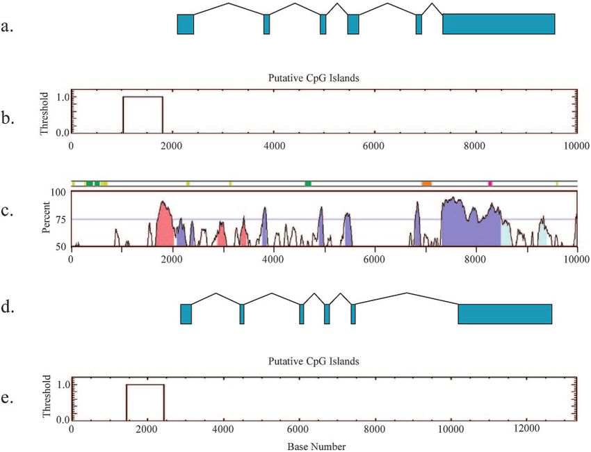
VOL. 25, 2005 ANALYSIS OF A NOVEL IMPRINTED GENE IN THE BRAIN 5515
TABLE 1. Probe set values TCC-3⬘. The probe for exons 13 and 14 was generated from primers 5⬘-TGAC
TCCATCAGCAGGCAGTATG-3⬘ and 5⬘-CTGTAGGCATCCTTGAACCGA
Signal log C-3⬘. A Northern blot with mouse brain and kidney RNA was probed with an
Probe set Region P Maternal signal Paternal signal
ratio
exon 1-specific probe generated from primers 5⬘-ATGCGACCATTGTCTCCG
1433542_at 3⬘ ⫺3.6 0.99998 3008.9 38804.5 TG-3⬘ and 5⬘-CATTCTGAAAACTGCTGCTTGAGC-3⬘.
1447757_x_at 3⬘ ⫺3.5 0.99997 380.7 5760.0 Bioinformatics. Expressed sequence tags (ESTs) were identified from the
1442100_at 5⬘ 0 0.111714 297.0 230.7 NCBI database, the TIGR database, and the UCSC genome browser. The mouse
genomic sequence for Inpp5f_v2 analyzed was 116,165,000 to 116,175,000 nucle-
otides (UCSC May 2004 assembly, NCBI build 33). The human genomic se-
quence obtained for chromosome 10 was nucleotides 121,566,025 to 121,579,332
bioarray labeling kit. Labeled probe was fragmented and hybridized to 430 A&B (NCBI build 35) determined by the Vistaplot program. The mouse sequence was
2.0 arrays for 16 h by using standard Affymetrix protocols. The arrays were then assembled from RACE product sequencing and from the ESTs with accession
washed on an Affymetrix fluidics station 450 and scanned on an Affymetrix numbers BB639524, BB646689, AW561896, and BE305393. The mouse genomic
scanner 3000, and data were extracted by using GCOS software. sequence was obtained by sequencing a BAC clone and from the UCSC genome
5ⴕ RACE for full-length clone identification. IMAGE clone 575575 (accession
browser sequences in the region of the ESTs listed above.
number AA124959) was identified by using Incyte Genomics GEM microarrays.
Programs used for sequence analysis. CpG plot (http://bioweb.pasteur.fr
Downloaded from http://mcb.asm.org/ on February 25, 2021 by guest
To further characterize this paternally expressed/maternally imprinted brain-
/seqanal/interfaces/cpgplot.html) was used to report the incidence of CpG is-
specific transcript, 5⬘ RACE (rapid amplification of cDNA ends) was performed
lands. The parameters used are a CG content of ⬎50%, an observed/expected
to obtain a full-length transcript. Poly(A)⫹ RNA was derived from newborn
ratio of ⬎0.6, and a window of 200 bp with a minimum length of 250 bp. A Vista
mouse brain and RACE-ready cDNA was made by using a SMART RACE
plot (http://www.gsd.lbl.gov/vista/index.shtml) was used for visualizing global
cDNA amplification kit (Clontech). The RACE primer was designed from the
DNA sequence alignments (10). The genome assemblies and regions used were
5⬘-most end of the AA124959 sequence, 5⬘-TCCCATGAGTTAGCCCAGCTT
116,165,000 to 116,175,000 bp for chromosome 7 from the May 2004 (mm 5)
G-3⬘. RACE was performed by using the GeneAmp 9700 PCR system (Perkin-
NCBI build 33 of the mouse genome and 121,566,025 to 121,579,332 bp for
Elmer): 5 cycles at 94°C for 5 s and 72°C for 3 min; 5 cycles at 94°C for 5 s, 70°C
chromosome 10 from the human May 2004 (hg 17) NCBI build 35 of the human
for 10 s, and 72°C for 3 min; 20 cycles at 94°C for 5 s, 68°C for 10 s, and 72°C for
genome prealigned with SLAGAN (6). Exon 1 was shaded manually because
3 min. The cDNA sequence from Inpp5f_v2 was compared to genomic sequence
Vistaplot autoshading does not currently contain the novel murine Inpp5f_v2
derived from a BAC clone identified by screening the Research Genetics geno-
mic BAC library with IMAGE clone 575575 cDNA. The BAC was sequenced gene. The sequence used for the CTCF binding site search was CCGCNNGG
with ABI sequencing technology and analyzed by using Sequencher (Ge- NGNC (25, 41), which was analyzed with the nucleic acid pattern search tool
necodes). Inpp5f_v2 was assigned this name in accordance with the mouse no- FUZZNUC in EMBOSS (http://ngfnblast.gbf.de/EMBOSS). Direct repeats
menclature committee (Lois Maltis at the Jackson Laboratories). were detected by using the Repeatmasker2 software from the University of
Allele-specific assays in intersubspecies hybrids. Newborn brain and kidney Washington (http://repeatmasker.genome.washington.edu/).
RNA from C57BL/6/J (B6), Mus mus castaneus (cast), cast ⫻ B6, and B6 ⫻ cast Section in situ hybridization. Section in situ hybridization was performed on
intersubspecies hybrids was reverse transcribed into cDNA. Polymorphisms were fixed CD1 mouse embryos harvested from 12.5 to 16.5 days postcoitum (dpc).
identified by sequencing Inpp5f_v2 in the B6 and cast strains. The Inpp5f_v2 exon The mouse embryos were fixed in 4% paraformaldehyde and embedded in
1 polymorphism is at 689 bp of exon 1 and was assayed by amplifying with paraffin wax, and sections were cut 7 m thick. Radioactive riboprobes were
primers 5⬘-ACCTAAGTCCGATGGCGTTCTC-3⬘ and 5⬘-TTTCTATTCTTTC transcribed in the presence of 35[S]UTP. Sense (control) and antisense (test)
CAGGTCTTCTAGG-3⬘ to distinguish a G in B6 and a T in cast sequence. probes were generated for the test gene. The EST IMAGE:575575 was used as
Inpp5f was assayed for imprinting by a polymorphism unique to this longer the template for the probe cloned into the EcoRI and NotI sites of a modified
transcript. This polymorphism was located in exon 5 at 252 bp, is an A in B6 and pT7T3 vector. The region of probe was exon 6 of Inpp5f_v2, and thus the signal
a G in cast, and is assayed by using the primers 5⬘-CATACTGCCTGCTGATG represents both long and short transcripts. The antisense template was linearized
GAGTCAC-3⬘ and 3⬘-AGAAAAGGAGAAACTGGAGCGG-3⬘. with EcoRI and transcribed with T3 polymerase. The sense template was linear-
Northern blot analysis. A MessageMap Northern blot (Stratagene) of adult ized with NotI and transcribed with T7 polymerase. The protocol for radioactive
BALB/c mouse tissues was probed, stripped, and rehybridized with probes spe- section in situ hybridization that was followed was essentially as described in
cific for different regions of Inpp5f_v2. The probe for exon 6 was generated with references 29 and 38. The slides were counterstained with Hoechst and the
primers 5⬘-TTCAGAAGAGTCCAGCAGAACCC-3⬘ and 5⬘-CCATATTCCAG signal, visualized by using fluorescence microscopy, was photographed.
GATGACTGCCTG-3⬘. The probe for exons 2 to 4 was generated with primers Bisulfite mutagenesis and sequencing. The methylation status of CpG dinucle-
5⬘-AAGTGCTGCTGCTGCTGTCTAAC-3⬘ and 5⬘-TTGCCATCTTCTTCAG otides within the two CpG islands associated with the promoters of Inpp5f
GACTACG-3⬘. An actin probe was used as a loading control. The probe for (CpG1) and Inpp5f_v2 (CpG2) (Fig. 1) were investigated in tissues from the F1
exons 11, 12, and 13 of Inpp5f (long transcript) was generated from primers progeny of C57BL/6J-Ei (B6) mothers and Mus mus castaneus (cast) fathers at 8
5⬘-AACCCTCACGGATGCCATTC-3⬘ and 5⬘-TCACCCTTTAGAGCAGCAG weeks of age. A total of 0.5 g of B6 ⫻ cast genomic DNA was digested with
FIG. 1. Schematic representation (not drawn to scale) of mouse Inpp5f_v2 and Inpp5f. (a) Mouse Inpp5f_v2 with six exons, including a unique
first exon located within an intron of Inpp5f. (b) Full-length Inpp5f with 20 exons. (c) The positions of two CpG islands and the SAC domain are
indicated. Putative CTCF binding sites in Inpp5f_v2 are indicated by horizontal bars.5516 CHOI ET AL. MOL. CELL. BIOL.
Downloaded from http://mcb.asm.org/ on February 25, 2021 by guest
FIG. 2. Allele-specific assays of Inpp5f_v2 and Inpp5f. The allele-specific assay uses intersubspecies hybrids to confirm Inpp5f_v2 imprinting in
brain and biallelic expression of Inpp5f in brain and kidney. (a) A polymorphism was detected in the unique exon 1 of Inpp5f_v2 at 665 bp (arrow
in B6 and cast traces) and assayed by PCR and sequencing to distinguish a G in B6 and a T in the cast sequence in brain. Inpp5f_v2 is maternally
imprinted/paternally expressed in brain (arrow in BXC and CXB traces). (b) Inpp5f was assayed for imprinting by a polymorphism unique to full-
length Inpp5f in exon 5 at 126 bp (arrow in B6 and cast traces). The SNP is an A in B6 and a G in cast. Brain and kidney were assayed by using PCR
and sequencing and, in both cases, biallelic expression was detected (BXC and CXB traces). (c) RT-PCR assay of exon 1 of Inpp5f_v2 indicates
amplification from brain but not kidney. Lane 1, marker; lane 2, blank; lane 3, Inpp5f_v2 cast ⫻ B6 brain; lane 4, Inpp5f_v2 cast ⫻ B6 brain ⫺RT;
lane 5, cast ⫻ B6 kidney; lane 6, cast ⫻ B6 kidney ⫺RT; lane 7, blank; lane 8, positive control cast ⫻ B6 brain; lane 9, positive control cast ⫻ B6 kidney.
EcoRI and converted with 3.25 M sodium meta-bisulfite in 0.93 M hydroxyqui- ing Inpp5f_v2 (Fig. 1) was differentially expressed in unipa-
none essentially as described in reference 8. Desulfonated converted DNA was rental duplicated proximal chromosome 7 maternal versus
amplified with primers specific for CpG1 and CpG2 (Fig. 1). A region from
CpG1 associated with the nonimprinted Inpp5f transcript was amplified in a
paternal brain RNA by using microarray analysis but was not
nested PCR amplification with the primers forward 5⬘-TATAGTTTTAYGTTG differentially expressed in newborn liver, heart, or kidney and
GGGAGG (where Y is a mix of C and T) and reverse 5⬘-AAAAAAATACAC thus was predicted to be tissue-specifically imprinted (7). An
TAAAAAAAATAACC-3⬘ for 35 cycles at 55°C and 1.5 mM MgCl2, followed by allele-specific assay was used to confirm from which allele(s)
a second round with forward primer 5⬘-GGTATGGAGTTTTTTTAGGTTAA
this gene was expressed. A polymorphism in the unique exon 1
GGAT-3⬘ and reverse primer 5⬘-CCTAAAACAAAAAAACTCCCC-3⬘ for 35
cycles at 55°C and 1.5 mM MgCl2. The product size is 269 bp with two C/A of this gene was used to distinguish the maternal and pater-
polymorphisms between B6 and cast at positions 162 and 165, respectively. The nal alleles in mouse intersubspecies hybrids (Fig. 2a) and
primers used for the CpG2 island upstream of the imprinted Inpp5f_v2 transcript confirmed that Inpp5f_v2 is paternally expressed in brain. The
were also nested and were forward primer 5⬘-TTAGGATTTAGAGTATTTGT
related, longer transcript Inpp5f is expressed but not imprinted
AGAAA-3⬘ and reverse primer TTTACAAAAAAAATACAACCCCACTA-3⬘
for 35 cycles at 55°C and 1.5 mM MgCl2, followed by forward primer 5⬘-TTTG in both brain and kidney. A polymorphism was assayed in exon
GTAGTTTTTTGTTTATTAAGT-3⬘ and reverse primer 5⬘-ACCCCACTAAC 5 of Inpp5f, which is unique to the larger transcript, this dem-
ACTTTAACCATAAAT-3⬘ for 35 cycles at 55°C and 1.5 mM MgCl2. This gives onstrated biallelic expression in brain and kidney (Fig. 2 b).
a 367-bp product with a T/G (B6/cast) polymorphism at position 36. The PCR The Inpp5f_v2 assay in kidney RNA failed to amplify a PCR
products were gel purified by using the QiaEXII (Qiagen) kit and cloned by using
the TOPO TA cloning system (Invitrogen). Individual clones were isolated by
product (Fig. 2c, lanes 5 and 6) using exon 1-specific primers.
using a Qiaprep minispin kit and sequenced from T3 and T7 primers by using Control reverse transcription-PCR (RT-PCR) of an unrelated
standard ABI sequencing technology (Big Dye v3.1) to determine the methyl- sequence confirmed the presence of template in both brain and
ation status of the CpG dinucleotides and the parental origin of each strand. kidney samples (Fig. 2c, lanes 7 and 8). Thus, the Inpp5f_v2
Only DNA strands ⬎95% converted were used for analysis. All strands were
transcript is not expressed in kidney at levels detectable by
known to be derived from unique template strands on the basis of either epi-
genotype or an unconverted cytosine occurring outside a CpG dinucleotide (data RT-PCR.
not shown). The variant transcript was characterized by a number of
approaches. First, the Inpp5f_v2 sequence was obtained by
RESULTS using 5⬘ RACE, followed by sequencing; the cDNA and geno-
Tissue-specific imprinting. We have used chromosome mic sequences were aligned, and the exon/intron boundaries
anomalies in combination with microarrays to identify a novel were assigned and revealed six exons (Fig. 1a and 3a). Al-
imprinted gene on mouse chromosome 7. A cDNA represent- though the Inpp5f_v2 gene may encode a protein, we have noVOL. 25, 2005 ANALYSIS OF A NOVEL IMPRINTED GENE IN THE BRAIN 5517
Downloaded from http://mcb.asm.org/ on February 25, 2021 by guest
FIG. 3. Comparison of mouse Inpp5f_v2 and human INPP5F_v2 genomic sequences. (a) Schematic diagram of the Inpp5f_v2 gene with exons
1 to 6 shown in green. (b) CpG island detection with thresholds: CpG ⬎ 0.6; % observed/expected ratio GC ⬎ 50; and window size, 200 bp. (c)
Conservation analysis of human to mouse by using VistaPlot with the mouse genome as the base genome. Conserved regions are shaded as follows:
exons in blue, 3⬘ untranslated region in light blue, and conserved noncoding sequence in pink (70% threshold). The feature bar above the graph
indicates repeat elements: LTR in pink, SINE in green, DNA in orange, and other repeats in light green. (d) Schematic representation of human
transcript variant INPP5F_v2, RefSeq NM_198330. (e) A conserved CpG island is also detected in the human region.
direct evidence that a protein is made. However, several up- (Affymetrix Genechips) hybridized with maternal versus pater-
stream ATGs are present in the imprinted transcript, and an nal duplicated chromosome 7 probes (Table 1). This illustrated
in-frame ATG near the 3⬘ end of exon 1 of Inpp5f_v2 is pre- the sensitivity of the microarray screen, since 3⬘ features rep-
dicted to code for the entire C-terminal half of Inpp5f. The resenting both Inpp5f and Inpp5f_v2 detect differential expres-
transcripts were further characterized by Northern blot analy- sion between the maternal and paternal duplications, whereas
ses and a brain-specific ⬃3.2-kb transcript was identified by 5⬘ features representing only the nonimprinted Inpp5f tran-
using probes containing exons 2 to 4 and exon 6 of Inpp5f_v2 script show a lack of differential expression (Table 1).
(Fig. 4a and b). An additional, larger transcript (⬃4.7 kb) was Comparative sequence analysis identified a gene in humans
detected on Northern blots in all tissues, including brain (Fig. with sequence similarity to Inpp5f called hSac2 or INPP5F
4a, b, d, and e). Probes specific to the larger transcript covering (19). No mouse orthologues of hSac2/INPP5F have been pre-
exons 11 to 14 detect the larger transcript in all tissues tested viously described. A human-mouse conservation analysis of
but do not detect the shorter transcript, as expected (Fig. 4d Inpp5f_v2 was performed by using VistaPlot. Five exons are
and e). Thus, the larger transcript represents full-length Inpp5f. highly conserved (Fig. 3c) in a region of conserved linkage
Figure 4f shows that exon 1 is specific to brain and absent in between mouse chromosome 7 and human chromosome 10.
kidney. The UCSC genome browser reveals two additional Exon 1 of Inpp5f_v2 is unique and begins between exons 15 to
putative brain-specific transcripts; these were not detected by 16 of the full-length Inpp5f gene (Fig. 1). Although exon 1
Northern blot analysis. shows a slightly lower degree of conservation between mice
Bioinformatics analysis. The sensitivity of the microarray and humans (Fig. 3c), a high degree of conservation is main-
screen used to detect differential expression of Inpp5f_v2 in tained in parts of the CpG island.
maternal versus paternal disomies was further validated by Imprinted genes are frequently associated with CTCF bind-
analysis of Inpp5f/Inpp5f_v2 features on a different platform ing sites, and multiple putative CTCF binding site sequences5518 CHOI ET AL. MOL. CELL. BIOL.
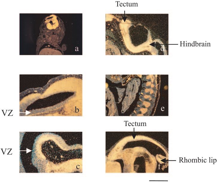
14.5dpc, expression is seen throughout the nervous system with
slightly less expression in the ventricular zones. Figure 5e
shows expression in the dorsal root ganglia lateral to the spinal
cord and separated by vertebral bodies. Expression is seen in
the tectum with a gradient of expression from the rostral to the
caudal region (Fig. 5f). Expression is again primarily outside
the ventricular zone, although the decreased expression cau-
dally may be due to less developmental progress. Strong ex-
pression is also seen in the ventral midbrain and hindbrain,
primarily outside the VZ. At this stage, relatively strong ex-
pression is seen in the rhombic lip, adjacent to the choroid
plexus. At 16.5-dpc expression is seen in the cerebral cortex,
the olfactory bulb, the ganglionic eminence, the thalamus, the
Downloaded from http://mcb.asm.org/ on February 25, 2021 by guest
hippocampus, and the retina. Slightly less expression is seen in
the inferior colliculus compared to the superior colliculus (data
not shown). The probe used was from exon 6 of Inpp5f_v2 so
it also reflects expression of Inpp5f.
Differential methylation in the brain-specific imprinted
transcript. CpG analysis of the mouse genomic sequence iden-
tified two CpG islands associated with the Inpp5f gene: one
near the 5⬘ end of Inpp5f (CpG1) and the other located in-
tronically to Inpp5f, but immediately 5⬘ of Inpp5f_v2 (CpG2)
(Fig. 1c and 3b). These two regions were investigated as
potential DMRs by using bisulfite conversion. Bisulfite se-
quencing of the CpG1 island shows that both alleles are hy-
pomethylated, as would be expected for a nonimprinted tran-
FIG. 4. Northern blot analyses of transcripts showing the brain scriptionally active gene (Fig. 6a). In the brain, CpG2, 5⬘ of
specific 3.2-kb Inpp5f_v2 band and a larger band present in all tissues.
Probes used are for exon 6 (a) and exons 2 to 4 (b) of Inpp5f_v2 and Inpp5f_v2 was differentially methylated between the maternal
-actin as a loading control (c), as well as probes from exons 11, 12, and paternal alleles. Most of the strands were methylated on
and 13 (d) and exons 13 and 14 (e) of the longer Inpp5f transcript. the maternal allele, whereas most strands were unmethylated
Exons 11 to 14 of the Inpp5f gene are outside of the Inpp5f_v2 tran- on the paternal allele (Fig. 6b). In kidney, all strands appeared
script and show no brain-specific ⬃3.2-kb band. (f) Exon 1 of Inpp5f_v2
almost completely methylated on the maternal allele. In con-
shows a 3.2-kb band specific to the brain and absent in kidney.
trast, on the paternal allele nine strands were unmethylated
and six were methylated (Fig. 6c). Statistical analysis showed
were detected within the CpG island associated with Inpp5f_v2 that the numbers of maternally methylated and unmethylated
by sequence searching (Fig. 1). In addition, a consensus CTCF strands in brain compared to kidney is not statistically signifi-
direct repeat (n ⫽ 2) is located further 3⬘ between exons 4 and cant. A standard goodness-of-fit with Yates’ continuity correc-
5 in an intronic sequence. The human INPP5F_v2 CpG island tion gives a 2 of 1.341 and a P value of 0.2469. Similarly, the
showed no exact matches to the CTCF consensus but, allowing Fisher’s exact test for count data, for the small sample size,
for two mismatches, 13 consensus sequences were detected in returns a P value of 0.22, a finding consistent with the 2 result.
the forward direction and 14 in the reverse orientation. The
structure of the human INPP5F_v2 transcript predicted from
DISCUSSION
Refseq clone NM_198330 (the orthologue of the mouse
Inpp5f_v2 gene) is shown in Fig. 3d and is similar to the mouse. Using chromosome anomalies and microarrays to detect
Spatial and temporal expression. A low-magnification 14.5- imprinted genes. Several approaches have been devised to
dpc embryo parasagittal section (lateral to the midline) illus- discover imprinted genes, and several have been detected by
trates the brain specific expression (Fig. 5a). A higher-power using microarrays to assay either parthenogenetic versus nor-
sagittal image (Fig. 5b) shows expression in the lateral cortex. mally fertilized embryos (16) or versus androgenetic embryos
There is strong labeling primarily outside the ventricular (pro- (20). A large-scale screen for imprinted transcripts has been
liferative) zone (VZ) dorsally and ventrally, although some VZ performed by using FANTOM cDNAs on RIKEN microarrays
labeling is also seen dorsally and frontally. A high-power view (23). Differentially expressed genes were identified, of which a
of a 12.5-dpc embryo highlighting the reduced expression in proportion will be nonimprinted genes due to differences in
the VZ and strong expression in the maturing regions of the development between androgenetic and parthenogenetic em-
midbrain and hindbrain is shown in Fig. 5c. There is an obvious bryos at 9.5 dpc (32, 37). Our experimental design has been
ventral gap in the expression extending from the VZ that could mindful of cell type differences and matches developmental
represent the isthmus. Expression is seen in the hindbrain and stage both for embryo and isolated tissue comparisons. Fur-
in a small part of the tectum. The area that will become the ther, the sensitivity of this screen is underscored by additional
brainstem is strongly labeled, whereas the rhombic lip (more microarray experiments that were able to discriminate between
dorsally located precursor of the cerebellum) shows less label- the transcriptional variants of the Inpp5f gene. Differences in
ing as does the more rostral portion of the tectum (Fig. 5d). At expression between maternal and paternal alleles for 3⬘ fea-VOL. 25, 2005 ANALYSIS OF A NOVEL IMPRINTED GENE IN THE BRAIN 5519
Downloaded from http://mcb.asm.org/ on February 25, 2021 by guest
FIG. 5. In situ hybridization of mouse embryo sections from 12.5 and 14.5 dpc. Signal is shown as yellow staining, and nonexpressing cells have
a blue appearance. (a) Parasagittal section (fairly far lateral) from 14.5-dpc embryo showing expression primarily in the brain. At the top (which
is highlighted in panel b) is the cerebral cortex. (b) Higher-power image of a 12.5-dpc embryo showing strong expression in the lateral cortex but
sparing the ventricular zone. (c) High-power view of a 12.5-dpc embryo highlighting the reduced expression in the ventricular zone and strong
expression in the maturing regions of the midbrain and hindbrain. (d) Expression is seen in the hindbrain and tectum. (e) Expression is seen in
the 12.5-dpc dorsal root ganglia lateral to the spinal cord separated by the vertebral disks (blue, nonstaining cells). (f) Expression is seen in the
tectum and largely outside the ventricular zone (14.5 dpc). Strong expression is also shown in the ventral midbrain and hindbrain and rhombic lip.
Scale bars: a, 200 m; b to f, 100 m. VZ, ventricular zone.
tures representing Inpp5f and Inpp5_v2 were detected, whereas the exons of Inpp5f_v2 as its C-terminal domain, the function
similar expression was detected for 5⬘ features representing of which is unknown.
Inpp5f alone (Table 1), further validating this approach. Transcript size, sequence features, and tissue distribution
Inpp5f_v2, Inpp5f, and hSac2/INPP5F. Inpp5f_v2 is located of Inpp5f_v2. Northern analyses reveal a brain-specific 3.2-kb
on distal mouse chromosome 7, band position 7F3 at 116.1 Mb transcript and a larger 4.7-kb transcript present in all tested
(UCSC Genome Browser using the Mouse May 2004 assem- tissues (Fig. 4). The 3.2-kb Inpp5f_v2 transcript is imprinted in
bly), ca. 14 Mb proximal of the H19/Igf2 cluster. This region brain (Fig. 2a) and the longer Inpp5f transcript is biallelically
of the genome shares conserved linkage with human chromo- expressed in both brain and kidney (Fig. 2b and c). In situ
some 10q26.13 in the region of the human gene hSac2/INPP5F. hybridization studies illustrate that Inpp5f_v2 and Inpp5f are
Comparative mouse and human sequence analysis has iden- expressed in brain structures and the developing nervous sys-
tified sequence similarity between Inpp5f_v2 and the hSac2/ tem over a range of developmental time points in the mouse
INPP5F gene. No mouse orthologue of hSac2/INPP5F has pre- embryo (Fig. 5).
viously been described. Inpp5f_v2 is a transcriptional variant of Approximately 40 to 60% of the multiexon genes in the
Inpp5f. Inositol phosphatases are involved in endocytosis and mouse and human transcriptomes exhibit alternative splicing
exocytosis at nerve terminals, a process essential for clathrin (21, 43). This facilitates increased complexity of the mamma-
coat shedding and synaptic vesicle recycling (1), which is im- lian proteome and altered protein function. In many cases, the
portant in the developing brain and in postnatal survival. It has alternative use of a first exon is regulated by alternative pro-
been shown that full-length human INPP5F has inositol 4,5- moters, conferring tissue specificity to the different isoforms of
bisphosphatase activity in vitro (19). However, unlike hSac2/ a gene. A recent genome survey reports that ⬎2,000 genes in
INPP5F, Inpp5f_v2 does not contain a Sac domain but rather is the mouse and ⬎3,000 genes in humans have alternative first
similar to the variable C-terminal domain of hSac2/INPP5F exons spliced to a common 3⬘ transcript (44) and, in many
(Fig. 1). It may be that Inpp5f_v2 originated independently of cases, conserved CpG islands are associated with the variable
the Sac domain of Inpp5f and that the larger transcript coopted exon. Inpp5f_v2 uses an alternative transcriptional start site5520 CHOI ET AL. MOL. CELL. BIOL.
Downloaded from http://mcb.asm.org/ on February 25, 2021 by guest
FIG. 6. Bisulfite mutagenesis of the methylation status of CpG dinucleotides in genomic DNA. Unfilled circles represent unmethylated CpGs,
and filled circles represent methylated CpGs; shaded circles indicate GpGs that could not be determined unequivocally. (a) BXC (B6 ⫻ cast) brain
DNA with primers specific for CpG1, maternal, and paternal alleles are hypomethylated. (b) BXC (B6 ⫻ cast) brain DNA with primers specific
for CpG2, the maternal allele appears hypermethylated and the paternal allele appears hypomethylated. (c) BXC (B6 ⫻ cast) kidney DNA with
primers specific for CpG2. The maternal allele is hypermethylated; the paternal allele has some methylated and some unmethylated CpGs.
between exons 15 and 16 of Inpp5f and thus is the first example specific RT-PCR assay. Although the in situ hybridization data
we are aware of in which the unique first exon is embedded suggest that expression of the Inpp5fv2 transcript varies in
within an intron near the 3⬘ end of the gene rather than the different regions of the brain (Fig. 5), this is unlikely to account
selection among first exons seen at the Gnas locus (26). At the for these findings since differential methylation is also present
Gnas locus, three CpG islands are associated with three alter- in nonexpressing kidney tissue (Fig. 6c), albeit to a lesser ex-
native start sites sharing common 3⬘ exons. The transcription tent. One possible explanation is that this DMR exerts its role
of Gnas is dependent on the methylation state of the CpG in the imprinting of Inpp5f_v2 at an early stage of embryonic
islands, as can be seen in the alternative use of the first exon of development and is not absolutely required for maintenance of
Nesp, Gnasxl (26), or Gnas1A (18). In humans, the alternative the imprint in neonatal and adult tissues. A similar situation
WT1 transcript AWT1 is similarly organized, is paternally ex- has been found in the Angelman/Prader-Willi Syndrome re-
pressed, and consists of exons 2 to 10 of the WT1 gene. How- gion, where differential methylation established in the germ
ever, like at the Gnas locus, this transcript uses a novel 5⬘ exon
line and maintained at 7.5 dpc is lost by adulthood (15). Bisul-
located in the first intron of the nonimprinted WT1 gene (11).
fite sequencing analysis of CpG2 in gametes and early-stage
Differential methylation. Differential methylation has been
embryos would determine whether this DMR is germ line
associated with most of the characterized imprinted genes,
derived and progressively lost during pre- and postnatal devel-
such as those in the H19/Igf2 region (36) and at the Dlk1/Gtl2
locus (34). In both of these domains, CpGs are hypermethyl- opment.
ated on the silent paternal allele and hypomethylated on the CpG1 is hypomethylated on both parental alleles (Fig. 6a),
expressing maternal allele. The methylation status of two CpG and Inpp5f is biallelically expressed in both kidney and brain.
islands associated with Inpp5f and Inpp5f_v2 was determined. The differential methylation pattern at CpG2 is maintained
The CpG2 is differentially methylated in brain, where the ex- in tissues where Inpp5f_v2 is not expressed, suggesting that a
pressing paternal allele is unmethylated and the silent mater- brain-specific transcriptional regulation mechanism other than
nal allele is methylated (Fig. 6b). This differentially methylated methylation may be involved and possibly imposed in addi-
region is likely to be involved in the imprinted expression of tion to the epigenetic silencing of the maternal allele. For
Inpp5f_v2 in brain. However, three maternal alleles out of Inpp5f_v2, methylation is related to imprinted gene expression
thirteen were hypomethylated (Fig. 6b), but no corresponding but not to tissue specificity; this is observed in the human NDN
maternally derived transcription was detected in the allele- gene, where a promoter-associated CpG island exhibits differ-VOL. 25, 2005 ANALYSIS OF A NOVEL IMPRINTED GENE IN THE BRAIN 5521
ential methylation independently of tissue-specific transcrip- 13. Guillemot, F., T. Caspary, S. M. Tilghman, N. G. Copeland, D. J. Gilbert,
N. A. Jenkins, D. J. Anderson, A. L. Joyner, J. Rossant, and A. Nagy. 1995.
tional status (17). Genomic imprinting of Mash2, a mouse gene required for trophoblast de-
One of the elements involved in the control of the reciprocal velopment. Nat. Genet. 9:235–241.
imprinting of the H19 and Igf2 genes is the vertebrate enhancer 14. Hark, A. T., C. J. Schoenherr, D. J. Katz, R. S. Ingram, J. M. Levorse, and
S. M. Tilghman. 2000. CTCF mediates methylation-sensitive enhancer-
blocking protein, CTCF, which binds to sites in DNA to block blocking activity at the H19/Igf2 locus. Nature 405:486–489.
promoter-enhancer interactions (4, 14, 33). Multiple CTCF 15. Kantor, B., Y. Kaufman, K. Makedonski, A. Razin, and R. Shemer. 2004.
binding site sequences were detected within the Inpp5f_v2 Establishing the epigenetic status of the Prader-Willi/Angelman imprinting
center in the gametes and embryo. Hum. Mol. Genet. 13:2767–2779.
CpG2 in mice (Fig. 1). However, functional CTCF binding 16. Kobayashi, S., H. Wagatsuma, R. Ono, H. Ichikawa, M. Yamazaki, H.
sites cannot be predicted from sequence alone and provide Tashiro, K. Aisaka, N. Miyoshi, T. Kohda, A. Ogura, M. Ohki, T. Kaneko-
Ishino, and F. Ishino. 2000. Mouse Peg9/Dlk1 and human PEG9/DLK1 are
only an indication of potential binding in vivo. paternally expressed imprinted genes closely located to the maternally ex-
Many imprinted genes are clustered, suggesting that they pressed imprinted genes: mouse Meg3/Gtl2 and human. MEG3. Genes Cells
reside in domains that are epigenetically modified, resulting in 5:1029–1037.
17. Lau, J. C. Y., M. L. Hanel, and R. Wevrick. 2004. Tissue-specific and im-
the coordinate regulation of more than one imprinted gene. printed epigenetic modifications of the human NDN gene. Nucleic Acids
Downloaded from http://mcb.asm.org/ on February 25, 2021 by guest
Although the identification of a DMR near Inpp5f_v2 is con- Res. 32:3376–3382.
sistent with an imprinted domain, one might expect one or 18. Liu, J., S. Yu, D. Litman, W. Chen, and L. S. Weinstein. 2000. Identification
of a methylation imprint mark within the mouse Gnas locus. Mol. Cell. Biol.
more additional imprinted genes to be present. Inspection of 20:5808–5817.
flanking sequence in combination with microarray differential 19. Minagawa, T., T. Ijuin, Y. Mochizuki, and T. Takenawa. 2001. Identification
and characterization of a Sac domain-containing phosphoinositide 5-phos-
expression data in this region revealed a cluster of differentially phatase. J. Biol. Chem. 276:22011–22015.
expressed transcripts 3⬘ to this gene. Further study of this 20. Mizuno, Y., Y. Sotomaru, Y. Katsuzawa, T. Kono, M. Meguro, M. Oshimuru,
flanking cluster of imprinted genes will shed light on whether J. Kawai, Y. Tomaru, H. Kiyosawa, I. Nikaido, H. Amanuma, Y. Hayash-
izaki, and Y. Okazaki. 2002. Asb4, Ata3, and Dcn are novel imprinted genes
Inpp5f_v2 resides alone or is coordinately regulated as part of identified by high-throughput screening using RIKEN cDNA microarray.
a larger region. Biochem. Biophys. Res. Commun. 290:1499–1505.
21. Modrek, B., and C. J. Lee. 2003. Alternative splicing in the human, mouse
ACKNOWLEDGMENTS and rat genomes is associated with an increased frequency of exon creation
and/or loss. Nat. Genet. 34:177–180.
This study was supported by PHS grant GM58759 from the National 22. Morison, I. M., C. J. Paton, and S. D. Cleverley. 2001. The imprinted gene
Institutes of Health (R.J.O.) and by The Wellcome Trust (R.J.O.). and parent of origin effect database. Nucleic Acids Res. 29:275–276.
J.D.C. is supported by an IRG fellowship from the GKT School of 23. Nikaido, I., C. Saito, Y. Mizuno, M. Meguro, H. Bono, M. Kadomura, T.
Medicine, King’s College London, and A.J.W. is supported by a stu- Kono, G. A. Morris, P. A. Lyons, M. Oshimura, Y. Hayashizaki, Y. Okazaki,
dentship from the Generation Trust. et al. 2003. Discovery of imprinted transcripts in the mouse transcriptome
using large-scale expression profiling. Genome Res. 13(Suppl. 6B):1402–
We thank Colin V. Beechey for the T65H mouse tissues and careful 1409.
reading of the manuscript. We thank Kathryn Woodfine for Affymetrix 24. Paulsen, M., O. El-Maarri, S. Engemann, M. Strodicke, O. Franck, K.
GeneChip hybridizations and Trevelyan Menheniott and Reiner Davies, R. Reinhardt, W. Reik, and J. Walter. 2000. Sequence conservation
Schulz for analysis of Affymetrix microarray data and statistical anal- and variability of imprinting in Beckwith-Wiedemann syndrome gene cluster
ysis. in human and mouse. Hum. Mol. Genet. 9:1829–1841.
25. Paulsen, M., S. Takada, N. A. Youngson, M. Bebnchaib, C. Charlier, K.
REFERENCES Segers, M. Georges, and A. C. Ferguson-Smith. 2001. Comparative sequence
1. Arai, Y., T. Ijuin, M. Itoh, T. Takenawa, S. Takashima, and L. E. Becker. analysis of the imprinted Dlk1-Gtl2 locus in three mammalian species reveals
2001. Developmental changes of synaptojanin expression in the human ce- highly conserved genomic elements and refines comparison with the Igf2-
rebrum and cerebellum. Dev. Brain Res. 129:1–9. H19 region. Genome Res. 11:2085–2094.
2. Barlow, D. P. 1995. Gametic imprinting in mammals. Science 270:1610–1613. 26. Peters, J., S. F. Wroe, C. A. Wells, H. J. Miller, D. Bodle, C. V. Beechey, C. M.
3. Beechey, C. V., B. M. Cattanach, and R. L. Selley. 2000. Standard ideogram/ Williamson, and G. Kelsey. 1999. A cluster of oppositely imprinted tran-
anomaly breakpoints of the Mouse MRC Mammalian Genetics Unit, Har- scripts at the Gnas locus in the distal imprinting region of mouse chromo-
well, Oxfordshire, United Kingdom. [Online.] http:www.mgu.har.mrc.ac.uk some 2. Proc. Natl. Acad. Sci. USA 96:3830–3835.
/imprinting/implink.html. 27. Reik, W., and J. Walter. 2001. Genomic imprinting: parental influence on the
4. Bell, A. C., and G. Felsenfeld. 2000. Methylation of a CTCF-dependent genome. Nat. Rev. Genet. 2:21–32.
boundary controls imprinted expression of the Igf2 gene. Nature 405:482– 28. Reik, W., and J. Walter. 1998. Imprinting mechanisms in mammals. Curr.
485. Opin. Genet. Dev. 8:154–164.
5. Blagitko, N., S. Mergenthaler, U. Schulz, H. A. Wollmann, W. Craigen, T. 29. Sassoon, D., and N. Rosenthal. 1993. Detection of messenger RNA by in situ
Eggermann, H. H. Ropers, and V. M. Kalscheuer. 2000. Human GRB10 is hybridization, p. 384–406. In P. M. Wassarman and M. L. DePamphilis (ed.),
imprinted and expressed from the paternal and maternal allele in a highly Methods in enzymology. Academic Press, London, England.
tissue- and isoform-specific fashion. Hum. Mol. Genet. 9:1587–1595. 30. Schoenherr, C. J., J. M. Levorse, and S. M. Tilghman. 2003. CTCF maintains
6. Brudno, M., C. B. Do, G. M. Cooper, M. F. Kim, E. Davydov, E. D. Green, differential methylation at the Igf2/H19 locus. Nature Genetics 33:66–69.
A. Sidow, and S. Batzoglou. 2003. NISC Comparative Sequencing Program 31. Smilinich, N. J., C. D. Day, G. V. Fitzpatrick, G. M. Caldwell, A. C. Lossie,
LAGAN and Multi-LAGAN: efficient tools for large-scale multiple align- P. R. Cooper, A. C. Smallwood, J. A. Joyce, P. N. Schofield, W. Reik, R. D.
ment of genomic DNA. Genome Res. 13:721–731. Nicholls, R. Weksberg, D. J. Driscoll, E. R. Maher, T. D. Shows, and M. J.
7. Choi, J. D., L. A. Underkoffler, J. C. Collins, S. M. Marcheginani, N. A. Higgins. 1999. A maternally methylated CpG island in KvLQT1 is associated
Terry, C. V. Beechey, and R. J. Oakey. 2001. Microarray expression profiling with an antisense paternal transcript and loss of imprinting in Beckwith-
of tissues from mice with uniparental duplications of chromosomes 7 and 11 Wiedemann syndrome. Proc. Natl. Acad. Sci. USA 96:8064–8069.
to identify imprinted genes. Mammalian Genome 12:758–764. 32. Surani, M. A., S. C. Barton, and M. L. Norris. 1986. Nuclear transplantation
8. Clark, S. J., J. Harrison, C. L. Paul, and M. Frommer. 1994. High sensitivity in the mouse: heritable differences between parental genomes after activa-
mapping of methylated cytosines. Nucleic Acids Res. 22:2990–2997. tion of the embryonic genome. Cell 45:127–136.
9. Constancia, M., B. Pickard, G. Kelsey, and W. Reik. 1999. Imprinting mech- 33. Szabo, P., S. H. Tang, Rentsendorj.A., G. P. Pfeifer, and J. R. Mann. 2000.
anisms. Genome Res. 8:881–900. Maternal-specific footprints at putative CTCF sites in the H19 imprinting
10. Couronne, O., A. Poliakov, N. Bray, T. Ishkhanov, D. Ryaboy, E. Rubin, L. control region give evidence for insulator function. Curr. Biol. 18:607–610.
Pachter, and I. Dubchak. 2003. Strategies and tools for whole-genome align- 34. Takada, S., M. Paulson, M. Trevendale, C.-E. Tsai, G. Kelsey, B. M. Catta-
ments. Genome Res. 13:73–80. nach, and A. C. Ferguson-Smith. 2002. Epigenetic analysis of the Dlk-Gtl2
11. Dallosso, A. R., A. L. Hancock, K. W. Brown, A. C. Williams, S. Jackson, and imprinted domain on mouse chromosome 12: implications for imprinting
K. Malik. 2003. Genomic imprinting at the WT1 gene involves a novel control from comparisons with Igf2-H19. Hum. Mol. Genet. 11:77–86.
coding transcript (AWT1) that shows deregulation in Wilms’ tumours. Hum. 35. Thorvaldson, J., J. L. Duran, and M. S. Bartolomei. 1998. Deletion of the
Mol. Genet. 13:405–415. H19 differentially methylated domain results in loss of imprinted expression
12. DeChiara, T. M., E. J. Robertson, and A. Efstratiadis. 1991. Parental im- of H19 and Igf2. Genes Dev. 12:3693–3702.
printing of the mouse insulin-like growth factor II gene. Cell 64:849–859. 36. Tremblay, K. D., J. R. Saam, R. S. Ingram, S. M. Tilghman, and M. S.5522 CHOI ET AL. MOL. CELL. BIOL.
Bartolomei. 1995. A paternal specific methylation imprint marks the alleles D. P. Barlow. 1997. Imprinted expression of the Igf2r gene depends on an
of the mouse H19 gene. Nat. Genet. 9:407–413. intronic CpG island. Nature 389:745–749.
37. Wilkins, A. S. 1993. Genetic analysis of animal development, 2nd ed. Wiley- 41. Wylie, A. A., S. K. Murphy, T. C. Orton, and R. L. Jirtle. 2000. Novel
Liss, New York, N.Y. imprinted DLJ1/GTL2 domain on human chromosome 14 contains motifs
38. Wilkinson, D. G., and M. A. Nieto. 1993. Detection of messenger RNA by in that mimic those implicated in IGF2/H19 regulation. Genome Res. 10:1711–
situ hybridization to tissue sections and whole mounts, p. 361–372. In P. M. 1718.
42. Yoon, B. J., H. Herman, A. Sikora, L. T. Smith, C. Plass, and P. D. Soloway.
Wassarman and M. L. DePamphilis (ed.), Methods in enzymology. Aca-
2002. Regulation of DNA methylation of Rasgrf1. Nat. Genet. 30:92–96.
demic Press, London, England.
43. Zavolan, M., S. Kondo, C. Schonbach, J. Adachi, D. A. Hume, Y. Hayash-
39. Wroe, S. F., G. Kelsey, J. A. Skinner, D. Bodle, S. T. Ball, C. V. Beechey, J. izaki, T. Gaasterland, et al. 2003. Impact of alternative initiation, splicing,
Peters, and C. M. Williamson. 2000. An imprinted transcript, antisense to and termination on the diversity of the mRNA transcripts encoded by the
Nesp, adds complexity to the cluster of imprinted genes at the mouse Gnas mouse transcriptome. Genome Res. 13(Suppl. 6B):1290–1300.
locus. Proc. Natl. Acad. Sci. USA 97:3342–3346. 44. Zhang, T., P. Haws, and Q. Wu. 2004. Multiple variable first exons: a mech-
40. Wutz, A., O. W. Smrzka, N. Schweifer, K. Schellander, E. F. Wagner, and anism for cell- and tissue-specific gene regulation. Genome Res. 14:79–89.
Downloaded from http://mcb.asm.org/ on February 25, 2021 by guestYou can also read