The Drosophila javelin Gene Encodes a Novel Actin-Associated Protein Required for Actin Assembly in the Bristle䌤
←
→
Page content transcription
If your browser does not render page correctly, please read the page content below
MOLECULAR AND CELLULAR BIOLOGY, Nov. 2011, p. 4582–4592 Vol. 31, No. 22
0270-7306/11/$12.00 doi:10.1128/MCB.05730-11
Copyright © 2011, American Society for Microbiology. All Rights Reserved.
The Drosophila javelin Gene Encodes a Novel Actin-Associated
Protein Required for Actin Assembly in the Bristle䌤
Shira Shapira, Anna Bakhrat, Amir Bitan, and Uri Abdu*
Department of Life Sciences and National Institute for Biotechnology in the Negev,
Ben-Gurion University, Beer-Sheva 84105, Israel
Received 1 June 2011/Returned for modification 23 June 2011/Accepted 7 September 2011
The Drosophila melanogaster bristle is a highly polarized cell that builds specialized cytoskeletal struc-
tures. Whereas actin is required for increasing bristle length, microtubules are essential for bristle axial
Downloaded from http://mcb.asm.org/ on January 5, 2021 by guest
growth. To identify new proteins involved in cytoskeleton organization during bristle development, we
focused on identifying and characterizing the javelin (jv) locus. We found that in a jv mutant, the bristle
tip is swollen and abnormal organization of bristle grooves is seen over the entire bristle. Using confocal
and electron microscopy, we found that in jv mutant bristles, actin bundles do not form properly due to
a loss of actin filaments within the bundle. We show that jv is an allele of the predicted CG32397 gene that
encodes a protein with no homologs outside insects. Expression of the Jv protein fused to a green
fluorescent protein (GFP) shows that the protein is colocalized with actin bundles in the bristle. Moreover,
expression of Jv-GFP within the germ line led to the formation of ectopic actin bundles that surround the
nucleus of nurse cells. Thus, we report that Jv is a novel actin-associated protein required for actin
assembly during Drosophila bristle development.
Changes in cell shape, driven by cytoskeleton structures In addition to actin bundles, a large population of microtu-
within the cell, are fundamental processes that allow cells and bules (MTs) is also found in the bristle shaft. It was shown that
tissues to adopt new shapes and functions. The highly polar- the majority of the MT array, which is shorter than the length
ized shape of the Drosophila melanogaster mechanosensory of a mature bristle, assembles early in development, although
bristle makes it a good model for studying the role of the the mechanism by which the MT network is organized remains
cytoskeleton during cell elongation. Since the cytoskeleton unknown (22). Since microtubule antagonists injected imme-
plays a crucial role in the development of these highly polar- diately prior to the time of bristle initiation led to the appear-
ized bristles, any changes in bristle organization would result in ance of shorter and fatter bristles than seen in untreated flies
an easily detectable phenotype in adult flies (22). Bristles (5), it was suggested that microtubules assume an important
sprout from the subepidermal layer during metamorphosis, role in maintaining bristle axial growth. Moreover, it was dem-
32 h after puparium formation (APF), and elongate over the onstrated that during bristle elongation, treatment with micro-
course of 16 h (24). Bristles can be divided into microchaetes tubule antagonists had no effects on microtubule network den-
and macrochaetes based on their size, number, and location. sity, suggesting that MTs within the bristle shaft are stable (22).
The 200 microchaete bristles, ordered in evenly spaced rows, Recently, we have shown that MTs are organized in a polarized
have an average length of 70 m. In contrast, the 22 macro- manner, i.e., minus end out, and that this MT organization is
chaetes on the fly’s dorsal thorax can reach lengths of up to require for the bristle axial shape (2).
400 m each (11, 19). javelin (jv) is a spontaneous mutation that was identified in
Bristle growth is driven by actin filament polymerization 1947. jv mutant bristles do not taper like bristles in wild-type
(22), with actin filaments being continually formed at the bris- flies, instead presenting a small enlargement before the tip. It
tle tip, where they are cross-bridged into modules. These mod- was shown that jv is cell autonomous and useful as a marker for
ules are attached end to end into firm bundles that lie against epidermal clones (14). Since jv affects bristle development, we
the membrane. Bundle formation is achieved in a three-stage focused on the further characterization of jv phenotypes and
process by at least two cross-linking proteins, forked and fascin
the identification of the encoding gene. We have shown that in
(19, 20, 23). Once elongation is complete, the actin bundles
jv mutants, the bristle tip becomes swollen, resembling a spear
decompose (⬃53 h APF) and a thick layer of chitin supports
(13), rather than assuming the pointed tip seen in wild-type
the cell in place of the bundles (9, 20). Thus, at the end of this
bristles. We showed, furthermore, that jv mutant bristles dis-
process, the bristle of the adult fly presents a grooved pattern.
play an irregular groove pattern with unparallel and shallower
The valleys of the bristle represent regions where membrane-
ridges. Now, closer examination of actin bundle arrangement
attached actin bundles were positioned, while the bristle ridges
in jv mutants using transmission electron microscopy (TEM)
correspond to regions that lacked actin bundles (19).
reveals that the loss of actin filaments within the bundles af-
fects the triangular structure of the bundles. We also demon-
strate that the jv mutant carries the predicted CG32397 gene
* Corresponding author. Mailing address: Ben-Gurion University,
Ben-Gurion Blvd., Beer-Sheva 84105, Israel. Phone: 972-8-6479072.
and that this gene encodes an actin-associated protein. Thus,
Fax: 972-8-6461710. E-mail: abdu@bgu.ac.il. our study identifies Jv as a novel actin-associated protein re-
䌤
Published ahead of print on 19 September 2011. quired for actin assembly.
4582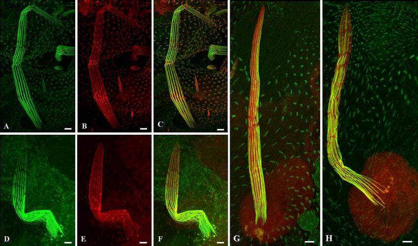
VOL. 31, 2011 JAVELIN, A NOVEL ACTIN-ASSOCIATED PROTEIN 4583
MATERIALS AND METHODS (5⬘-GGATGCAATTGCTCACTCAG-3⬘). Thus, we performed 5⬘ rapid amplifi-
cation of cDNA ends (RACE) as described below to identify the 5⬘ end of the
Drosophila stocks. The following mutant and transgenic fly strains were
used. The CG18769[c03230], CG18769[EY01803], jv1, nd11, nd14, Zpg96, Zpg107, gene. For this, we used a forward primer corresponding to the new predicted
Df(3L)BSC410, Df(3L)Exel9058, Df(3L)BSC437, Df(3L)Exel7210, Df(3L)BSC411, translation start site, FW1 New (5⬘-ATGGGCAACGGATATTTTCG-3⬘). The
Df(3L)Exel8101, Df(3L)ED211 Df(3L)Exel6109, Df(3L)BSC27, Df(3L)BSC224, rest of the sequence was amplified using primer pairs FW2 (5⬘-CAGCAGCAA
and w[1118]; CyO, P{Tub-PBacT}2/wg[Sp-1] lines were all obtained from the CATCAACAGCA-3⬘) and REV2 (5⬘-CCACTACAGCTAGGATATTC-3⬘),
Bloomington Stock Center. Germ line and bristle expression was performed with FW3 (5⬘-TCCTCTGTTAACCACCACAG-3⬘) and REV3 (5⬘-CTGAACGAGA
P{mat␣4-GAL-VP16}V37 (referred to as ␣-tub Gal4-3) and neur-Gal4, respec- TTACAAGTTG-3⬘), FW4 (5⬘-AGAAGGAGGCAATTCAGGAG-3⬘) and
tively. Ubiquitous expression was performed with actin5C-Gal4. All of the Gal4 REV4 (5⬘-GAACAGTCGTAGGCGTAGTTG-3⬘), FW5 (5⬘-CAACAATAATG
lines were obtained from the Bloomington Stock Center. The following upstream TGGTGATAG-3⬘) and REV5 (5⬘-ACACATTGCAGGCCTCGATTTG-3⬘),
activation sequence (UAS)-RNA interference (RNAi) lines, designated by trans- and FW6 (5⬘-TACTACGGAGGAAGAGTGCTG-3⬘) and REV6 (5⬘-CATTTT
formant identification number, were obtained from VDRC, Austria: CG18769 GTCATCCAGGC-3⬘).
RNAi, 9501; CG32397 RNAi, 103870; CG8368 RNAi, 108563; CG8398 RNAi, To identify the 5⬘ end of the CG32397 gene, single-stranded cDNA was
35927; and CG8398 RNAi, 35928. reverse transcribed from 1 to 3 g RNA extracted from wild-type pupae using
Developmental staging and pupa dissection. For examination of the bristles in BD PowerScript reverse transcriptase, 5⬘-CDS primer, and BD Smart II A oligo
wild-type and mutant flies, white prepupae were collected and placed on double- (BD Smart RACE cDNA amplification) according to the manufacturer’s instruc-
Downloaded from http://mcb.asm.org/ on January 5, 2021 by guest
sided Scotch tape in a petri dish placed in a 25°C incubator as previously tions (Clontech). 5⬘ RACE was performed using a gene-specific primer (5⬘-CG
described (22). At the appropriate time of incubation (38 to 48 h), the pupae AAAATATCCGTTGCCCATCGTG-3⬘) and a universal primer from the kit.
were dissected by removing the pupal case, and the pupae were filleted in The PCR conditions used were as follows. There were five initial PCR cycles
phosphate-buffered saline (PBS), pH 7.35 to 7.45, as described previously (21). consisting of 94°C for 30 s and 72°C for 3 min. Five additional cycles of 94°C for
The thoracic fillet was then placed on its back, and the internal organs and fat 30 s, 70°C for 30 s, and 72°C for 3 min followed. Finally, 25 PCR cycles consisting
bodies were removed (22). The clean tissue was fixed for light or electron of 94°C for 30 s, 68°C for 30 s, and 72°C for 3 min were conducted. The RACE
microscopy. product was analyzed by 1% agarose gel electrophoresis, and the fragment was
Bristle phalloidin and antibody staining. Clean thoraces were fixed in 4% purified using a DNA isolation kit (Biological Industries), subcloned into the
paraformaldehyde in PBS for 20 min and then in 4% paraformaldehyde con-
pGEM-T vector (Promega), and sequenced.
taining 0.3% Triton X-100 in PBS for an additional 20 min. The samples were
SEM. Adult Drosophila flies were fixed and dehydrated by immersion in
washed three times with 0.3% Triton X-100 in PBS for 10 min each time. For
increasing concentrations of ethanol (25%, 50%, 75%, and twice in 100%; 10 min
antibody staining, the thoraces were blocked in 0.1% Triton X-100 containing
each). The flies were then completely dehydrated using increasing concentra-
4% bovine serum albumin and 0.1% NaN3 for 1 h. The samples were then
tions of hexamethyldisilazane (HMDS) in ethanol (50%, 75%, and twice in
incubated with a primary antibody in the above-mentioned blocking solution
100%; 2 h each). The samples were air dried overnight, placed on stubs, and
overnight at 4°C, washed three times in 0.3% Triton X-100 in PBS, and incubated
coated with gold. The specimens were examined with a scanning electron mi-
with secondary antibodies and phalloidin in blocking solution for 2 h at room
temperature or at 4°C overnight in the dark. After such incubation, the samples croscope (SEM; JEOL model JSM-5610LV). Length measurements of adult
were washed three times with 0.3% Triton X-100 in PBS for 10 min each time bristles were performed using Image J (version 1.40j) software.
and then placed on a slide and mounted in Citifluor glycerol (Ted Pella, Redding, TEM. At 42 h APF, the dorsal surface of the thorax of each pupa was dissected
CA). A coverslip was placed on the sample, and the preparation was sealed with as described above and fixed for 20 min in 2% glutaraldehyde in 0.2 M PO4 (pH
nail polish. The slides were examined with an Olympus FV1000 laser-scanning 6.8) at room temperature and then for 1 h on ice. After 1 h, the specimens were
confocal microscope. The images are presented as z-axis projections of several transferred to a fixative comprising cold water, 0.2 M PO4 (pH 6.2), 4% osmium
optical sections that collectively cover the bristle diameter. Rabbit antiforked tetroxide (OsO4), and 50% glutaraldehyde and placed on ice for 1 h. The
(1:100), mouse antifascin (1:100) (provided by G. M. Guild and K. Cant, respec- specimens were then washed three times in cold water (20 min each) and
tively), mouse anti-spn-F (1:10) (1), and mouse anti-␣-tubulin (1:250) (Sigma) incubated in 1% uranyl acetate overnight at 4°C. This was followed by transfer to
primary antibodies were used. Goat anti-mouse Cy2 and Cy3 and goat anti-rabbit a dehydration series spanning from 30 to 100% acetone, with 10% increases
Cy3 (Jackson ImmunoResearch) secondary antibodies were used at a dilution of being made at each 15-min interval. Samples were then treated twice in propyl-
1:100. The goat anti-rabbit Cy3 (Molecular Probes) secondary antibodies were ene oxide for 15 min and soaked for 1 h in a 1:1 solution of propylene oxide and
used at a dilution of 1:500. For actin staining, we used Oregon Green 488- or araldite, followed by overnight incubation at 4°C in a 1:2 propylene oxide/araldite
Alexa Fluor 568-conjugated phalloidin (1:250) (Molecular Probes). mixture. The tissues were then transferred to araldite and incubated for 1 h,
Real-time RT-PCR. Total RNA was extracted after removing the pupal cases placed on araldite blocks (the blocks were polymerized a day before at 60°C), and
from pupae 48 h after pupariation by using the NucleoSpin RNA II kit, including embedded in araldite. These were then left at room temperature for 30 min after
DNase treatment, according to the manufacturer’s instructions (Macherey- embedding, at which point the samples were oriented and incubated at 60°C for
Nagel). cDNA was transcribed from 1 to 5 g total RNA using reverse trans- 24 h. Transverse sections were cut through the thorax using a Leica UltraCut
criptase and oligo(dT) (ABgene) according to the manufacturer’s instructions UCT ultramicrotome equipped with a diamond knife, stained with uranyl acetate
(Bio-Lab, Beit Haemek, Israel). Reverse-transcribed total RNA (100 ng) was and lead citrate, and then examined with a JEOL JEM-1230 transmission elec-
amplified in a 20-l reaction mixture containing 100 nM concentrations of each tron microscope operating at 120 kV.
primer and 10 l of SYBR green PCR master mix (Stratagene). Reverse tran-
Transgenic flies. To clone gene CG32397 in the pUASp vector containing
scription (RT)-PCR was performed to amplify CG32397 cDNA using primers
DNA encoding green fluorescent protein (GFP), total RNA was extracted
CG32397 Fwd (5⬘-AACGTTGACTTGGCCTACAC-3⬘) and CG32397 Rev (5⬘-
from wild-type pupae 48 h APF using a NucleoSpin RNA II kit and cDNA
AACTTCGCGCAGTTGTTTTC-3⬘). To normalize differences in total cDNA
was transcribed according to the manufacturer’s instructions (Bio-Lab). Clon-
between samples, cDNA from ribosomal protein 49 was amplified using primers
ing of the entire open reading frame was achieved in two steps, as follows. In
Rp49 Fwd (5⬘-CCGCTTCAAGGGACAGTATCTG-3⬘) and Rp49 Rev (5⬘-CA
the first step, the last 3,147 nucleotides of the 3⬘ end of the gene were PCR
CGTTGTGCACCAGGAACTT-3⬘). PCR conditions were as follows: the reac-
amplified and introduced into the XbaI site of pUASp-GFP as an XbaI-SpeI
tion mixtures were first kept at 95°C for 15 min, then 40 cycles of PCR (95°C for
30 s, 55°C for 1 min, and 72°C for 1 min) were performed, and finally, the fragment. In the second step, the first 2,538 nucleotides were added as an
mixtures were incubated at 95°C for 1 min, 55°C for 30 s, and 95°C for 30 s. All XbaI-XbaI fragment. To generate transgenic flies, a P-element-mediated
quantitative PCR analyses were performed in triplicate. Real-time PCR was germ line transformation of this construct was carried out according to
performed using the Mx3000p machine (Stratagene, La Jolla, CA), and the standard protocols (18).
amount of gene product in each sample was determined by the comparative Yeast two-hybrid assay. A yeast two-hybrid analysis was performed using the
quantification method using MxPro software (Stratagene). yeast two-hybrid Phagemid vector kit (Stratagene) according to the manufactur-
Genomic organization of CG32397 and 5ⴕ RACE. To amplify the gene er’s instructions. The pAD-CG32397 plasmid was used as bait, while plasmids
CG32397 from cDNA prepared from pupae 48 h APF, we used several primer encoding actin were used as prey. The YRG2 host strain (Stratagene) was
sets that cover the entire region of the gene. The first forward primer designed cotransformed with plasmid pAD-CG32397 and with pBD-actin plasmids using
from the predicted start codon, FW1 (5⬘-ATGACAAGCCGCAGTTTTATTA the lithium acetate (LiAc) method. Positive interactions were tested by selection
TC-3⬘), was unable to amplify a product together with the reverse primer, REV1 on plates with synthetic dextrose medium lacking histidine (SD⫺His).
4584 SHAPIRA ET AL. MOL. CELL. BIOL.
Downloaded from http://mcb.asm.org/ on January 5, 2021 by guest
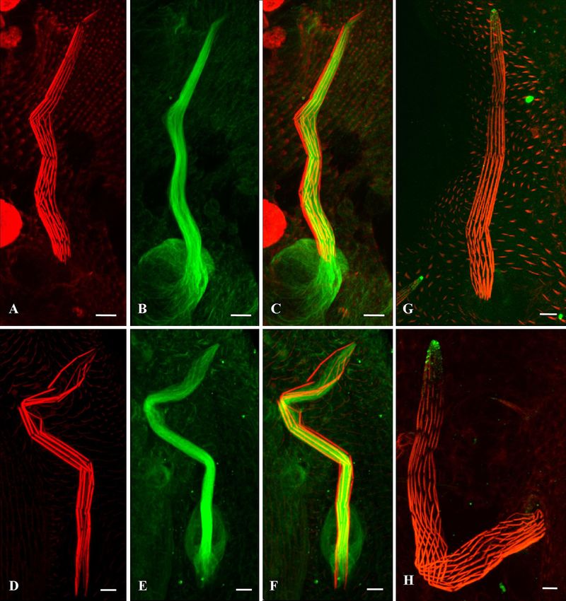
FIG. 1. Mutations in jv affect bristle development. Scanning electron micrographs of wild-type (A to D) and jv1 mutant (E to H) bristles are
shown. The ridges in the middle and lower parts of the jv1 bristle (G and H) are disorganized. They are shallower and thinner than wild-type bristles
(C and D). The jv1 bristle tips (F) are swollen and contain misaligned, smaller ridges than seen in the wild-type bristle tips (B). In addition, some
of the mutants present fused posterior scutellar bristles (J) that do not separate from one another, unlike in the wild type (I).
RESULTS each other (Fig. 1I); however, in the jv mutants, it seems that
these bristles fused together at their meeting points.
Analysis of the bristle defects in the javelin mutant. Since
Since variations in bristle length can indicate changes in the
bristle morphology reflects the organization of the cytoskele-
cytoskeleton (5, 22), we measured the lengths of the anterior
ton in the bristle (6, 8), we examined the external cuticular
and posterior scutellar macrochaetes. We found no significant
structure of macrochaetes by SEM. While the wild-type bristle
surface presented pronounced, straight ridges and valleys (Fig. difference between these lengths in the wild-type and mutant
1A to D), the mutants’ bristles appeared in a smoother cutic- bristles (Table 1).
ular pattern in which the ridges were shallower and thinner Actin bundles are disorganized in jv mutant bristles. Since
than in the wild-type bristles (Fig. 1E to H). In addition, in- jv mutant bristles exhibit an aberrant morphology that appears
stead of tapering toward the tip (Fig. 1B), the mutants’ bristles to be cytoskeleton related, we analyzed actin cytoskeleton or-
become bulbous at the tip, giving the bristle the shape of a ganization during pupal development. Initially, we examined
spear (Fig. 1F). Furthermore, the bristle tip has abnormally the bristles 42 h APF using confocal microscopy. Phalloidin
organized surface grooves, with the ridges not being parallel to staining of actin bundles revealed that while wild-type bristles
each other (Fig. 1F). In addition to these irregular groove taper in a straight direction toward the tip (Fig. 2A and B), jv
patterns, we also found that in 25% of the flies, the posterior mutant bristles remain wide (Fig. 2C and D). Next, we exam-
scutellar bristles are fused (Table 1 and Fig. 1J). In both wild- ined the bristles 48 h APF, when the bristles are fully elon-
type and jv mutants, these bristles grow diagonally and cross gated. During this developmental stage, the defective spatial
TABLE 1. Summary of bristle phenotypes from jv mutant and rescue fliesa
Value for genotypeb
Phenotype or Actin-Gal4;
parameter Actin-Gal4; Actin-Gal4:
Wild type jv1 jv1/jv2 pUAS-CG32397
jv1-pUAS-GFP-CG32397 jv1-pUAS-GFP-CG32397/jv2
RNAi
Fused bristle 0 (0/324) 25.8 (37/143) 14.9 (10/67) 13.3 (6/45) 2.9 (2/68) 2.3 (1/42)
Avg length of 313.6 ⫾ 44.7 (23) 308.5 ⫾ 58.1 (27) 312 ⫾ 34.2 (12) 310 ⫾ 55.3 (15) 309 ⫾ 47 (12) 314 ⫾ 39 (16)
macrochaetesc
Bulbous tip 0 (0/145) 62.5 (60/96) 53.9 (41/76) 56.6 (30/53) 30.6 (30/98) 34.9 (22/63)
a
All of the results presented in the table were taken from females. Similar results were also noted for males.
b
Values for fused bristle and bulbous tip phenotypes are percentages (number of flies with the genotype/total number of flies). Average lengths of macrochaetes and
standard deviations are in micrometers (number of macrochaetes measured).
c
Average length of anterior scutellar macrochaetes. Similar results were also found for posterior scutellar macrochaetes.VOL. 31, 2011 JAVELIN, A NOVEL ACTIN-ASSOCIATED PROTEIN 4585
which causes the poorly organized bundles to be less associated
with the membrane (Fig. 3C’ and C⬙).
Localization of the actin cross-linking proteins, forked and
fascin, is not affected in jv mutant bristles. Drosophila bristles
sprout 32 h APF during metamorphosis and elongate over the
course of 16 h (24). This elongation is driven by actin bundles
(22). Thus far, two known cross-linking proteins have been
discovered, forked and singed (a fascin homolog), which are
involved in actin construction in Drosophila bristles (20). Since
actin filaments in jv mutant bristles do not gather properly into
compact, maximally cross-linked bundles, we performed anti-
body staining of Drosophila fascin and forked proteins. Immu-
nofluorescent staining of developing mutant bristles in forked
proteins (Fig. 4E) revealed the localization of the protein to be
Downloaded from http://mcb.asm.org/ on January 5, 2021 by guest
similar to that of wild-type bristles (Fig. 4B). In both wild-type
and jv mutant bristles, forked is present along the entire length
of the actin bundles and is also concentrated in the gaps be-
tween the actin bundles (7) (Fig. 4 A to F). Fascin protein was
evenly diffused in the cytoplasm and in the socket cell of the
bristle in wild-type flies (4) (Fig. 4G). A similar distribution
was seen in the jv bristle (Fig. 4H).
The MT array in jv mutants is normal. In addition to actin
bundles, there is a large population of MTs, which run longi-
tudinally along the bristle shaft (2, 22). Like actin, these MTs
play an important role as cytoskeleton components in Drosoph-
ila bristles (22). Since inhibitor-based (5) and genetic (2) stud-
FIG. 2. Disorganization of actin bundles in the jv mutant. Confocal ies showed that disorganization of MTs can also lead to mor-
microscopy projections of macrochaetes from 42-h-old (A and B) and
48-h-old (E and F) wild-type pupae and from 42-h-old (C and D) and phological defects of the bristles, we investigated the
48-h-old (G and H) jv pupae. Actin bundles are stained with Oregon organization of MTs in jv mutant bristles. Specifically, we ex-
Green-phalloidin (gray). The bristle tip of a 42-h-old jv pupa (C; the amined how MTs are arranged in the mutant by immunostain-
area shown in the white rectangle is enlarged in panel D) is not ing developing bristles with anti-␣-tubulin antibodies. Exami-
tapered, as it is in the wild type (A and enlargement in panel B). At
48 h APF in the jv mutant, the actin bundles along the entire bristle run
nation of the jv mutant bristles revealed that the microtubule
in different directions (G). The bristle tip (enlargement in panel H) is array is properly organized (Fig. 5E), as in wild-type bristles
swollen, and the actin bundles in this region are not parallel to each (Fig. 5B).
other, as they are in the wild type (enlargement in panel F). Moreover, Next, we checked whether MT functionality is affected in the
some of the bundles are thinner (enlargement in panel H, arrowhead) jv mutants. The MT network serves as a transport system for
than others (enlargement in panel H, arrow). Scale bars, 5 m.
different proteins in bristles (2). Previous studies showed that
those several proteins (among them, spn-F and hook) are lo-
calized asymmetrically within the bristle shaft, where they ac-
cumulate at the bristle tip (2, 17). When the MT array is
organization of actin bundles is more pronounced than in bris- disorganized, spn-F and hook proteins do not reach their nat-
tles 42 h APF. At the later time point, the bundles are disor- ural positions but rather accumulate along the bristle shaft (2).
dered and run in different directions along the bristle shaft and Therefore, we used spn-F and hook proteins as markers to
some appear to be thinner than others (Fig. 2G). These actin inspect the polar transport system in the mutant bristles. Stain-
bundle defects seem to be more severe at the swollen bristle tip ing of wild-type bristles with anti-spn-F antibodies 42 h APF
(Fig. 2H), giving the bristle a spear-like shape. showed that the protein is localized at the bristle tip (2) (Fig.
To visualize the internal organization of actin bundles, we 5G). A localization pattern for spn-F was found to be similar to
examined thin transverse sections of bristles 42 h APF by that in the jv mutant bristle (Fig. 5H). Similar results were also
electron microscopy. While the actin membrane-associated found for the hook protein (data not shown).
bundles have a triangular shape in wild-type bristles (Fig. 3A to Molecular characterization of the javelin locus. The jv mu-
A⬙), in jv mutant bristles, the actin bundles were no longer tation was identified in 1947 and mapped to region 65A1-
triangular in shape (Fig. 3B’, B⬙, C’, and C⬙). It is of note that 65E1 (Flybase [http://flybase.org/]). However, the gene
in most cases, there were gaps in some of the bundles where no affected by this mutation was not defined. Using deficiency
actin filaments could be found (Fig. 3B’ and C’). In some mapping, we showed that the jv gene is found in region
extreme cases, the bundles in the mutants were larger than 65B4-65C1, which includes 7 different genes. To identify the
those of the wild type due to the more disordered arrangement sequence corresponding to the gene encoding Jv, we took two
of the filaments (Fig. 3C to C⬙). Groups of filaments tend to approaches. In the first, we tested whether jv mutants fail to
aggregate together, thus creating gaps or spaces within the complement known mutations in this region, namely, in the ndl
bundle, giving a perforated appearance. In addition, the actin and zpg genes. In the second approach, we performed RNAi
filaments are not fully attached to the plasma membrane, silencing of the other genes in the bristle using inducible RNAi in4586 SHAPIRA ET AL. MOL. CELL. BIOL.
Downloaded from http://mcb.asm.org/ on January 5, 2021 by guest
FIG. 3. Defects in the internal organization of actin bundles in a jv mutant. Thin transverse sections of microchaete bristles from 42-h-old
wild-type pupae (A, A’, and A⬙) and 42-h-old jv pupae (B to B⬙ and C to C⬙). In wild-type bristles, actin bundles are composed of a hexagonally
packed array of actin filaments and assume a triangular shape (higher magnification in panels A’ and A⬙). The bundles in jv mutants are not
organized properly and lack the triangular shape (B and C). The filaments are not tightly packed, thus creating gaps within the bundle (B’ and B⬙).
In extreme cases, the bundles are larger than those in the wild type and have more space between groups of filaments (C’). Also, these bundles
are not fully attached to the plasma membrane (C’ and C⬙). Scale bar sizes are indicated.
a Gal4/UAS system (3, 15). We found that downregulation of one To verify that CG32397 encodes Jv, we sequenced the
novel gene, CG32397, affected bristle morphology (Fig. 6D). CG32397 coding region from genomic DNA of the jv1 mutant
When this gene was affected, the bristle ridges were shallow, as in stock and compared the sequence obtained to that of the wild
jv mutants (Fig. 6B), and toward the bristle tip, the ridges became type. No mutations were found in the coding region of the
disorganized and nonparallel to one another. In addition, there CG32397 gene in the jv1 mutant.
was a spear-like shape to the bristle. We also found that in 13.3% Next, we performed real-time PCR to verify that our RNAi
of the flies, the posterior scutellar bristles were fused (Table 1). silencing indeed reduced the levels of the CG32397 transcript.
FIG. 4. Actin cross-linkers are properly localized in the jv mutant. (A to F) Confocal microscopy projections of bristles from 42-h-old wild-type
pupae (A to C) and 42-h-old jv pupae (D to F) stained with antiforked antibodies (red). (G and H) Confocal microscopy projections of bristles
from 42-h-old wild-type pupae (G) and 42-h-old jv pupae (H) stained with antifascin antibodies (red). The actin bundles in the bristles were stained
with Oregon Green-phalloidin (green). In jv mutants (E and F), as in wild-type bristles (B and C), the forked protein is localized along the actin
bundles and in the gaps between them. The fascin protein was evenly diffused in the cytoplasm and in the socket cell of the bristle in both jv mutant
(H) and wild-type (G) bristles. Scale bars, 5 m.VOL. 31, 2011 JAVELIN, A NOVEL ACTIN-ASSOCIATED PROTEIN 4587
Downloaded from http://mcb.asm.org/ on January 5, 2021 by guest
FIG. 5. Mutations in jv do not affect microtubule network organization. (A to F) Confocal microscopy projections of bristles from 45-h-old
wild-type pupae (A to C) and 45-h-old jv pupae (D to F) stained with anti-␣-tubulin antibodies (green) and Alexa Fluor-phalloidin (red). jv mutants
exhibit a microtubule array along the bristles that is properly organized (E), as in wild-type bristles (B). (G and H) Confocal microscopy images
of bristles from 42-h-old pupae (G and H) stained with anti-spn-F antibodies (green) and Alexa Fluor-phalloidin (red). In both jv mutant (H) and
wild-type (G) bristles, spn-F is localized to the bristle tip. Scale bars, 5 m.
We calculated the relative change (12) in CG32397 gene ex- subsequently refer to the spontaneous mutation in the original
pression after RNAi silencing and found that the transcript mutant stock of jv as jv1, while the CG18769[c03230] line will
level decreased almost 3.3-fold compared to that of the con- be referred as jv2. Next, we performed real-time PCR to ana-
trol. Next, we searched for several alleles of the CG32397 gene lyze the transcript levels of CG32397 in jv1 and in jv2 and found
and found 11 known transposon insertions in the gene area. that the transcript levels of CG32397 decreased 4.9-fold in jv1
Only two of these are available from the Bloomington collec- and 1.5-fold in jv2 compared to those of wild-type flies.
tion (CG18769[c03230] and CG18769[EY01803]). We found Since CG32397 is a predicted gene lacking molecular data,
that only homozygous CG18769[c03230] flies showed defects we decided to verify the predicted open reading frame of this
in bristle development which resemble the jv1 bristle pheno- gene. To this end, we designed several primer sets that cover
type (data not shown). Moreover, we found that jv1 fails to the entire predicted region of the gene (Fig. 7A). Using these
complement the bristle phenotype when crossed to flies bear- sets of primers (see Materials and Methods), we were able to
ing the P element (Fig. 6E). Next, we tested whether excision amplify almost the entire gene from cDNA prepared from
of the piggyBac element (CG18769[c03230]) from the pupae 48 h APF. However, we were unable to amplify the 5⬘
CG32397 gene could revert the bristle phenotype to a wild- end of the predicted gene. Thus, we performed 5⬘ RACE to
type-like phenotype. As expected, excision of the P element reveal the organization of this predicted gene. We found a
reverted the bristle phenotype to that of the wild type. More- discrepancy between the predicted gene and the 5⬘ RACE
over, after P element excision, this fly line complemented the result. We saw that the first translation start site is located 165
jv1 bristle phenotype (data not shown). For clarity, we will bp downstream of the predicted N-terminal methionine (Fig.4588 SHAPIRA ET AL. MOL. CELL. BIOL.
Downloaded from http://mcb.asm.org/ on January 5, 2021 by guest
FIG. 6. Jv is encoded by the CG32397 gene. Scanning electron micrographs represent bristles of wild-type (A), jv1 mutant (B), jv1 hemizygous
(C), Act-Gal4; pUAS-CG32397 RNAi (D), jv1/CG18769[c03230] (E), and actin-Gal4-jv1; pUAS-GFP-CG32397 (F) flies. RNAi silencing of
CG32397 in the bristles (D) has the same phenotype as in jv1 mutant bristles (B). (E) CG18769[c03230] fails to complement jv1 bristle defects.
(F) GFP-CG32397 rescues the jv1 bristle phenotype. Although the ridges are disorganized, they are more pronounced than in jv1 mutant bristles.
Moreover, the bristle tapers toward the tip and it is not swollen, as it is in jv1 mutants (B). Scale bar sizes are indicated.
7B and C) and that the open reading frame was unchanged but nine (Fig. 7B and C). Thus, our results reveal a new organiza-
predicted a product reduced by 55 amino acids. Moreover, we tion of CG32397 at the 5⬘ end of the gene. More importantly,
were able to show that the 5⬘ untranslated region (UTR) is our findings predict a gene product shortened from the previ-
expanded into 3 exons, the first containing 78 bp and located ously predicted gene product by 55 amino acids at the N
1.7 kbp from the second exon, the second located 3.8 kbp from terminus.
the new translation start site and containing 207 bp, and the Next, we performed rescue experiments by expressing the
third containing 130 bp directly upstream of the first methio- CG32397 protein in the jv1 or jv1/jv2 mutant background. WeVOL. 31, 2011 JAVELIN, A NOVEL ACTIN-ASSOCIATED PROTEIN 4589
Downloaded from http://mcb.asm.org/ on January 5, 2021 by guest
FIG. 7. Genomic organization of the CG32397 gene. (A) A schematic diagram of the predicted sequence of CG32397. The white rectangles
represent exons, while the black lines represent the introns of the gene. The small black arrows represent the six pairs of primers that were used
to amplify CG32397 from cDNA. The first primer pair failed to amplify the gene. The triangle between the fifth and sixth exons represents a
transposon insertion (CG18769[c03230]) that failed to complement jv1 bristle defects. (B) A schematic diagram of the sequence of CG32397
according to 5⬘ RACE. The gray rectangles represent the 3 exons in the 5⬘ UTR of the gene that were revealed by 5⬘ RACE. An enlargement of
this area (C) shows that there is a disparity between the predicted sequence and the 5⬘ RACE result. The first 35 bp, represented by a stippled
rectangle, do not exist in the sequence of the gene. Therefore, the first translation start site is located 165 bp downstream from the predicted
methionine.
created transgenic flies expressing nontagged and GFP-fused Next, we studied the effect of expression of GFP-Jv in ova-
CG32397 proteins (using the new, experimentally validated ries. For this, we expressed the GFP-fused protein in the germ
protein open reading frame) under the control of the UAS/ line using an ␣-tub Gal4-3 driver. We saw that in nurse cells,
Gal4 system. We addressed the ability of these flies to rescue expression of GFP-Jv led to the formation of an ectopic actin
the jv1 or jv1/jv2 bristle phenotype. We found that whereas 62% network that surrounded the nurse cell nucleus (Fig. 8E and F,
of the bristles from jv1 mutant flies (Fig. 6B) and about 54% of insets). This nurse cell nucleus-associated actin network is not
the bristles from jv1/jv2 flies had swollen tips (Fig. 6E, Table 1), normally found in wild-type cells (Fig. 8D). Moreover, at later
only 30.6% of flies expressing CG32397 in the jv1 mutant egg chamber developmental stages (stage 14) in some of the
background had swollen tips (Table 1 and Fig. 6F), and in nurse cells, GFP-Jv was also found in the actin network that
jv1/jv2, the percentage of flies having a swollen tip decreased to surrounds the nurse cell nucleus (Fig. 8G to I, insets). Thus,
about 35% (Table 1 and data not shown). Moreover, we found our results demonstrate that the Jv protein is associated with
that in 25% of jv1 flies and about 15% of jv1/jv2 flies, the the actin network. Yet, the ectopic actin network that sur-
posterior scutellar bristles are fused (Table 1). Expression of rounds the nurse cell nucleus had no effect on female fertility
CG32397 reduces the level of these fusion events to 3% in jv1 or embryo development.
and to 2.3% in jv1/jv2 flies. We noticed that in 95% of bristles Next, we analyzed whether CG32397 physically interacts
from flies expressing CG32397 in jv mutant backgrounds, the with actin monomers using a yeast two-hybrid assay. We found
ridges on the entire bristle length were not parallel to each that CG32397 failed to interact with actin, suggesting either
other as they are in the wild type (Fig. 6F). that CG32397 binds F-actin or that association of CG32397
Jv protein is associated with actin filaments. The CG32397 with actin is mediated by other proteins.
gene encodes a protein with no homologs outside insects.
Bioinformatics analysis revealed that the protein does not
DISCUSSION
contain any known domain. We noted that expression of
GFP-fused CG32397 rescues the jv1 bristle phenotype and Mutation in jv affects actin assembly in the bristle. To iden-
hence decided to analyze the localization pattern of this tify new players required for cytoskeleton organization during
GFP-fused protein in the bristle. We found that GFP-Jv Drosophila bristle development, we focused on the identifica-
(Fig. 8A) is localized along the actin bundles (Fig. 8B), as tion and characterization of the jv locus. Our results demon-
revealed by its colocalization with the actin-interacting dye strate that jv is involved in the assembly of actin bundles during
phalloidin (Fig. 8C). bristle development, as jv mutant flies display numerous de-4590 SHAPIRA ET AL. MOL. CELL. BIOL.
Downloaded from http://mcb.asm.org/ on January 5, 2021 by guest
FIG. 8. GFP-Jv is colocalized with the actin bundles in the bristle and forms an ectopic actin network in the nurse cell nucleus. (A to C)
Confocal microscopy projections of a 38-h-old pupa bristle from a neur-Gal4; pUAS-GFP-Jv transgenic fly (A, green) stained with Alexa
Fluor-phalloidin (B, red) are shown. Panel C is a merger of panels A and B, showing that GFP-Jv is localized along the actin bundles. Scale bars
for panels A to C, 5 m. (E to I) Confocal microscopy projections of an ovary from an ␣-tub Gal4-3; pUAS-GFP-Jv transgenic fly at stage 8 (E
and F, insets) and stage 14 (G to I). Both are stained with Alexa Fluor-phalloidin (red). In the nurse cells, GFP-Jv led to the formation of an ectopic
actin network, surrounding the nucleus (E and F; see enlargements of this region), which is not found in wild-type cells (D). In stage 14, GFP-Jv
is also colocalized with the actin that surrounds the nurse cell nucleus (G to I; insets are single-slice magnifications of this region). Scale bars for
panels D to I, 30 m (10 m for the insets within each panel).
fects in bristle morphology. In the mutants, the lower part of not fully attached to the plasma membrane. It is important to
the bristle is characterized by a smooth, grooved pattern, with mention that whereas confocal and SEM analysis was per-
the abnormality appearing more severe at the bristle tip. While formed on macrochaetes, the TEM analysis was conducted on
in wild-type flies, the bristle is widest at the base and tapers microchaetes. The reason for this was that defects in macro-
toward the tip, in jv mutants, the bristle tip becomes bulbous, chaetes are easy to detect in the confocal microscope, while
creating a spear-like shape. In addition, bristle ridges and val- microchaetes are easy to analyze by TEM due to the fact that
leys in this area are highly disordered and seem discontinuous. they are centrally positioned on the fly thorax and are more
Since abnormalities in bristle morphology can indicate defects numerous than the macrochaetes. Therefore, the probability of
in the organization of the cytoskeleton within the bristle shaft, getting thin transverse sections through the microchaetes is
we examined the organization and functionality of bristle actin high (22).
bundles and the microtubule array. We found that both the How do the defects in the organization of the actin filaments
function and organization of the MTs are not affected in jv in the bundles lead to aberrant bristle morphology in jv mu-
mutants. However, using confocal microscopy and actin stain- tants? During bristle development, the cytoplasm projects be-
ing, we revealed that the spatial organization of the actin bun- tween adjacent bundles, causing membrane protrusions. This
dles in the bristles is affected in jv mutants. Nonparallel actin creates the ridges seen in the adult bristle (20). In the areas
bundles, some of which appeared to be thinner than others, where actin bundles are attached to the membrane, such pro-
were observed along the bristle shaft and especially at the trusions cannot be accomplished, and as such, these regions
bristle tip. These results led us to examine the internal orga- will become the valleys seen in the cuticle (9, 20). Thus, while
nization of actin bundles in jv mutant bristles using TEM. the cytoplasm projects through the interspaces between actin
Transverse sections through jv1 bristles revealed defects in bundles in wild-type bristles, in jv mutants, the cytoplasm also
actin bundle formation. We noticed that the actin bundle no projects within the bundles, forming protrusions in the mem-
longer had the normal shape of a triangle that it had in a brane in areas where they are not supposed to be created. As
wild-type bristle. Instead, the actin bundles were disordered a result, the ridges that form are highly disordered and shal-
and presented gaps within them, leading to a perforated ap- lower and appear to be noncontinuous along the bristle. Thus,
pearance. In some cases, we found that the actin bundles were we suggest that the loss of actin filaments within the bundle inVOL. 31, 2011 JAVELIN, A NOVEL ACTIN-ASSOCIATED PROTEIN 4591
the jv mutant affects both actin bundle shape and bundle at- cell nucleus. Interestingly, examination of egg chambers at stage
tachment to the membrane, which in turn affects bristle shape 14 revealed that in the nurse cells, Jv-GFP is also colocalized with
and morphology. actin that surrounds the nurse cell nucleus. This actin network is
Another phenotype that we observed was fusion between the found in wild-type egg chambers and plays an important role in
posterior scutellar bristles. At their meeting points, these jv rapid cytoplasm transport from the nurse cell into the oocyte in a
mutant bristles have a shallower grooved pattern and a higher process termed dumping (10, 16). Thus, we suggest that expres-
number of contact points between them than do wild-type sion of CG32397 protein led to the premature formation of the
bristles. Similar results were observed in response to the ge- actin network, surrounding the nurse cell nucleus, which is re-
netic interaction between capping protein  (cpb) and arpc1 (6). quired at later stages of egg chamber development.
Bristles from these mutant flies are also characterized by an To conclude, the association of the Jv protein with the actin
irregular organization of ridges and valleys at the cuticle, as network, along with the effects of jv mutants on actin bundle
seen in jv mutants. Several processes could lead to bristle formation in the bristle, suggests that Jv is a novel protein
fusion, including secretion of chitin, as suggested by Frank which functions in actin assembly.
et al. (6), or membrane fusion.
Downloaded from http://mcb.asm.org/ on January 5, 2021 by guest
ACKNOWLEDGMENTS
Jv is encoded by CG32397. In this study, we show that jv
corresponds to the predicted gene termed CG32397. Several We thank Greg Guild, VDRC Austria, and the Bloomington Stock
lines of evidence support our claim. (i) Using fine deficiency Center for generously providing fly strains and reagents.
This research was supported by Israel Science Foundation Grant
mapping of jv mutants, we showed that jv is found in the
968/10 (to U.A.).
CG32397 region. (ii) We showed that downregulation of the
CG32397 gene led to defects in bristle morphology, similar to REFERENCES
the defects seen in jv mutants. (iii) A mutation in the CG32397 1. Abdu, U., D. Bar, and T. Schupbach. 2006. Spn-F encodes a novel protein
that affects oocyte patterning and bristle morphology in Drosophila. Devel-
gene (CG18769[c03230]) showed bristle defects similar to opment 133:1477–1484.
those of jv1 and also failed to complement the jv1 bristle phe- 2. Bitan, A., G. M. Guild, D. Bar-Dubin, and U. Abdu. 2010. Asymmetric
microtubule function is an essential requirement for polarized organization
notype, suggesting that jv1 and CG18769[c03230] are two dif- of the Drosophila bristle. Mol. Cell. Biol. 30:496–507.
ferent alleles of the same gene. (iv) The transcript levels of 3. Brand, A. H., and N. Perrimon. 1993. Targeted gene expression as a means
CG32397 are significantly reduced in jv mutants. (v) We found of altering cell fates and generating dominant phenotypes. Development
118:401–415.
that expression of the CG32397 protein in bristles partially 4. Cant, K., B. A. Knowles, M. S. Mooseker, and L. Cooley. 1994. Drosophila
rescued jv bristle defects. singed, a fascin homolog, is required for actin bundle formation during
Since CG32397 is a predicted gene, we analyzed its pre- oogenesis and bristle extension. J. Cell Biol. 125:369–380.
5. Fei, X., B. He, and P. N. Adler. 2002. The growth of Drosophila bristles and
dicted transcript. Using PCR primer sets that cover the gene, laterals is not restricted to the tip or base. J. Cell Sci. 115:3797–3806.
along with 5⬘ RACE, we found that the first translation start 6. Frank, D. J., R. Hopmann, M. Lenartowska, and K. G. Miller. 2006. Capping
protein and the Arp2/3 complex regulate nonbundle actin filament assembly
site is actually located 165 bp downstream of the previously to indirectly control actin bundle positioning during Drosophila melanogaster
predicted start site. bristle development. Mol. Biol. Cell 17:3930–3939.
As mentioned above, expression of the CG32397 protein 7. Grieshaber, S. S., D. H. Lankenau, T. Talbot, S. Holland, and N. S. Petersen.
2001. Expression of the 53 kD forked protein rescues F-actin bundle forma-
rescues most jv bristle defects. There are several possible ex- tion and mutant bristle phenotypes in Drosophila. Cell Motil. Cytoskeleton
planations for this partial rescue. (i) In the rescue experiments, 50:198–206.
we used actin-Gal4, which does not necessarily act as an en- 8. Guild, G. M., P. S. Connelly, L. Ruggiero, K. A. Vranich, and L. G. Tilney.
2003. Long continuous actin bundles in Drosophila bristles are constructed by
dogenous jv promoter. (ii) It is possible that CG32397 contains overlapping short filaments. J. Cell Biol. 162:1069–1077.
more alternative splice forms that we failed to detect in our 9. Guild, G. M., P. S. Connelly, K. A. Vranich, M. K. Shaw, and L. G. Tilney.
2002. Actin filament turnover removes bundles from Drosophila bristle cells.
molecular analysis. If this is indeed the case, it is possible that J. Cell Sci. 115:641–653.
the other variants are needed for full rescue. (iii) Our rescue 10. Gutzeit, H. O. 1986. The role of microfilaments in cytoplasmic streaming in
construct contained the entire open reading frame coding for Drosophila follicles. J. Cell Sci. 80:59–169.
11. Lees, A. D., and C. H. Waddington. 1942. The development of the bristles in
the protein but lacked both the 5⬘ and 3⬘ ends. It is possible normal and some mutant types of Drosophila melanogaster. Proc. R. Soc.
that these ends are required for the full function of the gene. Lond. B Biol. Sci. 131:87–110.
Still, we believe that our results described above together with 12. Livak, K. J., and T. D. Schmittgen. 2001. Analysis of relative gene expression
data using real-time quantitative PCR and the 2⫺⌬⌬CT method. Methods
our finding that CG32397 is an actin-associated protein (see 25:402–408.
below) demonstrate that the CG32397 gene encodes Jv. 13. Mitchell, H. K., and L. S. Lipps. 1978. Heat shock and phenocopy induction
in Drosophila. Cell 15:907–918.
Jv is an actin-associated protein. Bioinformatics analysis 14. Morata, G., and P. Ripoll. 1975. Minutes: mutants of Drosophila autono-
predicts that the jv gene encodes a protein that does not con- mously affecting cell division rate. Dev. Biol. 42:211–221.
tain any known domain. To better understand the function of 15. Negeri, D., H. Eggert, R. Gienapp, and H. Saumweber. 2002. Inducible RNA
interference uncovers the Drosophila protein Bx42 as an essential nuclear
the protein, we expressed Jv, fused to GFP, in fly bristles and cofactor involved in Notch signal transduction. Mech. Dev. 117:151–162.
ovaries. We determined the localization of this GFP-fused 16. Ogienko, A. A., S. A. Fedorova, and E. M. Baricheva. 2007. Basic aspects of
protein in bristles and found that it colocalized to the actin ovarian development in Drosophila melanogaster. Genetika 43:1341–1357.
17. Otani, T., et al. 2011. IKKε regulates cell elongation through recycling
bundles. These results reinforced our finding that CG32397 endosome shuttling. Dev. Cell 20:219–232.
encodes Jv, which is required for actin assembly during bristle 18. Spradling, A. C., and G. M. Rubin. 1982. Transposition of cloned P elements
into Drosophila germ line chromosomes. Science 218:341–347.
development. 19. Tilney, L. G., M. S. Tilney, and G. M. Guild. 1995. F-actin bundles in
Next, we analyzed the effects of ectopically expressed Jv in Drosophila bristles. I. Two filament cross-links are involved in bundling.
other tissues. Accordingly, we used the Drosophila egg chamber as J. Cell Biol. 130:629–638.
20. Tilney, L. G., P. Connelly, S. Smith, and G. M. Guild. 1996. F-actin bundles
a model tissue. We found that expressing Jv in the germ line led in Drosophila bristles are assembled from modules composed of short fila-
to formation of an ectopic actin network that surrounds the nurse ments. J. Cell Biol. 135:1291–1308.4592 SHAPIRA ET AL. MOL. CELL. BIOL.
21. Tilney, L. G., P. S. Connelly, K. A. Vranich, M. K. Shaw, and G. M. Guild. 1998. 23. Tilney, L. G., P. S. Connelly, K. A. Vranich, M. K. Shaw, and G. M. Guild.
Why are two different cross-linkers necessary for actin bundle formation in vivo 2000. Regulation of actin filament cross-linking and bundle shape in Dro-
and what does each cross-link contribute? J. Cell Biol. 143:121–133. sophila bristles. J. Cell Biol. 148:87–99.
22. Tilney, L. G., P. S. Connelly, K. A. Vranich, M. K. Shaw, and G. M. Guild. 24. Tilney, L. G., et al. 2004. The role actin filaments play in providing the
2000. Actin filaments and microtubules play different roles during bristle characteristic curved form of Drosophila bristles. Mol. Biol. Cell 15:5481–
elongation in Drosophila. J. Cell Sci. 113:1255–1265. 5491.
Downloaded from http://mcb.asm.org/ on January 5, 2021 by guestYou can also read