Evolution of the chelicera: a dachshunddomain is retained in the deutocerebral appendage of Opiliones (Arthropoda, Chelicerata)
←
→
Page content transcription
If your browser does not render page correctly, please read the page content below
EVOLUTION & DEVELOPMENT       14:6, 522–533 (2012)
                                                                                                              DOI: 10.1111/ede.12005

Evolution of the chelicera: a dachshund domain is retained in the
deutocerebral appendage of Opiliones (Arthropoda, Chelicerata)

Prashant P. Sharma,a,b,∗ Evelyn E. Schwager,b Cassandra G. Extavour,b and Gonzalo Giribeta,b
a
 Museum of Comparative Zoology, Harvard University, 26 Oxford Street, Cambridge, MA 02138, USA
b
 Department of Organismic and Evolutionary Biology, Harvard University, 26 Oxford Street, Cambridge, MA 02138,
USA
∗
    Author for correspondence (email: psharma@fas.harvard.edu)

SUMMARY The proximo-distal axis of the arthropod leg is            siomorphic three-segmented chelicera observed in “primitive”
patterned by mutually antagonistic developmental expression        chelicerate orders. Consistent with patterns reported in spi-
domains of the genes extradenticle, homothorax, dachshund,         ders, in the harvestman chelicera homothorax, extradenticle,
and Distal-less. In the deutocerebral appendages (the an-          and Distal-less have broadly overlapping developmental do-
tennae) of insects and crustaceans, the expression domain          mains, in contrast with mutually exclusive domains in the legs
of dachshund is frequently either absent or, if present, is        and pedipalps. However, unlike in spiders, the harvestman
not required to pattern medial segments. By contrast, the          chelicera bears a distinct expression domain of dachshund
dachshund domain is entirely absent in the deutocerebral ap-       in the proximal segment, the podomere that is putatively
pendages of spiders, the chelicerae. It is unknown whether         lost in derived arachnids. These data suggest that a tripar-
absence of dachshund expression in the spider chelicera is         tite proximo-distal domain structure is ancestral to all arthro-
associated with the two-segmented morphology of this ap-           pod appendages, including deutocerebral appendages. As a
pendage, or whether all chelicerates lack the dachshund do-        corollary, these data also provide an intriguing putative ge-
main in their chelicerae. We investigated gene expression          netic mechanism for the diversity of arachnid chelicerae: loss
in the harvestman Phalangium opilio, which bears the ple-          of developmental domains along the proximo-distal axis.

INTRODUCTION                                                       Rieckhof et al. 1997; Casares and Mann 1998; Abu-Shaar
                                                                   et al. 1999; Wu and Cohen 1999; Dong et al. 2001, 2002;
The articulated appendages of arthropods have facilitated          Rauskolb 2001; reviewed by Angelini and Kaufman 2005).
the tremendous diversity and evolutionary success of this             An interesting spatial reversal of exd and hth expression
phylum. Postulated to have evolved from a polyramous an-           domains has been documented as follows: exd is expressed
cestral condition, nearly every part of the arthropod leg has      throughout the legs in pancrustaceans (also termed tetra-
undergone extensive evolutionary modifications, enabling           conates), whereas it is restricted to the proximal part in myr-
adaptations to various ecological niches and environments          iapods and chelicerates; hth is expressed throughout the legs
(Snodgrass 1938; Cisne 1974; Waloszek et al. 2005). Inves-         in myriapods and chelicerates, but is restricted proximally in
tigation of genetic mechanisms of leg development, princi-         Pancrustacea (Abu-Shaar and Mann 1998; Abzhanov and
pally in the fruit fly Drosophila melanogaster, has implicated a   Kaufman 2000; Prpic et al. 2001, 2003; Inoue et al. 2002;
suite of four genes that pattern the proximo-distal (PD) axis:     Prpic and Tautz 2003; Angelini and Kaufman 2004, 2005;
Distal-less (Dll), dachshund (dac), extradenticle (exd), and ho-   Prpic and Damen 2004; Prpic and Telford 2008; Pechmann
mothorax (hth). In arthropod walking legs, at least three of       and Prpic 2009). Because onychophoran leg gap gene do-
these genes (Dll, dac, and either exd or hth) are expressed in     mains are comparable to those of pancrustaceans (Janssen
mutually antagonistic domains. Knockdown of these genes            et al. 2010), the spatial expression of exd and hth has been
results in loss of the podomeres (leg segments) patterned          interpreted as a potential synapomorphy for the sister group
by that particular gene, engendering the moniker, “leg gap         relationship of chelicerates and myriapods (termed Paradox-
genes” (Dong et al. 2001, 2002; Rauskolb 2001). Dll and dac        opoda or Myriochelata), a relationship recovered in many
pattern distal and medial podomeres respectively; proximal         molecular phylogenetic analyses (e.g., Hwang et al. 2001;
patterning requires the cofactors exd and hth (Sunkel and          Mallatt et al. 2004; Pisani et al. 2004; Mallatt and Giribet
Whittle 1987; Cohen and Jürgens 1989; Mardon et al. 1994;         2006; Dunn et al. 2008; von Reumont et al. 2009; Rehm et al.
González-Crespo and Morata 1996; Lecuit and Cohen 1997;           2011). However, this correlation of leg gap gene domains

522
                                                                                                           C 2012 Wiley Periodicals, Inc.
Sharma et al. Harvestman leg gap genes 523
remains to be tested in chelicerate and myriapod lineages activity is requisite for specification of cheliceral morphol-
other than spiders and millipedes. ogy in chelicerates (Prpic and Damen 2004; Pechmann et al.
In contrast with the walking leg, modified appendages are 2010).
associated with modified leg gap gene patterning. For exam- One limitation of this inference is that cheliceral morphol-
ple, the mandible of pancrustaceans and myriapods, and the ogy is quite variable. The chelicerae of spiders are comprised
maxilla of myriapods are considered gnathobasic (Snodgrass of two segments—the proximal basal segment and the dis-
1938; Popadic et al. 1996, 1998). In these appendages, Dll is tal fang—and are used for envenomation of prey and/or
not expressed in a manner consistent with PD axis formation manipulation of silk. Labidognathous chelicerae (with the
(Scholtz et al. 1998; Abzhanov and Kaufman 2000; Prpic and appendage perpendicular to the AP axis) do not occur out-
Tautz 2003). Similarly, leg gap gene expression in the thora- side of Araneomorphae (the group that includes orb weavers
copods of some crustaceans, and the antennae of insects and and jumping spiders). Orthognathous (with the appendage
millipedes, differs from that in the walking legs in that mutu- parallel to the AP axis) chelicerae occur in Mygalomorphae
ally antagonistic domains are not observed (e.g., Dong et al. (tarantula-like spiders) and Mesothelae (spiders with a seg-
2001; Williams et al. 2002; Prpic and Tautz 2003; Angelini mented opisthosoma), as well as three related arachnid or-
and Kaufman 2004). In the D. melanogaster antenna, hth, ders: Amblypygi, Uropygi, and Schizomida (the four form
dac, and Dll have overlapping expression domains and the the clade Tetrapulmonata). The chelicerae of these orders
dac medial domain is not functional (Dong et al. 2002; but are not chelate (forming a pincer), but rather shaped as a
see Angelini et al. 2009 for a case of a function antennal dac jackknife (Fig. 1). Another four lineages—Solifugae, Ricin-
domain in Tribolium castaneum). Comparable expression do- ulei, Pseudoscorpiones, and acariform Acari—bear two-
mains of leg gap genes occur in the antennae of other insects segmented chelicerae that are chelate, resembling a pair of
(Angelini and Kaufman 2004, 2005). scissors (acariform mites typically bear two cheliceral arti-
The leg gap genes also play a role in conferring antennal cles, but some lineages have a reduced third article, the nature
identity. In D. melanogaster knockdown of hth and Dll results of which is ambiguous; van der Hammen 1989; Evans 1992;
in antenna-to-leg transformations, and increasing dac ex- Shultz 2007). Finally, the “primitive” orders of Chelicerata—
pression induces medial leg structures in the antenna (Dong Pycnogonida, Xiphosura, Scorpiones, Opiliones, and the ex-
et al. 2001, 2002). A similar effect of hth knockdown has tinct Eurypterida (as well as Palpigradi and the parasiti-
been reported in the cricket antenna (Ronco et al., 2008), form Acari)—bear three-segmented chelicerae. In the con-
but in a hemipteran, hth knockdown resulted in the loss of text of chelicerate phylogeny, the spider chelicera is therefore
the antenna altogether (Angelini and Kaufman 2004). Addi- a derived structure (Fig. 1). Morphological and phylogenetic
tionally, Dll knockdown does not result in homeotic trans- studies have previously suggested that a three-segmented che-
formations in the hemipteran antenna (Angelini and Kauf- licera is the ancestral condition, and thus the two-segmented
man 2004). Knockdowns or mutations of some other genes morphology would have resulted from the loss of one of the
downstream of the leg gap genes can also result in homeotic segments, although this hypothesis has not been tested (e.g.,
antenna-to-leg transformations (Dong et al. 2002; Toegel Dunlop 1996; Wheeler and Hayashi 1998; Giribet et al. 2002;
et al. 2009; Angelini et al. 2009). Shultz 2007).
The chelicerate counterpart of the mandibulate antenna is The occurrence of a cheliceral type with an extra seg-
the chelicera, the namesake of this class of arthropods. Che- ment is particularly intriguing in the context of leg gap gene
licerae are the anterior-most pair of prosomal appendages domain evolution. However, the expression domains of leg
and are generally used for feeding. Homology of the anten- gap genes in chelicerate orders that bear three-segmented
nae of mandibulates and the chelicerae is based on their deu- chelicerae are not known. As a consequence, it is difficult to
tocerebral innervation and Hox gene boundaries (both are generalize patterns reported for leg gap genes in spiders to all
free of Hox expression; Telford and Thomas 1998; Hughes chelicerates. For example, the absence of the dachshund do-
and Kaufman 2002). However, investigation of leg gap gene main in the spider chelicera could be associated with the two-
expression in the appendages of spiders—including both my- segmented morphology of this appendage, implying the loss
galomorphs and araneomorphs—has demonstrated the lack of one segment, rather than with the chelicera itself. In order
of a dac domain altogether in the chelicera, as well as broadly to test this hypothesis, we examined gene expression of the leg
overlapping domains of hth, exd, and Dll (Abzhanov and gap genes in the harvestman Phalangium opilio, which bear
Kaufman 2000; Prpic et al. 2003; Prpic and Damen 2004; the plesiomorphic three-segmented chelicera, and compared
Pechmann and Prpic 2009). The similarity of overlapping these to data reported for spiders. We also sought similarities
expression domains in antennae and chelicerae is remark- in gene expression in the spider and harvestman chelicerae
able. Given the role of leg gap genes in specifying anten- to determine which aspects of PD axis specification are con-
nal identity in D. melanogaster (Dong et al. 2001, 2002), it served in chelicerates. We show that a dachshund domain is
has been suggested that leg gap gene domain overlap and present in the three-segmented harvestman chelicera and is524 EVOLUTION & DEVELOPMENT Vol. 14, No. 6, November–December 2012
Gene identification and whole mount in situ
hybridization
RNA was extracted from a range of embryonic stages using
Trizol (Invitrogen) and first strand cDNA synthesis was per-
formed using SuperScriptIII (Invitrogen). A developmental
transcriptome of P. opilio was generated by sequencing this
cDNA in a single flowcell on an Illumina GAII platform, us-
ing paired-end 150-bp-long reads. Thinning was performed
using 0.0496 as the limit (based on Phred quality scores), and
resulting quality of the thinned reads was visualized FastQC
(http://www.bioinformatics.bbsrc.ac.uk/projects/fastqc/).
After thinning, only those terminal bases with a Phred
quality score under 30 were trimmed. Assembly was con-
ducted using CLC Genomics Workbench 4.6.1 (CLC bio,
Aarhus, Denmark). All four genes were present in single
copy and sequences ranged in length from 661 to 1665 bp.
Gene identity was confirmed by protein BLAST (NCBI)
and visual inspection of amino acid alignments of orthologs
across Arthropoda. Sequences of all genes are deposited in
GenBank under accession numbers HE805503-HE805507.
Templates for riboprobe synthesis were generated as de-
scribed by Lynch et al. (2010): genes were amplified by PCR
using gene-specific primers (GSP) with an added linker se-
quence (5 -ggc cgc gg-3 for the forward primer end and 5 -ccc
ggg gc-3 for the reverse primer). A T7 polymerase binding
site for antisense or sense probe synthesis was generated in
a second PCR using the forward or reverse GSP and a uni-
versal primer binding to the 3 or 5 linker sequence with an
added T7 binding site, respectively. GSPs were designed from
the identified transcriptomic assembly. A list of the primers
used for generating sense and antisense probes is provided in
Table S1. Probe synthesis and in situ hybridization followed
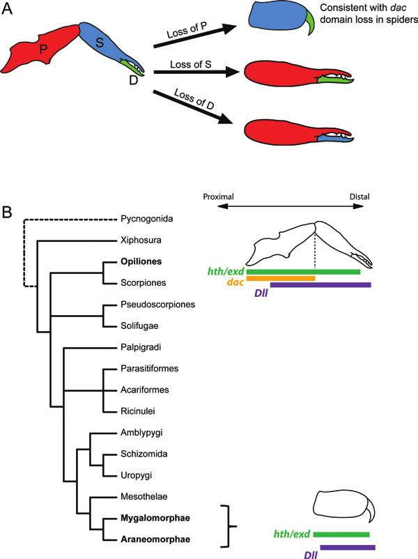
Fig. 1. Phylogeny of Chelicerata indicating relationships among the spider protocols for Cupiennius salei (Prpic et al. 2008).
orders and diversity of chelicerae. Constituent lineages of spi-
ders (order Araneae) as indicated. Orientation of the Araneo- The staining reactions for detection of transcripts lasted be-
morphae schematic indicates labidognathous chelicera (perpen- tween 20 min and 6 h at room temperature. Embryos were
dicular to body). Topology derived from Giribet et al. (2001), subsequently rinsed with 1× PBS + Tween-20 0.1% to stop
Shultz (2007), and Giribet and Edgecombe (2012). the reaction, counterstained with Hoechst 33342 (Sigma)
10 μg/ml to label nuclei, postfixed in 4% formaldehyde, and
stored at 4◦ C in glycerol. Embryos were mounted in glyc-
restricted to the proximal segment of this appendage, which erol and images were captured using an HrC AxioCam, a
is putatively lost in spiders. Lumar stereomicroscope driven by AxioVision v 4.8.2, and
an AxioImager compound microscope driven by AxioVision
v 4.8.2 (Zeiss, Oberkochen, Germany).
MATERIALS AND METHODS
RESULTS
Embryos
Adults of the synanthropic P. opilio (Arachnida, Opiliones, Expression of Po-hth and Po-exd
Eupnoi, Phalangiidae) were hand collected between 9 PM Po-hth is strongly expressed in the head lobes, the labrum,
and 3 AM from various sites in Weston and Woods Hole all of the appendages, and in the ventral ectoderm of all
(Falmouth), Massachusetts, USA in May through October segments (Fig. 2, Supporting information Fig. S1). In the
of 2009–2011. Adults were maintained and embryos collected pedipalps and walking legs of early embryos (stage 11), Po-
as previously described (Sharma et al. 2012). hth expression is concentrated in the proximal-most part ofSharma et al. Harvestman leg gap genes 525
Fig. 2. Expression of the Phalangium opilio homothorax gene in
the developing appendages. (A–C) Expression in the chelicera, Fig. 3. Expression of the Phalangium opilio extradenticle gene in
pedipalp, and L1, respectively, of a stage 11 embryo. Arrow- the developing appendages. (A–C) Expression in the chelicera,
heads indicate median ring of expression. (D–F) Expression in pedipalp, and L1, respectively, of a stage 12 embryo. Arrow-
the chelicera, pedipalp, and L2, respectively, of a stage 14 em- heads indicate distal ring in the patella of the pedipalp and legs.
bryo. Scale bars for all figures are 50 μm. px: proximal segment (D–F) Expression in the chelicera, pedipalp, and L1, respec-
of chelicera; 2nd: secondary article of chelicera; da: distal arti- tively, of a stage 15 embryo. Scale bars for all figures are 50 μm.
cle of chelicera; fe: femur; pa: patella; ti: tibia; mt: metatarsus; Abbreviations as in Fig. 2.
ta: tarsus.
contrast to the spider (the hth-1 paralog; Prpic et al. 2003).
the appendage, and in a separate and medial ring (Fig. 2, B In the pedipalp, Po-hth expression is observed throughout
and C). This ring of expression coincides with that of Po-exd the appendage, except for a distal portion of the tarsus
(see below), although the Po-hth medial domain is broader. (Fig. 2E). In the chelicera, Po-hth is expressed throughout
In the walking legs of older embryos (stage 14), the sepa- the appendage, except for the distal terminus, where expres-
rate expression domains are less marked; Po-hth is strongly sion is slightly weaker (Fig. 2, A and D).
expressed throughout the proximal-most part of the leg, in- Po-exd is expressed in the labrum, all of the appendages,
cluding the endites, to the tibia (Fig. 2F). In these older and in the ventral ectoderm of all prosomal and opistho-
stages, a more distal ring of expression is not observed, in somal segments (Fig. 3, Supporting information Fig. S1).526 EVOLUTION & DEVELOPMENT Vol. 14, No. 6, November–December 2012
In the appendages, Po-exd is strongly expressed in the
proximal-most parts of the pedipalps and walking legs, cor-
responding to the coxa and the endite, and a separate and
distinct ring of expression is observed in the patella of the
walking legs and pedipalp (Fig. 3, B and C). This ring is
retained in older stages, albeit wider and with weaker inter-
connecting expression in the femur and trochanter (Fig. 3,
E and F). In the chelicera, Po-exd is expressed throughout
the appendage except for the distal terminus; no rings of ex-
pression are observed, as in the other appendages (Fig. 3, A
and D).
Expression of Po-dac
Po-dac is expressed in the central nervous system, in several
groups of cells in the head lobes, and in the posterior termi-
nus. In older embryos, Po-dac is expressed in the developing
pleurites of the opisthosoma. Expression is never detected in
the labrum (Supporting information Fig. S2). All six pairs of
prosomal appendages express Po-dac (Fig. 4, A–F). In early
stages (stage 10), expression in all limb buds is similar and
occurs in a medial ectodermal ring (Fig. 4, A–C). In older
embryos (stage 14), the pedipalps and legs express Po-dac in a
domain encompassing the podomeres trochanter and femur
(Fig. 4, E and F). In the pedipalp and legs, Po-dac transcripts
are concentrated in the segmental boundaries delimiting the
femur, with slightly weaker interconnecting expression in the
femur (Fig. 4, E and F). Weak expression is observed more
proximally to the coxa; in spiders, this expression is associ-
ated with neural structures (Prpic and Damen 2004).
The chelicerae consistently express Po-dac as the ap-
pendage elongates (Fig. 4, D, Supporting information Fig.
S2). In early stages (stage 10), strong expression occurs in the
medial part of the cheliceral limb bud ectoderm, in a domain
highly comparable to other limb buds (Fig. 4A). This domain
does not include any part of the body wall. In older embryos,
strong expression of Po-dac is retained in the part of the Fig. 4. Expression of the Phalangium opilio dachshund gene in
chelicera that corresponds to the proximal segment, and no the developing appendages. (A–C) Expression in the chelicera,
pedipalp, and L1, respectively, of a stage 10 embryo. (D–F)
expression is detected in the secondary or distal articles (Fig.
Expression in the chelicera, pedipalp, and L1, respectively, of a
4D). Contrary to the other leg gap genes, the distal and prox- stage 14 embryo. Scale bars for all figures are 50 μm. cx: coxa;
imal boundaries of Po-dac expression in the chelicera appear tr: trochanter. Other abbreviations as in Fig. 2.
sharp rather than diffuse. Herein we consider an expression
boundary whose edge is straight and clear to be “sharp” (see,
e.g., Fig. 4, A–C) and otherwise to be “diffuse” (see, e.g., not expressed in the opisthosoma in a manner suggestive
Fig. 2, A–C). of rudimentary opisthosomal limb buds. In older embryos
(stage 14), a pair of strong expression domains is observed
in the head lobes, specifically in the part of the eye fields that
Expression of Po-Dll coalesce toward the midline during development (Support-
Po-Dll is expressed in all six prosomal appendages, as well ing information Fig. 2G). An additional and smaller pair
as in the developing labrum, the posterior terminus, and of expression domains is observed in the head, slightly pos-
the head lobes (Fig. 5, Supporting information Fig. S2). terior and lateral to the first pair (Supporting information
Unlike spiders, harvestmen do not have any opisthosomal Fig. 2G). Expression is also observed in the neuroectoderm
appendage-derived organs (e.g., spinnerets) and Po-Dll is along the ventral midline (Supporting information Fig. 2G),Sharma et al. Harvestman leg gap genes 527 Fig. 5. Expression of the Phalangium opilio Distal-less gene in the developing appendages. (A–C) Expression in the chelicera, pedipalp, and L1, respectively, of a stage 9 embryo. (D–I) Expression in the chelicera, pedipalp, and L1–L4, respectively, of a stage 14 embryo. Note the expression in the outgrown endites of the pedipalp and first leg (arrowheads), and the lack of these in the other legs. Scale bars for all figures are 50 μm. Abbreviations as in Fig. 2. comparable to that in a xiphosuran (Fig. 3J of Mittmann first walking legs (denoted L1), but not in L2–L4 (Fig. 5, and Scholtz 2001). E–I). In the pedipalps and legs of early embryos (stage 9), Po- In early stages (stage 9), Po-Dll is expressed in the distal Dll is strongly expressed in the distal part of the limb bud part of the cheliceral limb bud, comparably to the pedipalps (Fig. 5, B, C). Subsequently, Po-Dll appears throughout the and walking legs (Fig. 5A). This expression pattern is main- leg as strong rings of expression occurring coincidently with tained upon the differentiation of the distal podomere into the developing boundaries of the podomeres, while retaining the secondary and distal articles (stage 14). Po-Dll continues strong expression in the distal-most podomere (the tarsus; to be expressed mostly in the distal part of the appendage, Fig. 5, F–I). In older embryos (stage 14), Po-Dll is addition- which will form the second and distal articles; and tapering ally expressed in the outgrowing endites of the pedipalps and expression is observed extending into the proximal segment
528     EVOLUTION & DEVELOPMENT                   Vol. 14, No. 6, November–December 2012

(Fig. 5D). In contrast to the sharp expression boundaries in        similarly expressed in the legs, albeit without the medial ring
the other appendages, the proximal expression boundary of           domain (Prpic and Tautz 2003).
Po-Dll in the chelicera is diffuse.                                     In spite of these lineage-specific differences, the expression
                                                                    domains of Po-hth and Po-exd are comparable to those of spi-
                                                                    ders and a millipede (Abzhanov and Kaufman 2000; Prpic
DISCUSSION                                                          et al. 2003; Prpic and Tautz 2003; Prpic and Damen 2004;
                                                                    Pechmann and Prpic 2009). In general, hth is expressed
Here we examined gene expression of the single-copy or-             broadly in much of the developing appendage, whereas exd
thologs of the leg gap genes in the harvestman appendages.          is restricted to the proximal podomeres. Taken together
We observe that proximal PD axis patterning of the ap-              with the inverse spatial relationship of hth and exd in ony-
pendages is conserved in Opiliones and Araneae, and re-             chophorans and pancrustaceans (Prpic et al. 2003; Prpic and
sembles the patterning observed in a glomerid millipede.            Telford 2008; Janssen et al. 2010), the expression data ob-
These data are consistent with the Myriochelata hypothe-            served in P. opilio are consistent with a sister relationship of
sis. Second, we report novel expression domains of Dll in           chelicerates and myriapods.
apomorphic structures of harvestmen, namely the outgrown                The Myriochelata hypothesis is controversial, owing to
endites that form the stomotheca and the portion of the eye         discordance with morphological and paleontological data,
fields that form the ocularium. Most significantly, in the har-     as well as numerous phylogenetic and phylogenomic stud-
vestman chelicera, the genes hth, exd, and Dll have broadly         ies that have recovered chelicerates as sister to the remain-
overlapping expression domains, as in the spider chelicera          ing arthropods (e.g., Giribet et al. 2001; Regier et al. 2008,
and the mandibulate antenna, but these are independent of           2010). However, other studies, some with deeper gene sam-
the retention of a dac domain, which patterns the proximal          pling, have recovered the monophyly of chelicerates and myr-
segment of the harvestman chelicera.                                iapods (Hwang et al. 2001; Mallatt et al. 2004; Pisani et al.
                                                                    2004; Mallatt and Giribet 2006; Dunn et al. 2008; Hejnol
                                                                    et al. 2009; von Reumont et al. 2009; Rehm et al. 2011). Con-
Proximal patterning in harvestmen legs is                           sequently, although the spatial relationship of hth and exd
consistent with the Myriochelata hypothesis                         in arthropods constitutes a poorly sampled, one-character
The expression domains of the leg gap genes hth and exd in P.       system, it is plausible that PD axis patterning in myriapod
opilio are comparable, but not identical, to those of spiders. In   and chelicerate appendages constitutes a homologous condi-
older stages, Po-hth is expressed continuously throughout the       tion. Myriochelata is also supported by detailed similarities
appendage, from the coxa and endite to a distal podomere,           in chelicerate and myriapod neurogenesis (Dove and Stollew-
such as the tibia (P. opilio legs) or the tarsus (P. opilio pedi-   erk 2003; Kadner and Stollewerk 2004; Mayer and Whiting-
palps). This expression domain approximates that of the spi-        ton 2009), which contrasts with the neuroblast-driven system
der paralog hth-1, which is similarly broadly expressed from        present in insects and crustaceans (Ungerer et al. 2011).
proximal-most segments to part of the tarsus in the pedi-
palps and walking legs (Prpic and Damen, 2004; Pechmann
and Prpic 2009). A second paralog common to spiders, hth-2,         A role for Dll in patterning harvestman
is expressed in multiple rings and is believed to be involved       apomorphies
in leg segmentation (Prpic et al. 2003; Pechmann et al. 2009),      Consistent with its role in patterning outgrowths, Dll is ex-
but such rings corresponding to podomere boundaries were            pressed in the distal parts of all appendages. Additional ex-
not observed in P. opilio. Some lineage-specific differences        pression domains occur in the labrum and telson, which have
exist between the expression domains of Po-hth and spider           been reported in various other arthropod species (e.g., Pan-
hth-1. For example, a separate and more distal ring of hth          ganiban et al. 1995; Popadic et al. 1998; Thomas and Telford
expression is not observed in P. opilio, but has been reported      1999; Abzhanov and Kaufman 2000). Like the other leg gap
for the hth-1 paralog of spiders (Prpic et al. 2003; Prpic and      genes, Dll is known to have additional roles in development
Damen 2004). The pedipalps and the walking legs of P. opilio        beyond the PD axis, such as patterning sensory organs and
also have differing distal boundaries of hth expression, unlike     bristles (Sunkel and Whittle 1987; Cohen and Jürgens 1989;
spiders. The significance of these differences is not known.        Mittmann and Scholtz 2001; Williams et al. 2002), and even
    In contrast to hth, Po-exd is restricted to the proximal        gap gene function in spiders (Pechmann et al. 2011). Here
segments and a discrete ring of expression in the patella,          we observed two additional domains of Dll function that are
which closely resembles the expression domain of the exd-1          unique to the harvestman.
paralog in multiple spider species (Abzhanov and Kaufman               First, Dll is expressed in the endites of both the pedi-
2000; Prpic et al. 2003; Prpic and Damen 2004; Pechmann             palps and the first walking legs. These domains of expression
and Prpic 2009). In the millipede Glomeris marginata, exd is        are similar to the Dll expression domains in the endites of
Sharma et al. Harvestman leg gap genes 529
crustaceans (Panganiban et al. 1995). In P. opilio, the endites 2000; Prpic and Damen 2004; Pechmann and Prpic 2009).
of these two appendages elongate in the adult, forming a pre- This late-stage expression was previously postulated to be
oral cavity called the stomotheca—a structure that occurs of neural nature, as comparable expression also occurred in
only in harvestmen and scorpions, and the putative synapo- the coxae of all other appendages of older C. salei embryos
morphy of this clade (Stomothecata sensu Shultz 2007; (Prpic and Damen 2004). In that study, Prpic and Damen
Fig. 1). The other endites of the harvestman neither elongate (2004) observed that the broadly overlapping domains of
nor express Dll (Fig. 5, E–I). In the spider, the pedipalpal hth, exd, and Dll in the spider chelicera resembled the ex-
endite expresses Dll, but other endites do not (Schoppmeier pression of these genes in the antenna of D. melanogaster. It
and Damen 2001; Prpic and Damen 2004; Pechmann and was also conjectured that the lack of antagonistic hth, exd,
Prpic 2009). As in the harvestman, the Dll-expressing endite and Dll domains in the spider chelicera was associated with
of spiders is retained in the adult, forming the spider’s “max- complete loss of the cheliceral dac domain.
illa” (not homologous to the mandibulate maxilla). Taken Unlike the chelicerae of spiders, the plesiomorphic, three-
together, these data suggest that Dll is involved in patterning segmented chelicera of the harvestman expresses dac in a
the endites that form gnathobasic mouthparts in chelicerates. manner consistent with PD axis patterning during early de-
Expression data from mouthparts of scorpions, which could velopment. In early embryos, the dac domain in the che-
further test this hypothesis, are not presently available. licera is topologically indistinguishable from that in the other
Second, Dll is expressed in a pair of domains in the center appendage types. However, as the distal portion of the ap-
of the each eye field. Dll expression in the head lobes has been pendage forms an asymmetrical chela, dac is consistently and
observed in other chelicerates, but Dll expression in spider strongly expressed in the proximal portion of the chelicera.
and mites is either peripheral or diffuse, in comparison to Even after the chelicera has formed the three constituent
the harvestman (Thomas and Telford 1999; Abzhanov and segments, dac is expressed strongly throughout the proximal
Kaufman 2000; Schoppmeier and Damen 2001; Pechmann segment. This may imply that the segment missing in the
and Prpic 2009). Moreover, the pair of domains that strongly spider chelicera is the proximal-most one, which is consis-
express Dll in P. opilio subsequently form a fused outgrowth tent with traditional hypotheses of chelicera evolution based
called the ocularium, a stalk-like structure that bears a single upon morphology (Dunlop 1996; Wheeler and Hayashi 1998;
pair of simple ocelli, in the adult (Juberthie 1964). A similar Shultz 2007).
eye mound also occurs in pycnogonids, but as with scorpi- Although we do not currently have the tools to test func-
ons, expression data for the pycnogonid eye mound are not tionally putative mutual antagonisms of the leg gap genes in
presently available. As with the endites, the co-occurrence the harvestman chelicera, we observe that the expression do-
of the expression domains and subsequent outgrowth in the mains of Po-hth, -exd, and -Dll are broadly overlapping in this
locality of the expression suggest that Dll is involved in ocu- appendage in spite of the presence of a dac domain, much
larium formation. like their corresponding orthologs in the spider chelicera.
Functional tests of Dll activity by dsRNAi-mediated These data suggest that the lack of antagonistic domains
knockdown have been conducted in a spider and in a mite of hth, exd, and Dll in the spider chelicera is not associ-
(Schoppmeier and Damen 2001; Khila and Grbic 2007). In ated with the absence of the dac domain, but rather with the
the mite, the knockdown is reported to result in truncation specification of chelicerae and antennae generally. If these
of the pedipalpal endite (Khila and Grbic 2007), whereas in broadly overlapping domains are involved in conferring che-
the spider, the effect of the knockdown on the pedipalpal liceral identity—as with the D. melanogaster antenna—mild
endite (or maxilla) was not specified, but this structure is ap- knockdown phenotypes of one or more of these three genes
parently lost as well (Fig. 4, B and D of Schoppmeier and could result in chelicera-to-leg transformations. Future work
Damen 2001). Functional methods to test Dll activity in the could examine this testable hypothesis, taking advantage of
endites and ocularium of harvestmen are not yet developed, functional genetic tools available in spiders (Hilbrant et al.
but are of significant interest, given other reported cases of 2012).
Dll cooption to form nonappendage structures (e.g., butter- The retention of dac in the three-segmented chelicera
fly wing spots, McMillan et al. 2002; beetle horns, Moczek is remarkable, insofar as dac also occurs in homologous
and Rose 2009). appendages (the antenna) of some insects, such as D.
melanogaster (Dong et al. 2001), Oncopeltus fasciatus (An-
gelini and Kaufman 2004), T. castaneum (Prpic et al. 2001),
A dac domain is present in the three-segmented and Gryllus bimaculatus (Ronco et al. 2008), but not other pa-
chelicera narthropods, such as the isopod Porcellio scaber (Abzhanov
In spiders, dac is initially not observed in the two-segmented and Kaufman 2000). dac is also not observed in the frontal
chelicera, but is expressed proximally and within the ap- appendage or jaw of the onychophoran Euperipatoides
pendage in older stages of C. salei (Abzhanov and Kaufman kanangrensis (Janssen et al. 2010). Intriguingly, a large dac530 EVOLUTION & DEVELOPMENT Vol. 14, No. 6, November–December 2012 Fig. 6. Alternative hypotheses of chelicera evolution and summary of known leg gap gene expression domains in chelicerae throughout Chelicerata. (A) From a three-segmented ancestral state, a two-segmented chelicera could be obtained by loss of the proximal [P], secondary [S], or the distal [D] segment. If dac is considered a marker of the proximal segment, expression data from spiders and the harvestman support a transition of chelicera types by loss of the dac domain, and therefore of the proximal segment. (B) Fragmentary leg gap gene expression data are available for the chelicerae of Chelicerata. The complete suite of expression domain is known only for opisthothele spiders and harvestmen. For Xiphosura and Acariformes, only the Dll domains have been reported, and are not depicted. domain comparable that of P. opilio has only been observed basal chelicerate orders, but is a matter of interest for future in one other arthropod: the millipede G. marginata (Prpic and investigation. Tautz 2003). The relevance of this observation to the Myri- The presence of the dac domain in some insects has been ochelata hypothesis cannot be assessed given the presently interpreted as a possible retained rudiment of an ances- limited data on deutocerebral dac domains in myriapods and tral tripartite domain structure (Prpic and Damen 2004).
Sharma et al.                                                                                 Harvestman leg gap genes             531

However, labile deployment of the leg gap genes in modi-            as spiders and other tetrapulmonates (Fig. 6A). Such an ex-
fied appendages precludes the assignment of homology of             perimental result, if tested among several major lineages of
structures on the basis of gene expression domains alone            Chelicerata, would support a clear mechanism for the evo-
(Williams 1998; Abzhanov and Kaufman 2000). Neverthe-               lutionary transition from the three-segmented chelicera to
less, our observation that a dac domain occurs in the medial        the two-segmented types: loss of the dac domain along the
portion of the deutocerebral appendage in a plesiomorphic           proximo-distal axis.
order of chelicerates—in addition to a myriapod and some                However, it is presently unknown whether different lin-
pancrustaceans—lends credibility to the hypothesized tri-           eages of chelicerates with two-segmented chelicerae (e.g.,
partite domain structure of this appendage in the common            solifuges, pseudoscorpions, amblypygids) pattern this ap-
ancestor of arthropods, with subsequent losses of particu-          pendage in the same way (Fig. 6B). Phylogenetic approaches
lar domains upon modification (as have occurred in other            have previously coded the two- and three-segmented che-
modified appendage types, such as mandibles and maxillae;           licera as two to three separate character states, presuming
Scholtz et al. 1998; Abzhanov and Kaufman 2000; Angelini            homology among these types (Shultz 1990, 2007; Wheeler
and Kaufman 2005). However, it is also intriguing that dur-         and Hayashi 1998; Giribet et al. 2002). It remains to be tested
ing later developmental stages, both the cofactors hth and          whether a two-segmented chelicera can be obtained by alter-
exd are expressed continuously and more distally than the           native modifications of the three-segmented ancestral state,
dac domain in the harvestman chelicera. To our knowledge,           that is by deletions of the second or the distal articles, as
the chelicera of P. opilio constitutes the first arthropod ap-      alternatives to the proximal article. A survey of leg gap gene
pendage wherein this phenomenon occurs, discording with             expression across Chelicerata, with emphasis on dac, may aid
patterns previously observed for appendage regionalization          in testing the hypothesis of multiple cheliceral dac domain
via the leg gap genes (Kojima 2004; Angelini and Kaufman            losses in derived arachnids as a mechanism for transition to
2005).                                                              the two-segmented chelicera.
    Comparative functional data are limited for dac, but ac-
tivity in the deutocerebral appendage appears to vary among
                                                                    CONCLUSION
species. For example, in D. melanogaster, the antennal dac
domain is small, limited to the third antennal segment (Mar-
                                                                    The ancient history and plesiomorphic morphology of har-
don et al. 1994). Null dac mutants bear fusion of the a5-arista
                                                                    vestmen, here represented by P. opilio, lend itself to inves-
joint, but not the loss of any segments, whereas overexpres-
                                                                    tigation of many aspects of early arthropod evolution. We
sion of the dac domain results in medial leg structures in
                                                                    observed that dac is expressed in the proximal segment of the
the antenna (Dong et al. 2001, 2002). Similarly, dac is weakly
                                                                    chelicera in the harvestman, whereas neither the dac domain
expressed in the proximal antenna of O. fasciatus, and knock-
                                                                    nor this segment is retained in spiders. This correlation sug-
down of dac has no observable effect on the antenna at all
                                                                    gests that cheliceral segment number is determined by the
(Angelini and Kaufman 2004). By contrast, despite modest
                                                                    presence of the dac domain, providing a putative mechanism
antennal expression levels (Prpic et al. 2001), knockdown
                                                                    for the evolutionary transitions in chelicera morphology.
of dac in T. castaneum induces truncation of the antenna,
owing to the reduction of funicle (medial) segments and fu-
sion of antennal segments in this region, as well as homeotic       Acknowledgments
transformation of the distal funicle articles toward a club-like    We are indebted to Sónia C.S. Andrade, Ana Riesgo, and Alicia
                                                                    Pérez-Porro for technical assistance with the transcriptome of P.
(distal) identity (Angelini et al. 2009). Thus, in the deutocere-
                                                                    opilio. Dave Smith and Bernhard Götze at the Harvard Center for
bral appendage of at least one arthropod lineage, dac acts as       Biological Imaging facilitated use of microscopes. Comments from
a leg gap gene, as well as confers segmental identity along         Elizabeth L. Jockusch and Frank Smith refined some of the ideas
the PD axis.                                                        presented. EES was supported by DFG fellowship SCHW 1557/1-
    In the harvestman, dac is initially strongly expressed in the   1. This work was partially supported by NSF grant IOS-0817678 to
                                                                    CGE, and by internal MCZ funds to GG. Comments from editor
median portion of the cheliceral limb bud, and this domain
                                                                    Lisa M. Nagy and two anonymous reviewers improved an earlier
is later constrained to the part of the appendage that forms        version of the manuscript.
the proximal segment in P. opilio. Definitive determination
of the role of dac in harvestmen must await the development
of functional genetic tools for this system. However, one in-       REFERENCES
triguing possibility is that, if the cheliceral dac domain of P.    Abu-Shaar, M., and Mann, R. S. 1998. Generation of multiple antag-
opilio functions in a manner similar to that of T. castaneum,         onistic domains along the proximodistal axis during Drosophila leg
a knockdown of this gene may result in the loss of the proxi-         development. Development 125: 3821–3830.
                                                                    Abu-Shaar, M., Ryoo, H. D., and Mann, R. S. 1999. Control of the
mal segment, and therefore, in a two-segmented chelicera—             nuclear localization of extradenticle by competing nuclear import
the condition that occurs in derived arachnid orders, such            and export signals. Genes Dev. 13: 935–945.
532 EVOLUTION & DEVELOPMENT Vol. 14, No. 6, November–December 2012 Abzhanov, A., and Kaufman, T. C. 2000. Homologs of Drosophila ap- chus urticae: dsRNA and siRNA parental silencing of the Distal-less pendage genes in the patterning of arthropod limbs. Dev. Biol. 227: gene. Dev. Genes Evol. 217: 241–251. 673–689. Kojima, T. 2004. The mechanism of Drosophila leg development along Angelini, D. R., and Kaufman, T. C. 2004. Functional analyses in the the proximodistal axis. Develop. Growth Differ. 46: 115–129. hemipteran Oncopeltus fasciatus reveal conserved and derived aspects Lecuit, T., and Cohen, S. M. 1997. Proximal-distal axis formation in the of appendage patterning in insects. Dev. Biol. 271: 306–321. Drosophila leg. Nature 388: 139–145. Angelini, D. R., and Kaufman, T. C. 2005. Insect appendages and com- Lynch, J. A., Peel, A. D., Drechsler, A., Averof, M., and Roth, S. 2010. parative ontogenetics. Dev. Biol. 286: 57–77. EGF signaling and the origin of axial polarity among the insects. Angelini, D. R., Kikuchi, M., and Jockusch, E. L. 2009. Genetic pattern- Curr. Biol. 20: 1042–1047. ing in the adult capitate antenna of the beetle Tribolium castaneum. Mallatt, J. M., Garey, J. R., and Shultz, J. W. 2004. Ecdysozoan phy- Dev. Biol. 327: 240–251. logeny and Bayesian inference: First use of nearly complete 28S and Casares, F., and Mann, R. S. 1998. Control of antennal versus leg de- 18S rRNA gene sequences to classify the arthropods and their kin. velopment in Drosophila. Nature 392: 723–726. Mol. Phylogenet. Evol. 31: 178–191. Cisne, J. L. 1974. Evolution of the world fauna of aquatic free-living Mallatt, J., and Giribet, G. 2006. Further use of nearly complete, 28S arthropods. Evolution 28: 337–366. and 18S rRNA genes to classify Ecdysozoa: 37 more arthropods and Cohen, S. M., and Jürgens, G. 1989. Proximal-distal pattern formation a kinorhynch. Mol. Phylogenet. Evol. 40: 772–794. in Drosophila: Graded requirement for Distal-less gene activity during Mardon, G., Solomon, N. M., and Rubin, G. M. 1994. dachshund en- limb development. Wilhelm Roux’s Arch. Dev. Biol. 198: 157–169. codes a nuclear protein required for normal eye and leg development Dong, P. D. S., Chu, J., and Panganiban, G. 2001. Proximodistal domain in Drosophila. Development 120: 3473–3486. specification and interactions in developing Drosophila appendages. Mayer, G., and Whitington, P. M. 2009. Velvet worm development links Development 128: 2365–2372. myriapods with chelicerates. Proc. R. Soc. Lond. B 276: 3571–3579. Dong, P. D. S., Dicks, J. S., and Panganiban, G. 2002. Distal-less and ho- McMillan, W. O., Monteiro, A., and Kapan, D. D. 2002. Development mothorax regulate multiple targets to pattern the Drosophila antenna. and evolution on the wing. Trends Ecol. Evol. 17: 125–133. Development 129: 1967–1974. Mittmann, B., and Scholtz, G. 2001. Distal-less expression in embryos Dove, H., and Stollewerk, A. 2003. Comparative analysis of neurogen- of Limulus polyphemus (Chelicerata, Xiphosura) and Lepisma sac- esis in the myriapod Glomeris marginata (Diplopoda) suggests more charina (Insecta, Zygentoma) suggests a role in the development of similarities to chelicerates than to insects. Development 130: 2161– mechanoreceptors, chemoreceptors, and the CNS. Dev. Genes Evol. 2171. 211: 232–243. Dunlop, J. A. 1996. Evidence for a sister group relationship between Moczek, A. P., and Rose, D. J. 2009. Differential recruitment of limb Ricinulei and Trigonotarbida. Bull. Br. Arachnol. Soc. 10: 193– patterning genes during development and diversification of beetle 204. horns. Proc. Natl. Acad. Sci. USA 106: 8992–8997. Dunn, C. W., et al. 2008. Broad phylogenomic sampling improves reso- Panganiban, G., Sebring, A., Nagy, L., and Carroll, S. 1995. The devel- lution of the animal tree of life. Nature 452: 745–749. opment of crustacean limbs and the evolution of arthropods. Science Evans, G. O. 1992. Principles of Acarology. CABI, Wallingford, UK. 270: 1363– 1366. Giribet, G., and Edgecombe, G. D. 2012. Reevaluating the arthropod Pechmann, M., Khadjeh, S., Sprenger, F., and Prpic, N.-M. 2010. tree of life. Annu. Rev. Entomol. 57: 167–186. Patterning mechanisms and morphological diversity of spider ap- Giribet, G., Edgecombe, G. D., and Wheeler, W. C. 2001. Arthropod pendages and their importance for spider evolution. Arthropod Struct. phylogeny based on eight molecular loci and morphology. Nature Dev. 39: 453–467. 413: 157–161. Pechmann, M., Khadjeh, S., Turetzek, N., McGregor, A. P., Damen, W. Giribet, G., Edgecombe, G. D., Wheeler, W. C., and Babbitt, C. 2002. G. M., and Prpic, N.-M. 2011. Novel function of Distal-less as a gap Phylogeny and systematic position of Opiliones: A combined analysis gene during spider segmentation. PLoS Genet. 7: e1002342. of chelicerate relationships using morphological and molecular data. Pechmann, M., and Prpic, N.-M. 2009. Appendage patterning in the Cladistics 18: 5–70. South American bird spider Acanthoscurria geniculata (Araneae: My- González-Crespo, S., and Morata, G. 1996. Genetic evidence for the galomorphae). Dev. Genes Evol. 219:189–198. subdivision of the arthropod limb into coxopodite and telopodite. Pisani, D., Poling, L. L., Lyons-Weiler, M., and Hedges, S. B. 2004. The Development 122: 3921–3928. colonization of land by animals: Molecular phylogeny and divergence Hilbrant, M., Damen, W. G. M., and McGregor, A. P. 2012. Evolu- times among arthropods. BMC Biol. 2: 1–10. tionary crossroads in developmental biology: the spider Parasteatoda Popadic, A., Panganiban, G., Abzhanov, A., Rusch, D., Shear, W. A., tepidariorum. Development 139: 2655–2662. and Kaufman, T. C. 1998. Molecular evidence for the gnathobasic Hejnol, A., et al. 2009. Assessing the root of bilaterian animals with derivation of arthropod mandibles and the appendicular origin of the scalable phylogenomic methods. Proc. R. Soc. Lond. B 276: 4261– labrum and other structures. Dev. Genes Evol. 208: 142–150. 4270. Popadic, A., Rusch, D., Peterson, M., Rogers, B. T., and Kaufman, T. Hughes, C. L., and Kaufman, T. C. 2002. Hox genes and the evolution C. 1996. Origin of the arthropod mandible. Nature 380: 395. of the arthropod body plan. Evol. Dev. 4: 459–499. Prpic, N.-M., and Damen, W. G. M. 2004. Expression patterns of leg Hwang, U. W., Friedrich, M., Tautz, D., Park, C. J., and Kim, W. 2001. genes in the mouthparts of the spider Cupiennius salei (Chelicerata: Mitochondrial protein phylogeny joins myriapods with chelicerates. Arachnida). Dev. Genes Evol. 214: 296–302. Nature 413: 154–157. Prpic, N.-M., Janssen, R., Wigand, B., Klingler, M., and Damen, W. Inoue, Y., et al. 2002. Correlation of expression patterns of homothorax, G. M. 2003. Gene expression in spider appendages reveals reversal of dachshund, and Distal-less with the proximodistal segmentation of exd/hth spatial specificity, altered leg gap gene dynamics, and suggests the cricket leg bud. Mech. Dev. 113: 141–148. divergent distal morphogen signaling. Dev. Biol. 264: 119–140. Janssen, R., Eriksson, B. J., Budd, G. E., Akam, M., and Prpic, Prpic, N.-M., Schoppmeier, M., and Damen, W. G. M. 2008. Whole- N.-M. 2010. Gene expression patterns in an onychophoran reveal mount in situ hybridization of spider embryos. CSH Protocols 1–4, that regionalization predates limb segmentation in pan-arthropods. doi:10.1101/pdb.prot506. Evol. Dev. 12: 363–372. Prpic, N.-M., and Tautz, D. 2003. The expression of the proximodistal Juberthie, C. 1964. Recherches sur la biologie des Opilions. Dissertation, axis patterning genes Distal-less and dachshund in the appendages of Université de Toulouse, Toulouse, France. Glomeris marginata (Myriapoda: Diplopoda) suggests a special role Kadner, D., and Stollewerk, A. 2004. Neurogenesis in the chilopod of these genes in patterning the head appendages. Dev. Biol. 260: Lithobius forficatus suggests more similarities to chelicerates than to 97–112. insects. Dev. Genes Evol. 214: 367–379. Prpic, N.-M., and Telford, M. J. 2008. Expression of homothorax and Khila, A., and Grbic, M. 2007. Gene silencing in the spider mite Tetrany- extradenticle mRNA in the legs of the crustacean Parhyale hawaiensis:
Sharma et al.                                                                                           Harvestman leg gap genes                533

  Evidence for a reversal of gene expression regulation in the pancrus-       genes shows chelicerate arthropods retain their deutocerebral seg-
  tacean lineage. Dev. Genes Evol. 218: 333–339.                              ment. Proc. Natl. Acad. Sci. USA 95: 10671–10675.
Prpic, N.-M., Wigand, B., Damen, W. G. M., and Klinger, M. 2001. Ex-        Thomas, R. H., and Telford, M. J. 1999. Appendage development in em-
  pression of dachshund in wild-type and Distal-less mutant Tribolium         bryos of the oribatid mite Archegozetes longisetosus (Acari, Oribatei,
  corroborates serial homologies in insect appendages. Dev. Genes Evol.       Trhypochthoniidae). Acta Zool. 80: 193–200.
  211: 467–477.                                                             Toegel, J. P., Wimmer, E. A., and Prpic, N.-M. 2009. Loss of spineless
Rauskolb, C. 2001. The establishment of segmentation in the Drosophila        function transforms the Tribolium antenna into a thoracic leg with
  leg. Development 128: 4511–4521.                                            pretarsal, tibiotarsal and femoral identity. Dev. Genes Evol. 219: 53–
Regier, J. C., et al. 2008. Resolving arthropod phylogeny: Exploring phy-     58.
  logenetic signal within 41 kb of protein-coding nuclear gene sequence.    Ungerer, P., Eriksson, B. J., and Stollewerk, A. 2011. Neurogenesis in the
  Syst. Biol. 57: 920–938.                                                    water flea Daphnia magna (Crustacea, Branchiopoda) suggests differ-
Regier, J. C., et al. 2010. Arthropod relationships revealed by phy-          ent mechanisms of neuroblast formation in insects and crustaceans.
  logenomic analysis of nuclear protein-coding sequences. Nature 463:         Dev. Biol. 357: 42–52.
  1079–1084.                                                                Van der Hammen, L. 1989. An Introduction to Comparative Arachnology.
Rehm, P., et al. 2011. Dating the arthropod tree based on large-scale         SPB, Leiden, UK.
  transcriptome data. Mol. Phylogenet. Evol. 61: 880–887.                   von Reumont, B. M., et al. 2009. Can comprehensive background knowl-
Rieckhof, G. E., Casares, F., Ryoo, H. D., Abu-Shaar, M., and Mann, R.        edge be incorporated into substitution models to improve phyloge-
  S. 1997. Nuclear translocation of extradenticle requires homothorax,        netic analyses? A case study on major arthropod relationships. BMC
  which encodes an extradenticle-related homeodomain protein. Cell            Evol. Biol. 9: 119.
  91: 171–183.                                                              Waloszek, D., Chen, J., Maas, A., and Wang, X. 2005. Early Cambrian
Ronco, M., Uda, T., Mito, T., Minelli, A., Noji, S., and Klingler, M.         arthropods—New insights into arthropod head and structural evolu-
  2008. Antenna and all gnathal appendages are similarly transformed          tion. Arthropod Struct. Dev. 34: 189–205.
  by homothorax knock-down in the cricket Gryllus bimaculatus. Dev.         Wheeler, W. C., and Hayashi, C. Y. 1998. The phylogeny of the extant
  Biol. 313: 80–92.                                                           chelicerate orders. Cladistics 14: 173–192.
Scholtz, G., Mittmann, B., and Gerberding, M. 1998. The pattern of          Williams, T. A. 1998. Distalless expression in crustaceans and the pat-
  Distal-less expression in the mouthparts of crustaceans, myriapods          terning of branched limbs. Dev. Genes Evol. 207: 427–434.
  and insects: New evidence for a gnathobasic mandible and the com-         Williams, T. A., Nulsen, C., and Nagy, L. M. 2002. A complex role
  mon origin of Mandibulata. Int. J. Dev. Biol. 42: 801–810.                  for Distal-less in crustacean appendage development. Dev. Biol. 241:
Schoppmeier, M., and Damen, W. G. M. 2001. Double-stranded RNA                302–312.
  interference in the spider Cupiennius salei: The role of Distal-less      Wu, J., and Cohen, S. M. 1999. Proximodistal axis formation in the
  is evolutionarily conserved in arthropod appendage formation. Dev.          Drosophila leg: Subdivision into proximal and distal domains by ho-
  Genes Evol. 211: 76–82.                                                     mothorax and distal-less. Development 126: 109–117.
Sharma, P. P., Schwager, E. E., Extavour, C. G., and Giribet, G. 2012.
  Hox gene expression in the harvestman Phalangium opilio reveals
  divergent patterning of the chelicerate opisthosoma. Evol. Dev. 14:       SUPPORTING INFORMATION
  450–463.
Shultz, J. W. 1990. Evolutionary morphology and phylogeny of Arach-
  nida. Cladistics 6: 1–38.                                                 Additional Supporting Information may be found in the on-
Shultz, J. W. 2007. A phylogenetic analysis of the arachnid orders          line version of this article:
  based on morphological characters. Zool. J. Linn. Soc. 150: 221–
  265.                                                                         Fig. S1. Expression of the Phalangium opilio homothorax
Snodgrass, R. E. 1938. Evolution of the Annelida, Onychophora and
  Arthropoda. Smithson. Misc. Collns. 97: 1–159.                            and extradenticle genes.
Sunkel, C. E., and Whittle, J. R. S. 1987. Brista: A gene involved in          Fig. S2. Expression of the Phalangium opilio dachshund
  the specification and differentiation of distal cephalic and thoracic     and Distal-less genes.
  structures in Drosophila melanogaster. Wilhelm Roux’s Arch. Dev.
  Biol. 196: 124–132.                                                          Table S1 List of primer sequences used for riboprobe syn-
Telford, M. J., and Thomas, R. H. 1998. Expression of homeobox              thesis.
You can also read