A myosin II nanomachine mimicking the striated muscle - Nature
←
→
Page content transcription
If your browser does not render page correctly, please read the page content below
ARTICLE
                  DOI: 10.1038/s41467-018-06073-9                OPEN

                  A myosin II nanomachine mimicking the striated
                  muscle
                  Irene Pertici       1,   Lorenzo Bongini1, Luca Melli1,4, Giulio Bianchi1, Luca Salvi1,5, Giulia Falorsi1, Caterina Squarci1,
                  Tamás Bozó          2,    Dan Cojoc3, Miklós S.Z. Kellermayer 2, Vincenzo Lombardi 1 & Pasquale Bianco 1
1234567890():,;

                  The contraction of striated muscle (skeletal and cardiac muscle) is generated by ATP-
                  dependent interactions between the molecular motor myosin II and the actin filament. The
                  myosin motors are mechanically coupled along the thick filament in a geometry not
                  achievable by single-molecule experiments. Here we show that a synthetic one-dimensional
                  nanomachine, comprising fewer than ten myosin II dimers purified from rabbit psoas, per-
                  forms isometric and isotonic contractions at 2 mM ATP, delivering a maximum power of 5
                  aW. The results are explained with a kinetic model fitted to the performance of mammalian
                  skeletal muscle, showing that the condition for the motor coordination that maximises the
                  efficiency in striated muscle is a minimum of 32 myosin heads sharing a common mechanical
                  ground. The nanomachine offers a powerful tool for investigating muscle contractile-protein
                  physiology, pathology and pharmacology without the potentially disturbing effects of the
                  cytoskeletal—and regulatory—protein environment.

                  1 PhysioLab, University of Florence, Florence 50019, Italy. 2 Department of Biophysics and Radiation Biology, Semmelweis University, Budapest H-1094,

                  Hungary. 3 IOM-CNR, Trieste 34149, Italy. 4Present address: F. Hoffmann-La Roche Ltd, Basel 4053, Switzerland. 5Present address: Department of
                  Biochemistry, University of Munich, Munich 81377, Germany. Correspondence and requests for materials should be addressed to
                  V.L. (email: vincenzo.lombardi@unifi.it)

                  NATURE COMMUNICATIONS | (2018)9:3532 | DOI: 10.1038/s41467-018-06073-9 | www.nature.com/naturecommunications                                            1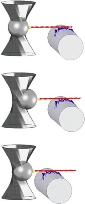
ARTICLE                                                                          NATURE COMMUNICATIONS | DOI: 10.1038/s41467-018-06073-9

M
           yosin II is a dimeric mechanoenzyme that uses the free-         mode optical fibre chemically etched to a diameter of ~4 µm. A
           energy change associated with the binding and hydro-            three-way piezoelectric nanopositioner brings the HMM ensem-
           lysis of ATP in either of its motor domains (the heads)         ble to interact with a fluorescently labelled actin filament (length
to generate force and displacement along filamentous actin via a            range 7–11 µm), attached with the correct polarity (bead-tailed
10-nm working stroke1. In the striated-muscle sarcomere, the               actin (BTA))16 to a polystyrene bead held with DLOT17 in the
motors emerge as two antiparallel arrays from a bipolar thick              middle of a 180-µm-deep flow chamber (Figs. 1a and 2a and
filament. As a result, steady force and shortening in the sarcomere         Supplementary Fig. 1). Thus the single actin filament selects a
are generated by cyclic ATP-driven interactions of the heads that          functional one-dimensional array out of the fibre-bound HMM
pull the actin filament from the opposite extremities of the sar-           molecules. The mechanical output of the nanomachine is mea-
comere towards the centre2–4. The motors of each half thick                sured by means of the DLOT, which acts as a force transducer
filament are mechanically coupled via the thick filament back-               (range 0.5–200 pN, compliance 3.7 nm pN−1), and the nanopo-
bone, which allows for steady contraction in spite of the fact that        sitioner carrying the HMM-coated fibre, which acts as a dis-
the individual myosin heads interact with actin only briefly and            placement transducer (range 1–75,000 nm). The system operates
remain detached throughout most of their ATP hydrolysis cycle.             either in position- or in force-feedback mode18 (see Methods and
The duty ratio, that is the fraction of time the myosin head is            Supplementary Fig. 2). Methylcellulose (0.5% w/v, 400 cP) was
bound to actin during its kinetic cycle, is as low as 0.05 in an           present in the solutions used for the experiment to inhibit the
unloaded contraction, which implies that no more than 5% of the            lateral diffusion of F-actin6, thereby minimising the probability
available heads are attached to actin at any moment5,6. The                that, in 2 mM ATP, the interaction terminates during mechanical
energetic cost of low-load contraction is thus minimised, due to           protocols that minimise the number of actin-attached heads. All
the strain dependence of the chemo-mechanical steps of the                 experiments were conducted at room temperature (23 °C).
ATPase cycle in the attached heads2,7. In the intact cell envir-
onment, the half-sarcomere is able to work across a wide range of
                                                                           Number of myosin molecules available for actin interaction.
externally applied loads by tuning the number of heads attached
                                                                           The number of HMM molecules on the fibre surface able to
to actin in proportion to the global filament load, while the force
                                                                           interact with the actin filament is initially determined by mea-
of the individual attached head is maintained similar to that
                                                                           suring the number of mechanical rupture events in ATP-free
developed under isometric conditions by the progression of the
                                                                           solution19,20 (Fig. 1). First the actin filament is brought to interact
working stroke8. Cell-mechanics experiments, however, cannot
                                                                           with the HMM-coated surface to form rigor bonds, then it is
reveal the motor coupling mechanism because of the large
                                                                           pulled away at constant velocity in a direction orthogonal to the
ensemble of the motors and the unavoidable presence of
                                                                           motor surface (z, parallel to the axis of the trapping laser, velocity
mechanically coupled cytoskeletal proteins and filaments. By
                                                                           25 nm s−1) to break the rigor bonds one at a time while avoid-
contrast, single-molecule mechanics experiments on purified
                                                                           ing that the detached HMM binds back (Fig. 1a). The number
proteins9–11 provide insights into the motor function, but they
                                                                           of rupture events in a single mechanical trial increases with
suffer from the intrinsic limit in that they cannot detect the
                                                                           the [HMM] used to coat the fibre (Fig. 1b) according to
unique performance emerging from the collective nature of motor
                                                                           a relation that attains a saturating value of 8.2 ± 1.2 at [HMM] =
action within the architecture of the half-sarcomere. Only myosin
                                                                           100 µg ml−1 (Fig. 1c), which is the concentration adopted for the
II motors working in ensemble can fit the task, but so far the
                                                                           experiments. The length of the functional nanoarray, estimated
various attempts failed to produce steady force and shortening in
                                                                           by summing the distances travelled following the rupture events
solution with physiological [ATP]12–15.
                                                                           (Supplementary Fig. 3) is 523 ± 175 nm (mean ± SD), one order
   Here we demonstrate the performance of a one-dimensional
                                                                           of magnitude smaller than the average length of the actin fila-
machine titrated to contain the minimum number of motor
                                                                           ment. This architecture of the machine implies that for a given
molecules needed to reproduce the collective mechanics of
                                                                           [HMM] the measured number of rupture events does not sig-
muscle myosin II in situ. The mechanical output of the
                                                                           nificantly change from experiment to experiment, therefore there
ensemble of myosin II molecules purified from the psoas
                                                                           is no need to normalise the mechanical data by actin filament
muscle of the rabbit and brought to interact with a correctly
                                                                           length.
oriented actin filament is measured by means of counter-
propagating dual laser optical tweezers (DLOT) in either
position or force clamp. The design of the nanomachine                     Isometric and isotonic performance of the nanomachine. In
ensures that, like in the three-dimensional lattice of the sar-            experiments on the active nanoarray (Fig. 2), a solution with 2
comere, the array of motors interacting with the actin filament             mM ATP is flowed into the chamber. The BTA is brought
lies on a plane parallel to the actin filament, overcoming a main           towards the motor array by moving the nanopositioner in the z
limit of all existing designs12–15. In 2 mM ATP, the nano-                 direction in position feedback. Once an acto-myosin interaction is
machine reproduces the steady force and shortening velocity                established, the position is clamped and the ensemble of motors
typical of the isometric and isotonic contractions of the sar-             starts to develop force (F) (phase 1) up to a maximum steady
comere in vivo. The power of the nanomachine is interpreted                value (F0) (isometric condition). Subsequently, the control is
with a simple model, the mechanical–kinetic features of which              switched to force clamp (phase 2) and a staircase of stepwise
are constrained by preliminarily fitting the performance of fast            reductions in force (4 pN per step in the experiment in Fig. 2b
mammalian muscle. We find that, to attain the efficiency of                  and 5 pN in the experiment in Fig. 2c) separated by 1 s intervals is
striated muscle with a one-dimensional machine, a minimum                  imposed (phases 3–6, isotonic conditions). The nanomachine
of 32 myosin heads sharing a common mechanical ground must                 responds with actin filament sliding in the direction of shortening
be available for interaction with the actin filament.                       at a constant velocity (V), which is greater the smaller the level of
                                                                           constant force between steps.
                                                                              In Fig. 2b, the interaction is terminated after ~2 µm of total
Results                                                                    sliding because of force-feedback failure due to the interference of
Constructing a one-dimensional myosin II nanomachine. The                  the optical fibre with the laser beam forming the optical trap. In
nanomachine is an ensemble of fast vertebrate skeletal-muscle              Fig. 2c, following the fifth step that drops the force to almost zero
heavy meromyosin (HMM) molecules attached onto a single-                   (phase 7), the control is switched to position feedback and the

2                        NATURE COMMUNICATIONS | (2018)9:3532 | DOI: 10.1038/s41467-018-06073-9 | www.nature.com/naturecommunications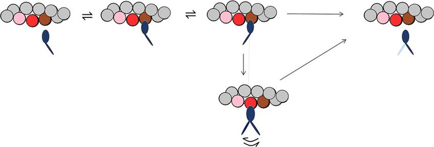
NATURE COMMUNICATIONS | DOI: 10.1038/s41467-018-06073-9                                                                                            ARTICLE

  a                                b                                                                 c
                                             10
                       1

                                   ΔZ (μm)
                                              8
                                                                                                                       10
                                              6
                                             20

                                    F (pN)
                                             10                                                                         8

                                                                                                      Rupture events
                                              0
                                                                                                                        6
                                             10

                                    F (pN)
                                             5
                       2
                                              0                                                                        4

                                             15
                                    F (pN)

                                             10                                                                        2
                                              5
                                              0
                                             15
                                    F (pN)

 y                                           10                                                                             0   20   40      60        80   100
             x
                                              5                                                                                      [HMM] (μg ml–1)
                                              0
       z                                          0   20       40           60       80       100
                                                                    t (s)

Fig. 1 Assessing the number of HMMs available for actin interaction from rigor rupture events. a Schematics of the protocol: (1) formation of rigor bonds
between the HMM array (blue) by the optical fibre (grey) and the actin filament (red) attached to the trapped bead (dark grey) via gelsolin (yellow); (2)
optical fibre moved away from the actin filament in the direction (z) orthogonal to the actin–myosin interface. b Records of the nanopositioner movement
(black, ΔZ velocity 25 nm s−1) and force with different [HMM] (colours refer to different concentrations as indicated in c; green: 25 µg ml−1; violet: 38 µg
ml−1; red: 50 µg ml−1; blue: 100 µg ml−1). The small vertical bars indicate the rupture events (force drop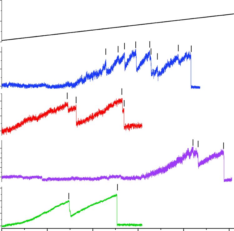
ARTICLE NATURE COMMUNICATIONS | DOI: 10.1038/s41467-018-06073-9
a b c
1
1
1 1 2 3 4 5 6 7 8 9 10 11
1 2 3 4 5 6 0
0
L (μm)
L (μm)
–1
–1
–2
2
–2 –3
30 40
20 F0 30
F (pN)
F0
F (pN) 10 20
3
0 10
0
–10
1 2 3 4 5 6 7 8 2 4 6 8 10 12
t (s) t (s)
d e f
12 4 6
10 5
Number of events
3
8 4
V (μm s–1)
P (aW)
6 2 3
4 2
1
2 1
0 5 10 15 20 25 30 35 0 4 8 12 16 20 0 4 8 12 16 20
F0 (pN) F (pN) F (pN)
Fig. 2 Mechanical output of the myosin-II nanomachine in 2 mM ATP. a Schematic representation of three snapshots during the phases of the interaction
between the actin filament and the motors numbered as in b. Components of the system coloured as in Fig. 1a. b Recording of the relative sliding (red
trace) and force (blue trace) during the interaction. Phase 1, following the formation of the first bonds between the actin filament and myosin motors, the
force rises in position feedback to the maximum isometric value (F0 ~ 17 pN). Phases 2–6, shortening response to a staircase of stepwise reductions in force
(4 pN) separated by 1 s imposed in force feedback. c Another experiment in which in phase 1 in position feedback isometric force rises to F0 (~21 pN) with
some force fluctuations and in frames 2–7 the staircase in force feedback is made by 5-pN steps separated by 1 s (3–5) and 0.5 s (6, 7). In phase 8, after a
total active sliding of 2.7 µm, the control is switched back to position feedback, and the force redevelops up to the original F0. Phases 9–11, force response to
a series of rapid lengthenings and shortenings of ~500 nm superimposed on F0. In both b, c, black lines are superimposed on the length trace to better
appreciate the slope. d Frequency distribution of F0. Data are plotted in classes of 2 pN and fitted with a Gaussian (continuous line): centre = 15.5 ± 0.4 pN,
σ = 4.7 ± 0.4 pN (mean ± SEM, n = 46). e Force–velocity (F–V) relation. The black filled symbol on the ordinate is the Vf value from IVMA (see related
Supplementary Fig. 4). The dashed line is Hill’s hyperbolic fit to the data. f Power–force (P–F) relation from the fit in e
trap (3.7 nm pN−1), and (ii) the motors are randomly oriented in function of the angle formed with the correct orientation, by a
the array, so that the force of HMM is reduced by a factor of two factor ranging between 1 (0°) and 0.1 (180°)10, which results in a
with respect to that on a correctly oriented myosin rod reduction of F0 generated by the randomly oriented attached
cofilament10. With the trap compliance in series, the addition heads by 45% and a corresponding leftward shift in the calculated
or subtraction of the force contribution by a single head induces F–V relation for the half-sarcomere (Supplementary Fig. 6, dotted
substantial sliding of the actin filament and a corresponding line). With these assumptions, the model is tested for the ability
change in the strain of the other attached heads, undermining the to simulate the machine performance by keeping unaltered all the
condition of independent force generators that characterises the mechano-kinetic parameters selected for muscle half-sarcomere
action of the myosin heads in isometric contraction in situ and using as the only free parameter the number of heads
(Supplementary Fig. 7, upper row left panel). The strain- available for the actin interaction, N. According to in situ
dependent kinetics of the attached heads is governed by the mechanical and structural evidence that in active muscle the two
push–pull experienced by all the other attached heads when actin heads of each dimer work independently36, N is twice the number
slides away towards the bead for the addition–subtraction of the of molecules and thus is 294 in the half-thick filament.
force contribution by one head (upper row, right panel), and The F–V and P–F relations of the nanomachine are best fitted
under these conditions, the isometric ATPase rate (φ0) and with N = 16 (Fig. 4b, c: continuous line, model; symbols and
slipping rate (rs,0) increase (Table 1). During isotonic shortening, dashed line, experiment). Notably, N = 16 is expected from the
the perturbation of the attached head induced by the force number of HMM molecules (8) estimated from rupture events in
contribution from each single head is still present (Supplementary rigor (Fig. 1), if, as in situ, also in the machine at 2 mM ATP and
Fig. 7, lower row), but the effect on both φ and rs vanishes thus at a correspondingly low duty ratio (≤0.3), both heads of
(Table 1), because the reduction of head strain increases anyway each dimer work independently. It must be noted that this
the head cycling kinetics. As for the correction for the effect of the conclusion appears in contrast with single molecule experiments
random orientation of the heads in the array, the force of each showing a cooperative behaviour between the two heads of the
attached head of the nanomachine is assumed to vary, as a myosin dimer at low ATP37.
4 NATURE COMMUNICATIONS | (2018)9:3532 | DOI: 10.1038/s41467-018-06073-9 | www.nature.com/naturecommunications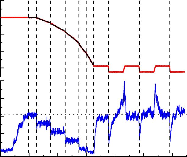
NATURE COMMUNICATIONS | DOI: 10.1038/s41467-018-06073-9                                                                                   ARTICLE

                             a                                                   b
                                       20
                                                                                           20

                                       15
                                                                                           15

                              F (pN)

                                                                                 F (pN)
                                       10
                                                                                           10

                                        5                                                  5

                                        0                                                  0
                                            0.1   0.2       0.3   0.4   0.5                     0.1   0.2       0.3   0.4   0.5
                                                        t (s)                                               t (s)

                             c                                                   d
                                                                                           25
                                       20
                                                                                           20

                                       15
                                                                                           15

                                                                                  F (pN)
                              F (pN)

                                       10
                                                                                           10

                                        5                                                   5

                                        0                                                   0
                                            0.1   0.2      0.3    0.4   0.5                     0.1   0.2      0.3    0.4   0.5
                                                        t (s)                                               t (s)

Fig. 3 Rate of force development. Upper row: time course of initial force development (a, Frame 1 from Fig. 2b) and force redevelopment following a large
release (b, Frame 8 from Fig. 2c). The different trace thickness in a and b is due to different acquisition rates (500 and 5000 points s−1, respectively).
Dashed lines are single exponential fits. Lower row: Model simulation of initial force development (c) and force redevelopment following a large release
(d), using 16 available myosin heads and a series compliance of 3.7 nm pN−1. Dashed lines are single exponential fits. τ, calculated from fits to experimental
traces as in a and b (0.15 ± 0.09 s, mean ± SEM, n = 34), is not significantly different from that calculated from fits to simulated traces in c and d (0.11 ±
0.06 s, mean ± SEM, n = 34) (see related Supplementary Fig. 5)

   In Fig. 4d, the various simulated mechanical parameters are                 Discussion
plotted as a function of N (filled circles). The relations for F0 and           The nanomachine described in this paper sets the forefront of the
Pmax intersect the dashed lines (experimental values) at N = 16.               research on the mechanics of a limited ensemble of muscle
By contrast, independent of N, the predicted V0 remains greater                myosins. The performance of our system emerges clearly from
than the experimental values. This discrepancy is likely due to a              the comparison with the ongoing research based on alternative
V0-quenching effect caused by the random orientation of the                    approaches.
motors. A similar conclusion has been drawn for actin sliding                     In theory, the best way to obtain an oriented ensemble of
velocity in IVMA experiments10,38,39, but that conclusion was                  myosin motors consists in using either a native isolated thick
contradicted by other evidence5,6,40.                                          filament or a synthetic cofilament made by rods and myosin
   The time courses of force rise and redevelopment (upper row in              molecules. With this motor design and the laser trap Kaya and
Fig. 3) are reproduced with a remarkable agreement by the model                collaborators obtained, in ATP-free solution, static stiffness
simulation with the series compliance equal to 3.7 nm pN−1 (lower              measurements in agreement with fibre measurements8,12,22.
row) (see also Supplementary Fig. 5). Noteworthy, as detailed in               However, the active performance of their motor system (in 1 mM
Supplementary Fig. 5, the number of interactions necessary to rise             ATP)13 consisted only in transient displacements of the actin
the force to the maximum isometric value against the trap                      filament abruptly interrupted after variable extent, without any
compliance is compatible with the number allowed in probabilistic              production of steady force and shortening. Other approaches that
terms by an ensemble of 16 independent myosin heads, further                   exploited the Three Bead Assay geometry (originally designed for
proving the solidity of the kinetic and methodological analysis used           single molecule mechanics9) by increasing the myosin density on
for interpreting the nanomachine performance.                                  the surface of the fixed bead to have multiple motor
   The number of attached heads (Na) predicted by the model                    interactions41,44 did not achieve any physiological machine per-
during isometric contraction of the nanomachine fluctuates                      formance, as the [ATP] was systematically kept at least ten times
between 5 and 6, each generating an average force of 2.8 pN,                   smaller than the millimolar concentration to increase the lifetime
which is much higher than that reported in most cases for single,              of the interactions.
randomly oriented HMM11,41, except when measurements were                         The design of our machine, which exploits the dual laser beam
made with a relatively high trap stiffness9,42,43. In isotonic                 technique, appears a decisive choice for the success of the assay.
contraction, Na decreases almost in proportion to F (Fig. 4e,                  Beyond the large dynamic range, the DLOT geometry ensures
continuous line), in remarkable agreement with in situ                         that the array of interacting motors lies on a plane that is parallel
experiments8.                                                                  to the BTA, in this way preserving their condition of “in parallel

NATURE COMMUNICATIONS | (2018)9:3532 | DOI: 10.1038/s41467-018-06073-9 | www.nature.com/naturecommunications                                              5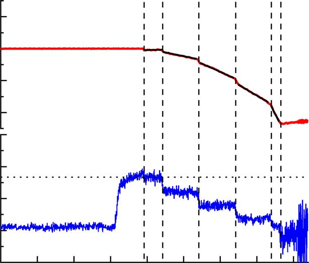
ARTICLE                                                                                           NATURE COMMUNICATIONS | DOI: 10.1038/s41467-018-06073-9

                       a                                                                                    b
                        +                     –                                                                           5
                                                  1              2                   3
                                                                                                                          4

                                                                                                             V (μm s–1)
                                      D               A1              A2                        D
                                                                                                                          3
                                                                                    3′
                                                                       Slip
                                                                                                                          2

                                                                                                                          1
                                                                     A1′-A2′

                                                                                                                               0    4     8     12      16    20
                                                                                                            c                              F (pN)
                                                                                                                          6
                       d
                                      50                                                                                  5
                                      40
                       F0 (pN)

                                                                                                                          4

                                                                                                             P (aW)
                                      30
                                      20                                                                                  3
                                      10                                                                                  2

                                      25                                                                                   1
                       Pmax (aW)

                                      20
                                      15                                                                                       0    4     8        12   16    20
                                      10                                                                                                      F (pN)
                                       5                                                                    e
                                                                                                                          13
                                       9                                                                                  11
                        V0 (μm s–1)

                                       7                                                                                  9
                                       5                                                                                  7

                                                                                                            Na
                                       3                                                                                  5
                                            10 12 14 16 18 20 22 24 26 28 30 32 34 36 38 40 42                            3
                                                                    N                                                     1

                                                                                                                               0   0.2   0.4      0.6   0.8   1.0
                                                                                                                                               F/F0

Fig. 4 Model simulation of the mechanical output of the machine. a Kinetic scheme, with three states of the myosin head (blue): D, detached; A1 and A2,
attached to one monomer of the actin filament (brown). The scheme includes the possibility that during shortening the attached head in the A2 state slips
to the next actin monomer farther from the centre of the sarcomere (red). The probability of a second slipping to the pink monomer is limited to 1/10 of
that of the first slipping. Details on the strain dependence of the rate functions and how they are constrained by fitting skeletal muscle performance are in
Supplementary Fig. 6. The corresponding equations are listed in Supplementary Table 1. The effect of trap compliance on the nanomachine performance is
detailed in Supplementary Fig. 7. b F–V relation calculated for a number of available heads, N = 16 (continuous line) and experimental relation from Fig. 2e
(open circles and dashed line). c Corresponding P–F relations. d Dependence on N of the three parameters featuring the machine performance, as indicated
in the ordinate of each plot. The dashed lines are the respective experimental values. e Relation between number of attached heads (Na) and force relative
to F0 calculated with the model for N = 16 (continuous line) and 32 (dotted line)

force generators”. The lack of alignment between the actin fila-                             constant for a total relative sliding distance of 2–3 µm, that is, one
ment and the motor ensemble in the single laser trap and in any                             order of magnitude larger than in the native system in vivo,
other existing system based on single actin-single myosin fila-                              allowing the whole force–velocity relation to be determined
ment13–15 is likely the reason of their limited performance, as the                         during the same interaction.
attached head closest to the bead experiences an additional stress                             The model simulation of the nanomachine performance pre-
generated by the out-of-axis vertical component of the ensemble                             dicts that, during shortening at the maximum power (Pmax, 5.4
force. In ref. 13, the interactions between actin and myosin fila-                           aW, attained at 0.3 F0, Figs. 2f and 4c), the rate of ATP hydrolysis
ments produce transient displacements, lasting only ~100 ms,                                per myosin head (φPmax) is ~30 s−1 (Table 1). N being 16 in our
during which a force–velocity relation can be calculated using the                          machine, the total ATP split per second by the machine at Pmax is
time derivative of the displacement against the trap compliance,                            (30 s−1 × 16 = ) 480 s−1. The ratio between Pmax and the corre-
while force is continuously changing. However, a force–velocity                             sponding total ATP split per second is the mechanical energy
relation calculated in this way is based on the not verified                                 delivered per ATP, that is (5400 zJ s−1/480 s−1 = ) 11.2 zJ. Thus,
assumption that the number of available heads is constant during                            with a free energy of the ATP hydrolysis of 110 zJ24, the efficiency
the whole interaction, and moreover, it does not fulfil the                                  of the nanomachine (ε) would be (11.2/110 = ) 0.1, at least three
requirement that the force and the corresponding shortening                                 times lower than that reported for fast mammalian muscle
velocity should be steady-state quantities produced by a collective                         (0.3–0.4)24. Taking into account that the random orientation of
motor based on muscle myosin.                                                               the motors reduces the force by 45%, the machine with correctly
   Noteworthy, in contrast with the results from the two-filament                            oriented motors would produce a work per ATP of 20 zJ, with ε
design12,13, but in agreement with in situ results35, in our HMM-                           still significantly lower than that in the muscle. This can be
made machine low-load shortening does not imply any need of                                 explained considering that, with N = 16, during shortening
assuming a large reduction of stiffness in negatively strained                              against a load ≤0.3 F0, Na decreases to
NATURE COMMUNICATIONS | DOI: 10.1038/s41467-018-06073-9 ARTICLE
Table 1 Simulated mechanical and energetic parameters of the half-sarcomere and their modulation by the conditions imposed
by the nanomachine
F0 (pN) a/F0 Pmax (aW) φ0 (s−1 per φPmax (s−1 per rs,0 (s−1 per rs,Pmax (s−1 per
head) head) head) head)
Compliance 0.01 nm pN−1 433 ± 5 0.365 462 11.65 35.50 0.60 31.24
Compliance 3.7 nm pN−1 552 ± 1 0.234 437 14.50 33.82 15.60 23.87
Compliance 3.7 nm pN−1 + 312 ± 7 0.188 237 14.48 33.70 13.52 24.63
random
Upper row: in vivo series compliance; middle row: trap compliance; lower row: trap compliance with random orientation of motors
F0 force per half-thick filament, a/F0 relative value of the parameter a of Hill’s hyperbolic equation21, Pmax maximum power, φ flux through step 1 of the cycle in Fig. 4a, corresponding to the ATP
hydrolysis rate either in isometric condition (φ0) or at Pmax (φPmax), rs slipping rate within the same ATPase cycle (step “slip” in Fig. 4a) in isometric condition (rs,0) and at Pmax (rs,Pmax)
φPmax is independent of N (Fig. 5a), ε increases with N up to a a treat for force as it provides a corresponding increase in the
value of 0.17 attained for N > 32 (Fig. 5b). In this case, the cor- number of force-generating motors acting in parallel on the actin
rection for the effect of the random orientation of motors would filament.
increase ε to 0.31, similar to muscle values24. The reduction of ε We conclude that the coordination between myosin motors
for N < 32 is accounted for by a corresponding reduction in the that maximises power and efficiency in striated muscle is based
power delivered per head, (Pmax/N = ) Pmax,h (Fig. 5c) and is on an architecture, shared by the half-sarcomere and our one-
likely explained by considering that only for N > 32 even at low dimensional nanomachine, which simply requires that the target
load Na remains >3 (Fig. 4e, dotted line), which is the condition sites on the actin filament are exposed to at least 16 HMMs linked
for at least one attached head at any time6,45. through their rod to a common mechanical ground without any
The performance of our synthetic nanomachine, made of less specific geometry such as that of the myosin molecules on the
than ten HMM molecules, and its interpretation with a kinetic thick filament. It is the actin filament that makes the functional
model based on mammalian muscle performance24–27 allow a selection, which, in the case of a one-dimensional machine,
complete recapitulation of muscle mechanics and energetics at consists in one binding site every 36 nm. Notably, a similar
the molecular level. The conditions for coordination between conclusion was drawn for the unloaded sliding velocity of actin
myosin motors that lead the half-sarcomere to an efficient power filaments over engineered filaments of non-muscle myosin46. In
generation emerge explicitly: once the different working condi- muscle myosin-based machines, a specific property emerges from
tions of the nanomachine (series compliance and orientation of motor coordination: during shortening an attached head can
the motors in the ensemble) are taken into account, the efficiency rapidly regenerate the working stroke within the same ATPase
and the power per head are identical to those in the muscle, cycle, by slipping to the next actin monomer farther from the
provided that the number of heads available for the interaction minus end (Fig. 4a), while the actin filament slides along for the
with the actin filament (N) are ≥32, that is, assuming that the two action of the other attached heads. This function is preserved in
heads of each dimer work independently, the number of HMM our nanomachine, which, starting from purified proteins, allows
molecules is ≥16. As the motor array in our nanomachine spans the mechanics and energetics of the collective myosin II motor to
523 ± 175 nm, the structural condition for performance optimi- be studied in the absence of cytoskeleton and regulatory proteins,
sation is one HMM available every ((523 ± 175)/15 = ) 34.9 ± the effects of which can then be selectively tested with different
11.7 nm of actin filament. This spatial frequency corresponds to degrees of reconstitution. Eventually, the machine provides a new
the half-pitch of filamentous actin (36 nm), which, in a one- quantitative assay for investigating muscle diseases related to
dimensional machine, is the periodicity at which actin monomers mutations in sarcomeric proteins and testing small molecule
become accessible to myosin heads. Considering the limits of the effectors as potential therapeutic tools.
present method of constructing the motor array, which provides
an average spatial frequency of one HMM every ~70 nm (Fig. 1 Methods
and Supplementary Fig. 3), 32 available heads could be achieved Animals and ethical approval. Adult male New Zealand white rabbits (4–5 kg),
only by doubling the span of the myosin array. How do the provided by Envigo, were housed at CeSAL, University of Florence, under con-
structural constraints for maximum power and efficiency found trolled temperature (20 ± 1°C), humidity (55 ± 10%) and illumination (light on for
12 h daily, from 7 a.m. to 7 p.m.). Food and water were provided ad libitum. All
here for the one-dimensional machine apply to the three- animals were treated in accordance with both the Italian regulation on animal
dimensional architecture of the muscle sarcomere? The double- experimentation (authorisation no. 956/2015 PR) in compliance with Decreto
hexagonal lattice formed by myofilaments provides a 2:1 ratio Legislativo 26/2014 and the EU regulation (directive 2010/63). For the preparation
between actin and myosin filaments, with the actin filament of proteins, rabbits were euthanised by injection of an overdose of sodium pen-
tobarbitone (150 mg kg−1) in the marginal ear vein, in accordance with Italian
running in the centre of a triangular column formed by three regulation on animal experimentation (authorisation no. 956/2015-PR).
neighbouring myosin filaments; along 700 nm of the myosin
filament, there are (700 nm/14.3 nm × 3 = ) 147 HMM dimers,
Preparation of proteins. Myosin was purified from rabbit psoas muscle according
that, at full overlap, yield (147/2 ~ ) 73 HMM dimers per actin to protocols based on established methods47; HMM was prepared by chymotryptic
filament with a spatial frequency of (73/700 nm ~ ) 0.1 HMM per digestion of myosin48 and stored at –80°C after quick freezing in liquid nitrogen.
nm. Thus the minimum length of the functional unit for max- Protein concentrations were determined spectrophotometrically using the extinc-
imum efficiency in situ is predicted to be (16/0.1 = ) 160 nm, that tion coefficients ϵ280 nm (ml mg−1 cm−1) = 0.56 and 0.65 for myosin and HMM,
respectively. The absorption was corrected for light scattering using the apparent
is (160/523 = ) 1/3 of that of the one-dimensional machine. This absorption at 320 nm. Actin was prepared from acetone-extracted rabbit muscle
conclusion shows that, given the three-dimensional architecture powder49. For stabilisation against depolymerisation and for fluorescence imaging,
of the sarcomere with respect to the one-dimensional machine, F-actin was labelled with a molar excess of tetramethylrhodamine-isothiocyanate-
the length of the functional unit for collective motor action in the phalloidin (Sigma-Aldrich)50. The correct orientation of the actin filament relative
to the myosin ensemble was achieved by using the BTA method established in
half-sarcomere at full filament overlap is (700/160 ~ ) 4 times Ishiwata laboratory16 and improved by Attila Nagy (National Institutes of Health,
greater than that necessary for a myosin head to work at max- NIH, Bethesda, MD). The method exploits the property of gelsolin to cap the
imum efficiency and power. A four times longer thick filament is barbed (+end) of the filament. Gelsolin (plasma, porcine, Hypermol, Germany)
NATURE COMMUNICATIONS | (2018)9:3532 | DOI: 10.1038/s41467-018-06073-9 | www.nature.com/naturecommunications 7ARTICLE NATURE COMMUNICATIONS | DOI: 10.1038/s41467-018-06073-9
a b c
40 0.25 0.8
Pmax (s–1 per head)
35 0.20
0.6
Pmax,h (aW)
30
0.15
25 0.4
0.10
20
0.2
15 0.05
10 0 0
10 15 20 25 30 35 40 10 15 20 25 30 35 40 10 15 20 25 30 35 40
N N N
Fig. 5 Model predictions of the energetic parameters. Dependence on N of the various parameters is calculated at the maximum power (Pmax). a Rate of
ATP hydrolysis per myosin head, φPmax. b Efficiency, ε. c Power per myosin head, Pmax,h
was covalently bound to polystyrene beads with 1-ethyl-3-(3-dimethylaminopro- fibre, which following the etching acquired the desired dimension, had to be dis-
pyl)-carbodiimide in order to bind the +end of a single actin filament. Protein carded owing to both the roughness of the surface (Supplementary Fig. 8) and the
purity was evaluated with sodium dodecyl sulphate-polyacrylamide gel electro- absence of any evidence of actin sliding in IVMA. Other kinds of optical fibres also
phoresis. The functionality of actin, myosin and HMM was tested with IVMA. failed to fulfil both criteria, very likely depending on the different composition of
the glass that produced, after etching, a deterioration in the smoothness of the
surface, compromising the possibility to have a regular HMM distribution. A
Mechanical set-up. The mechanics of the synthetic machine was measured using a
planar surface with the desired dimension could not be achieved by using rods of
DLOT apparatus17 that allows measurements of force up to 200 pN (resolution 0.3
squared section, because the etching introduced concavity of the surface.
pN) and of movement up to 75,000 nm (resolution 1.1 nm). In the DLOT, two
The functionalised optical fibre was positioned in the lower compartment just
counter-propagating diode lasers (250 mW, 808 nm, Lumics GmbH., Germany) are
before the confluence of the upper compartment, and the chamber was sealed, by
focussed to form a single optical trap in the flow chamber51. The two laser beams
heating the two parafilm layers, to prevent leakage (Supplementary Fig. 1). The
have orthogonal polarisations, which allow their optical paths to be separated using
lower compartment was filled by means of a syringe pump (flow velocity 400 μl
polarising beam splitters, and are focussed through two water-immersion objective
min−1) with 100 μg ml−1 HMM and 1 µM G-actin dissolved in buffer A (25 mM
lenses (Olympus, UPLSAPO 60XW, NA 1.20) facing each other. The light exiting
imidazole pH 7.4, 33 mM KCl, 0.1 mM CaCl2, 5 mM MgCl2, 10 mM DTT and 2
from either objective is projected on two position-sensitive detectors, which
mM ATP). Actin was introduced in order to neutralise the dead (rigor-like) HMM.
monitor the XY force components acting on a trapped bead (diameter 3 µm). The
After 5 min, the time required for the deposition of the HMM molecules on the
force measurement is based on the conservation of light momentum flux, so that it
fibre surface, the unbound HMM were washed out with ATP-free buffer A plus
is unaffected by bead size, shape, refractive index and location52. The actin filament
0.5 mg ml−1 bovine serum albumin (BSA). BTAs, dissolved in buffer A, were
is attached to the trapped bead acting as a force transducer and the support of the
introduced using the upper compartment and trapped, with the optical tweezers,
array of myosin motors is integral to the flow chamber (Supplementary Fig. 1), the
one by one at the intersection of the flows. A BTA with a single actin filament at
position of which is controlled by a three-way piezoelectric nanopositioner (nano-
least 6–7-μm long was selected. Then the flow chamber was moved across the x–y
PDQ375, Mad City Lab, Madison, WI, USA) acting as a motor-displacement
planes by means of the micro-positioner to bring the BTA close to the optical fibre.
transducer. With our National Instruments board controller, the nanopositioner
The final, precise positioning of the support with the motor array relative to the
resolution isNATURE COMMUNICATIONS | DOI: 10.1038/s41467-018-06073-9 ARTICLE
Data analysis. The velocity of shortening (V, µm s−1) in the responses to 2 kinetics29,30. Detachment from either A1’ or A2’ (step 3’) implies ATP hydrolysis
reduction of force to F values (pN) below the isometric value (F0) was measured by (Supplementary Fig. 6e).
the slope of the length trace (interpolated with a black line for clarity in Fig. 2b, c). The F–V relation of a half-thick filament at full overlap (in which 294 motors
The F–V data were fitted with the hyperbolic Hill equation21: (F + a)(V + b) = (V0 can interact with the nearby actin filaments) calculated from the model simulation
+ b)a, where a and b are the distances of the asymptotes from the ordinate and (Supplementary Fig. 6f, continuous line) perfectly fits that derived from published
abscissa, respectively, and V0 (the ordinate intercept) estimates the maximum or data on fast mammalian muscle25,26 (filled circles), entailing a Pmax at 0.3 F0
unloaded shortening velocity. a is a parameter that is used to express the degree of (Supplementary Fig. 6g, continuous line). The ATPase rate (φ), calculated by the
curvature of the relation (the greater the curvature the smaller the value of a). a has flux through step 1, is 11.65 s−1 per myosin head at F0 (φ0) and 35.5 s−1 at Pmax
the dimension of a force (pN) and, when normalised for F0, is an index of the (φPmax) that is three times φ0, in agreement with the literature24,27 (Table 1).
relative maximum power that can be delivered at intermediate forces21. The power Applying the model simulation to the synthetic machine implies the introduction
output (P) at any force was calculated by the product between F and V and of a large compliance in series, which causes the strain-dependent kinetics of the
expressed as (µm s−1)·pN = 10−18 W = aW. Dedicated programs written in Lab- attached motors to be governed by the push–pull experienced when actin slides away
VIEW (National Instruments) and Origin 2015 (OriginLab Corporation) were used from–toward the bead for the addition–subtraction of the force contribution by each
for the analysis. All data are expressed as mean ± SEM unless otherwise stated. motor (Supplementary Fig. 7, upper row, right panel). With the series compliance
equal to 3.7 nm pN−1, F0 and φ0 increase by ~25% and the slipping rate (rs,0), which
is near 0 with the small (muscle) series compliance, increases up to ~14 s−1 per head
Model simulation. The results were simulated numerically by implementing a (Table 1). During isotonic shortening, both φ and rs increase and the effect of the
simplified version of the mechanical–kinetic model used to simulate transient and large compliance on both parameters vanishes (Table 1), because in any case
steady-state response of the muscle fibre28,30. The stochastic model estimates the the reduction of motor strain increases the motor cycling kinetics. However, in
probability distributions of potential results by allowing for random variation in the compliant system, the perturbation of the motor strain induced by the force
inputs over time until the standard deviation of the result isARTICLE NATURE COMMUNICATIONS | DOI: 10.1038/s41467-018-06073-9
17. Smith, S. B., Cui, Y. & Bustamante, C. Overstretching B-DNA: the elastic 45. Harada, Y., Sakurada, K., Aoki, T., Thomas, D. D. & Yanagida, T.
response of individual double-stranded and single-stranded DNA molecules. Mechanochemical coupling in actomyosin energy transduction studied by
Science 271, 795–799 (1996). in vitro movement assay. J. Mol. Biol. 5, 49–68 (1990).
18. Bianco, P. et al. in Novel Approaches for Single Molecule Activation and 46. Hariadi, R. F. et al. Mechanical coordination in motor ensembles revealed
Detection (eds Benfenati, F., Di Fabrizio, E. & Torre, V.) 123–147 (Springer, using engineered artificial myosin filaments. Nat. Nanotechnol. 10, 696–700
Berlin, Heidelberg, 2014). (2015).
19. Nishizaka, T., Seo, R., Tadakuma, H., Kinosita, K. Jr & Ishiwata, S. 47. Margossian, S. S. & Lowey, S. Preparation of myosin and its subfragments
Characterization of single actomyosin rigor bonds: load dependence of from rabbit skeletal muscle. Methods Enzymol. 85, 55–71 (1982).
lifetime and mechanical properties. Biophys. J. 79, 962–974 (2000). 48. Kron, S. J., Toyoshima, Y. Y., Uyeda, T. Q. & Spudich, J. A. Assays for actin
20. Suzuki, M. & Ishiwata, S. Quasiperiodic distribution of rigor cross-bridges sliding movement over myosin-coated surfaces. Methods Enzymol. 196,
along a reconstituted thin filament in a skeletal myofibril. Biophys. J. 101, 399–416 (1991).
2740–2748 (2011). 49. Pardee, J. D. & Spudich, J. A. Purification of muscle actin. Methods Enzymol.
21. Hill, A. V. The heat of shortening and the dynamic constants of muscle. Proc. 85, 164–181 (1982).
R. Soc. B 126, 136–195 (1938). 50. Yanagida, T., Nakase, M., Nishiyama, K. & Oosawa, F. Direct observation of
22. Linari, M., Caremani, M., Piperio, C., Brandt, P. & Lombardi, V. Stiffness and motion of single F-actin filaments in the presence of myosin. Nature 307,
fraction of myosin motors responsible for active force in permeabilized muscle 58–60 (1984).
fibers from rabbit psoas. J. Physiol. 92, 2476–2490 (2007). 51. Kellermayer, M. S., Smith, S. B., Granzier, H. L. & Bustamante, C. Folding-
23. Lewalle, A., Steffen, W., Stevenson, O., Ouyang, Z. & Sleep, J. Single-molecule unfolding transitions in single titin molecules characterized with laser
measurement of the stiffness of the rigor myosin head. Biophys. J. 94, tweezers. Science 276, 1112–1116 (1997).
2160–2169 (2008). 52. Smith, S. B., Cui, Y. & Bustamante, C. Optical-trap force transducer that
24. Barclay, C. J. Energetics of contraction. Compr. Physiol. 5, 961–995 (2015). operates by direct measurement of light momentum. Methods Enzymol. 361,
25. Ranatunga, K. W. Temperature-dependence of shortening velocity and rate of 134–162 (2003).
isometric tension development in rat skeletal muscle. J. Physiol. 329, 465–483
(1982).
26. Ranatunga, K. W. The force-velocity relation of rat fast- and slow-twitch Acknowledgements
muscles examined at different temperatures. J. Physiol. 351, 517–529 (1984). We thank Gabriella Piazzesi and Massimo Reconditi for insightful comments on the
27. Woledge, R. C., Curtin, N. A. & Homsher, E. Energetics Aspects Of Muscle manuscript; Marco Capitanio for bead displacement imaging analysis; Mario Dolfi and
Contraction (Academic Press, London, 1985). the staff of the mechanical workshop of the Department of Physics and Astronomy
28. Piazzesi, G. & Lombardi, V. A cross-bridge model that is able to explain (University of Florence) for electronic and mechanical engineering support and James
mechanical and energetic properties of shortening muscle. Biophys. J. 68, Sellers and Attila Nagy (NIH, Bethesda, USA) for initial support for the bead-tailed actin
1966–1979 (1995). preparation. This work was supported by Istituto Italiano di Tecnologia, SEED-2009
29. Huxley, A. F. & Simmons, R. M. Proposed mechanism of force generation in (Italy), Ente Cassa di Risparmio di Firenze Project 2010.1402 and 2015.0902 (Italy),
striated muscle. Nature 233, 533–538 (1971). PRIN 2010/2011 Ministero dell’Istruzione, dell’Università e della Ricerca (Italy) and
30. Caremani, M., Melli, L., Dolfi, M., Lombardi, V. & Linari, M. The working NVKP-16-1-2016-0017 National Heart Program (Hungary).
stroke of the myosin II motor in muscle is not tightly coupled to release of
orthophosphate from its active site. J. Physiol. 591, 5187–5205 (2013).
31. Higuchi, H. & Goldman, Y. E. Sliding distance between actin and myosin Author contributions
filaments per ATP molecule hydrolysed in skinned muscle fibres. Nature 352, V.L., P.B. and M.S.Z.K. conceived the study. I.P., L.M., V.L., D.C. and P.B. designed the
352–354 (1991). experiment and implemented the procedure. I.P., P.B., L.M., L.S. and G.F. executed the
32. Irving, M., Lombardi, V., Piazzesi, G. & Ferenczi, M. A. Myosin head experiments and analysed the data. L.B., V.L., P.B., G.B. and C.S. implemented the model
movements are synchronous with the elementary force-generating process in simulation. D.C. and P.B. designed and fabricated the myosin supports and the sample
muscle. Nature 357, 156–158 (1992). chamber. M.S.Z.K. and T.B. performed atomic force microscopic analyses. V.L. wrote the
33. Lombardi, V., Piazzesi, G. & Linari, M. Rapid regeneration of the actin- paper with input from the other authors.
myosin power stroke in contracting muscle. Nature 355, 638–641 (1992).
34. Yanagida, T., Arata, T. & Oosawa, F. Sliding distance of actin filament induced
by a myosin crossbridge during one ATP hydrolysis cycle. Nature 316,
Additional information
Supplementary Information accompanies this paper at https://doi.org/10.1038/s41467-
366–369 (1985).
018-06073-9.
35. Brunello, E. et al. The contributions of filaments and cross-bridges to
sarcomere compliance in skeletal muscle. J. Physiol. 592, 3881–3899 (2014).
Competing interests: The authors declare no competing interests.
36. Reconditi, M. et al. Sarcomere-length dependence of myosin filament
structure in skeletal muscle fibres of the frog. J. Physiol. 592, 1119–1137
Reprints and permission information is available online at http://npg.nature.com/
(2014).
reprintsandpermissions/
37. Tyska, M. J. et al. Two heads of myosin are better than one for generating
force and motion. Proc. Natl Acad. Sci. USA 96, 4402–4407 (1999).
Publisher's note: Springer Nature remains neutral with regard to jurisdictional claims in
38. Sellers, J. R. & Kachar, B. Polarity and velocity of sliding filaments: control of
published maps and institutional affiliations.
direction by actin and of speed by myosin. Science 249, 406–408 (1990).
39. Yamada, A., Ishii, N. & Takahashi, K. Direction and speed of actin filaments
moving along thick filaments isolated from molluscan smooth muscle. J.
Biochem. 108, 341–343 (1990).
Open Access This article is licensed under a Creative Commons
40. Toyoshima, Y. Y., Toyoshima, C. & Spudich, J. A. Bidirectional movement of
Attribution 4.0 International License, which permits use, sharing,
actin filaments along tracks of myosin heads. Nature 341, 154–156 (1989).
41. Debold, E. P., Patlak, J. B. & Warshaw, D. M. Slip sliding away: load- adaptation, distribution and reproduction in any medium or format, as long as you give
dependence of velocity generated by skeletal muscle myosin molecules in the appropriate credit to the original author(s) and the source, provide a link to the Creative
laser trap. Biophys. J. 89, L34–L36 (2005). Commons license, and indicate if changes were made. The images or other third party
42. Capitanio, M. et al. Ultrafast force-clamp spectroscopy of single molecules material in this article are included in the article’s Creative Commons license, unless
reveals load dependence of myosin working stroke. Nat. Methods 9, indicated otherwise in a credit line to the material. If material is not included in the
1013–1019 (2012). article’s Creative Commons license and your intended use is not permitted by statutory
43. Takagi, Y., Homsher, E. E., Goldman, Y. E. & Shuman, H. Force generation in regulation or exceeds the permitted use, you will need to obtain permission directly from
single conventional actomyosin complexes under high dynamic load. Biophys. the copyright holder. To view a copy of this license, visit http://creativecommons.org/
J. 90, 1295–1307 (2006). licenses/by/4.0/.
44. Kad, N. M., Kim, S., Warshaw, D. M., VanBuren, P. & Baker, J. E. Single-
myosin crossbridge interactions with actin filaments regulated by troponin-
© The Author(s) 2018
tropomyosin. Proc. Natl Acad. Sci. USA 102, 16990–16995 (2005).
10 NATURE COMMUNICATIONS | (2018)9:3532 | DOI: 10.1038/s41467-018-06073-9 | www.nature.com/naturecommunicationsYou can also read