Clinical and MRI changes of puborectalis and iliococcygeus after a short period of intensive pelvic floor muscles training with or without ...
←
→
Page content transcription
If your browser does not render page correctly, please read the page content below
bioRxiv preprint first posted online Jan. 16, 2018; doi: http://dx.doi.org/10.1101/248823. The copyright holder for this preprint
 (which was not peer-reviewed) is the author/funder, who has granted bioRxiv a license to display the preprint in perpetuity.
                                    All rights reserved. No reuse allowed without permission.

       version: 16 Jan 2018

      Clinical and MRI changes of puborectalis and
      iliococcygeus after a short period of intensive
      pelvic floor muscles training with or without
      instrumentation
      A randomized study

      Frédéric Dierick · Ekaterina Galtsova ·
      Clara Lauer · Fabien Buisseret ·
      Anne-France Bouché · Laurent Martin

      Abstract Purpose: This study evaluates the impact of a 3-week period of
      intensive pelvic floor muscles training (PFMT), with or without instrumen-
      tation, on clinical and static magnetic resonance imaging (MRI) changes of
      puborectalis (PR) and iliococcygeus (IL) muscles. Methods: 24 healthy young
  5   women were enrolled in the study and 17 achieved the 9 sessions of 30 minutes
      training exercises and conducted all assessments. Participants were randomly
      assigned in two training groups: voluntary contractions combined with hy-
      popressive exercises (HYPO) or biofeedback exercises combined with transvagi-
      nal electrical stimulations (ELEC). Clinical and T2-weighted MRI assessments
 10   were realized before and after training. Results: Modified Oxford Grading Sys-
      tem (MOGS) scores for left PR and perineal body significantly increased in
      the two groups (p=0.039, p=0.008), but MOGS score for right PR signifi-
      cantly increased only in HYPO (p=0.020). Muscle volumes of right and left
      IL significantly decreased (p=0.040, p=0.045) after training as well as signal
 15   intensities of right and left PR (p=0.040, p=0.021) and thickness of right and
      left IL at mid-vagina location (p=0.012, p=0.011). Conclusions: A short pe-
      riod of intensive PFMT induces clinical and morphological changes in PFMs
      at rest suggesting a decrease in IL volume and adipose content of PR. Given
      the difference in cost and accessibility between HYPO and ELEC approaches,

      Frédéric Dierick
      Forme & Fonctionnement Humain Lab, Department of Physical Therapy, CERISIC, Haute
      Ecole Louvain en Hainaut, rue Trieu Kaisin, 136, 6061 Montignies-sur-Sambre, Belgium
      Tel.: +32-71-159801
      E-mail: frederic.dierickf@gmail.com
      Laurent Martin
      Grand Hôpital de Charleroi, site Saint-Jospeh, rue Marguerite Depasse, 6, 6060 Gilly, Bel-
      gium
      Tel.: +32-71-107550
      E-mail: laurent.martin@ghdc.be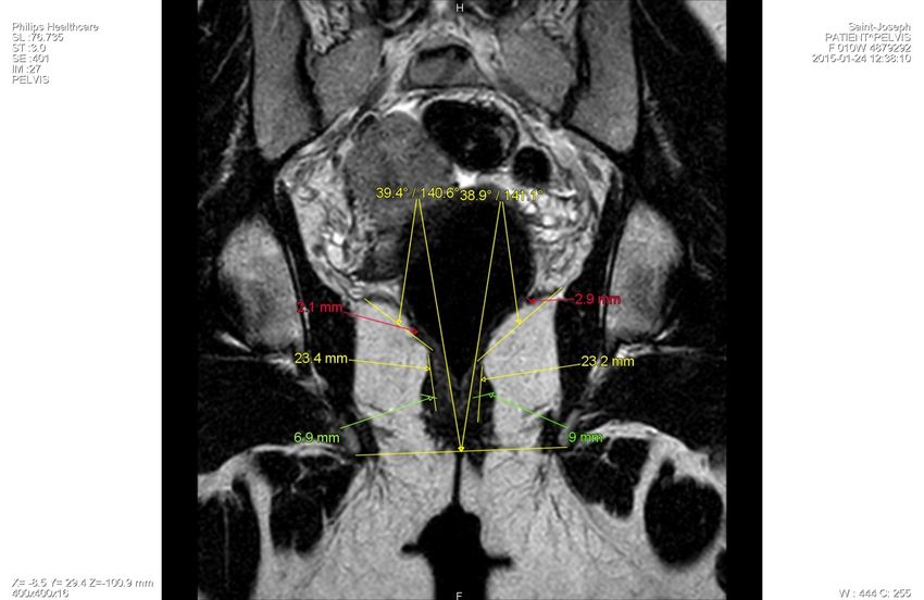
bioRxiv preprint first posted online Jan. 16, 2018; doi: http://dx.doi.org/10.1101/248823. The copyright holder for this preprint
 (which was not peer-reviewed) is the author/funder, who has granted bioRxiv a license to display the preprint in perpetuity.
                                    All rights reserved. No reuse allowed without permission.

      2                                                                          Frédéric Dierick et al.

 20   PFMT should be based primarily on non-instrumented exercises.

      Keywords anatomy · levator ani · transversus abdominis · strengthening ·
      morphology · hypopresssive · biofeedback · electrical stimulation

      1 Introduction

 25   Pelvic floor dysfunction (PFD), in particular stress urinary incontinence (UI)
      caused by structural defects in connective tissue and muscles that support
      pelvic organs (Petros, 2010; Memon and Handa, 2013), is a common and dis-
      tressing condition affecting young and middle-aged women (Kim et al, 2003;
      Lose, 2005; Robinson and Cardozowan, 2014; Schreiber Pedersen et al, 2017).
 30   Several randomized controlled trials have demonstrated that pelvic floor mus-
      cles training (PFMT) is effective to reduce stress and any type of UI in women,
      therefore it is recommended as a first-line conservative therapy (Dumoulin
      et al, 2014, 2015). Nowadays, PFMT can be achieved with instrumentation,
      using vaginal/ anal probe electrode delivering biofeedback exercises or electri-
 35   cal stimulations, and weighted vaginal cones or other resistance devices. It can
      be also achieved without any instrumentation, using direct voluntary contrac-
      tions, also known as Kegel’s exercises (Harvey, 2003), or indirect contractions
      with transversus abdominis muscle activation through hypopressive exercises
      (Caufriez, 1997; Resende et al, 2012). Previous studies on women with PFD
 40   show that PFMT without the use of electrical stimulation (Bø et al, 1999) or
      biofeedback (Fitz et al, 2012) is more efficient that with it, but this is contro-
      versial (Castro et al, 2008; Ibrahim et al, 2015). Uncertainty about which of
      these strategies are most effective in training women is one of the key clinical
      questions which needs to be prioritized because training using instrumentation
 45   is more costly than without. Therefore, further studies are needed to assess
      PFMT strategies efficacy.
          The most common type of involuntary leakage of urine is stress urinary in-
      continence (SUI), a worldwide problem that affects the quality of life of millions
      of women since is typically experienced when coughing, sneezing, laughing, or
 50   exercising. In SUI, urine leakage is explained by a lack of closure pressure in
      the urethra during exertion, that could be attributed to anatomic changes in
      the bladder and urethra and/or PFM, endopelvic fascia and connective tissue
      supports (Petros and Ulmsten, 1990; Tunn et al, 2006; Hay-Smith et al, 2011).
      From a physiological point of view, we believe that it is relevant to study the
 55   impact of PFMT in young asymptomatic women with “non-pathological” leva-
      tor ani (LA) muscle signals that are preserved from the consequences of parity
      or loco-regional surgery. However, to the best of our knowledge, no study has
      investigated clinical and morphological changes in LA muscle subdivisions be-
      fore and after PFMT in young asymptomatic nulliparous women, even though
 60   a recent study reported a high prevalence of 20% for involuntary loss of urine
      in a group of 159 young presumably healthy women aged 18–30 years (van
      Breda et al, 2015).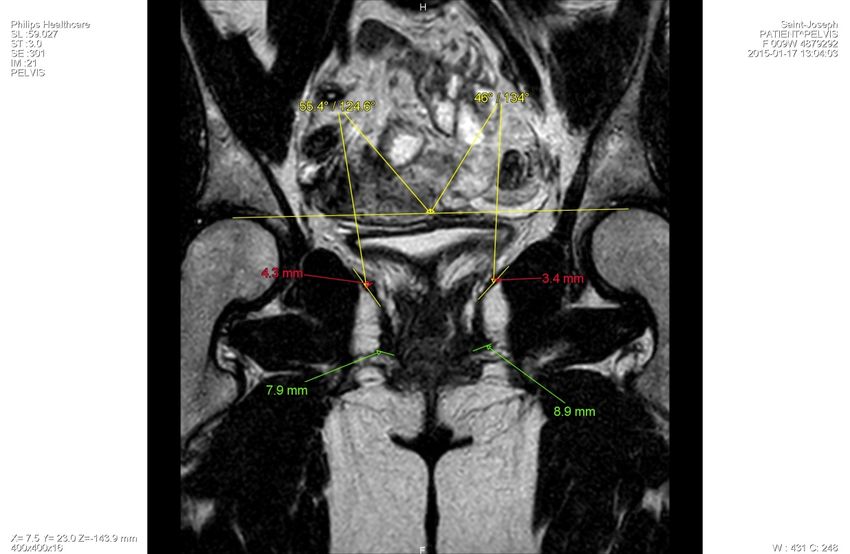
bioRxiv preprint first posted online Jan. 16, 2018; doi: http://dx.doi.org/10.1101/248823. The copyright holder for this preprint
 (which was not peer-reviewed) is the author/funder, who has granted bioRxiv a license to display the preprint in perpetuity.
                                    All rights reserved. No reuse allowed without permission.

      Intensive training of pelvic floor muscles                                                       3

          The purpose of this randomized study was to evaluate the impact of a 3-
      week period of intensive PFMT, with or without instrumentation based on
 65   biofeedback and electrical stimulation, on clinical and static MRI changes
      of puborectalis (PR) and iliococcygeus (IL) muscles. The protocol without
      biofeedback and electrical stimulation was entirely realized without any in-
      strumentation and based on voluntary PFM contractions combined with hy-
      popressive exercises.

 70   2 Methods

      2.1 Study participants and PFM exercises

      A convenience sample of 24 healthy young nulliparous women (age: 22.9±1.6
      yrs, weight: 61.5±6.9 kg, height: 167.7±7.9 cm, body mass index (BMI):
      21.8±1.5 kg m−2 ), was recruited through advertisement and snowball sam-
 75   pling at our physiotherapy department. Before clinical and MRI assessments,
      all participants were questioned about PFD including symptoms of urinary
      and fecal continence, using a validated french version (de Tayrac et al, 2007)
      of Pelvic Floor Distress Inventory (PFDI-20) and Pelvic Floor Impact Ques-
      tionnaire (PFIQ-7) (Barber et al, 2005). Additionally, we asked them if they
 80   were aware of their PFM function and if they known or already practiced PFM
      exercises. To be included in the study, participant had to have no MRI incom-
      patibility (intrauterine device, pregnancy) or have practiced PFM exercises
      within the last year. Exclusion criteria were: pelvic floor troubles, diarrhea or
      chronic constipation, intensive sports practices, claustrophobia, neuroleptic or
 85   antidepressant medication, abortion. Inclusion criteria were: BMI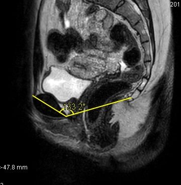
bioRxiv preprint first posted online Jan. 16, 2018; doi: http://dx.doi.org/10.1101/248823. The copyright holder for this preprint
 (which was not peer-reviewed) is the author/funder, who has granted bioRxiv a license to display the preprint in perpetuity.
                                    All rights reserved. No reuse allowed without permission.

      4                                                                          Frédéric Dierick et al.

105   tain this position in apnea for 20 seconds. A standing, quadrupedic, and lying
      posture with active movements were selected, and the sequence of postures
      was left to the physiotherapist’s discretion. After diaphragmatic aspiration,
      a voluntary contraction of PFM was achieved and sustained throughout the
      active movements. A complete session lasted 30 min.
110       In ELEC group, a commercially available electrical stimulator (Myomed
      632, Enraf Nonius, Delft, the Netherlands) was used to deliver transvaginal
      electrical stimulations (excitomotor, bidirectional, rectangular, symmetric cur-
      rent) via vaginal probes (Goode et al, 2003) and lasts 15 min (450 s on right
      PR and 450 s on left PR) and biofeedback training lasts 15 min (6 s mus-
115   cle contraction and 12 s of rest). Frequency stimulation was set between 20
      and 50 Hz. Biphasic pulses had duration of 1 ms. The current intensity was
      adjusted to the maximum level that could be tolerated comfortably, up to
      maximum 100 mA.
          Written informed consent was obtained from all individual participants in-
120   cluded in the study. No financial compensation was provided to participate to
      the study. Institutional review board approval is not required at our institution
      for MRI using standard pulse sequences. A Consolidated Standards of Report-
      ing Trials (CONSORT) diagram showing enrollment, training allocation, and
      follow-up is presented in Fig. 1.

125   2.2 Clinical and MRI assessments

      Clinical assessment was realized in gynecological position (with the thighs
      flexed to 90 degrees) by a physiotherapist who has specialized in uro-gynecology;
      before and after PFMT. At the first assessment, all participants were in-
      structed on how to correctly perform their PFM using vaginal palpation. The
130   physiotherapist was blinded to group allocation.
          PFM strength was measured through an arbitrary 0–12 points scale using
      a highly reliable (Isherwood and Rane, 2000) mechanical perineometer (PFX2,
      Cardio Design, Castle Hill, New South Wales, Australia) with a vaginal probe
      (26–28 mm diameter, active surface: 55 mm long, overall: 108 mm long). The
135   cranial movement of the vaginal probe during measurement of PFM contrac-
      tion was observed. Any contraction for which a retroversion of the hip or a
      Valsava maneuver was noticed were discounted. PFM strength was also as-
      sessed by bi-digital palpation (Bø and Sherburn, 2005) with the two distal
      phalanges inside the introitus vagina using the Modified Oxford Grading Sys-
140   tem (MOGS). It was individually assessed for right and left parts of PR and
      perineal body (PB), and consists of a 0–5 points scale: 0 = no contraction,
      1 = flicker, 2 = weak, 3 = moderate, 4 = good (with lift) and 5 = strong.
      Three consecutive maximal contractions sustained for 5 seconds were recorded
      for perineometer and digital assessments, with a 30-second interval between
145   efforts and the best of the 3 was registered.
          PFM resting tone (RT) was quantified by digital palpation of left and
      right PR muscles (Dietz and Shek, 2008) on a 6-point scale with the following
bioRxiv preprint first posted online Jan. 16, 2018; doi: http://dx.doi.org/10.1101/248823. The copyright holder for this preprint
(which was not peer-reviewed) is the author/funder, who has granted bioRxiv a license to display the preprint in perpetuity.
All rights reserved. No reuse allowed without permission.
Intensive training of pelvic floor muscles 5
grades: 0 = muscle not palpable, 1 = muscle palpable but very flaccid, wide
hiatus, minimal resistance to distension, 2 = hiatus wide but some resistance
150 to distension, 3 = hiatus fairly narrow, fair resistance to palpation but easily
distended, 4 = hiatus narrow, muscle can be distended but high resistance
to distension, no pain, and 5 = hiatus very narrow, no distension possible,
‘woody’ feel, possibly with pain: ‘vaginismus’. PFM RT was computed as the
sum of the grades of left and right parts of PR. Diaphragmatic aspiration
155 (DA) (Caufriez, 1997) was rated on a scale of 0–5–10 (0 = no movement, 5
= mobilization of the perineum or viscera, 10 = mobilization of the perineum
and viscera). The steps of DA are as follows: slow diaphragmatic inspiration,
followed by total expiration and, after glottal closure, a gradual contraction
of the abdominal wall muscles, with superior displacement of the diaphragm
160 cupola (Caufriez, 1997; Resende et al, 2012; Santa Mina et al, 2015).
For MRI acquisition, each participant underwent an assessment performed
in a supine position using a 1.5 Tesla MRI scanner (Ingenia 1.5T, Phillips,
The Netherlands) and surface coils. Static T2-weighted turbo spin echo (TSE)
techniques without fat saturation were used to image the sagittal, coronal, and
165 axial planes during 20 minutes (spatial resolution 0.52×0.52 mm, repetition
time (TR) 3745-4815 ms , echo time (TE) 90 ms, no gap, slices thickness
3 mm and water phase shift minimum). Seventy images were obtained in
sagittal plane, fifty in coronal and thirty-five in the PR paraxial plane. All
participants were instructed by a medical imaging technologist to relax their
170 PFM while breathing in and out normally. MRI assessments were performed
before and after PFMT. All images were obtained at our nuclear magnetic
resonance department. Different measurements were performed on images and
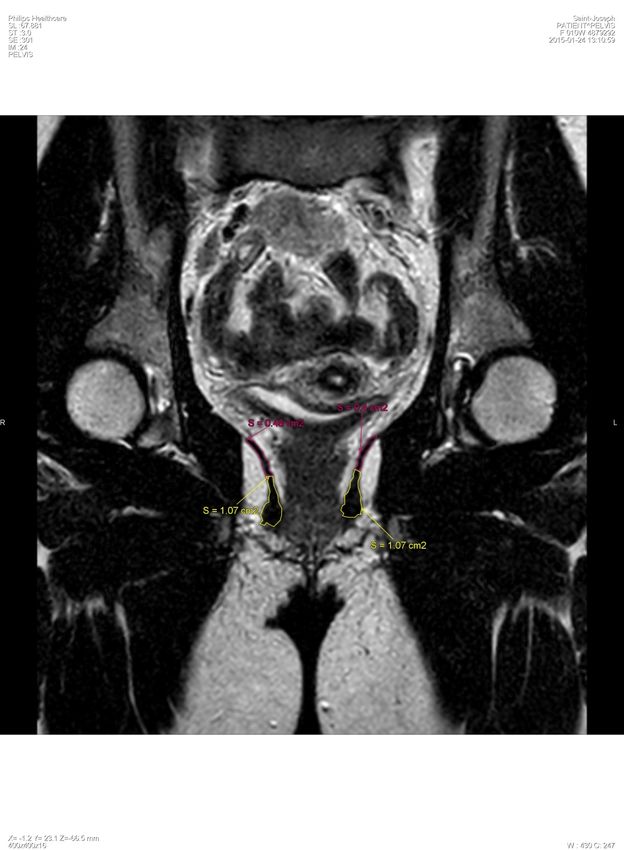
are detailed in Table 1, Fig. 2 and 3. Mid-pubic line (MPL) (Singh et al, 2001),
pubococcygeal line (PCL) (Yang et al, 1991), and posterior levator plate (PLP)
175 (Hsu et al, 2006) were determined (see Fig. 2 and Table 1).
Measurements required about 6 hours for each participant and were per-
formed by a radiologist who has specialized in pelvic floor MRI (L.M.) and
two physiotherapists (E.G. and C.L.).
2.3 Statistical methods
180 All statistical procedures were performed with SigmaPlot software version 11.0
(Systat Software, San Jose, CA). The p-value was considerate significant when
less than 0.05. Clinical results at baseline between HYPO and ELEC groups
were compared using Mann-Whitney U tests and MRI results with t-tests.
Wilcoxon signed-ranked tests were performed to examine the effects of train-
185 ing protocols on clinical results in HYPO and ELEC groups. A two-way RM
ANOVA (before-after, training groups) with Holm-Sidak method for pairwise
multiple comparisons was performed to examine the effect of PFMT and train-
ing protocols on MRI results. All data are presented as means and standard
deviations (SD) and were checked for normality (Shapiro-Wilk) and equal vari-
190 ance (Brown-Forsythe) tests.bioRxiv preprint first posted online Jan. 16, 2018; doi: http://dx.doi.org/10.1101/248823. The copyright holder for this preprint
(which was not peer-reviewed) is the author/funder, who has granted bioRxiv a license to display the preprint in perpetuity.
All rights reserved. No reuse allowed without permission.
6 Frédéric Dierick et al.
For PR and IL volumes, both interobserver and intraobserver reliability
were assessed using intraclass correlation coefficient (ICC(2,k): 2-way random
effects, absolute agreement, multiple raters (k=2): E.G. and L.M.; ICC(2,1):
2-way random effects, absolute agreement, single rater: E.G.) (Shrout and
195 Fleiss, 1979). Each observer’s first measure recorded from each subject was
used in the calculation of interobserver reliability. Intraobserver reliability was
calculated using a second measurement recorded from each subject. ICCs were
computed with R software (version 3.4.1) and irr package (version 0.84).
3 Results
200 3.1 Clinical results
None of the participants had PFD symptoms as determined by responses to
questionnaires PFID-20 and PFQ-7. No significant differences between groups
were observed at baseline for all clinical results. MOGS scores for left PR
and PB significantly increased in the two groups but MOGS score for right
205 PR significantly increased only in HYPO group (Table 2). PFM strength and
resting tone, and diaphragmatic aspiration were not modified.
3.2 MRI results
No significant differences between groups were observed at baseline for all MRI
results. The ICC values were 0.70 (inter-) and 0.95 (intra-) for PR volume and
210 0.80 and 0.83 for IL volume, showing moderate to very good reliability results.
Muscle volumes of right and left IL significantly decreased after training and
signal intensities of right and left PR significantly decreased after training (Ta-
ble 3). Muscle thickness of right and left IL at mid-vagina location significantly
decreased after training (Table 3). MPL–PCL and PLP–PCL angles, PR vol-
215 ume, IL signal intensities, and PR and IL measurements done at mid-vagina,
mid-rectum, and at 9 & 3 o’clock the vagina were not modified.
4 Discussion
LA muscle complex is one of the most important and complicated anatomical
structure of the human body and as a result also one of the most poorly
220 understood (Lammers et al, 2013). However, understanding the basic anatomy
of the LA is essential when formulating a clinical opinion as injuries occur in
13–36% of women who have a vaginal delivery (Schwertner-Tiepelmann et al,
2012). Until now, no study explore the effects of a short period of intensive
PFMT on the morphology of the pelvic muscles and related clinical scores
225 in healthy nulliparous women, and the produced changes are uncertain. Our
hypothesis was that the protocol based on biofeedback combined with vaginal
stimulations would lead to less important morphological and clinical changesbioRxiv preprint first posted online Jan. 16, 2018; doi: http://dx.doi.org/10.1101/248823. The copyright holder for this preprint
(which was not peer-reviewed) is the author/funder, who has granted bioRxiv a license to display the preprint in perpetuity.
All rights reserved. No reuse allowed without permission.
Intensive training of pelvic floor muscles 7
of PFM than voluntary contractions combined with hypopressive exercises.
Results show the presence of clinical and morphological changes in different
230 parts of LA muscle at rest after PFMT. However, changes were not significantly
higher in the group that do not use instrumentation.
Historically, MRI studies have outlined the anatomy of PFM much more
clearly than was possible with anatomical dissection studies (Raizada and Mit-
tal, 2008), and different subdivisions of the LA were identified (Kearney et al,
235 2004; Margulies et al, 2006). Terminologia Anatomica (Federative Commitee
on Anatomical Terminology, 1998), identifies three major subdivisions of LA:
IL, PR, and pubococcygeus (PC). Over the years, these subdivisions have been
given several names, making understanding even more difficult (Lammers et al,
2013). Among female, PC muscle, also called pubovisceralis (PV), is further
240 divided into the puboperinealis, pubovaginalis, and puboanalis (Lawson, 1974;
Kearney et al, 2004). Even if different parts of PV and enclosing urogenital
hiatus, could be observed with standard MRI techniques, they can not be
distinguished at their origin of the pubic bone (Zijta et al, 2013). Only MRI
studies with diffusion tensor imaging (DTI) and fiber tractography allowed to
245 provide a three-dimensional overall appearance of muscular fibers of PV (Zijta
et al, 2011; Rousset et al, 2012), and it was therefore decided not to study PV.
From a morphological viewpoint, PR is thicker than IL and forms a band
around the urethra, vagina and rectum (Singh et al, 2002). Our results show
that PR thickness values before PFMT were greater at mid-vagina level com-
250 pared to other levels, with mean values between 8.7±0.6 and 9.4±0.7 mm, and
IL thickness values were greater at mid-rectum level, with mean values between
3.6±0.4 and 4.0±0.4 mm. Singh et al (2002) observed lower thickness values
for PR (6.5±2.0) and IL (2.9±0.8), but their sample was composed of healthy
nulliparous women aged between 23–42 yrs. Dumoulin et al (2007) showed in
255 women with SUI that LA surface area at rest after PFMT was significantly
smaller than before training; however, during a voluntary contraction, LA sur-
face was significantly higher than before training. Even in healthy women, our
results are in agreement with this study since we observed a decreased volume
and thickness of IL muscle at mid-vagina level, both for PFMT with and with-
260 out instrumentation. A dynamic MRI study is therefore needed to complete
our static results. Furthermore, PR muscle volume and thickness were un-
changed but signal intensity of PR compared with that of OI decreased, both
for PFMT with and without instrumentation. The signal intensity of healthy
muscles, i.e. muscles showing no muscle edema, fatty infiltration, or mass le-
265 sion, is well lower than that of fat for T2-weighted images (May et al, 2000).
Hence it can be argued that the decreased signal intensity of PR indicates a
decrease in fatty composition of this muscle, in favor of an increase of muscle
fibers. Since SUI, apart of ligamentous and fascial lesions, is associated with
increased signal intensity (Kirschner-Hermanns et al, 1993), this observation
270 could be of major importance in pathological conditions. From a clinical view-
point, MOGS scores significantly increased after PFMT, indicating a global
increase of PFM strength. Since a 3-week period of intensive PFMT, based on
90 minutes/week, was sufficient to increase muscle strength in the 2 trainingbioRxiv preprint first posted online Jan. 16, 2018; doi: http://dx.doi.org/10.1101/248823. The copyright holder for this preprint
(which was not peer-reviewed) is the author/funder, who has granted bioRxiv a license to display the preprint in perpetuity.
All rights reserved. No reuse allowed without permission.
8 Frédéric Dierick et al.
groups, we believe that, in a preventive purpose, it is not necessary to use an
275 expensive and more invasive method than combined voluntary and hypopres-
sive exercises (Bernardes et al, 2012).
PFM strength changes observed after PFMT and assessed with perineome-
ter were not significant. Perineometer values represent a global measurement of
vaginal closure force that also includes the “noise” caused by the rise in intra-
280 abdominal pressure that often accompanies a LA contraction (Ashton-Miller
et al, 2014). Even if we systematically checked the presence of a cranial move-
ment of the vaginal probe during the measurement, we can exclude an increase
in intra-abdominal pressure that could have masked the finding of a significant
increase in the vaginal closure force after PFMT. The use of an instrumented
285 speculum designed to minimize the effect of intra-abdominal pressure during
measurement of PFM strength and developed by Ashton-Miller et al (2014),
may be a solution to overcome this limitation. Another explanation of these
results could be that the global maximal vaginal closure force was not modi-
fied but that some muscles have increased their strength to the detriment of
290 others. Our hypothesis is based on the following observations after PFMT: (1)
the decreasing volume and thickness of IL at the level of mid-vagina, without
any change in signal intensity; and (2) the decrease of signal intensity in PR.
Therefore, IL force may have been reduced while PR force increased. If this
is the case, PFMT could be effective in modifying the resulting forces of the
295 different parts of LA since the lines of action of IL (PV) and PR muscles are
quite different: the first one lifts the pelvic floor while the second closes it
(Betschart et al, 2014). The absence of a change in signal intensity of IL must
be balanced according to the training groups. Surprisingly, signal intensity
tends to increase in ELEC group while it decrease in HYPO. This finding also
300 support the use of non-instrumented PFMT.
The present study has several limitations. The first limitation is the small
number of the participants; therefore, our results may be not representative
of the general population. To reduce bias about group homogeneity, several
inclusion criteria were met. The first inclusion criterion was that all partici-
305 pants were young and nulliparous women. According to Slieker-ten Hove et al
(2009), voluntary muscle contraction decrease with age, but there is no rela-
tion with parity. DeLancey et al (2003) found abnormalities in the LA muscle
on MRI after vaginal delivery, that are not found in nulliparous. In women
over age 60 years, the PFMs are significantly thinner both at rest and during
310 contraction, compared to younger women (Bernstein, 1997). The second crite-
rion was that participants were European women to avoid ethnic variability.
Howard et al found a functional and morphological differences in the urethral
sphincter and support system of nulliparous black and white women (Howard
et al, 2000; Handa et al, 2008). The second limitation is that PFMs were only
315 assessed with static MRI and a dynamic MRI study is needed to complete our
results (Cai et al, 2013; Dumoulin et al, 2007). Others limitations relate to
MRI acquisition. Each participant underwent MRI adapted at supine position
with a mid-full bladder repletion. In the supine position, PFMs do not undergo
organ weight and gravity (Fielding et al, 1998). A mid-full bladder repletionbioRxiv preprint first posted online Jan. 16, 2018; doi: http://dx.doi.org/10.1101/248823. The copyright holder for this preprint
(which was not peer-reviewed) is the author/funder, who has granted bioRxiv a license to display the preprint in perpetuity.
All rights reserved. No reuse allowed without permission.
Intensive training of pelvic floor muscles 9
320 condition is difficult to obtain because of the ability of each bladder. During
image analysis, we observed different degree of repletion of the bladder, which
could influence our results. Finally, signal measurements were difficult to do
coronally in IL (very thin) and in some PR and IL frequently fasciculated with
fat striations.
325 5 Conclusions
This study suggests that 270 minutes of PFMT in healthy nulliparous women
produces clinical and morphological changes in PFMs at rest. Muscle volume
and thickness of IL at mid-vagina location decreased after training. PR volume
was not modified but its signal intensity decreased, indicating a decrease in
330 fatty composition of this muscle, in favor of an increase of muscle fibers. Ad-
ditionally, MOGS scores increased after training, indicating that participants
improved voluntary force contraction of their PFMs. Static MRI appears to be
a relevant tool to highlight the impact of PFMT but is not sufficient to fully
explain all clinical improvement and morphological changes. Further dynamic
335 MRI studies and/or DTI-based tractography are needed to better understand
the impact of training on the morphology of PFMs, and determine if these
changes are sustainable and applicable to women with PFD.
Acknowledgements The authors would like to thank Caroline Lahaye and Jean-Louis
Greffe for their assistance in the development of the protocol, and Jean-Claude Malherbe
(Enraf-Nonius) for making the stimulation/biofeedback material available throughout the
experiment. They also thank the financial support of Grand Hôpital de Charleroi (GHdC
asbl), Philips SA, and the Haute Ecole Louvain en Hainaut for the MRI.
References
Ashton-Miller JA, Zielinski R, DeLancey JOL, Miller JM (2014) Validity
and reliability of an instrumented speculum designed to minimize the
effect of intra-abdominal pressure on the measurement of pelvic floor
muscle strength. Clin Biomech (Bristol, Avon) 29(10):1146–50, DOI
10.1016/j.clinbiomech.2014.09.011
Barber M, Walters M, Bump R (2005) Short forms of two
condition-specific quality-of-life questionnaires for women with
pelvic floor disorders (PFDI-20 and PFIQ-7). Am J Obstet
Gynecol 193(1):103–113, DOI 10.1016/j.ajog.2004.12.025, URL
http://dx.doi.org/10.1016/j.ajog.2004.12.025
Bernardes BT, Resende P A, Stüpp L, Oliveira E, Castro A R, Bella ZI,
Girão MJ, Sartori MG (2012) Efficacy of pelvic floor muscle training and
hypopressive exercises for treating pelvic organ prolapse in women: random-
ized controlled trial. Sao Paulo Med J 130(1):5–9
Bernstein IT (1997) The pelvic floor muscles: muscle thickness in healthy
and urinary-incontinent women measured by perineal ultrasonography withbioRxiv preprint first posted online Jan. 16, 2018; doi: http://dx.doi.org/10.1101/248823. The copyright holder for this preprint
(which was not peer-reviewed) is the author/funder, who has granted bioRxiv a license to display the preprint in perpetuity.
All rights reserved. No reuse allowed without permission.
10 Frédéric Dierick et al.
reference to the effect of pelvic floor training. Estrogen receptor studies.
Neurourol Urodyn 16(4):237–275
Betschart C, Kim J, Miller JM, Ashton-Miller JA, DeLancey JOL
(2014) Comparison of muscle fiber directions between different lev-
ator ani muscle subdivisions: in vivo mri measurements in women.
Int Urogynecol J 25(9):1263–1268, DOI 10.1007/s00192-014-2395-9, URL
http://dx.doi.org/10.1007/s00192-014-2395-9
Bø K, Sherburn M (2005) Evaluation of female pelvic-floor muscle function
and strength. Phys Ther 85(3):269–282
Bø K, Talseth T, Holme I (1999) Single blind, randomised controlled trial
of pelvic floor exercises, electrical stimulation, vaginal cones, and no
treatment in management of genuine stress incontinence in women. BMJ
318(7182):487–493
van Breda HMK, Bosch JLHR, de Kort LMO (2015) Hidden prevalence of
lower urinary tract symptoms in healthy nulligravid young women. Int Urog-
ynecol J 26(11):1637–43, DOI 10.1007/s00192-015-2754-1
Cai XR, Qiu L, Wu HJ, R LS (2013) Assessment of levator ani morphology
and function in asymptomatic nulliparous women via static and dynamic
magnetic resonance imaging. Int J Gynaecol Obstet 121(3):233–239, DOI
10.1016/j.ijgo.2013.01.022
Castro R, Arruda R, Zanetti M, Santos P, Sartori M, MJ G (2008) Single-
blind, randomized, controlled trial of pelvic floor muscle training, electri-
cal stimulation, vaginal cones, and no active treatment in the management
of stress urinary incontinence. Clinics (Sao Paulo) 63(4):465–472, DOI
10.1590/S1807-59322008000400009
Caufriez M (1997) Gymnastique abdominale hypopressive. Editions M.
Caufriez, Bruxelles
DeLancey JO, Kearney R, Chou Q, Speights S, Binno S (2003) The appear-
ance of levator ani muscle abnormalities in magnetic resonance images after
vaginal delivery. Obstet Gynecol 101(1):46–53
Dietz HP, Shek KL (2008) The quantification of levator muscle resting tone by
digital assessment. Int Urogynecol J 19(11):1489–1493, DOI 10.1007/s00192-
008-0682-z, URL http://dx.doi.org/10.1007/s00192-008-0682-z
Dumoulin C, Peng Q, Stodkilde-Jorgensen H, Shishido K, Con-
stantinou C (2007) Changes in levator ani anatomical configura-
tion following physiotherapy in women with stress urinary inconti-
nence. J Urol 178(3):970–977, DOI 10.1016/j.juro.2007.05.023, URL
http://dx.doi.org/10.1016/j.juro.2007.05.023
Dumoulin C, Hay-Smith EJC, Mac Habée-Séguin G (2014) Pelvic floor
muscle training versus no treatment, or inactive control treatments, for
urinary incontinence in women (review). Cochrane Database Syst Rev
14(5):CD005,654, DOI 10.1002/14651858.CD005654.pub3.
Dumoulin C, Hay-Smith J, Habée-Séguin GM, Mercier J (2015) Pelvic
floor muscle training versus no treatment, or inactive control treatments,
for urinary incontinence in women: A short version Cochrane system-
atic review with meta-analysis. Neurourol Urodyn 34(4):300–308, DOIbioRxiv preprint first posted online Jan. 16, 2018; doi: http://dx.doi.org/10.1101/248823. The copyright holder for this preprint
(which was not peer-reviewed) is the author/funder, who has granted bioRxiv a license to display the preprint in perpetuity.
All rights reserved. No reuse allowed without permission.
Intensive training of pelvic floor muscles 11
10.1002/nau.22700, URL http://dx.doi.org/10.1002/nau.22700
Fielding JR, Griffiths DJ, Versi E, Mulkern RV, Lee ML, Jolesz FA
(1998) MR imaging of pelvic floor continence mechanisms in the supine
and sitting positions. AJR Am J Roentgenol 171(6):1607–1610, DOI
10.2214/ajr.171.6.9843296
Fitz FF, Resende APM, Stüpp L, Sartori MGF, Girão MJBC, Cas-
tro RA (2012) Biofeedback for the treatment of female pelvic
floor muscle dysfunction: a systematic review and meta-analysis. Int
Urogynecol J 23(11):1495–1516, DOI 10.1007/s00192-012-1707-1, URL
http://dx.doi.org/10.1007/s00192-012-1707-1
Goode PS, Burgio KL, Locher JL, Roth DL, Umlauf MG, Richter HE, Varner
RE, Lloyd LK (2003) Effect of behavioral training with or without pelvic
floor electrical stimulation on stress incontinence in women: a randomized
controlled trial. JAMA 290(3):345–352, DOI 10.1001/jama.290.3.345
Handa VL, Lockhart ME, Fielding JR, Bradley CS, Brubaker L, Cundiff GW,
Ye W, Richter HE, Pelvic Floor Disorders Network (2008) Racial differ-
ences in pelvic anatomy by magnetic resonance imaging. Obstet Gynecol
111(4):914–20, DOI 10.1097/AOG.0b013e318169ce03
Harvey MA (2003) Pelvic floor exercises during and after pregnancy: a sys-
tematic review of their role in preventing pelvic floor dysfunction. J Obstet
Gynaecol Can 25(6):487–498
Hay-Smith EJC, Herderschee R, Dumoulin C, Herbison GP (2011) Com-
parisons of approaches to pelvic floor muscle training for urinary incon-
tinence in women (review). Cochrane Database of Systematic Reviews Issue
12:CD009,508, DOI 10.1002/14651858.CD009508
Slieker-ten Hove MP, Pool-Goudzwaard AL, Eijkemans MJC, Steegers-
Theunissen RPM, Burger CW, Vierhout ME (2009) Pelvic floor muscle
function in a general female population in relation with age and parity and
the relation between voluntary and involuntary contractions of the pelvic
floor musculature. Int Urogynecol J 20(12):1497–1504, DOI 10.1007/s00192-
009-0978-7, URL http://dx.doi.org/10.1007/s00192-009-0978-7
Howard D, DeLancey JOL, Tunn R, Ashton-Miller JA (2000) Racial differences
in the structure and function of the stress urinary continence mechanism.
Obstet Gynecol 95(5):713–717
Hoyte L, Jakab M, Warfield SK, Shott S, Flesh G, Fielding JR (2004) Le-
vator ani thickness variations in symptomatic and asymptomatic women
using magnetic resonance-based 3-dimensional color mapping. Am J
Obstet Gynecol 191(3):856–861, DOI 10.1016/j.ajog.2004.06.067, URL
http://dx.doi.org/10.1016/j.ajog.2004.06.067
Hsu Y, Summers A, Hussain HK, Guire KE, Delancey JO (2006) Le-
vator plate angle in women with pelvic organ prolapse compared to
women with normal support using dynamic MR imaging. Am J Ob-
stet Gynecol 194(5):1427–1433, DOI 10.1016/j.ajog.2006.01.055, URL
http://dx.doi.org/10.1016/j.ajog.2006.01.055
Ibrahim IK, Hameed MMA, Taher EM, Shaheen EM, Elsawy MSAG (2015)
Efficacy of biofeedback-assisted pelvic floor muscle training in females withbioRxiv preprint first posted online Jan. 16, 2018; doi: http://dx.doi.org/10.1101/248823. The copyright holder for this preprint
(which was not peer-reviewed) is the author/funder, who has granted bioRxiv a license to display the preprint in perpetuity.
All rights reserved. No reuse allowed without permission.
12 Frédéric Dierick et al.
pelvic floor dysfunction. Alexandria J Med 51:137–142
Isherwood PJ, Rane A (2000) Comparative assessment of pelvic floor strength
using a perineometer and digital examination. BJOG 107(8):1007–1011
Kearney R, Sawhney R, DeLancey JOL (2004) Levator ani muscle anatomy
evaluated by origin-insertion pairs. Obstet Gynecol 104(1):168–173, DOI
10.1097/01.AOG.0000128906.61529.6b
Kim JK, Kim YJ, Choo MS, Cho KS (2003) The urethra and its supporting
structures in women with stress urinary incontinence: MR imaging using an
endovaginal coil. AJR 180:1037–1044, DOI 10.2214/ajr.180.4.1801037
Kirschner-Hermanns R, Wein B, Niehaus S, Schaefer W, Jakse G (1993) The
contribution of magnetic resonance imaging of the pelvic floor to the under-
standing of urinary incontinence. Br J Urol 72(5 Pt 2):715–718
Lammers K, Prokop M, Vierhout ME, Kluivers KB, Fütterer JJ (2013) A
pictorial overview of pubovisceral muscle avulsions on pelvic floor magnetic
resonance imaging. Insights Imaging 4(4):431–441, DOI 10.1007/s13244-013-
0261-9, URL http://dx.doi.org/10.1007/s13244-013-0261-9
Lawson JO (1974) Pelvic anatomy. i. pelvic floor muscles. Ann R Coll Surg
Engl 54(5):244–252
Lose G (2005) The burden of stress urinary incontinence. Eur
Urol Suppl 4(1):5–10, DOI 10.1016/j.eursup.2004.10.002, URL
http://dx.doi.org/10.1016/j.eursup.2004.10.002
Margulies RU, Hsu Y, Kearney R, Stein T, Umek WH, DeLancey
JOL (2006) Appearance of the levator ani muscle subdivisions in
magnetic resonance images. Obstet Gynecol 107(5):1064–1069, DOI
10.1097/01.AOG.0000214952.28605.e8
May DA, Disler DG, Jones EA, Balkissoon AA, Manaster BJ (2000)
Abnormal signal intensity in skeletal muscle at MR imaging:
patterns, pearls, and pitfalls. Radiographics 20:S295–S315, DOI
10.1148/radiographics.20.suppl 1.g00oc18s295
Memon HU, Handa VL (2013) Vaginal childbirth and pelvic floor disorders.
Womens Health (Lond Engl) 9(3):265–277, DOI 10.2217/whe.13.17
Petros PEP (2010) The integral theory system. a simplified clinical approach
with illustrative case histories. Pelviperineology 29:37–51
Petros PEP, Ulmsten UI (1990) An integral theory of female urinary inconti-
nence. Acta Obstet Gynecol Scand 69(S153):7–31
Raizada V, Mittal RK (2008) Pelvic floor anatomy and ap-
plied physiology. Gastroenterology Clinics of North Amer-
ica 37(3):493–509, DOI 10.1016/j.gtc.2008.06.003, URL
http://dx.doi.org/10.1016/j.gtc.2008.06.003
Resende APM, Stüpp L, Bernardes BT, Oliveira E, Castro RA, Girão MJBC,
Sartori MGF (2012) Can hypopressive exercises provide additional ben-
efits to pelvic floor muscle training in women with pelvic organ pro-
lapse? Neurourol Urodyn 31(1):121–125, DOI 10.1002/nau.21149, URL
http://dx.doi.org/10.1002/nau.21149
Robinson D, Cardozowan L (2014) Urinary incontinence in the young woman:
treatment plans and options available. Womens Health (Lond) 10(2):201–17,bioRxiv preprint first posted online Jan. 16, 2018; doi: http://dx.doi.org/10.1101/248823. The copyright holder for this preprint
(which was not peer-reviewed) is the author/funder, who has granted bioRxiv a license to display the preprint in perpetuity.
All rights reserved. No reuse allowed without permission.
Intensive training of pelvic floor muscles 13
DOI 10.2217/whe.14.1
Rousset P, Delmas V, Buy JN, Rahmouni A, Vadrot D, Deux JF (2012) In
vivo visualization of the levator ani muscle subdivisions using MR fiber
tractography with diffusion tensor imaging. J Anat 221(3):221–228, DOI
10.1111/j.1469-7580.2012.01538.x, URL http://dx.doi.org/10.1111/j.1469-
7580.2012.01538.x
Garcı́a del Salto L, de Miguel Criado J, Aguilera del Hoyo L, Gutiérrez Ve-
lasco L, Fraga Rivas P, Manzano Paradela M, Dı́ez Pérez de las Va-
cas M, Marco Sanz A, Fraile Moreno E (2014) MR imaging–based as-
sessment of the female pelvic floor. Radiographics 34(5):1417–1439, DOI
10.1148/rg.345140137
Santa Mina D, Au D, Alibhai SMH, Jamnicky L, Faghani N, Hilton WJ, Ste-
fanyk LE, Ritvo P, Jones J, Elterman D, et al (2015) A pilot randomized
trial of conventional versus advanced pelvic floor exercises to treat urinary
incontinence after radical prostatectomy: a study protocol. BMC Urol 15(1),
DOI 10.1186/s12894-015-0088-4, URL http://dx.doi.org/10.1186/s12894-
015-0088-4
Schreiber Pedersen L, Lose G, Høybye MT, Elsner S, Waldmann A, Rudnicki
M (2017) Prevalence of urinary incontinence among women and analysis of
potential risk factors in germany and denmark. Acta Obstet Gynecol Scand
96(8):939–948, DOI 10.1111/aogs.13149
Schwertner-Tiepelmann N, Thakar R, Sultan AH, Tunn R (2012)
Obstetric levator ani muscle injuries: current status. Ultrasound
Obstet Gynecol 39(4):372–383, DOI 10.1002/uog.11080, URL
http://dx.doi.org/10.1002/uog.11080
Shrout PE, Fleiss JL (1979) Intraclass correlations: uses in assessing rater
reliability. Psychol Bull 86(2):420–428
Singh K, Reid WM, Berger LA (2001) Assessment and grading of pelvic
organ prolapse by use of dynamic magnetic resonance imaging. Am
J Obstet Gynecol 185(1):71–77, DOI 10.1067/mob.2001.113876, URL
http://dx.doi.org/10.1067/mob.2001.113876
Singh K, Reid WMN, Berger LA (2002) Magnetic resonance imaging of normal
levator ani anatomy and function. Obstet Gynecol 99(3):433–438
de Tayrac R, Deval B, Fernandez H, Marès P (2007) Validation
linguistique en français des versions courtes des questionnaires de
symptômes (PFDI-20) et de qualité de vie (PFIQ-7) chez les pa-
tientes présentant un trouble de la statique pelvienne. J Gynecol Obstet
Biol Reprod (Paris) 36(8):738–748, DOI 10.1016/j.jgyn.2007.08.002, URL
http://dx.doi.org/10.1016/j.jgyn.2007.08.002
Federative Commitee on Anatomical Terminology (1998) Terminologia
Anatomica: International Anatomical Terminology. Thieme, Stuttgart
Tunn R, DeLancey JO, Howard D, Ashton-Miller JA, Quint LE (2003)
Anatomic variations in the levator ani muscle, endopelvic fascia,
and urethra in nulliparas evaluated by magnetic resonance imaging.
Am J Obstet Gynecol 188(1):116–121, DOI 10.1067/mob.2003.58, URL
http://dx.doi.org/10.1067/mob.2003.58bioRxiv preprint first posted online Jan. 16, 2018; doi: http://dx.doi.org/10.1101/248823. The copyright holder for this preprint
(which was not peer-reviewed) is the author/funder, who has granted bioRxiv a license to display the preprint in perpetuity.
All rights reserved. No reuse allowed without permission.
14 Frédéric Dierick et al.
Tunn R, Goldammer K, Neymeyer J, Gauruder-Burmester A, Hamm B, Bey-
ersdorff D (2006) MRI morphology of the levator ani muscle, endopelvic
fascia, and urethra in women with stress urinary incontinence. Eur J Obstet
Gynecol Rep Biol 126(2):239–245, DOI 10.1016/j.ejogrb.2005.10.018, URL
http://dx.doi.org/10.1016/j.ejogrb.2005.10.018
Yang A, Mostwin JL, Rosenshein NB, Zerhouni EA (1991) Pelvic floor descent
in women: dynamic evaluation with fast mr imaging and cinematic display.
Radiology 179(1):25–33, DOI 10.1148/radiology.179.1.2006286
Zijta FM, Froeling M, van der Paardt MP, Lakeman MME, Bipat S, Mon-
tauban van Swijndregt AD, Strijkers GJ, Nederveen AJ, Stoker J (2011) Fea-
sibility of diffusion tensor imaging (dti) with fibre tractography of the normal
female pelvic floor. Eur Radiol 21(6):1243–1249, DOI 10.1007/s00330-010-
2044-8, URL http://dx.doi.org/10.1007/s00330-010-2044-8
Zijta FM, Froeling M, Nederveen AJ, Stoker J (2013) Diffusion ten-
sor imaging and fiber tractography for the visualization of the fe-
male pelvic floor. Clin Anat 26(1):110–114, DOI 10.1002/ca.22184, URL
http://dx.doi.org/10.1002/ca.22184bioRxiv preprint first posted online Jan. 16, 2018; doi: http://dx.doi.org/10.1101/248823. The copyright holder for this preprint
(which was not peer-reviewed) is the author/funder, who has granted bioRxiv a license to display the preprint in perpetuity.
All rights reserved. No reuse allowed without permission.
Intensive training of pelvic floor muscles 15
Assessed for Excluded (n=4):
eligibility (n=24) Declined to participate (n=4)
Randomized
(n=20)
HYPO group ELEC group
Allocated to intervention (n=10): Allocated to intervention (n=10):
Received intervention (n=9) Received intervention (n=9)
Did not receive intervention (n=1) Did not receive intervention (n=1)
Could not find time to participate Declined to participate
Post-intervention measurement:
Post-intervention measurement:
Lost to follow-up (n=1)
Lost to follow-up (n=0)
Did not complete measurement
Analyzed (n=9) Analyzed (n=8)
Fig. 1 Flowchart of participants’ progress through the phases of the trialbioRxiv preprint first posted online Jan. 16, 2018; doi: http://dx.doi.org/10.1101/248823. The copyright holder for this preprint
(which was not peer-reviewed) is the author/funder, who has granted bioRxiv a license to display the preprint in perpetuity.
All rights reserved. No reuse allowed without permission.
16 Frédéric Dierick et al.
b
d
a
c
Fig. 2 Different IRM views showing measurements. (a) midsagittal plane: MPL–PCL angle;
(b) midsagittal plane: PLP–PCL angle; (c) coronal plane to the mid vagina: PR thickness
(green), IL thickness(red) and angle (yellow); (d) para-axial view: PR thickness. Abbrevi-
ations: PR: puborectalis; IL: iliococcygeus; MPL: mid-pubic line; PCL: pubococcygeal line;
PLP: posterior levator plate.bioRxiv preprint first posted online Jan. 16, 2018; doi: http://dx.doi.org/10.1101/248823. The copyright holder for this preprint
(which was not peer-reviewed) is the author/funder, who has granted bioRxiv a license to display the preprint in perpetuity.
All rights reserved. No reuse allowed without permission.
Intensive training of pelvic floor muscles 17
b
d
a
c
Fig. 3 Different IRM views showing measurements. (a) coronal plane to the mid rectum:
PR thickness (green) and height (yellow), IL thickness (red) and angle (yellow); (b) coronal
plane: PR (yellow) and IL (red) contouring; (c) axial plane: PR signal intensity; (d) coronal
plane: IL signals intensity. Abbreviations are the same as in Fig. 2.bioRxiv preprint first posted online Jan. 16, 2018; doi: http://dx.doi.org/10.1101/248823. The copyright holder for this preprint
(which was not peer-reviewed) is the author/funder, who has granted bioRxiv a license to display the preprint in perpetuity.
All rights reserved. No reuse allowed without permission.
18 Frédéric Dierick et al.
Table 1 Measurements done on MRI. Illustrations are given in Fig. 2 and 3.
Measurements Measurement Sites
Plane Fig. #
(units) sites checked
Between lower
edge of symphysis Through the
MPL–PCL Angle
Midsagittal pubis and last bladder, urethra, 2a
angle (deg)
sacrococcygeal disc vagina and rectum
space
Between the point
of intersection Through the
PLP–PCL Angle
Midsagittal of levator plate- bladder, urethra, 2b
angle (deg)
PCL and distal vagina and rectum
insertion
Para-axial
(in PR axis,
Mid-rectum and
at the level of Maximum
PR mid-vagina. 2c
the inferior thickness Sagittal
thickness At 9 & 3 o’clock 3a
border of the (mm)
the vagina
pubic
symphysis)
Maximum
PR
Coronal thickness Mid-rectum Sagittal 3a
height
(mm)
Maximum
IL Mid-rectum and 2c
Coronal thickness Sagittal
thickness mid-vagina 3a
(mm)
Mid-rectum and
mid-vagina.
IL Angle 2c
Coronal Angle formed by IL Sagittal
angle (deg) 3a
with horizontal
plane of pelvis
PR Contouring All images were
Coronal Sagittal 3b
volume (cm3 ) the muscle appear
IL Contouring All images where
Coronal Sagittal 3b
volume (cm3 ) the muscle appear
Relative signal
PR signal At 9 & 3 o’clock
Axial PR/OI×100 3c
intensity the vagina and OI
(%)
Relative signal
IL signal Through back of
Coronal IL/OI×100 3d
intensity rectum
(%)
PR: puborectalis; IL: iliococcygeus; OI: obturator internus; MPL: mid-pubic line; PCL:
pubococcygeal line; PLP: posterior levator plate.bioRxiv preprint first posted online Jan. 16, 2018; doi: http://dx.doi.org/10.1101/248823. The copyright holder for this preprint
(which was not peer-reviewed) is the author/funder, who has granted bioRxiv a license to display the preprint in perpetuity.
All rights reserved. No reuse allowed without permission.
Intensive training of pelvic floor muscles 19
Table 2 Clinical results.
Before (B) After (A) B versus A
Training groups HYPO ELEC HYPO ELEC W/p (HYPO) W/p (ELEC)
PFM strength 4.5[4-7.25] 4[2.5-6] 5[3-5.5] 5.25[3.625-7.750] 12/ 0.461 17/ 0.094
MOGS rPR 3[1.5-3.5] 4[2-4] 4[3.75-4.25] 4[3.125-4.875] 38/ 0.020 21/ 0.078
MOGS lPR 2.5[1.5-3.5] 3[2-3.375] 3.5[3-4.25] 3.75[3-4.5] 35/ 0.039 28/ 0.016
MOGS PB 3[1-4] 4[1.25-4.375] 4.5 [3.75-5] 4[3.5-5] 36/ 0.008 24/ 0.047
PFM RT 6[4.25-8] 7.5[5.25-8.875] 7[4.5-8] 7[7-8] 1/ 0.945 5/ 0.688
DA 10[5-10] 7.5[5-10] 10[5-10] 7.5[5-10] 0/ 1.000 0/ 1.000
PFM: pelvic floor muscles, MOGS: Modified Oxford Grading System, rPR: right pub-
orectalis, lPR: left puborectalis, PB: perineal body, RT: resting tone, DA: diaphragmatic
aspiration.bioRxiv preprint first posted online Jan. 16, 2018; doi: http://dx.doi.org/10.1101/248823. The copyright holder for this preprint
(which was not peer-reviewed) is the author/funder, who has granted bioRxiv a license to display the preprint in perpetuity.
All rights reserved. No reuse allowed without permission.
20 Frédéric Dierick et al.
Table 3 MRI results.
Before (B) After (A) p
Training
Training groups HYPO ELEC HYPO ELEC Training B–A
× B–A
Angle
MPL–PCL 124.5±2.0 127.3±2.1 124.5±2.0 125.7±2.1 0.484 0.269 0.276
PLP–PCL 3.9±0.7 5.6±0.7 4.1±0.7 5.5±0.7 0.103 0.959 0.717
Volume
rPR 5.908±0.5 6.987±0.6 5.879±0.5 6.626±0.6 0.195 0.576 0.634
lPR 5.970±0.6 6.882±0.6 6.171±0.6 6.311±0.6 0.482 0.604 0.288
rIL 5.563±0.4 5.625±0.4 4.309±0.4 4.509±0.4 0.727 0.040 0.897
lIL 5.720±0.4 5.529±0.4 4.487±0.4 4.578±0.4 0.900 0.045 0.781
Signal intensity
rPR 223.8±14 228.4±16 187.4±14 194.2±16 0.722 0.040 0.945
lPR 223.3±13 221.4±15 185.6±13 176.5±15 0.695 0.021 0.826
rIL 291.9±36 262.5±41 270.4±36 328.4±41 0.681 0.632 0.353
lIL 326.3±33 242.2±36 286.0±36 279.4±37 0.198 0.967 0.330
Mid-vagina
rPR thickness 9.2±0.7 9.4±0.7 8.6±0.7 9.0±0.7 0.630 0.436 0.802
lPR thickness 8.7±0.6 9.3±0.7 8.4±0.6 9.3±0.7 0.636 0.742 0.762
rIL thickness 3.5±0.3 4.0±0.4 2.5±0.3 2.9±0.4 0.224 0.012 0.970
lIL thickness 3.8±0.4 3.6±0.4 2.7±0.4 2.9±0.4 0.955 0.011 0.398
rIL angle 55.0±2.6 55.7±2.8 54.3±2.6 61.1±2.8 0.292 0.191 0.098
lIL angle 58.1±2.8 53.7±2.9 55.1±2.8 56.3±2.9 0.674 0.896 0.081
Mid-rectum
rPR thickness 8.7±0.9 8.0±1.0 8.7±0.9 8.0±1.0 0.562 0.973 0.973
lPR thickness 8.4±0.8 8.3±0.9 8.5±0.8 8.0±0.9 0.728 0.890 0.794
rPR height 16.8±2.2 20.7±2.4 18.2±2.2 24.1±2.4 0.075 0.254 0.636
lPR height 16.7±2.1 20.5±2.2 18.6±2.1 23.6±2.2 0.118 0.103 0.690
rIL thickness 3.6±0.4 4.0±0.4 3.2±0.4 4.2±0.4 0.091 0.666 0.358
lIL thickness 4.0±0.4 4.2±0.4 3.1±0.4 4.3±0.4 0.149 0.258 0.119
rIL angle 46.4±2.7 44.5±2.9 44.4±2.7 41.3±2.9 0.485 0.143 0.716
lIL angle 46.0±2.7 43.5±2.8 46.5±2.7 39.6±2.8 0.167 0.419 0.296
9 & 3 o’clock vagina
rPR thickness 8.0±0.9 7.8±0.9 7.4±0.9 8.0±0.9 0.854 0.717 0.585
lPR thickness 8.2±0.9 8.0±1.0 8.1±0.9 7.8±1.0 0.848 0.820 0.971
All angles are expressed in degrees, volumes in cm3 , signal intensities in %, and distances
in mm.You can also read