Phosphorylation and Ubiquitylation Regulate Protein Trafficking, Signaling, and the Biogenesis of Primary Cilia
←
→
Page content transcription
If your browser does not render page correctly, please read the page content below
MINI REVIEW
                                                                                                                                            published: 12 April 2021
                                                                                                                                     doi: 10.3389/fcell.2021.664279

                                            Phosphorylation and Ubiquitylation
                                            Regulate Protein Trafficking,
                                            Signaling, and the Biogenesis of
                                            Primary Cilia
                                            Elena A. May 1,2† , Tommy J. Sroka 1,2† and David U. Mick 1,2*
                                            1
                                              Center of Human and Molecular Biology (ZHMB), Saarland University School of Medicine, Homburg, Germany, 2 Center
                                            for Molecular Signaling (PZMS), Department of Medical Biochemistry and Molecular Biology, Saarland University School
                                            of Medicine, Homburg, Germany

                                            The primary cilium is a solitary, microtubule-based membrane protrusion extending from
                                            the surface of quiescent cells that senses the cellular environment and triggers specific
                                            cellular responses. The functions of primary cilia require not only numerous different
                                            components but also their regulated interplay. The cilium performs highly dynamic
                         Edited by:
                                            processes, such as cell cycle-dependent assembly and disassembly as well as delivery,
         Francisco Sanchez-Madrid,
    Autonomous University of Madrid,        modification, and removal of signaling components to perceive and process external
                              Spain         signals. On a molecular level, these processes often rely on a stringent control of
                      Reviewed by:          key modulatory proteins, of which the activity, localization, and stability are regulated
                  Cosima T. Baldari,
            University of Siena, Italy      by post-translational modifications (PTMs). While an increasing number of PTMs on
               Miguel Angel Alonso,         ciliary components are being revealed, our knowledge on the identity of the modifying
 Consejo Superior de Investigaciones
                                            enzymes and their modulation is still limited. Here, we highlight recent findings on
           Científicas (CSIC), Spain
                                            cilia-specific phosphorylation and ubiquitylation events. Shedding new light onto the
                   *Correspondence:
                         David U. Mick      molecular mechanisms that regulate the sensitive equilibrium required to maintain and
                    david.mick@uks.eu       remodel primary cilia functions, we discuss their implications for cilia biogenesis, protein
     † These   authors have contributed     trafficking, and cilia signaling processes.
                    equally to this work
                                            Keywords: primary cilia, post-translational modification, cell signaling, ciliogenesis, Hedgehog signaling,
                    Specialty section:      phosphorylation, ubiquitylation
         This article was submitted to
         Cell Adhesion and Migration,
                a section of the journal   INTRODUCTION
  Frontiers in Cell and Developmental
                                Biology    Primary cilia are dynamic cellular signaling compartments of the plasma membrane (Garcia
        Received: 04 February 2021         et al., 2018; Anvarian et al., 2019) composed of a membrane-surrounded microtubule core,
         Accepted: 09 March 2021           termed the axoneme. The axoneme emerges from a matured mother centriole, the so-called basal
           Published: 12 April 2021
                                           body, that connects to the plasma membrane via distinct appendages, the transition fibers (see
                              Citation:    Figure 1). Primary cilia are indispensable for embryonic development and cell differentiation.
     May EA, Sroka TJ and Mick DU          Consequently, defective primary cilia give rise to severe human diseases, known as ciliopathies,
              (2021) Phosphorylation
                                           that are commonly caused by aberrant ciliary signaling processes (Baker and Beales, 2009; Reiter
  and Ubiquitylation Regulate Protein
                 Trafficking, Signaling,
                                           and Leroux, 2017). On a molecular level, observed defects comprise not only signaling components
  and the Biogenesis of Primary Cilia.     but also the protein machinery that is required to build and maintain cilia (Sánchez and Dynlacht,
      Front. Cell Dev. Biol. 9:664279.     2016; Breslow and Holland, 2019). Therefore, ciliopathy genes also include protein trafficking
     doi: 10.3389/fcell.2021.664279        components, such as the cilia-specific intraflagellar transport (IFT) complexes, IFT-A and IFT-B

Frontiers in Cell and Developmental Biology | www.frontiersin.org                1                                             April 2021 | Volume 9 | Article 664279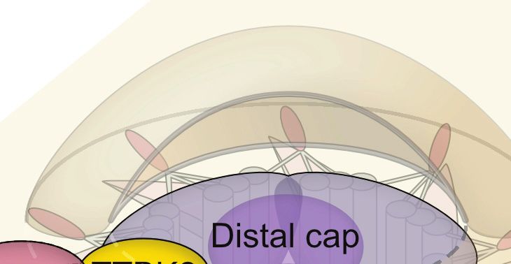
May et al.                                                                                                Phosphorylations and Ubiquitylations in Cilia

(Webb et al., 2020), and all eight subunits of the BBSome, defects         detyrosinated, glutamylated, and glycylated, which reflects
of which cause Bardet–Biedl Syndrome (Jin et al., 2010; Forsythe           axoneme maturation and affects axoneme assembly, protein
et al., 2018). IFT complexes transport cargoes along the axoneme           interaction, and stability (Janke and Magiera, 2020). In the
in an anterograde and retrograde fashion with the help of specific         following, we focus on phosphorylation and ubiquitylation and
kinesin and dynein motors, respectively (Satir and Christensen,            discuss recent findings on their involvement in regulating cilia
2007). The ciliary membrane does not fully enclose the ciliary             formation and signaling.
compartment at the proximal end, where it is separated from the
cytosol by the transition zone (Yang et al., 2015). A concerted
interplay of IFT complexes, the BBSome, transition fibers, and the         CILIARY SIGNALING
transition zone enables select proteins to enter or exit the cilium
(Garcia-Gonzalo and Reiter, 2017; Gonçalves and Pelletier, 2017).          Conceptually, primary cilia are believed to function as cell
    Post-translational modification (PTM) is a fundamental                 type-specific micro-compartments with diverse compositions
principle in molecular biology referring to the modulation of              including receptors to receive, mediators to process, and effectors
protein properties by covalent attachment of small molecules.              to transmit signals to the rest of the cell (Sung and Leroux,
PTMs are catalyzed by various antagonistic enzymatic activities            2013; Nachury and Mick, 2019). Despite a large variety of
that modify target proteins at specific locations (Vu et al., 2018).       receptors, far fewer mediators are commonly used in cellular
For instance, phosphorylation can modulate interaction surfaces            signaling processes. Cyclic nucleotides or calcium ions are
or lead to intramolecular rearrangements that alter enzymatic              second messengers, the concentrations of which are interpreted
activities. Protein kinases phosphorylate their substrates at              by specific enzymes to further transmit signals via PTMs
specific consensus sites consisting of only a few amino                    (Hilgendorf et al., 2019; Sherpa et al., 2019; Tajhya and Delling,
acids. Moreover, they are often targets of phosphorylation                 2020). To communicate with the rest of the cell, effectors
themselves, which results in phosphorylation cascades that                 are transported into and out of cilia in a dynamic fashion,
are typically found in cellular signaling processes (Miller and            which allows their modification according to the signaling status
Turk, 2018). Opposingly, protein phosphatases act on hundreds              (Niewiadomski et al., 2019). This general principle highlights the
of different substrates to revert phosphorylations (Bertolotti,            tight connection between cilia signaling and protein trafficking.
2018). Compared to phosphorylation, ubiquitylation requires                Apart from the IFT complexes, cilia require a multitude of
a more elaborate machinery. Ubiquitin is a small, 8.5-kDa                  additional factors to convey ciliary signals, which involves
protein that is usually attached to lysine residues of target              not only common protein trafficking components, such as
proteins (Swatek and Komander, 2016; Yau and Rape, 2016).                  β-arrestins, but also cilia-specific machinery, including the
The enzymatic cascade of ubiquitylation involves E1 activating,            BBSome or the Tubby family of proteins (Mukhopadhyay and
E2 conjugating, and E3 ligating enzymes. While the E1 and E2               Jackson, 2011). While the inventory of primary cilia continues to
enzymes supply reactive ubiquitin molecules, the vast number               expand (Mick et al., 2015; Kohli et al., 2017; May et al., 2021),
of different E3 ubiquitin ligases determines substrate specificity.        the number of enzymes, which catalyze PTMs and have been
Similarly, deubiquitylating enzymes (DUBs) are highly specific             unambiguously shown to localize to primary cilia, is limited.
with only a few substrates per enzyme (Clague et al., 2019).               Nonetheless, we are beginning to unravel how ciliary signaling
Ubiquitin contains seven lysine residues, to which further                 dynamics can be established as we identify more and more targets
ubiquitin molecules can be added to generate poly-ubiquitin                of PTMs in cilia.
chains. Depending on the lysine residue, ubiquitin chains are
differentiated into several linkage types that have been implicated        Hedgehog Signaling
in specific functions. K48- and K29-linked ubiquitins, for                 One hallmark ciliary signaling pathway that highlights the
example, are the main linkage types associated with proteasomal            dynamics in PTMs is Hedgehog signaling in vertebrates
degradation of target proteins, while the K63 chains and mono-             (Figure 1; Gigante and Caspary, 2020). An elegantly orchestrated
ubiquitin are often times involved in protein trafficking events           interplay of positive and negative regulators in Hedgehog
(Swatek and Komander, 2016).                                               signaling allows for the correct patterning of the developing
    The dynamic nature of PTMs is critical for most cellular               embryo, in addition to maintaining adult tissue homeostasis
processes and is extensively studied in protein trafficking                (Shimada et al., 2019). Gradients of the hedgehog morphogens
and cell signaling (Patwardhan et al., 2021). The central role             ultimately result in finely tuned levels of active GLI transcription
of the primary cilium as a cellular signaling hub suggests                 factors that determine target gene expression (Briscoe and
that PTMs regulate core ciliary functions. In addition to                  Novitch, 2008). In the absence of Hedgehog morphogens, their
phosphorylation and ubiquitylation, ciliary proteins are targets           receptor Patched (PTCH1) localizes to the primary cilium
of diverse modifications, such as acetylation (Kerek et al.,               (Rohatgi et al., 2007), while the key Hedgehog effector and G
2021), SUMOylation (McIntyre et al., 2015), and methylation                protein-coupled receptor (GPCR) Smoothened (SMO) surveys
(Yeyati et al., 2017). Several lipid modifications (including              the primary cilium by shuttling in and out without appreciable
acylation, myristoylation, palmitoylation, and prenylation) of             local accumulation (Figure 1A; Kim et al., 2009; Goetz and
ciliary proteins have also been involved in protein trafficking,           Anderson, 2010). A second GPCR, the constitutively active
membrane tethering, and protein stability (Roy and Marin,                  GPR161, stimulates ciliary adenylyl cyclases to increase cAMP
2019). Moreover, ciliary microtubules are extensively acetylated,          levels within cilia and thereby activates the cAMP-dependent

Frontiers in Cell and Developmental Biology | www.frontiersin.org      2                                        April 2021 | Volume 9 | Article 664279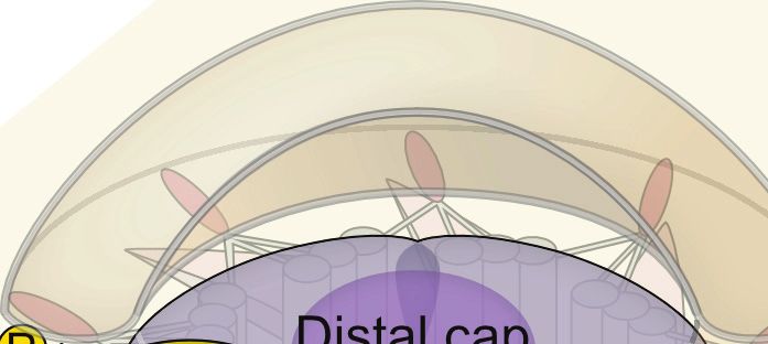
May et al.                                                                                                               Phosphorylations and Ubiquitylations in Cilia

             A                                                                     B

  FIGURE 1 | Post-translational modifications (PTMs) regulating Hedgehog signaling. (A) In unstimulated cells, the GPCR SMO constantly surveys the cilium without
  accumulation, due to constant removal in a ubiquitin (Ub) and BBSome-dependent manner. The Hedgehog receptor PTCH1 suppresses SMO, and the constitutively
  active GPCR GPR161 is retained in cilia. GPR161 stimulates adenylyl cyclases (AC) to generate cAMP. High cAMP is sensed by the regulatory PKA subunit Iα (RIα),
  which releases the PKA catalytic subunit (C) to phosphorylate target proteins, such as GLI transcription factors. GLI phosphorylation leads to ubiquitylation and
  proteolytic cleavage to GLI repressor forms (GLI-R) that repress target gene expression in the nucleus. The ubiquitin ligase MEGF is recruited to the plasma
  membrane by MGRN1 where it ubiquitinylates SMO for subsequent degradation. (B) In the presence of Hedgehog ligands, PTCH1 exits the primary cilium,
  presumably in a Ub-dependent fashion, leading to SMO activation and accumulation. GPR161 in turn is phosphorylated by GRK2 (and PKA). GPR161
  phosphorylation is sensed by β-arrestin2, which leads to ubiquitylation and BBSome-mediated removal of GPR161 together with PKA from the primary cilium.
  Together with a drop in cAMP levels, GLIs are no longer phosphorylated and full-length GLIs activate target genes in the nucleus. After removal from cilia, the
  mechanism by which GPR161 is internalized remains unclear.

protein kinase (PKA) (Mukhopadhyay et al., 2013). GPR161 also                        cilia (Bachmann et al., 2016). Here, it tethers to the cilia-resident
fulfills a second function in PKA signaling, as it serves as an                      PKA regulatory subunit RIα that senses ciliary cAMP (Mick
atypical A-kinase anchoring protein (AKAP) that targets PKA to                       et al., 2015). At high ciliary cAMP levels in unstimulated cells,

Frontiers in Cell and Developmental Biology | www.frontiersin.org                3                                             April 2021 | Volume 9 | Article 664279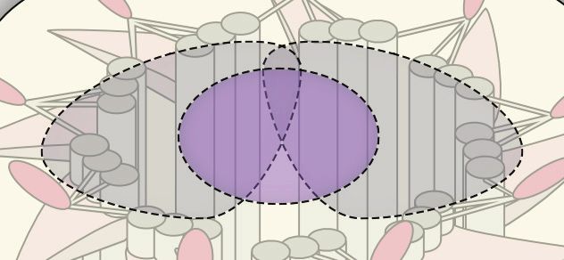
May et al.                                                                                                 Phosphorylations and Ubiquitylations in Cilia

PKA-RIα binds cAMP and releases the catalytic PKA-C subunit                 E, have been unambiguously shown to localize to primary cilia,
(Figure 1A; Taylor et al., 2004). Free PKA-C can phosphorylate              where they modulate ciliary signal transduction by regulating
and regulate target proteins, such as the GLI transcription factors         protein trafficking (Chávez et al., 2015; Garcia-Gonzalo et al.,
that convey the signaling status to the nucleus (Tuson et al., 2011;        2015). Ciliary lipid phosphatase activities create a specific
Niewiadomski et al., 2014). PKA-mediated phosphorylation                    phosphatidylinositide phosphate environment that is required
of GLIs is a pre-requisite for their proteolytical cleavage to              for efficient ciliary signaling.
yield repressor forms that block the transcription of target                    A recent study investigated the involvement of ubiquitin in
genes (Figure 1A). GLI transcription factors are precisely                  Hedgehog signaling by fusing mono-ubiquitin to the C-terminus
regulated by a variety of activating and deactivating PTMs,                 of SMO (Desai et al., 2020). The SMO-Ub fusion accumulated
which include activating phosphorylations at the N-terminal                 in cilia in the absence of stimulation in IFT and BBSome
repressor domain, and two clusters of PKA phosphorylation                   mutants but failed to accumulate in cilia after Hedgehog pathway
sites on the activator domain. PKA phosphorylation precedes                 activation in wild type cells (Desai et al., 2020). These findings
further phosphorylation by CK1 and GSK3β, which in turn                     indicate that ubiquitin is required for the removal of SMO from
recruits the SCF E3 ubiquitin ligase that marks GLIs for                    cilia by a process involving IFT and the BBSome. Moreover,
proteolytic processing by the proteasome (Kong et al., 2019;                β-arrestin2 was shown to mediate the ubiquitylation of GPR161
Niewiadomski et al., 2019).                                                 in response to Hedgehog pathway activation (Shinde et al., 2020),
    Upon Hedgehog ligand binding, PTCH1 exits the primary                   before GPR161 exits the primary cilium in a BBSome-dependent
cilium and SMO is activated and retained in cilia, whereas                  fashion (Ye et al., 2018). More evidence for the central role
GPR161 is removed (Rohatgi et al., 2007; Gigante and Caspary,               of ubiquitylation for cilia trafficking comes from mutational
2020). As the adenylyl cyclase inhibitory SMO replaces the                  analysis of the Hedgehog receptor PTCH1. PTCH1 harbors
stimulating GPR161, ciliary cAMP decreases (Mukhopadhyay                    two E3 ubiquitin ligase recognition motifs and remains in the
et al., 2013). Consequently, PKA activity ceases and the GLI                cilium when both motifs are mutated, even upon stimulation
transcription factors are no longer phosphorylated and further              with Hedgehog ligands (Kim J.C. et al., 2015). SMO has been
processed, such that they can function as activators to initiate            reported to be a target of the ubiquitin ligase HERC4 (Jiang
target gene expression in the nucleus (Figure 1B).                          et al., 2019). Furthermore, ubiquitylation of SMO by a complex
    One central element in Hedgehog signaling is the dynamic re-            of the E3 ubiquitin ligase MGRN1 and the plasma membrane
localization of the components involved. Similar to other cellular          protein MEGF8 serves as a signal for proteasomal degradation
protein trafficking mechanisms, PTMs control the localization of            (Kong et al., 2020). Yet, whether these ubiquitin ligases are
Hedgehog signaling proteins, which is particularly well-studied             directly involved in the IFT-dependent retrieval of SMO awaits
for GPR161 (Mukhopadhyay et al., 2013; Pal et al., 2016).                   experimental validation.
GPR161’s C-terminal tail not only contains the AKAP binding                     Molecular dissection of ubiquitylation may help to decipher
domain for PKA but also several protein kinase consensus                    the different functions of ubiquitin in regulating ciliary proteins.
sites, including one for PKA (Bachmann et al., 2016). Upon                  Upon Hedgehog pathway activation, specifically K63-linked
Hedgehog pathway activation, the C-terminal tail of GPR161                  ubiquitin chains increase in primary cilia upon GPCR activation
is phosphorylated by GRK2 and presumably PKA (Bachmann                      or in BBSome mutants (Shinde et al., 2020). This suggests
et al., 2016; Pal et al., 2016; May et al., 2021). GRK-mediated             that K63 ubiquitin chains function as export signals for ciliary
phosphorylation recruits the molecular sensor of activated                  proteins, which are recognized by the BBSome (Desai et al., 2020;
GPCRs, specifically β-arrestin2, which is required for the removal          Shinde et al., 2020). In BBSome mutant mice, photoreceptor
of activated GPCRs from cilia (Figure 1B; Pal et al., 2016).                outer segments, which are uniquely modified cilia that harbor the
Consequently, GPR161 exits cilia together with its binding                  entire signaling cascade for visual phototransduction, accumulate
partner PKA (May et al., 2021). Thereby, PKA activity in cilia is           more than 100 proteins that are absent in wild types (Datta
inhibited by two mechanisms: (i) reducing cAMP levels and (ii)              et al., 2015). Based on these findings, the BBSome has been
removing PKA itself.                                                        proposed to mediate the removal of unwanted proteins from
    As exemplified by the GLI transcription factors and                     cilia and, therefore, may function as an important mediator
GPR161, specific phosphorylations are often catalyzed by                    of a ciliary protein quality control network (Shinde et al.,
individual kinases; however, our knowledge of specific protein              2020). Additional components, such as the AAA-ATPase VCP
phosphatases that antagonize these phosphorylations in cilia                or the ubiquitin-regulatory X domain protein UBXN10, have
is still rudimentary. The protein phosphatases PP1 and PP2A                 been shown to localize to primary cilia (Mick et al., 2015;
have been reported to dephosphorylate SMO to dampen Hh                      Raman et al., 2015). While data from trypanosomes indicate
signaling in Drosophila (Su et al., 2011; Liu et al., 2020). However,       that the BBSome may directly recognize ubiquitin as it can be
since primary cilia are dispensable for Drosophila Hh signal                enriched on ubiquitin-agarose resin (Langousis et al., 2016), it
transduction, it remains unclear whether PP1 and PP2A also                  does not contain canonical ubiquitin binding domains. How
function within primary cilia. Mass spectrometric analyses have             ubiquitylated proteins are recognized in cilia on a molecular
identified PP1 subunits in isolated Chlamydomonas cilia (Pazour             level and what enzymatic activities regulate ubiquitylation within
et al., 2005) and PP2A subunits in primary cilia of kidney                  cilia remains to be established. The E3 ubiquitin ligase CBL is
epithelial cells (Ishikawa et al., 2012), but these findings still          recruited to cilia in response to PDGFRα signaling (Schmid et al.,
await confirmation by independent methods. In contrast, lipid               2018) and the deubiquitylase UBPY/USP8 has been reported to
phosphatases, such as the inositol polyphosphate-5-phosphatase              antagonize SMO ubiquitylation in Drosophila (Ma et al., 2016).

Frontiers in Cell and Developmental Biology | www.frontiersin.org       4                                        April 2021 | Volume 9 | Article 664279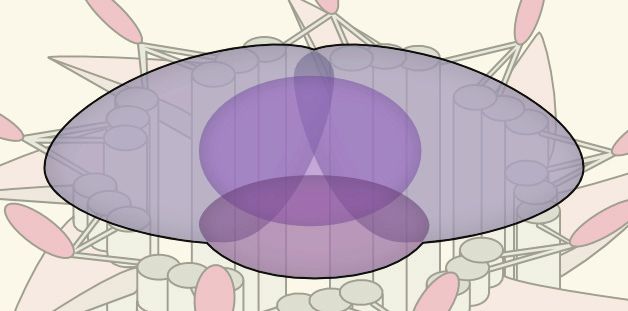
May et al. Phosphorylations and Ubiquitylations in Cilia
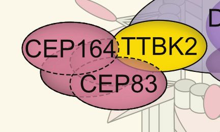
These findings seem promising starting points for future studies appendage proteins, such as CEP164 and CEP83 (Bernatik
elucidating the cilia-specific ubiquitylation network. et al., 2020), which is required for efficient vesicle recruitment
(Figure 2B; Lo et al., 2019). Notably, TTBK2 phosphorylates
MPP9 resulting in the loss of MPP9 and the CEP97–CP110
CILIUM DYNAMICS complex from the distal centriolar end (Figure 2C; Huang et al.,
2018). Moreover, with the onset of cilia formation, MPP9 is
Cilium Assembly—A Primary Cilium Is ubiquitylated and degraded by the proteasome. Although the
(Re)born precise ubiquitin linkage type has not been determined yet, many
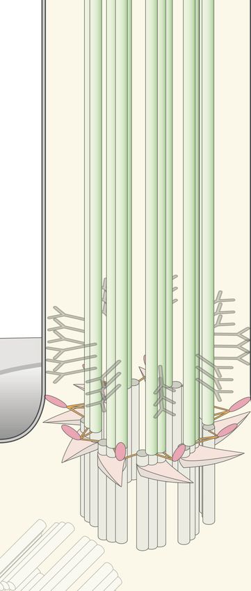
Ciliogenesis, i.e., the formation of cilia, is another dynamic molecular details of MPP9 PTM have been resolved. Intriguingly,
process that is regulated by specific phosphorylation and one identified ubiquitylation site in MPP9 is flanked by two
ubiquitylation events (Cao et al., 2009; Shearer and Saunders, phosphorylation sites. Phosphorylation-deficient mutants show
2016). A specialized maternal centriole, the so-called basal body, reduced ubiquitylation and consequently stabilize MPP9 (Huang
templates the cilium. Yet, mother and daughter centrioles also et al., 2018). This finding highlights a typical PTM cascade
form centrosomes required for spindle apparatus formation and and suggests that the phosphorylation status determines MPP9
chromosome segregation in metaphase. These two alternative stability. Similar to MPP9, the CEP97–CP110 complex is subject
roles of the mother centriole necessitate a cell cycle-dependent to proteasomal degradation when ciliation is initiated (Spektor
assembly and disassembly of primary cilia (Wang and Dynlacht, et al., 2007; Nagai et al., 2018). CP110 has been shown to be
2018; Breslow and Holland, 2019). Depending on cell type, the a target of the SCF ubiquitin ligase complex and a substrate of
mother centriole takes one of two different routes to form a the E3 ubiquitin ligase UBR5 in an in vitro ubiquitylation assay
cilium, starting either directly at the plasma membrane (termed (D’Angiolella et al., 2010; Hossain et al., 2017). Additionally,
extracellular pathway) or within the cell (Bernabé-Rubio and CEP97 degradation is suppressed after knockdown of the CUL3
Alonso, 2017; Kumar and Reiter, 2021). Here, we will be focusing E3 ligase, and therefore, it remains bound to CP110 at centrioles
on the intracellular pathway, which occurs in several steps (see and inhibits ciliogenesis (Nagai et al., 2018). UBR5 has been
Figure 2): (i) maturation of the mother centriole and acquisition found at centrosomes and CUL3 has been suggested to localize
of so-called distal and subdistal appendages, (ii) recruitment of specifically to mother centrioles (Moghe et al., 2012; Nagai et al.,
a growing ciliary vesicle (the future ciliary membrane) to the 2018), where it may ubiquitylate Aurora kinase A, a central
mother centriole, (iii) separation of the ciliary compartment regulator of the cell cycle and promoter of cilium disassembly
by the formation of the transition zone, (iv) extension of the (Pugacheva et al., 2007). It will be interesting to investigate
ciliary axoneme, and (v) docking of the basal body and final whether these ubiquitin ligases converge on the same targets
fusion with the plasma membrane. While this process has been and whether ubiquitylation is the cause or consequence of
described on an ultrastructural level more than half a century ago CEP97–CP110 removal. Also, how precisely ubiquitylation can
(Sorokin, 1968), we are still discovering an increasing number be regulated and what role DUBs, such as USP33 that targets
of the required factors such as RABs and EHD family proteins CP110 (Li et al., 2013), are playing in ciliogenesis need to be
that are involved in membrane recruitment (Lu et al., 2015; addressed in future studies.
Blacque et al., 2018) and are just beginning to understand
their regulation. Cilium Disassembly
A central kinase that determines cilium formation is the Tau In contrast to cilia formation, we are just beginning to understand
tubulin kinase 2 (TTBK2) (Tomizawa et al., 2001; Goetz et al., the molecular details of how cilia are dismantled to allow cell
2012). TTBK2 loss was originally reported to allow basal body cycle re-entry (Liang et al., 2016; Breslow and Holland, 2019).
docking to the plasma membrane, while blocking transition zone NEK2, a kinase predominantly expressed in the S and G2
formation and ciliary shaft elongation. In actively proliferating phases of the cell cycle, has been proposed to promote cilium
cells, the distal ends of both mother and daughter centrioles are disassembly (Figure 2D; Kim S. et al., 2015). Among several
capped by protein complexes of CP110 and CEP97 that suppress targets, NEK2 phosphorylates and stimulates the microtubule-
cilia formation (Spektor et al., 2007; Schmidt et al., 2009). depolymerizing KIF24 at the distal centriolar ends (Kim S. et al.,
Recruitment of these caps seems to follow a hierarchical scheme, 2015). This may not only block unwanted cilium assembly but
the precise order of which awaits clarification (Ye et al., 2014; also shift the balance toward disassembly when resting cells
Tsai et al., 2019). One central component involved in ciliogenesis re-enter the cell cycle (Kim S. et al., 2015; Viol et al., 2020).
is the microtubule-depolymerizing kinesin KIF24 (Kobayashi In support of a central role for KIF24 in cilium disassembly,
et al., 2011). KIF24 recruits the M-Phase phosphoprotein MPP9, a recent study identified FLS2 as a CDK-like kinase that
which is required for the assembly of CEP97–CP110 complexes phosphorylates the KIF24 ortholog CrKIF13 in Chlamydomonas,
at the distal ends of centrioles (Figure 2A; Huang et al., 2018). allowing efficient cilia disassembly (Figure 2D; Zhao et al., 2020).
The specific removal of the CEP97–CP110 complex relies on In turn, phosphorylated FLS2 showed lower activity and appears
TTBK2 phosphorylation (Figures 2B,C; Goetz et al., 2012; to be dephosphorylated upon cilia disassembly when it enters
Čajánek and Nigg, 2014; Huang et al., 2018). To ensure specificity cilia by binding to the IFT-B component IFT70 (Zhao et al.,
of distal end uncapping and thereby cilium formation at the 2020). While the precise mechanisms of regulation still need to
mother centriole, it is the distal appendage protein CEP164 be established, it is tempting to speculate that dephosphorylation
that recruits TTBK2 (Schmidt et al., 2012; Čajánek and Nigg, of FLS2 may not only alter its kinase activity but also unmask
2014). TTBK2 has recently been shown to phosphorylate distal targeting signals for IFT. This suggests a mechanism by which an
Frontiers in Cell and Developmental Biology | www.frontiersin.org 5 April 2021 | Volume 9 | Article 664279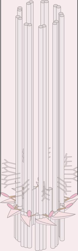
May et al.                                                                                                                  Phosphorylations and Ubiquitylations in Cilia

      A                                                                                     D

                                                                                        B
      C

  FIGURE 2 | Post-translational modifications (PTMs) in primary cilia assembly and disassembly. (A) Mother centriolar distal appendage components CEP164 and
  CEP83 are shown in red. The distal end proteins KIF24 and MPP9 recruit the capping protein complex CEP97–CP110 to block axoneme extension. Note that only
  one cap is shown for simplicity, while each microtubule triplet is capped by one complex. Unphosphorylated CEP83 diminishes ciliary vesicle recruitment (B) CEP164
  recruits the kinase TTBK2 that phosphorylates CEP164 and CEP83. Recruitment and activity of TTBK2 enables subsequent steps of cilia assembly such as
  formation of the ciliary vesicle (CV). (C) TTBK2 phosphorylates MPP9, which results in ubiquitylation and dissociation of MPP9 and the remaining CEP97–CP110
  complex from the distal centriolar end. Several E3 ubiquitin ligase complexes have been implicated in modifying distal cap components, while the precise location of
  ubiquitylation has not been determined (see text for details). Ultimately, MPP9, CEP97, and CP110 are degraded by the proteasome (UPS) and ciliary growth can be
  initiated. (D) Diagram of fully assembled primary cilium. The microtubule depolymerizing kinesin KIF24 has been implicated in microtubule disassembly.
  Phosphorylation of KIF24 by NEK2 stimulates KIF24 activity. In Chlamydomonas, dephosphorylated FLS2 enters cilia by binding to IFT-B and phosphorylates the
  KIF24 homolog. Upon disassembly, tubulins are ubiquitylated by unknown mechanisms. IFT-A binds to K63-linked ubiquitin chains and mediates removal.

active kinase can be directed into cilia to promote disassembly                        has only recently been demonstrated. Despite a massive rise
by phosphorylating specific targets such as the microtubule-                           in the ubiquitin levels in shortening cilia, semi-quantitative
depolymerizing kinesin KIF24.                                                          mass spectrometric analysis of a temperature-sensitive
   In cilia of Chlamydomonas, a ubiquitin conjugation system                           Chlamydomonas model has only detected an increase in
has been identified more than a decade ago (Huang et al.,                              ubiquitylation of α-tubulin and ubiquitin itself (Wang et al.,
2009), yet the involvement of ubiquitin in cilia disassembly                           2019). The study further revealed α-tubulin poly-ubiquitylation

Frontiers in Cell and Developmental Biology | www.frontiersin.org                  6                                              April 2021 | Volume 9 | Article 664279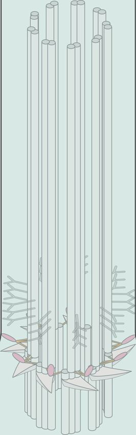
May et al.                                                                                                                         Phosphorylations and Ubiquitylations in Cilia

by K63 chains, which allows binding to the IFT-A subunit                                     AUTHOR CONTRIBUTIONS
IFT139 for tubulin removal via retrograde IFT (Figure 2D; Wang
et al., 2019). Intriguingly, the authors also observed an increase                           EM wrote the first draft of the manuscript. TS conceptualized and
in K11 and K48 chains in response to cilia shortening. K11                                   prepared the figures. DM wrote sections of the manuscript. All
chains are also assembled by the anaphase-promoting complex                                  authors contributed to the conception of the article, manuscript
to drive proteasomal degradation of substrates during mitosis,                               revision, and read and approved the submitted version.
suggesting potential mechanisms for cell cycle-dependent
regulation (Matsumoto et al., 2010).
                                                                                             FUNDING
OUTLOOK                                                                                      The authors acknowledge support by the Deutsche
                                                                                             Forschungsgemeinschaft (DFG, German Research Foundation)
As we are gathering increasing evidence for the existence                                    and Saarland University within the funding programme Open
of a ciliary ubiquitylation machinery involved in protein                                    Access Publishing. This work was supported by the DFG grant
trafficking, signaling, disassembly, and potentially protein                                 SFB894/TPA-22 (to DM).
quality control, its identity remains elusive. Similarly,
antagonistic cilia-specific DUBs as well as protein
phosphatases that counterbalance known kinases await their                                   ACKNOWLEDGMENTS
identification. Powerful unbiased genetic and proteomic
screening technologies have been applied to primary cilia                                    The authors thank D. Breslow and B. Schrul for comments
(Mick et al., 2015; Kohli et al., 2017; Breslow et al.,                                      on the manuscript and N. Byers and V. Chaumet for
2018; Pusapati et al., 2018) and promise to reveal the                                       critically reading the manuscript. They apologize to all
missing links that modulate manifold dynamic processes                                       colleagues whose work could not be mentioned due to length
in cilia by PTM.                                                                             restrictions.

REFERENCES                                                                                      Cilia, Chap. 17, ed. R. D. Sloboda (Cambridge, MA: Academic Press), 333–346.
                                                                                                doi: 10.1016/S0091-679X(08)94017-6
Anvarian, Z., Mykytyn, K., Mukhopadhyay, S., Pedersen, L. B., and Christensen,               Chávez, M., Ena, S., Van Sande, J., de Kerchove d’Exaerde, A., Schurmans, S.,
    S. T. (2019). Cellular signalling by primary cilia in development, organ function           and Schiffmann, S. N. (2015). Modulation of ciliary phosphoinositide content
    and disease. Nat. Rev. Nephrol. 15, 199–219. doi: 10.1038/s41581-019-0116-9                 regulates trafficking and sonic hedgehog signaling output. Dev. Cell 34, 338–
Bachmann, V. A., Mayrhofer, J. E., Ilouz, R., Tschaikner, P., Raffeiner, P., Röck,              350. doi: 10.1016/j.devcel.2015.06.016
    R., et al. (2016). Gpr161 anchoring of PKA consolidates GPCR and cAMP                    Clague, M. J., Urbé, S., and Komander, D. (2019). Breaking the chains:
    signaling. Proc. Natl. Acad. Sci. U.S.A. 113, 7786–7791. doi: 10.1073/pnas.                 deubiquitylating enzyme specificity begets function. Nat. Rev. Mol. Cell Biol.
    1608061113                                                                                  20, 338–352. doi: 10.1038/s41580-019-0099-1
Baker, K., and Beales, P. L. (2009). Making sense of cilia in disease: The human             D’Angiolella, V., Donato, V., Vijayakumar, S., Saraf, A., Florens, L., Washburn,
    ciliopathies. Am. J. Med. Genet. C Semin. Med. Genet. 151, 281–295. doi: 10.                M. P., et al. (2010). SCF Cyclin F controls centrosome homeostasis and
    1002/ajmg.c.30231                                                                           mitotic fidelity through CP110 degradation. Nature 466, 138–142. doi: 10.1038/
Bernabé-Rubio, M., and Alonso, M. A. (2017). Routes and machinery of primary                    nature09140
    cilium biogenesis. Cell. Mol. Life Sci. CMLS 74, 4077–4095. doi: 10.1007/s00018-         Datta, P., Allamargot, C., Hudson, J. S., Andersen, E. K., Bhattarai, S., Drack,
    017-2570-5                                                                                  A. V., et al. (2015). Accumulation of non-outer segment proteins in the
Bernatik, O., Pejskova, P., Vyslouzil, D., Hanakova, K., Zdrahal, Z., and Cajanek,              outer segment underlies photoreceptor degeneration in Bardet-Biedl syndrome.
    L. (2020). Phosphorylation of multiple proteins involved in ciliogenesis by Tau             Proc. Natl. Acad. Sci. U.S.A. 112, E4400–E4409. doi: 10.1073/pnas.151011
    Tubulin kinase 2. Mol. Biol. Cell 31, 1032–1046. doi: 10.1091/mbc.E19-06-0334               1112
Bertolotti, A. (2018). The split protein phosphatase system. Biochem. J. 475,                Desai, P. B., Stuck, M. W., Lv, B., and Pazour, G. J. (2020). Ubiquitin links
    3707–3723. doi: 10.1042/BCJ20170726                                                         smoothened to intraflagellar transport to regulate Hedgehog signaling. J. Cell
Blacque, O. E., Scheidel, N., and Kuhns, S. (2018). Rab GTPases in cilium formation             Biol. 219:e201912104. doi: 10.1083/jcb.201912104
    and function. Small GTPases 9, 76–94. doi: 10.1080/21541248.2017.1353847                 Forsythe, E., Kenny, J., Bacchelli, C., and Beales, P. L. (2018). Managing bardet-
Breslow, D. K., and Holland, A. J. (2019). Mechanism and regulation of centriole                biedl syndrome-now and in the future. Front. Pediatr. 6:23. doi: 10.3389/fped.
    and cilium biogenesis. Annu. Rev. Biochem. 88, 691–724. doi: 10.1146/annurev-               2018.00023
    biochem-013118-111153                                                                    Garcia, G., Raleigh, D. R., and Reiter, J. F. (2018). How the ciliary membrane is
Breslow, D. K., Hoogendoorn, S., Kopp, A. R., Morgens, D. W., Vu, B. K., Kennedy,               organized inside-out to communicate outside-in. Curr. Biol. 28, R421–R434.
    M. C., et al. (2018). A CRISPR-based screen for Hedgehog signaling provides                 doi: 10.1016/j.cub.2018.03.010
    insights into ciliary function and ciliopathies. Nat. Genet. 50, 460–471. doi:           Garcia-Gonzalo, F. R., Phua, S. C., Roberson, E. C., Garcia, G., Abedin,
    10.1038/s41588-018-0054-7                                                                   M., Schurmans, S., et al. (2015). Phosphoinositides regulate ciliary protein
Briscoe, J., and Novitch, B. G. (2008). Regulatory pathways linking progenitor                  trafficking to modulate hedgehog signaling. Dev. Cell 34, 400–409. doi: 10.1016/
    patterning, cell fates and neurogenesis in the ventral neural tube. Philos. Trans.          j.devcel.2015.08.001
    R. Soc. Lond. B. Biol. Sci. 363, 57–70. doi: 10.1098/rstb.2006.2012                      Garcia-Gonzalo, F. R., and Reiter, J. F. (2017). Open sesame: how transition fibers
Čajánek, L., and Nigg, E. A. (2014). Cep164 triggers ciliogenesis by recruiting                and the transition zone control ciliary composition. Cold Spring Harb. Perspect.
    Tau tubulin kinase 2 to the mother centriole. Proc. Natl. Acad. Sci. U.S.A. 111,            Biol. 9:a028134. doi: 10.1101/cshperspect.a028134
    E2841–E2850. doi: 10.1073/pnas.1401777111                                                Gigante, E. D., and Caspary, T. (2020). Signaling in the primary cilium through
Cao, M., Li, G., and Pan, J. (2009). “Regulation of cilia assembly, disassembly,                the lens of the Hedgehog pathway. Wiley Interdiscip. Rev. Dev. Biol. 9:e377.
    and length by protein phosphorylation,” in Methods in Cell Biology Primary                  doi: 10.1002/wdev.377

Frontiers in Cell and Developmental Biology | www.frontiersin.org                        7                                               April 2021 | Volume 9 | Article 664279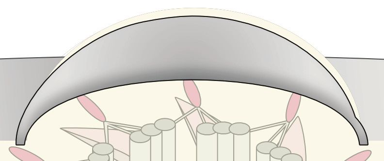
May et al.                                                                                                                                Phosphorylations and Ubiquitylations in Cilia

Goetz, S. C., and Anderson, K. V. (2010). The primary cilium: a signaling center                  Li, J., D’Angiolella, V., Seeley, E. S., Kim, S., Kobayashi, T., Fu, W., et al. (2013).
    during vertebrate development. Nat. Rev. Genet. 11, 331–344. doi: 10.1038/                        USP33 regulates centrosome biogenesis via deubiquitination of the centriolar
    nrg2774.The                                                                                       protein CP110. Nature 495, 255–259. doi: 10.1038/nature11941
Goetz, S. C., Liem, K. F. J., and Anderson, K. V. (2012). The spinocerebellar ataxia-             Liang, Y., Meng, D., Zhu, B., and Pan, J. (2016). Mechanism of ciliary disassembly.
    associated gene Tau tubulin kinase 2 controls the initiation of ciliogenesis. Cell                Cell. Mol. Life Sci. 73, 1787–1802. doi: 10.1007/s00018-016-2148-7
    151, 847–858. doi: 10.1016/j.cell.2012.10.010                                                 Liu, M., Liu, A., Wang, J., Zhang, Y., Li, Y., Su, Y., et al. (2020). Competition
Gonçalves, J., and Pelletier, L. (2017). The ciliary transition zone: finding the pieces              between two phosphatases fine-tunes Hedgehog signaling. J. Cell Biol.
    and assembling the gate. Mol. Cells 40, 243–253. doi: 10.14348/molcells.2017.                     220:e202010078. doi: 10.1083/jcb.202010078
    0054                                                                                          Lo, C.-H., Lin, I.-H., Yang, T. T., Huang, Y.-C., Tanos, B. E., Chou, P.-C., et al.
Hilgendorf, K. I., Johnson, C. T., Mezger, A., Rice, S. L., Norris, A. M., Demeter, J.,               (2019). Phosphorylation of CEP83 by TTBK2 is necessary for cilia initiation.
    et al. (2019). Omega-3 fatty acids activate ciliary FFAR4 to control adipogenesis.                J. Cell Biol. 218, 3489–3505. doi: 10.1083/jcb.201811142
    Cell 179, 1289–1305.e21. doi: 10.1016/j.cell.2019.11.005                                      Lu, Q., Insinna, C., Ott, C., Stauffer, J., Pintado, P. A., Rahajeng, J., et al. (2015).
Hossain, D., Javadi Esfehani, Y., Das, A., and Tsang, W. Y. (2017). Cep78 controls                    Early steps in primary cilium assembly require EHD1/EHD3-dependent ciliary
    centrosome homeostasis by inhibiting EDD-DYRK2-DDB1VprBP. EMBO Rep.                               vesicle formation. Nat. Cell Biol. 17, 228–240. doi: 10.1038/ncb3109
    18, 632–644. doi: 10.15252/embr.201642377                                                     Ma, G., Li, S., Han, Y., Li, S., Yue, T., Wang, B., et al. (2016). Regulation of
Huang, K., Diener, D. R., and Rosenbaum, J. L. (2009). The ubiquitin conjugation                      smoothened trafficking and hedgehog signaling by the SUMO pathway. Dev.
    system is involved in the disassembly of cilia and flagella. J. Cell Biol. 186,                   Cell 39, 438–451. doi: 10.1016/j.devcel.2016.09.014
    601–613. doi: 10.1083/jcb.200903066                                                           Matsumoto, M. L., Wickliffe, K. E., Dong, K. C., Yu, C., Bosanac, I., Bustos, D.,
Huang, N., Zhang, D., Li, F., Chai, P., Wang, S., Teng, J., et al. (2018). M-                         et al. (2010). K11-Linked polyubiquitination in cell cycle control revealed by
    Phase Phosphoprotein 9 regulates ciliogenesis by modulating CP110-CEP97                           a K11 linkage-specific antibody. Mol. Cell 39, 477–484. doi: 10.1016/j.molcel.
    complex localization at the mother centriole. Nat. Commun. 9:4511. doi: 10.                       2010.07.001
    1038/s41467-018-06990-9                                                                       May, E. A., Kalocsay, M., Galtier, D., Auriac, I., Schuster, P. S., Gygi, S. P., et al.
Ishikawa, H., Thompson, J. III, Yates, J. R., and Marshall, W. F. (2012). Proteomic                   (2021). Time-resolved proteomics profiling of the ciliary Hedgehog response.
    analysis of mammalian primary cilia. Curr. Biol. 22, 414–419. doi: 10.1016/j.cub.                 J. Cell Biol. doi: 10.1083/jcb.202007207 [Epub ahead of print].
    2012.01.031                                                                                   McIntyre, J. C., Joiner, A. M., Zhang, L., Iñiguez-Lluhí, J., and Martens, J. R. (2015).
Janke, C., and Magiera, M. M. (2020). The tubulin code and its role in controlling                    SUMOylation regulates ciliary localization of olfactory signaling proteins. J. Cell
    microtubule properties and functions. Nat. Rev. Mol. Cell Biol. 21, 307–326.                      Sci. 128, 1934–1945. doi: 10.1242/jcs.164673
    doi: 10.1038/s41580-020-0214-3                                                                Mick, D. U., Rodrigues, R. B., Leib, R. D., Adams, C. M., Chien, A. S., Gygi, S. P.,
Jiang, W., Yao, X., Shan, Z., Li, W., Gao, Y., and Zhang, Q. (2019). E3 ligase Herc4                  et al. (2015). Proteomics of primary cilia by proximity labeling. Dev. Cell 35,
    regulates Hedgehog signalling through promoting Smoothened degradation.                           497–512. doi: 10.1016/j.devcel.2015.10.015
    J. Mol. Cell Biol. 11, 791–803. doi: 10.1093/jmcb/mjz024                                      Miller, C. J., and Turk, B. E. (2018). Homing in: mechanisms of substrate targeting
Jin, H., White, S. R., Shida, T., Schulz, S., Aguiar, M., Gygi, S. P., et al. (2010).                 by protein kinases. Trends Biochem. Sci. 43, 380–394. doi: 10.1016/j.tibs.2018.
    The conserved Bardet-Biedl syndrome proteins assemble a coat that traffics                        02.009
    membrane proteins to cilia. Cell 141, 1208–1219. doi: 10.1016/j.cell.2010.05.015              Moghe, S., Jiang, F., Miura, Y., Cerny, R. L., Tsai, M.-Y., and Furukawa, M. (2012).
Kerek, E. M., Yoon, K. H., Luo, S. Y., Chen, J., Valencia, R., Julien, O., et al. (2021).             The CUL3−KLHL18 ligase regulates mitotic entry and ubiquitylates Aurora-A.
    A conserved acetylation switch enables pharmacological control of tubby-like                      Biol. Open 1, 82–91. doi: 10.1242/bio.2011018
    protein stability. J. Biol. Chem. 296:100073. doi: 10.1074/jbc.RA120.015839                   Mukhopadhyay, S., and Jackson, P. K. (2011). The tubby family proteins. Genome
Kim, J., Kato, M., and Beachy, P. A. (2009). Gli2 trafficking links Hedgehog-                         Biol. 12:225. doi: 10.1186/gb-2011-12-6-225
    dependent activation of Smoothened in the primary cilium to transcriptional                   Mukhopadhyay, S., Wen, X., Ratti, N., Loktev, A., Rangell, L., Scales, S. J., et al.
    activation in the nucleus. Proc. Natl. Acad. Sci. U.S.A. 106, 21666–21671. doi:                   (2013). The ciliary G-protein-coupled receptor Gpr161 negatively regulates the
    10.1073/pnas.0912180106                                                                           Sonic hedgehog pathway via cAMP signaling. Cell 152, 210–223. doi: 10.1016/j.
Kim, J. C., Hsia, E. Y., Brigui, A., Plessis, A., Beachy, P. A., and Zheng, X. (2015).                cell.2012.12.026
    The role of ciliary trafficking in Hedgehog receptor signaling. Sci. Signal. 8,               Nachury, M. V., and Mick, D. U. (2019). Establishing and regulating the
    ra55–ra55. doi: 10.1126/scisignal.aaa5622                                                         composition of cilia for signal transduction. Nat. Rev. Mol. Cell Biol. 20,
Kim, S., Lee, K., Choi, J.-H., Ringstad, N., and Dynlacht, B. D. (2015). Nek2                         389–405. doi: 10.1038/s41580-019-0116-4
    activation of Kif24 ensures cilium disassembly during the cell cycle. Nat.                    Nagai, T., Mukoyama, S., Kagiwada, H., Goshima, N., and Mizuno, K. (2018).
    Commun. 6:8087. doi: 10.1038/ncomms9087                                                           Cullin-3-KCTD10-mediated CEP97 degradation promotes primary cilium
Kobayashi, T., Tsang, W. Y., Li, J., Lane, W., and Dynlacht, B. D. (2011). Centriolar                 formation. J. Cell Sci. 131:jcs219527. doi: 10.1242/jcs.219527
    kinesin Kif24 interacts with CP110 to remodel microtubules and regulate                       Niewiadomski, P., Kong, J. H., Ahrends, R., Ma, Y., Humke, E. W., Khan, S.,
    ciliogenesis. Cell 145, 914–925. doi: 10.1016/j.cell.2011.04.028                                  et al. (2014). Gli protein activity is controlled by multisite phosphorylation in
Kohli, P., Höhne, M., Jüngst, C., Bertsch, S., Ebert, L. K., Schauss, A. C., et al. (2017).           vertebrate hedgehog signaling. Cell Rep. 6, 168–181. doi: 10.1016/j.celrep.2013.
    The ciliary membrane-associated proteome reveals actin-binding proteins as                        12.003
    key components of cilia. EMBO Rep. 18, 1521–1535. doi: 10.15252/embr.                         Niewiadomski, P., Niedziółka, S. M., Markiewicz, Ł, Uśpieński, T., Baran, B., and
    201643846                                                                                         Chojnowska, K. (2019). Gli proteins: regulation in development and cancer.
Kong, J. H., Siebold, C., and Rohatgi, R. (2019). Biochemical mechanisms of                           Cells 8:147. doi: 10.3390/cells8020147
    vertebrate hedgehog signaling. Development 146:dev166892. doi: 10.1242/dev.                   Pal, K., Hwang, S., Somatilaka, B., Badgandi, H., Jackson, P. K., DeFea, K., et al.
    166892                                                                                            (2016). Smoothened determines β-arrestin–mediated removal of the G protein–
Kong, J. H., Young, C. B., Pusapati, G. V., Patel, C. B., Ho, S., Krishnan, A.,                       coupled receptor Gpr161 from the primary cilium. J. Cell Biol. 212, 861–875.
    et al. (2020). A ubiquitin-based mechanism for the oligogenic inheritance of                      doi: 10.1083/jcb.201506132
    heterotaxy and heart defects. bioRxiv [Preprint] doi: 10.1101/2020.05.25.113944               Patwardhan, A., Cheng, N., and Trejo, J. (2021). Post-translational modifications of
Kumar, D., and Reiter, J. (2021). How the centriole builds its cilium: of mothers,                    G Protein–Coupled receptors control cellular signaling dynamics in space and
    daughters, and the acquisition of appendages. Curr. Opin. Struct. Biol. 66,                       time. Pharmacol. Rev. 73, 120–151. doi: 10.1124/pharmrev.120.000082
    41–48. doi: 10.1016/j.sbi.2020.09.006                                                         Pazour, G. J., Agrin, N., Leszyk, J., and Witman, G. B. (2005). Proteomic analysis of
Langousis, G., Shimogawa, M. M., Saada, E. A., Vashisht, A. A., Spreafico, R.,                        a eukaryotic cilium. J. Cell Biol. 170, 103–113. doi: 10.1083/jcb.200504008
    Nager, A. R., et al. (2016). Loss of the BBSome perturbs endocytic trafficking                Pugacheva, E. N., Jablonski, S. A., Hartman, T. R., Henske, E. P., and Golemis,
    and disrupts virulence of Trypanosoma brucei. Proc. Natl. Acad. Sci. U.S.A. 113,                  E. A. (2007). HEF1-dependent aurora a activation induces disassembly of the
    632–637. doi: 10.1073/pnas.1518079113                                                             primary cilium. Cell 129, 1351–1363. doi: 10.1016/j.cell.2007.04.035

Frontiers in Cell and Developmental Biology | www.frontiersin.org                             8                                                  April 2021 | Volume 9 | Article 664279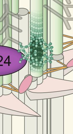
May et al.                                                                                                                               Phosphorylations and Ubiquitylations in Cilia

Pusapati, G. V., Kong, J. H., Patel, B. B., Krishnan, A., Sagner, A., Kinnebrew, M.,            Taylor, S. S., Yang, J., Wu, J., Haste, N. M., Radzio-Andzelm, E., and Anand,
   et al. (2018). CRISPR screens uncover genes that regulate target cell sensitivity to            G. (2004). PKA: a portrait of protein kinase dynamics. Biochim. Biophys.
   the morphogen sonic hedgehog. Dev. Cell 44, 113–129.e8. doi: 10.1016/j.devcel.                  Acta BBA Proteins Proteom. 1697, 259–269. doi: 10.1016/j.bbapap.2003.
   2017.12.003                                                                                     11.029
Raman, M., Sergeev, M., Garnaas, M., Lydeard, J. R., Huttlin, E. L., Goessling,                 Tomizawa, K., Omori, A., Ohtake, A., Sato, K., and Takahashi, M. (2001). Tau-
   W., et al. (2015). Systematic proteomics of the VCP–UBXD adaptor network                        tubulin kinase phosphorylates tau at Ser-208 and Ser-210, sites found in paired
   identifies a role for UBXN10 in regulating ciliogenesis. Nat. Cell Biol. 17,                    helical filament-tau. FEBS Lett. 492, 221–227. doi: 10.1016/S0014-5793(01)
   1356–1369. doi: 10.1038/ncb3238                                                                 02256-6
Reiter, J. F., and Leroux, M. R. (2017). Genes and molecular pathways                           Tsai, J.-J., Hsu, W.-B., Liu, J.-H., Chang, C.-W., and Tang, T. K. (2019). CEP120
   underpinning ciliopathies. Nat. Rev. Mol. Cell Biol. 18, 533–547. doi: 10.1038/                 interacts with C2CD3 and Talpid3 and is required for centriole appendage
   nrm.2017.60                                                                                     assembly and ciliogenesis. Sci. Rep. 9:6037. doi: 10.1038/s41598-019-42577-0
Rohatgi, R., Milenkovic, L., and Scott, M. P. (2007). Patched1 regulates hedgehog               Tuson, M., He, M., and Anderson, K. V. (2011). Protein kinase A acts at the basal
   signaling at the primary cilium. Science 317, 372–376. doi: 10.1126/science.                    body of the primary cilium to prevent Gli2 activation and ventralization of
   1139740                                                                                         the mouse neural tube. Dev. Camb. Engl. 138, 4921–4930. doi: 10.1242/dev.
Roy, K., and Marin, E. P. (2019). Lipid modifications in cilia biology. J. Clin. Med.              070805
   8, 921–921. doi: 10.3390/jcm8070921                                                          Viol, L., Hata, S., Pastor-Peidro, A., Neuner, A., Murke, F., Wuchter, P., et al. (2020).
Sánchez, I., and Dynlacht, B. D. (2016). Cilium assembly and disassembly. Nat. Cell                Nek2 kinase displaces distal appendages from the mother centriole prior to
   Biol. 18, 711–717. doi: 10.1038/ncb3370                                                         mitosis. J. Cell Biol. 219:e201907136. doi: 10.1083/jcb.201907136
Satir, P., and Christensen, S. T. (2007). Overview of structure and function                    Vu, L. D., Gevaert, K., and De Smet, I. (2018). Protein language: post-translational
   of mammalian cilia. Annu. Rev. Physiol. 69, 377–400. doi: 10.1146/annurev.                      modifications talking to each other. Trends Plant Sci. 23, 1068–1080. doi: 10.
   physiol.69.040705.141236                                                                        1016/j.tplants.2018.09.004
Schmid, F. M., Schou, K. B., Vilhelm, M. J., Holm, M. S., Breslin, L., Farinelli,               Wang, L., and Dynlacht, B. D. (2018). The regulation of cilium assembly and
   P., et al. (2018). IFT20 modulates ciliary PDGFRα signaling by regulating the                   disassembly in development and disease. Dev. Camb. Engl. 145:dev151407.
   stability of Cbl E3 ubiquitin ligases. J. Cell Biol. 217, 151–161. doi: 10.1083/jcb.            doi: 10.1242/dev.151407
   201611050                                                                                    Wang, Q., Peng, Z., Long, H., Deng, X., and Huang, K. (2019). Polyubiquitylation
Schmidt, K. N., Kuhns, S., Neuner, A., Hub, B., Zentgraf, H., and Pereira, G. (2012).              of α-tubulin at K304 is required for flagellar disassembly in Chlamydomonas.
   Cep164 mediates vesicular docking to the mother centriole during early steps of                 J. Cell Sci. 132:jcs229047. doi: 10.1242/jcs.229047
   ciliogenesis. J. Cell Biol. 199, 1083–1101. doi: 10.1083/jcb.201202126                       Webb, S., Mukhopadhyay, A. G., and Roberts, A. J. (2020). Intraflagellar transport
Schmidt, T. I., Kleylein-Sohn, J., Westendorf, J., Le Clech, M., Lavoie, S. B., Stierhof,          trains and motors: insights from structure. Semin. Cell Dev. Biol. 107, 82–90.
   Y.-D., et al. (2009). Control of centriole length by CPAP and CP110. Curr. Biol.                doi: 10.1016/j.semcdb.2020.05.021
   CB 19, 1005–1011. doi: 10.1016/j.cub.2009.05.016                                             Yang, T. T., Su, J., Wang, W.-J., Craige, B., Witman, G. B., Tsou, M.-F. B., et al.
Shearer, R. F., and Saunders, D. N. (2016). Regulation of primary cilia formation                  (2015). Superresolution pattern recognition reveals the architectural map of the
   by the ubiquitin-proteasome system. Biochem. Soc. Trans. 44, 1265–1271. doi:                    ciliary transition zone. Sci. Rep. 5:14096. doi: 10.1038/srep14096
   10.1042/BST20160174                                                                          Yau, R., and Rape, M. (2016). The increasing complexity of the ubiquitin code. Nat.
Sherpa, R. T., Mohieldin, A. M., Pala, R., Wachten, D., Ostrom, R. S., and Nauli,                  Cell Biol. 18, 579–586. doi: 10.1038/ncb3358
   S. M. (2019). Sensory primary cilium is a responsive cAMP microdomain in                     Ye, F., Nager, A. R., and Nachury, M. V. (2018). BBSome trains remove activated
   renal epithelia. Sci. Rep. 9:6523. doi: 10.1038/s41598-019-43002-2                              GPCRs from cilia by enabling passage through the transition zone. J. Cell Biol.
Shimada, I. S., Somatilaka, B. N., Hwang, S.-H., Anderson, A. G., Shelton, J. M.,                  217, 1847–1868. doi: 10.1083/jcb.201709041
   Rajaram, V., et al. (2019). Derepression of sonic hedgehog signaling upon                    Ye, X., Zeng, H., Ning, G., Reiter, J. F., and Liu, A. (2014). C2cd3 is critical for
   Gpr161 deletion unravels forebrain and ventricular abnormalities. Dev. Biol.                    centriolar distal appendage assembly and ciliary vesicle docking in mammals.
   450, 47–62. doi: 10.1016/j.ydbio.2019.03.011                                                    Proc. Natl. Acad. Sci. U.S.A. 111, 2164–2169. doi: 10.1073/pnas.1318737111
Shinde, S. R., Nager, A. R., and Nachury, M. V. (2020). Ubiquitin chains earmark                Yeyati, P. L., Schiller, R., Mali, G., Kasioulis, I., Kawamura, A., Adams, I. R.,
   GPCRs for BBSome-mediated removal from cilia. J. Cell Biol. 219:e202003020.                     et al. (2017). KDM3A coordinates actin dynamics with intraflagellar transport
   doi: 10.1083/jcb.202003020                                                                      to regulate cilia stability. J. Cell Biol. 216, 999–1013. doi: 10.1083/jcb.201607
Sorokin, S. P. (1968). Centriole formation and ciliogenesis. Aspen Emphysema                       032
   Conf. 11, 213–216.                                                                           Zhao, Q., Li, S., Shao, S., Wang, Z., and Pan, J. (2020). FLS2 is a CDK-like
Spektor, A., Tsang, W. Y., Khoo, D., and Dynlacht, B. D. (2007). Cep97 and CP110                   kinase that directly binds IFT70 and is required for proper ciliary disassembly
   suppress a cilia assembly program. Cell 130, 678–690. doi: 10.1016/j.cell.2007.                 in Chlamydomonas. PLoS Genet. 16:e1008561. doi: 10.1371/journal.pgen.10
   06.027                                                                                          08561
Su, Y., Ospina, J. K., Zhang, J., Michelson, A. P., Schoen, A. M., and Zhu, A. J.
   (2011). Sequential phosphorylation of smoothened transduces graded hedgehog                  Conflict of Interest: The authors declare that the research was conducted in the
   signaling. Sci. Signal. 4:ra43. doi: 10.1126/scisignal.2001747                               absence of any commercial or financial relationships that could be construed as a
Sung, C.-H., and Leroux, M. R. (2013). The roles of evolutionarily conserved                    potential conflict of interest.
   functional modules in cilia-related trafficking. Nat. Cell Biol. 15, 1387–1397.
   doi: 10.1038/ncb2888                                                                         Copyright © 2021 May, Sroka and Mick. This is an open-access article distributed
Swatek, K. N., and Komander, D. (2016). Ubiquitin modifications. Cell Res. 26,                  under the terms of the Creative Commons Attribution License (CC BY). The use,
   399–422. doi: 10.1038/cr.2016.39                                                             distribution or reproduction in other forums is permitted, provided the original
Tajhya, R., and Delling, M. (2020). New insights into ion channel-dependent                     author(s) and the copyright owner(s) are credited and that the original publication
   signalling during left-right patterning. J. Physiol. 598, 1741–1752. doi: 10.1113/           in this journal is cited, in accordance with accepted academic practice. No use,
   JP277835                                                                                     distribution or reproduction is permitted which does not comply with these terms.

Frontiers in Cell and Developmental Biology | www.frontiersin.org                           9                                                   April 2021 | Volume 9 | Article 664279
You can also read