Retinoblastoma Tumor Suppressor Protein Roles in Epigenetic Regulation - MDPI
←
→
Page content transcription
If your browser does not render page correctly, please read the page content below
cancers
Review
Retinoblastoma Tumor Suppressor Protein Roles in
Epigenetic Regulation
Frederick Guzman 1 , Yasamin Fazeli 2 , Meagan Khuu 2 , Kelsey Salcido 2 , Sarah Singh 2                and
Claudia A. Benavente 1,2,3,4, *
 1   Department of Pharmaceutical Sciences, University of California, 101 Theory #100, Irvine, CA 92617, USA;
     guzmanfg@uci.edu
 2   School of Biological Sciences, University of California, 5120 Natural Sciences II, Irvine, CA 92697, USA;
     yfazeli@uci.edu (Y.F.); mskhuu@uci.edu (M.K.); salcidok@uci.edu (K.S.); singhss1@uci.edu (S.S.)
 3   Department of Developmental and Cell Biology, University of California, 2011 Biological Sciences III, Irvine,
     CA 92697, USA
 4   Chao Family Comprehensive Cancer Center, University of California, 839 Medical Sciences Rd, Irvine,
     CA 92697, USA
 *   Correspondence: claudia.benavente@uci.edu

 Received: 1 September 2020; Accepted: 27 September 2020; Published: 29 September 2020

 Simple Summary: Loss of function of the retinoblastoma gene (RB1) is the rate-limiting step in the
 initiation of both the hereditary and sporadic forms of retinoblastoma tumor. Furthermore, loss of
 function of the retinoblastoma tumor suppressor protein (pRB) is frequently found in most human
 cancers. In retinoblastoma, tumor progression is driven by epigenetic changes following pRB loss.
 This review focuses on the diverse functions of pRB in epigenetic regulation.

 Abstract: Mutations that result in the loss of function of pRB were first identified in retinoblastoma
 and since then have been associated with the propagation of various forms of cancer. pRB is best
 known for its key role as a transcriptional regulator during cell cycle exit. Beyond the ability of pRB to
 regulate transcription of cell cycle progression genes, pRB can remodel chromatin to exert several of its
 other biological roles. In this review, we discuss the diverse functions of pRB in epigenetic regulation
 including nucleosome mobilization, histone modifications, DNA methylation and non-coding RNAs.

 Keywords: retinoblastoma; RB1; E2F; epigenetic; DNA methylation; histone modification;
 nucleosomes; miRNA; ncRNA; chromatin

1. Introduction
     Mutations leading to the loss of the retinoblastoma transcriptional corepressor 1 gene (RB1) or
deregulation of its gene product, the retinoblastoma tumor suppressor protein (pRB), are associated
with several forms of cancer including retinoblastoma, osteosarcoma, adenocarcinomas, small cell
lung cancer, breast cancer, prostate cancer, and others [1]. pRB controls transcription and is a negative
regulator of cell proliferation through repression of the E2F family of transcription factors (E2Fs).
Transcriptional repression in this model occurs through the binding of pRB’s “pocket” domain to
E2Fs’ C-terminal transactivation domain [2]. In addition to its interactions with E2Fs, pRB can further
regulate transcription through the binding to chromatin remodelers. Current models of pRB functions
are increasingly complex and yet to be completely elucidated. The pRB-E2F association is known to
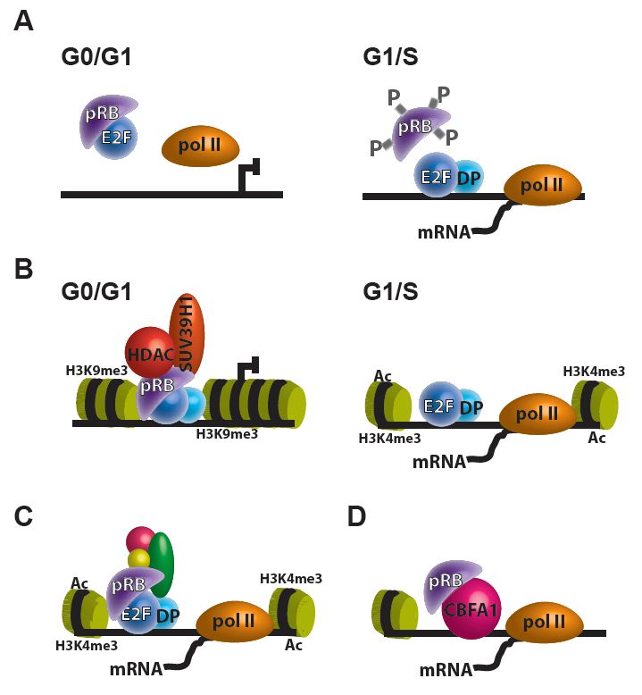
play a role in transcriptional regulation of E2F targets in at least three different mechanism: (1) direct
pRB repression on E2F transcription (Figure 1A), (2) pRB recruitment of transcriptional corepressors to
E2F targets (Figure 1B), and (3) association of the pRB-E2F complex with transcriptional coactivators,

Cancers 2020, 12, 2807; doi:10.3390/cancers12102807                                 www.mdpi.com/journal/cancers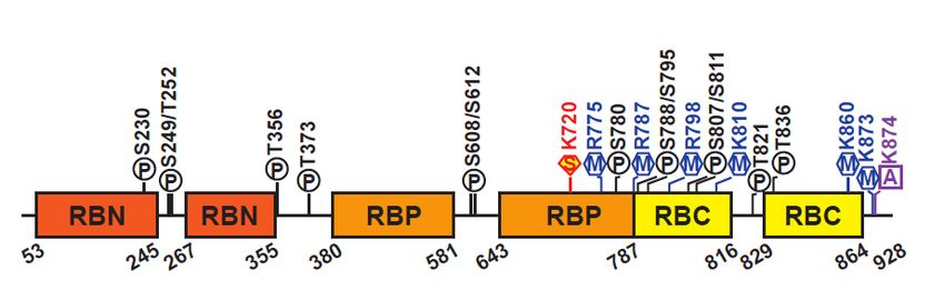
Cancers 2020, 12, 2807                                                                                                  2 of 14
Cancers 2020, 12, x FOR PEER REVIEW                                                                                      2 of 15

which  result in increased
 also noteworthy            expression
                    that beyond        of E2F
                                  its E2F      targets (Figure
                                           transcriptional     1C) [3]. ItpRB
                                                            regulation,    is also
                                                                                may noteworthy    that beyond
                                                                                       regulate transcription
its E2F transcriptional  regulation, pRB  may   regulate transcription through     its interaction
 through its interaction with other transcription factors and has transcription-independent         with other
                                                                                                           roles
transcription  factors and has transcription-independent
 throughout the cell (Figure 1D) [4].                       roles throughout    the  cell (Figure  1D) [4].

      Figure 1. Transcriptional regulation by pRB. (A–C) The pRB can regulate E2F targets in at least
      three
       Figuredifferent mechanisms:
               1. Transcriptional       (A) direct
                                   regulation       pRB (A–C)
                                                 by pRB.  repression on E2F
                                                                The pRB    can transcription;   (B) pRB
                                                                                regulate E2F targets        recruitment
                                                                                                        in at least three
      of transcriptional
       different          corepressors,
                  mechanisms:    (A) directlike pRB
                                                HDACs    and histone
                                                     repression       methyltransferases
                                                                 on E2F                      (e.g.,
                                                                            transcription; (B)   pRBSUV39H1)     to E2F
                                                                                                         recruitment    of
      targets; and (C) association
       transcriptional  corepressors,of the
                                         likepRB-E2F
                                              HDACscomplex      with transcriptional
                                                       and histone   methyltransferasescoactivators,   which results
                                                                                            (e.g., SUV39H1)            in
                                                                                                                  to E2F
      increased   expression
       targets; and           of E2F of
                     (C) association   targets.  (D) pRBcomplex
                                          the pRB-E2F     can regulate
                                                                  with transcription
                                                                        transcriptionalthrough    its interaction
                                                                                         coactivators,              with
                                                                                                          which results
      other  transcription factors  (e.g., CBFA1).
       in increased expression of E2F targets. (D) pRB can regulate transcription through its interaction with
      other transcription factors (e.g., CBFA1).
    Within the cell cycle, CYCLIN D: CDK4/6 mono-phosphorylates pRB at active sites and
unstructured   regions
      Within the     cell ofcycle,
                              the protein
                                    CYCLIN  during   early G1 mono-phosphorylates
                                                 D: CDK4/6       phase, CYCLIN E: CDK2     pRB hyper-phosphorylates
                                                                                                   at active sites and
pRB  in late G1,  and  CYCLIN        B: CDK1    maintain     pRB  in  a hyper-phosphorylated
unstructured regions of the protein during early G1 phase, CYCLIN E: CDK2 hyper-phosphorylates      state during S, G2,
and
pRB in late G1, and CYCLIN B: CDK1 maintain pRB in a hyper-phosphorylated G0
    M   phases  [5]. pRB     mono-phosphorylation         inactivates   unphosphorylated      pRB       functions.
                                                                                                    state during S,pRB
                                                                                                                     G2,
hyper-phosphorylation        inhibits its ability to bind   to E2Fs,  allowing  E2Fs  to
and M phases [5]. pRB mono-phosphorylation inactivates unphosphorylated pRB G0 functions.drive  transcription  of genes
                                                                                                                    pRB
necessary  for G1/S phaseinhibits
hyper-phosphorylation           cell cycle
                                         itsprogression.
                                             ability to bindWhile   hyper-phosphorylation
                                                                to E2Fs,  allowing E2Fs to drive largely  disrupts pRB
                                                                                                       transcription  of
binding  to  other proteins,     some   protein   interactions    persist following    pRB  hyper-phosphorylation.
genes necessary for G1/S phase cell cycle progression. While hyper-phosphorylation largely disrupts
The
pRBprotein–protein
       binding to other interactions    that co-exist
                                  proteins,            with pRBinteractions
                                              some protein          in a hyper-phosphorylated
                                                                                   persist following form pRB
                                                                                                           suggest that
                                                                                                                 hyper-
pRB hyper-phosphorylation
phosphorylation.                   does not inhibit
                     The protein–protein              all pRB that
                                                interactions    functions  [6].with
                                                                     co-exist   While
                                                                                    pRBpRB
                                                                                         in abinding   to E2Fs occur at
                                                                                               hyper-phosphorylated
form suggest that pRB hyper-phosphorylation does not inhibit all pRB functions [6]. While pRB
binding to E2Fs occur at the pocket domain, pRB interactions with epigenetically-relevant proteins
Cancers 2020,12,
Cancers2020, 12,2807
x FOR PEER REVIEW 33 of
of 14
15
involve the LxCxE-binding domain [7]. The availability of the LxCxE binding site, even when pRB is
the pocket
bound domain,
to E2F, pRB expanded
presents interactions with epigenetically-relevant
functions of pRB in the recruitment proteins
of involve
chromatin the remodelers
LxCxE-bindingand
domain [7]. The availability of the LxCxE binding site, even when pRB
other protein complexes. Indeed, pRB has been reported to interact with over 300 proteins is bound to E2F,and
presents
many
expanded functions of
protein interactions pRB
are, in the
or are recruitment
involved of chromatin
with, chromatin remodelers
modifier and[8].
proteins other
pRBprotein complexes.
therefore extends
Indeed,
relevance into protein complexes beyond those within the canonical pRB/E2F pathway. Studiesare,
pRB has been reported to interact with over 300 proteins and many protein interactions in
or are involved with,
retinoblastoma chromatin
indicate that thesemodifier
tumorsproteins
develop[8]. pRB therefore
quickly as a resultextends relevance
of epigenetic into protein
deregulation of
complexes
key cancer beyond
pathways those
as a within the canonical
direct result pRB/E2F
of pRB loss pathway.
[9]. Studies in theStudies in retinoblastoma
last decade indicate
have found significant
that
evidence regarding the relationship between pRB and chromatin remodeling proteins, pushingasthe
these tumors develop quickly as a result of epigenetic deregulation of key cancer pathways a
direct result of
boundaries of knowledge
pRB loss [9]. Studies in
regarding pRBthefunctions.
last decade have
This found
review significant
provides evidenceexamination
an in-depth regarding theof
relationship between pRB and chromatin remodeling proteins, pushing
the literature involving pRB in epigenetic regulation, including nucleosome mobilization,the boundaries of knowledge
histone
regarding
acetylation,pRB functions.
histone This review
methylation, DNA provides an in-depth
methylation, examinationRNAs.
and non-coding of the literature involving pRB
in epigenetic regulation, including nucleosome mobilization, histone acetylation, histone methylation,
DNA
2. pRB methylation, and non-coding RNAs.
Protein Structure
2. pRBHuman
ProteinpRB consists of 928 amino acids, which can be divided into three domains: the N-
Structure
terminal domain (RBN), the A/B region or “pocket” domain (RBP), and the C-terminal domain (RBC)
Human pRB consists of 928 amino acids, which can be divided into three domains: the N-terminal
(Figure 2) [10,11]. The RBP contains a Leu-X-Cys-X-Glu (LxCxE) binding cleft which mediates
domain (RBN), the A/B region or “pocket” domain (RBP), and the C-terminal domain (RBC)
interactions with hundreds of proteins, including oncoproteins and transcription factors. Among
(Figure 2) [10,11]. The RBP contains a Leu-X-Cys-X-Glu (LxCxE) binding cleft which mediates
these, the best characterized interaction is that between pRB and the E2F family of transcription
interactions with hundreds of proteins, including oncoproteins and transcription factors. Among these,
factors. The interaction between pRB and other proteins depends on the pRB’s structure and its post-
the best characterized interaction is that between pRB and the E2F family of transcription
translational modifications, which play a central role in regulating pRB’s function in many different
factors. The interaction between pRB and other proteins depends on the pRB’s structure and its
developmental processes (reviewed in [8,12,13]). The RBN domain closely resembles the pocket
post-translational modifications, which play a central role in regulating pRB’s function in many
structure and can physically interact with the RBP [14]. The RBC domain is intrinsically disordered
different developmental processes (reviewed in [8,12,13]). The RBN domain closely resembles the
and is required for the interaction between pRB and the E2F/DP complex [15].
pocket structure and can physically interact with the RBP [14]. The RBC domain is intrinsically
The three pRB domains are connected by unstructured linker sequences that provide flexibility
disordered and is required for the interaction between pRB and the E2F/DP complex [15].
to the protein. These linker sequences are subjected to post-translational modifications, most notably
The three pRB domains are connected by unstructured linker sequences that provide flexibility to
CDK-dependent phosphorylation, affecting pRB interaction with other proteins and its functional
the protein. These linker sequences are subjected to post-translational modifications, most notably
roles [16,17]. In general, pRB phosphorylation results in inactivation of pRB functions, resulting in
CDK-dependent phosphorylation, affecting pRB interaction with other proteins and its functional
transcriptional derepression and cell cycle progression [18]. Human pRB has 14 phosphorylation, 2
roles [16,17]. In general, pRB phosphorylation results in inactivation of pRB functions, resulting in
acetylation, and 6 methylation sites, and is also subject to ubiquitination and SUMOylation (Figure
transcriptional derepression and cell cycle progression [18]. Human pRB has 14 phosphorylation,
2) [8,13]. These post-translational modifications can promote or prevent occurrence of other
2 acetylation, and 6 methylation sites, and is also subject to ubiquitination and SUMOylation (Figure 2) [8,13]
modifications, controlling pRB functions, including its ability to recruit chromatin remodeling
These post-translational modifications can promote or prevent occurrence of other modifications, controlling
factors.
pRB functions, including its ability to recruit chromatin remodeling factors.
Figure 2. Schematic of pRB structure. Representation of the human pRB domain organization and
Figure 2. Schematic
post-translational of pRB structure.
modifications. Representation
Structured domains of
arethe humanaspRB
depicted domain
colored organization
boxes, and
unstructured
post-translational modifications. Structured domains are depicted as colored boxes, unstructured
domains are depicted as a line in black. Phosphorylation (P) sites are shown in black, methylation (M)
domains
sites are acetylation
in blue, depicted as(A)
a line in in
sites black. Phosphorylation
purple, (P) sites
and sumoylation are shown
(S) sites in red. in black, methylation (M)
sites in blue, acetylation (A) sites in purple, and sumoylation (S) sites in red.
3. Role of pRB in Nucleosome Mobilization
3. Role of pRBDNA
Genomic in Nucleosome
is packaged Mobilization
by histones and non-histone proteins into nucleosomes, the structural
units Genomic
of the eukaryotic
DNA ischromatin
packagedand
bychromosomes.
histones andSWI/SNF complexes
non-histone are multiprotein
proteins chromatin
into nucleosomes, the
remodeling complexes found in eukaryotes that rearrange nucleosomes in an ATP-dependent
structural units of the eukaryotic chromatin and chromosomes. SWI/SNF complexes are multiprotein
manner
chromatin [19–22]. Thesecomplexes
remodeling modifications
foundcan
in involve changing
eukaryotes the location
that rearrange and/or changing
nucleosomes the
in an ATP-
dependent manner [19–22]. These modifications can involve changing the location and/or changingCancers 2020, 12, 2807 4 of 14
conformation of nucleosomes, thus reorganizing chromatin structure in order to facilitate transcription
factor binding [23–28]. The nucleosomal changes mediated by SWI/SNF can result in transcriptional
activation or repression, depending on the components of the complex. In mammals, SWI/SNF
complexes are driven by the ATPase subunits Brahma (BRM) and Brahma-related gene 1 (BRG1) [29,30].
Both BRM and BRG1 contain a LxCxE binding motif and are known to interact with pRB [31–34].
BRG1 and BRM binding to pRB is crucial for pRB-mediated repression of E2F1 activity and the
resulting cell cycle arrest [31,34]. BRM and BRG1-deficient cells are resistant to pRB-mediated arrest,
an effect that is reversible through ectopic expression of BRM and BRG1 to re-sensitize cells to growth
inhibition [35–39]. As BRM and BRG1 share some functional redundancy, BRM expression is sufficient
for sensitizing cells to pRB-mediated arrest when BRG1 is lost [39]. It is therefore an established
consensus that physical interaction between pRB and BRM/BRG1 is required for pRB-mediated cell
cycle arrest and that the SWI/SNF complex is directly involved in cell cycle regulation. However,
a study showing that cellular growth and senescence could still be achieved when the LxCxE motif
is mutated in BRG1, brought into question whether the direct interaction between BRG1 and pRB is
essential [40]. Although this finding could be disputed by an earlier study indicating that pRB and
BRG1 do not interact through the LxCxE motif but through an adjacent site [41], the former study
suggests that the induction of pRB dephosphorylation needed for cell cycle arrest is the result of
BRG1-facilitated upregulation of the cyclin-dependent kinase inhibitor, p21 [40].
Despite being predominantly associated with transcriptional activation, BRG1 forms pRB-SWI/SNF
and pRB-SWI/SNF-HDAC repressor complexes, acting as cell cycle checkpoints during G1-S transition
by inhibiting CYCLIN A and CYCLIN E, respectively [31,33,34]. The formation and activity of these
repressors are regulated by CDK-mediated phosphorylation of pRB [41] and BRM/BRG1 [42,43].
In addition to transcriptional regulation, pRB and BRG1 also interact during DNA double strand break
repair [44]. Upon DNA damage, a phosphorylated E2F1-TopBP1 complex forms to recruit pRB. Then,
pRB subsequently recruits BRG1 to the double strand break in order to decrease nucleosome density at
the site of the break and stimulate DNA end resection [44]. In this complex, pRB also plays a stabilizing
role by shielding phosphorylated E2F1 from proteasomal degradation [45].
It is incontestable that the interplay between pRB and SWI/SNF is critical for cellular homeostasis.
SWI/SNF complexes are considered tumor suppressors and are mutated in nearly 20% of human
malignancies [46]. Specifically, BRG1 is reported to be a target for mutation in 10–30% of human cancer
cell lines [38,47,48]. In pRB wild-type cancers, harboring loss of or mutated BRG1 could contribute
to pRB signaling pathway inactivation and thus provide growth advantage [36,37]. While loss of
BRM/BRG1 is associated with clinical poor prognosis in non-small cell lung cancers [48], many others
have suggested that elevated BRM/BRG1 levels is a prognostic factor for various types of cancers [49–51].
In addition, loss of Brg1 in combination with pRB-loss enhances retinoblastoma tumorigenesis,
suggesting that BRG1 acts as a tumor suppressor in the developing retina [52]. In summary, mammalian
SWI/SNF complexes and their individual subunits BRM and BRG1 are closely associated with cellular
processes regulated by pRB; however, the activity and mechanism of actions of these complexes remain
highly context-dependent.
4. pRB and Histone Acetylation
Histone acetylation involves the transfer of an acetyl group from acetyl coenzyme A to lysine
residues on histone tails by histone acetyltransferases (HATs). Histone acetylation relaxes the chromatin
structure, allowing access of transcriptional activators to gene promoter regions to stimulate transcription.
Several HATs have been identified, including transcriptional coactivators CBP/p300, PCAF, GCN5L, SRC1,
ACTR and TAFII250. Removal of these acetyl groups from the tails of histone octamers is mediated by
any of the 18 histone deacetylases (HDACs 1 to 18), ultimately influencing heterochromatinization of
the nucleosome [53]. These modifications of the acetylation state of histone tails can be facilitated by the
interaction of pRB protein with class I HDACs (HDAC1-3) within its role in E2F transcriptional repression
during cell cycle regulation and terminal differentiation [54,55]. Furthermore, pRB also forms part of HATCancers 2020, 12, 2807 5 of 14
complexes to facilitate HAT inactivation to repress transcription during terminal differentiation [56,57] or
to facilitate histone acetylation required for DNA break repair [58].
The binding to HDACs and other chromatin remodelers is facilitated by the LxCxE motif within the
pocket structure of pRB [7]. Since binding of E2F and pRB is independent of the LxCxE domain, HDAC1
and HDAC2 can interact with the pRB/E2F complex to help in the long-term repression of E2F target
genes [54,59]. The interaction between pRB and HDAC1 can also be indirect: HDAC1-3 can bind to pRB
using pRB-binding proteins, like RBP1, as bridging molecules [60] and HDAC1 is part of the SIN3 and
CTBP/CTIP complexes, which are also pRB-interacting proteins [61,62]. The deacetylation activity of
HDACs is required for pRB repression of genes normally activated during G1 [41]. Inhibition of HDAC
activity, using inhibitors like Trichostatin A, was shown to inhibit the pRB-mediated inactivation of G1
cell cycle genes [41,63]. HDACs deacetylase activity when interacting with pRB is also important for the
placement of further repressive chromatin modifications, as histone deacetylation is required prior to
pRB’s association with SUV39H1 and HP1 for H3K9 trimethylation, discussed in the next section [64,65].
Beyond gene expression repression, the interactions between HDAC1/2 and the pRB/E2F complex
mediate epigenetic silencing of transposable elements including long interspersed nuclear elements
(LINE); thus, helping maintain genomic stability [66].
As mentioned previously, E2F1 and pRB also participate in transcription-independent functions in
DNA repair [44,67]. In this process, interactions between HATs and pRB are an important component
of the DNA damage response. Following the E2F1 and pRB recruitment of BRG1-containing SWI/SNF
complex to double-strand breaks to decrease nucleosome density at the site of the DNA breaks [44],
E2F1 and pRB recruit p300/CBP to sites of DNA damage [58]. p300 and CBP then mediate the
acetylation of multiple lysine residues on histone H3 in nucleosomes flanking double-strand breaks.
Further, E2F1 and pRB are also required for the recruitment of Tip60 and induction of H4K16 acetylation
at DNA breaks, although this may involve an indirect mechanism rather than direct interaction between
Tip60 and pRB [58].
5. pRB and Histone Methylation
Another important mechanism for chromatin regulation involves histone methylation.
Histone methylation is catalyzed by histone methyltransferases (HMTs) and removal of these methyl
groups is mediated by histone demethylases (KDMs). In humans, there are over 100 predicted
HMTs and more than 20 KDMs. Histone methylation comprises an elaborate network that can
activate or repress chromatin depending on the amino acid residue modified and the proteins that
bind to these modified tails. Among the most studied histone methylation marks associated with
repressed chromatin are histone H3 lysine 9 di- and tri-methylation (H3K9me2/3), histone H4 lysine 20
trimethylation (H4K20me3), and histone H3 lysine 27 trimethylation (H3K27me3). Several studies
indicate that pRB can interact with HMTs and KDMs independent of the LxCxE motif to repress
transcription as well as to maintain heterochromatin in intergenic zones, centromeres, peri-centromeres,
and telomeres [64,68–70].
SUV39H1 is an HMT that functionally interacts with the heterochromatin protein 1 (HP1) to
repress transcription at heterochromatic and euchromatic sites. SUV39H1 trimethylates H3K9, creating
a repressive histone mark that serves as a binding site for HP1. The interaction between H3K9me3
and HP1 changes the nucleosome structure into a packed, transcriptionally inactive conformation.
pRB can associate with SUV39H1 and HP1 through the pocket domain to repress transcription,
including cell cycle genes cyclin E and cyclin A2 [64,69]. In this process, pRB is both necessary for
the direct methylation of H3K9 by SUV39H1 and the binding of HP1 to repress gene expression [64].
The interaction between pRB and HP1, and the resulting changes in heterochromatin organization,
also contribute to the changes in gene expression associated with the induction of senescence and the
permanence of the senescent state [71].
pRB also binds to HMTs SUV4-20H1 and SUV4-20H2, two H4K20 trimethylating enzymes [70].
Trimethylation of H4K20 by SUV4-20H marks pericentric heterochromatin, a fraction of theCancers 2020, 12, 2807 6 of 14
heterochromatin located on both sides of centromeres that is crucial for preserving the integrity
of the genome and promoting correct chromosomal segregation. The formation of this constitutive
heterochromatin requires pRB and its family members, p107 and p130, for the stabilization of H4K20me3
by SUV4-20Hs, in a process that is independent of the E2F family [70]. Disruption of this heterochromatin
structure due to pRB loss can result in centromere fusions, chromosome mis-segregation, and genomic
instability [72].
The pRB/E2F complex can recruit enhancer of zeste homolog 2 (EZH2) to mediate H3K27me3
deposition to repress transcription during differentiation and stress [73]. H3K27me3 is also an important
histone mark that represses heterochromatin at repetitive genomic regions, including tandem sequence
repeats and interspersed repeats [74]. In this role, pRB, E2F1, and EZH2 can form a complex that directs
H3K27me3 deposition to silence genomic repeat elements [75]. This cell-cycle-independent interaction
between pRB and E2F1 initially recruits EZH2 to diverse repeat sequences, but pRB presence does
not appear to be necessary for subsequent spreading of EZH2 and H3K27me3 [75]. Disrupting the
ability of pRB to recruit EZH2 in mouse models results in cancer susceptibility, suggesting a role for
pRB-EZH2 in tumor suppression [75].
The role of pRB in regulating histone methylation also extends to interactions with KDMs.
pRB can bind to KDM1A (also known as LSD1) and KDM5A (also known as RBP2 or JARID1A),
two KDMs that catalyze the removal of methyl groups from histone H3 lysine 4 (H3K4) [68,76].
Methylated H3K4 is a histone mark commonly found at actively transcribed promoter regions.
KDM5A can demethylate H3K4me2/3, while KDM1A can demethylate H3K4me1/2. pRB binding to
KMD1A represses transcription by recruiting it to demethylate H3K4 to pRB-regulated genes [68].
On the other hand, pRB binding to KDM5A displaces KDM5A and inhibits H3K4 demethylation,
activating—rather than repressing—transcription [76]. Interestingly, the capacity of pRB to induce
differentiation correlates with its ability to activate transcription together with transcription factors such
as MYOD, CBFA1, and GRα [4,76]. KDM5A release from genes encoding mitochondrial proteins occurs
early during differentiation and is required for the activation of differentiation markers. pRB can directly
bind to and activate these KDM5A targets with mitochondrial functions potentially by sequestering
KDM5A away from these genes [77]. Thus, pRB loss may also contribute to tumorigenesis through
defects in cellular metabolism and terminal differentiation associated with its role in histone methylation.
6. pRB in DNA Methylation
DNA methylation involves the addition of a methyl group to the 5-carbon position of cytosine,
forming 5-methylcytosine, at CpG dinucleotides [78,79]. Methylation of DNA is generally associated
with an inactive chromatin through the inhibition of transcription factor binding [80], as well as
allowing the occupation of methyl-CpG-binding proteins that facilitate transcriptional repression [81].
DNA methylation is catalyzed by enzymes in the DNA methyltransferase family (DNMTs), including
DNMT1, which is associated with the maintenance of DNA methylation [82,83], and DNMT3A and
DNMT3B, which are responsible for de novo DNA methylation [84]. pRB not only transcriptionally
represses the expression of DNMT1 and DNMT3A/B [85–87], but also physically interacts with DNMT1
and chromatin remodelers that can recruit DNMT1 and DNMT3A/B to affect DNA methylation [88–91].
As was discussed previously for histone modifiers, pRB transcriptional repression of E2F-regulated
genes can also occur through direct recruitment of DNMT1. pRB can form a stable complex with E2F1,
HDAC1, and DNMT1 and repress E2F-driven transcription [92]. Even though it is not clear whether
the catalytic activity of DNMT1 is necessary for the E2F transcriptional repression of this complex,
DNA methylation at E2F-dependent promoters is known to prevent the binding of E2F proteins [93].
The DNMT1/pRB interaction could serve as a way to enhance pRB-mediated repression of cell cycle
progression during terminal differentiation as a mechanism to ensure genes involved in cell division
are permanently turned off. Thus, it is likely that loss of functional pRB may contribute to aberrant
DNMT1 localization and the resulting changes in global methylation observed in many cancer cells,
including retinoblastoma.Cancers 2020, 12, 2807 7 of 14
The areas of the genome to be silenced by DNA methylation are recognized by
chromatin-remodeling complexes that recruit DNMTs to the site. In this context, pRB is known
to regulate the expression of chromatin remodeling proteins UHRF1 (ubiquitin-like, containing PHD
and RING finger domains 1) and HELLS (helicase, lymphoid specific), which recruit DNMT1 and
DNMT3, respectively. UHRF1 is a direct downstream target of the pRB/E2F pathway [88,89]. As a
chromatin remodeler, UHRF1 recruits HDACs, HMTs, and DNMT1 to maintain heterochromatin
during cell division (reviewed in [94]). pRB has an interesting connection with UHRF1 that goes
beyond its transcriptional regulation as UHRF1 contains two LxCxE binding sequences that allow
it to interact with pRB at the protein level, and UHRF1 can repress pRB gene transcription during
S-phase entry [95]. UHRF1 overexpression is observed in multiple types of cancer with pRB loss,
including retinoblastoma [96], with loss of UHRF1 capable of reversing the adverse effects associated
with pRB loss [88]. Unpublished studies from our lab using genetic engineered mouse models of
retinoblastoma also indicate that UHRF1 overexpression is required for the epigenetic changes that
drive retinoblastoma tumor progression and in the absence of UHRF1, retinoblastoma tumors fail
to form. HELLS is also transcriptionally regulated by the pRB/E2F pathway and overexpressed in
multiple types of cancer, including retinoblastoma [90,96,97]. HELLS remodels chromatin to render
DNA accessible to DNMT3 to support de novo DNA methylation and stable gene silencing during
cellular differentiation. In the HELLS-mediated transcriptional repression, HELLS also acts as a
recruiting factor for DNMT1 and HDACs to establish transcriptionally repressive chromatin [98].
In retinoblastoma, HELLS overexpression is critical for ectopic proliferation and tumor progression [91].
Consequently, inactivation of the pRB pathway alters DNMTs activity, leading to aberrant genomic
DNA methylation patterns and malignant progression.
7. pRB Regulation of Non-Coding RNAs
The majority of transcriptional outcomes depend on non-coding RNAs (ncRNAs), including
microRNAs (miRNAs) and long non-coding RNAs (lncRNAs). MiRNAs are short nucleotide sequences
(~22 nucleotides) known to regulate gene expression post-transcriptionally. MiRNAs are transcribed
by RNA polymerase II, cleaved in the nucleus by the endonuclease, Droscha, and then exported
to the cytoplasm for further processing by another endonuclease, Dicer, allowing the transcript to
become a mature miRNA duplex (reviewed in [99]). The miRNA can base pair to its corresponding
regulatory site, forming the RNA-inducing silencing complex (RISC). LncRNAs are longer nucleotide
sequences (>200 bp) that regulate gene expression at the transcriptional and post-transcriptional level.
Primary lncRNAs are transcribed and transported into the nucleus similarly to miRNA transcripts but
display similar properties of gene structure to protein-coding genes [100]. LncRNAs act as chromatin
regulators by recruiting and interacting with chromatin remodeling enzymes, and as transcriptional
regulators by binding to RNA-binding factors that can promote or repress gene transcription [101].
The transcriptional, post-transcriptional and epigenetic regulatory functions of ncRNAs can either be
oncogenic or tumor suppressive. Here, we present pRB regulation of ncRNA, particularly during cancer.
Loss of pRB function and derepression of E2F-target genes results in deregulation of several
ncRNA in RB (reviewed in [102]). However, whether or not several of these RNA molecules are
directly regulated by pRB has not been thoroughly examined. One of the strongest evidence that
pRB regulation of ncRNAs can have critical effects in tumorigenesis upon loss of pRB is portrayed
by the miR-17~92 cluster [103]. In mouse models of retinoblastoma, loss of miR-17~92 completely
suppresses retinoblastoma tumor formation [104], strongly supporting that miR-17~92 is regulated
by pRB and critical for tumor development. In the absence of pRB, the cyclin-dependent kinase
inhibitor p21Cip1 is upregulated; however, the overexpression of miR-17~92 caused by pRB-deficiency
counteracts the upregulation of p21Cip1 , leading to tumor formation [105]. Several other miRNAs that
are transcriptionally controlled by activating E2Fs (E2F1, E2F2, and E2F3) during G1 are generally
accepted to be transcriptionally repressed by pRB, as part of cell-cycle regulation by the pRB/E2F
pathway, including miR-106b~25, miR-15b~16-2 and the let-7 family [106].Cancers 2020, 12, 2807 8 of 14
Several ncRNAs participate in auto-regulatory feedback loops. Therefore, many ncRNAs that
regulate pRB may also be regulated by the pRB/E2F pathway. Research revealing ncRNA regulation by
pRB should be further investigated for future engineering of potential therapeutic targets. In addition
to the uncertainty regarding pRB regulation of ncRNAs, the degree of how much the regulatory
networks differ between cancers compared to those in retinoblastoma is still unknown. A ncRNA
biomarker for a specific cancer type may be different than those for retinoblastoma, even if that ncRNA
is regulated by the pRB-E2F pathway. Further investigations on tissue specific expression patterns
should be performed to reveal tissue-specific mechanisms of pRB regulation of ncRNAs.
8. Conclusions
Even though pRB and the pRB/E2F pathway have been extensively studied for decades,
the molecular mechanisms behind the wide-ranging roles of this protein and pathway are still
yet to be fully elucidated. As reviewed here, several of pRB functions, both within its canonical role in
cell cycle control and other non-canonical cellular functions, are exerted through its interactions with
epigenetic modifiers (summarized in Table 1). The intricacy of these multiprotein complexes and the
cellular context under which they interact with pRB depict the diversity of pRB cellular roles and may
explain why pRB inactivation is sufficient to cause cancer in some tissues (e.g., retinoblastoma) but not
in others. Thus, understanding the context of these pRB interactions with multi-protein complexes that
control nucleosome and chromatin modifications has the potential of providing alternative strategies
for cancer therapy in a pathway that, at least to this day, remains largely undruggable.
Table 1. Epigenetic regulators regulated by the pRB/E2F pathway
Epigenetic Regulation Type Component Relation with pRB/E2F Pathway References
Nucleosome Mobilization
BRG1 E2F target and binds to pRB [31]
BRM Binds to pRB [34]
Histone Acetylation
HDAC1–3 Binds to pRB [54,55]
SIN3 Binds to pRB [61,62]
CTBP/CTIP Binds to pRB [61,62]
CBP/p300 Binds to pRB [58]
Histone Methylation
SUV39H1 Binds to pRB [64,69]
SUV4-20H1 Binds to pRB [70]
SUV4-20H2 Binds to pRB [70]
HP1 Binds to pRB [64]
EZH2 Binds to pRB [73,75]
KMD1A Binds to pRB [68]
KMD5A Binds to pRB [76]
DNA Methylation
DNMT1 E2F target and binds to pRB. [86]
DNMT3 E2F target and binds to pRB. [85]
UHRF1 E2F target and binds to pRB. [88,89,95]
HELLS E2F target and binds to pRB. [90,96,97]
Non-coding RNAs
miR-17–92 E2F target [103–105]
miR-106b–25 E2F target [106]
miR-15b–16-2 E2F target [106]
let-7 family E2F target [106]
As almost all human cancers carry abnormalities in the pRB/E2F pathway components including
INK4A, CCND, CDK4/6, RB1, or E2Fs, genetic of functional inactivation of the pRB/E2F pathway
seems to be indispensable for deregulated proliferation in most cancer settings [107]. Mutations in theCancers 2020, 12, 2807 9 of 14
RB1 gene are found in retinoblastoma, osteosarcoma, and small cell lung cancer at a high frequency,
and with less frequency in other types of cancer [6,107,108]. Mutations in upstream components such
as INK4A are frequently detected in pancreatic cancer and non-small cell lung cancer, and CCND
amplification is often found in breast cancer [6,107,109]. While these differences may be related to
different tissue-specific mutational mechanisms, these observations also suggest that the pRB/E2F
pathway is not strictly linear, and that loss of function due to genetic ablation of the RB1 gene is
not completely synonymous to loss of E2F binding activity due to hyper-phosphorylation of pRB.
From the multiple examples presented, it is evident that many of the pRB functions are likely preserved
when pRB is hyper-phosphorylated. Thus, not every one of pRB’s function should be considered as
integral for its role as a tumor suppressor. Recent studies on the interplay between pRB and chromatin
remodelers have been able to explain, in part, the role of pRB in the epigenetic mechanisms that lead
to tumorigenesis. Whether these can be harnessed to develop novel cancer therapeutics remains to
be determined.
Author Contributions: Conceptualization, writing, and reviewing—all authors, editing and supervision—C.A.B.
All authors have read and agreed to the published version of the manuscript.
Funding: pRB research in our laboratory is supported by the NIH (CA229696), the American Cancer Society
(129801-IRG-16-187-13), and the AACR-Aflac, Inc. Career Development Award for Pediatric Cancer Research
(18-20-10-BENA). F.G., K.S., and S.S. are supported by the NIH-MARC U-STAR training grant (T34GM136498).
Conflicts of Interest: The authors declare no conflict of interest.
References
1. Harbour, J.W.; Dean, D.C. The Rb/E2F pathway: Expanding roles and emerging paradigms. Genes Dev. 2000,
14, 2393–2409. [CrossRef]
2. Lee, C.; Chang, J.H.; Lee, H.S.; Cho, Y. Structural basis for the recognition of the E2F transactivation domain
by the retinoblastoma tumor suppressor. Genes Dev. 2002, 16, 3199–3212. [CrossRef] [PubMed]
3. Ianari, A.; Natale, T.; Calo, E.; Ferretti, E.; Alesse, E.; Screpanti, I.; Haigis, K.; Gulino, A.; Lees, J.A. Proapoptotic
function of the retinoblastoma tumor suppressor protein. Cancer Cell 2009, 15, 184–194. [CrossRef] [PubMed]
4. Thomas, D.M.; Carty, S.A.; Piscopo, D.M.; Lee, J.S.; Wang, W.F.; Forrester, W.C.; Hinds, P.W. The retinoblastoma
protein acts as a transcriptional coactivator required for osteogenic differentiation. Mol. Cell 2001, 8, 303–316.
[CrossRef]
5. Narasimha, A.M.; Kaulich, M.; Shapiro, G.S.; Choi, Y.J.; Sicinski, P.; Dowdy, S.F. Cyclin D activates the Rb
tumor suppressor by mono-phosphorylation. eLife 2014, 3. [CrossRef]
6. Dyson, N.J. RB1: A prototype tumor suppressor and an enigma. Genes Dev. 2016, 30, 1492–1502. [CrossRef]
7. Magnaghi-Jaulin, L.; Groisman, R.; Naguibneva, I.; Robin, P.; Lorain, S.; Le Villain, J.P.; Troalen, F.; Trouche, D.;
Harel-Bellan, A. Retinoblastoma protein represses transcription by recruiting a histone deacetylase. Nature
1998, 391, 601–605. [CrossRef]
8. Sanidas, I.; Morris, R.; Fella, K.A.; Rumde, P.H.; Boukhali, M.; Tai, E.C.; Ting, D.T.; Lawrence, M.S.; Haas, W.;
Dyson, N.J. A Code of Mono-phosphorylation Modulates the Function of RB. Mol. Cell 2019, 73, 985–1000.
[CrossRef] [PubMed]
9. Zhang, J.; Benavente, C.A.; McEvoy, J.; Flores-Otero, J.; Ding, L.; Chen, X.; Ulyanov, A.; Wu, G.; Wilson, M.;
Wang, J.; et al. A novel retinoblastoma therapy from genomic and epigenetic analyses. Nature 2012, 481,
329–334. [CrossRef]
10. Wang, J.Y.; Knudsen, E.S.; Welch, P.J. The retinoblastoma tumor suppressor protein. Adv. Cancer Res. 1994,
64, 25–85. [CrossRef] [PubMed]
11. Welch, P.J.; Wang, J.Y. Disruption of retinoblastoma protein function by coexpression of its C pocket fragment.
Genes Dev. 1995, 9, 31–46. [CrossRef] [PubMed]
12. Dick, F.A.; Rubin, S.M. Molecular mechanisms underlying RB protein function. Nat. Rev. Mol. Cell Biol. 2013,
14, 297–306. [CrossRef] [PubMed]
13. Macdonald, J.I.; Dick, F.A. Posttranslational modifications of the retinoblastoma tumor suppressor protein as
determinants of function. Genes Cancer 2012, 3, 619–633. [CrossRef] [PubMed]Cancers 2020, 12, 2807 10 of 14
14. Hassler, M.; Singh, S.; Yue, W.W.; Luczynski, M.; Lakbir, R.; Sanchez-Sanchez, F.; Bader, T.; Pearl, L.H.;
Mittnacht, S. Crystal structure of the retinoblastoma protein N domain provides insight into tumor
suppression, ligand interaction, and holoprotein architecture. Mol. Cell 2007, 28, 371–385. [CrossRef]
15. Rubin, S.M.; Gall, A.L.; Zheng, N.; Pavletich, N.P. Structure of the Rb C-terminal domain bound to E2F1-DP1:
A mechanism for phosphorylation-induced E2F release. Cell 2005, 123, 1093–1106. [CrossRef]
16. Burke, J.R.; Deshong, A.J.; Pelton, J.G.; Rubin, S.M. Phosphorylation-induced conformational changes in the
retinoblastoma protein inhibit E2F transactivation domain binding. J. Biol. Chem. 2010, 285, 16286–16293.
[CrossRef]
17. Burke, J.R.; Hura, G.L.; Rubin, S.M. Structures of inactive retinoblastoma protein reveal multiple mechanisms
for cell cycle control. Genes Dev. 2012, 26, 1156–1166. [CrossRef]
18. Weinberg, R.A. The retinoblastoma protein and cell cycle control. Cell 1995, 81, 323–330. [CrossRef]
19. Phelan, M.L.; Sif, S.; Narlikar, G.J.; Kingston, R.E. Reconstitution of a core chromatin remodeling complex
from SWI/SNF subunits. Mol. Cell 1999, 3, 247–253. [CrossRef]
20. Muchardt, C.; Yaniv, M. ATP-dependent chromatin remodelling: SWI/SNF and Co. are on the job. J. Mol. Biol.
1999, 293, 187–198. [CrossRef]
21. Tyler, J.K.; Kadonaga, J.T. The "dark side" of chromatin remodeling: Repressive effects on transcription. Cell
1999, 99, 443–446. [CrossRef]
22. Laurent, B.C.; Treitel, M.A.; Carlson, M. Functional interdependence of the yeast SNF2, SNF5, and SNF6
proteins in transcriptional activation. Proc. Natl. Acad. Sci. USA 1991, 88, 2687–2691. [CrossRef] [PubMed]
23. Dingwall, A.K.; Beek, S.J.; McCallum, C.M.; Tamkun, J.W.; Kalpana, G.V.; Goff, S.P.; Scott, M.P. The Drosophila
snr1 and brm proteins are related to yeast SWI/SNF proteins and are components of a large protein complex.
Mol. Biol. Cell 1995, 6, 777–791. [CrossRef]
24. Papoulas, O.; Beek, S.J.; Moseley, S.L.; McCallum, C.M.; Sarte, M.; Shearn, A.; Tamkun, J.W. The Drosophila
trithorax group proteins BRM, ASH1 and ASH2 are subunits of distinct protein complexes. Development
1998, 125, 3955–3966. [PubMed]
25. Kwon, H.; Imbalzano, A.N.; Khavari, P.A.; Kingston, R.E.; Green, M.R. Nucleosome disruption and
enhancement of activator binding by a human SW1/SNF complex. Nature 1994, 370, 477–481. [CrossRef]
26. Wang, W.; Xue, Y.; Zhou, S.; Kuo, A.; Cairns, B.R.; Crabtree, G.R. Diversity and specialization of mammalian
SWI/SNF complexes. Genes Dev. 1996, 10, 2117–2130. [CrossRef]
27. Sif, S.; Stukenberg, P.T.; Kirschner, M.W.; Kingston, R.E. Mitotic inactivation of a human SWI/SNF chromatin
remodeling complex. Genes Dev. 1998, 12, 2842–2851. [CrossRef]
28. Hamiche, A.; Sandaltzopoulos, R.; Gdula, D.A.; Wu, C. ATP-dependent histone octamer sliding mediated by
the chromatin remodeling complex NURF. Cell 1999, 97, 833–842. [CrossRef]
29. Khavari, P.A.; Peterson, C.L.; Tamkun, J.W.; Mendel, D.B.; Crabtree, G.R. BRG1 contains a conserved domain
of the SWI2/SNF2 family necessary for normal mitotic growth and transcription. Nature 1993, 366, 170–174.
[CrossRef]
30. Muchardt, C.; Yaniv, M. A human homologue of Saccharomyces cerevisiae SNF2/SWI2 and Drosophila brm
genes potentiates transcriptional activation by the glucocorticoid receptor. EMBO J. 1993, 12, 4279–4290.
[CrossRef]
31. Dunaief, J.L.; Strober, B.E.; Guha, S.; Khavari, P.A.; Alin, K.; Luban, J.; Begemann, M.; Crabtree, G.R.; Goff, S.P.
The retinoblastoma protein and BRG1 form a complex and cooperate to induce cell cycle arrest. Cell 1994, 79,
119–130. [CrossRef]
32. Singh, P.; Coe, J.; Hong, W. A role for retinoblastoma protein in potentiating transcriptional activation by the
glucocorticoid receptor. Nature 1995, 374, 562–565. [CrossRef] [PubMed]
33. Strober, B.E.; Dunaief, J.L.; Guha; Goff, S.P. Functional interactions between the hBRM/hBRG1 transcriptional
activators and the pRB family of proteins. Mol. Cell. Biol. 1996, 16, 1576–1583. [CrossRef] [PubMed]
34. Trouche, D.; Le Chalony, C.; Muchardt, C.; Yaniv, M.; Kouzarides, T. RB and hbrm cooperate to repress the
activation functions of E2F1. Proc. Natl. Acad. Sci. USA 1997, 94, 11268–11273. [CrossRef] [PubMed]
35. Zhu, L.; van den Heuvel, S.; Helin, K.; Fattaey, A.; Ewen, M.; Livingston, D.; Dyson, N.; Harlow, E. Inhibition
of cell proliferation by p107, a relative of the retinoblastoma protein. Genes Dev. 1993, 7, 1111–1125. [CrossRef]
36. Strobeck, M.W.; Knudsen, K.E.; Fribourg, A.F.; DeCristofaro, M.F.; Weissman, B.E.; Imbalzano, A.N.;
Knudsen, E.S. BRG-1 is required for RB-mediated cell cycle arrest. Proc. Natl. Acad. Sci. USA 2000, 97,
7748–7753. [CrossRef]Cancers 2020, 12, 2807 11 of 14
37. Reisman, D.N.; Strobeck, M.W.; Betz, B.L.; Sciariotta, J.; Funkhouser, W., Jr.; Murchardt, C.; Yaniv, M.;
Sherman, L.S.; Knudsen, E.S.; Weissman, B.E. Concomitant down-regulation of BRM and BRG1 in human
tumor cell lines: Differential effects on RB-mediated growth arrest vs CD44 expression. Oncogene 2002, 21,
1196–1207. [CrossRef]
38. Bartlett, C.; Orvis, T.J.; Rosson, G.S.; Weissman, B.E. BRG1 mutations found in human cancer cell lines
inactivate Rb-mediated cell-cycle arrest. J. Cell. Physiol. 2011, 226, 1989–1997. [CrossRef]
39. Strobeck, M.W.; Reisman, D.N.; Gunawardena, R.W.; Betz, B.L.; Angus, S.P.; Knudsen, K.E.; Kowalik, T.F.;
Weissman, B.E.; Knudsen, E.S. Compensation of BRG-1 function by Brm: Insight into the role of the core
SWI-SNF subunits in retinoblastoma tumor suppressor signaling. J. Biol. Chem. 2002, 277, 4782–4789.
[CrossRef]
40. Kang, H.; Cui, K.; Zhao, K. BRG1 controls the activity of the retinoblastoma protein via regulation of
p21CIP1/WAF1/SDI. Mol. Cell. Biol. 2004, 24, 1188–1199. [CrossRef]
41. Zhang, H.S.; Gavin, M.; Dahiya, A.; Postigo, A.A.; Ma, D.; Luo, R.X.; Harbour, J.W.; Dean, D.C. Exit from
G1 and S phase of the cell cycle is regulated by repressor complexes containing HDAC-Rb-hSWI/SNF and
Rb-hSWI/SNF. Cell 2000, 101, 79–89. [CrossRef]
42. Muchardt, C.; Reyes, J.C.; Bourachot, B.; Leguoy, E.; Yaniv, M. The hbrm and BRG-1 proteins, components of
the human SNF/SWI complex, are phosphorylated and excluded from the condensed chromosomes during
mitosis. EMBO J. 1996, 15, 3394–3402. [CrossRef]
43. Shanahan, F.; Seghezzi, W.; Parry, D.; Mahony, D.; Lees, E. Cyclin E associates with BAF155 and BRG1,
components of the mammalian SWI-SNF complex, and alters the ability of BRG1 to induce growth arrest.
Mol. Cell. Biol. 1999, 19, 1460–1469. [CrossRef]
44. Velez-Cruz, R.; Manickavinayaham, S.; Biswas, A.K.; Clary, R.W.; Premkumar, T.; Cole, F.; Johnson, D.G.
RB localizes to DNA double-strand breaks and promotes DNA end resection and homologous recombination
through the recruitment of BRG1. Genes Dev. 2016, 30, 2500–2512. [CrossRef] [PubMed]
45. Campanero, M.R.; Flemington, E.K. Regulation of E2F through ubiquitin-proteasome-dependent degradation:
Stabilization by the pRB tumor suppressor protein. Proc. Natl. Acad. Sci. USA 1997, 94, 2221–2226. [CrossRef]
[PubMed]
46. Kadoch, C.; Hargreaves, D.C.; Hodges, C.; Elias, L.; Ho, L.; Ranish, J.; Crabtree, G.R. Proteomic and
bioinformatic analysis of mammalian SWI/SNF complexes identifies extensive roles in human malignancy.
Nat. Genet. 2013, 45, 592–601. [CrossRef]
47. Wong, A.K.; Shanahan, F.; Chen, Y.; Lian, L.; Ha, P.; Hendricks, K.; Ghaffari, S.; Iliev, D.; Penn, B.;
Woodland, A.M.; et al. BRG1, a component of the SWI-SNF complex, is mutated in multiple human tumor
cell lines. Cancer Res. 2000, 60, 6171–6177.
48. Reisman, D.N.; Sciarrotta, J.; Wang, W.; Funkhouser, W.K.; Weissman, B.E. Loss of BRG1/BRM in human lung
cancer cell lines and primary lung cancers: Correlation with poor prognosis. Cancer Res. 2003, 63, 560–566.
49. Jubierre, L.; Soriano, A.; Planells-Ferrer, L.; Paris-Coderch, L.; Tenbaum, S.P.; Romero, O.A.; Moubarak, R.S.;
Almazan-Moga, A.; Molist, C.; Roma, J.; et al. BRG1/SMARCA4 is essential for neuroblastoma cell viability
through modulation of cell death and survival pathways. Oncogene 2016, 35, 5179–5190. [CrossRef]
50. Bai, J.; Mei, P.; Zhang, C.; Chen, F.; Li, C.; Pan, Z.; Liu, H.; Zheng, J. BRG1 is a prognostic marker and potential
therapeutic target in human breast cancer. PLoS ONE 2013, 8, e59772. [CrossRef]
51. Muthuswami, R.; Bailey, L.; Rakesh, R.; Imbalzano, A.N.; Nickerson, J.A.; Hockensmith, J.W. BRG1 is a
prognostic indicator and a potential therapeutic target for prostate cancer. J. Cell. Physiol. 2019. [CrossRef]
[PubMed]
52. Aldiri, I.; Ajioka, I.; Xu, B.; Zhang, J.; Chen, X.; Benavente, C.; Finkelstein, D.; Johnson, D.; Akiyama, J.;
Pennacchio, L.A.; et al. Brg1 coordinates multiple processes during retinogenesis and is a tumor suppressor
in retinoblastoma. Development 2015, 142, 4092–4106. [CrossRef] [PubMed]
53. Roth, S.Y.; Denu, J.M.; Allis, C.D. Histone acetyltransferases. Annu. Rev. Biochem. 2001, 70, 81–120. [CrossRef]
[PubMed]
54. Brehm, A.; Miska, E.A.; McCance, D.J.; Reid, J.L.; Bannister, A.J.; Kouzarides, T. Retinoblastoma protein
recruits histone deacetylase to repress transcription. Nature 1998, 391, 597–601. [CrossRef] [PubMed]
55. Puri, P.L.; Iezzi, S.; Stiegler, P.; Chen, T.T.; Schiltz, R.L.; Muscat, G.E.; Giordano, A.; Kedes, L.; Wang, J.Y.;
Sartorelli, V. Class I histone deacetylases sequentially interact with MyoD and pRb during skeletal myogenesis.
Mol. Cell 2001, 8, 885–897. [CrossRef]Cancers 2020, 12, 2807 12 of 14
56. MacLellan, W.R.; Xiao, G.; Abdellatif, M.; Schneider, M.D. A novel Rb- and p300-binding protein inhibits
transactivation by MyoD. Mol. Cell. Biol. 2000, 20, 8903–8915. [CrossRef]
57. Siegert, J.L.; Robbins, P.D. Rb inhibits the intrinsic kinase activity of TATA-binding protein-associated factor
TAFII250. Mol. Cell. Biol. 1999, 19, 846–854. [CrossRef]
58. Manickavinayaham, S.; Velez-Cruz, R.; Biswas, A.K.; Bedford, E.; Klein, B.J.; Kutateladze, T.G.; Liu, B.;
Bedford, M.T.; Johnson, D.G. E2F1 acetylation directs p300/CBP-mediated histone acetylation at DNA
double-strand breaks to facilitate repair. Nat. Commun. 2019, 10, 4951. [CrossRef]
59. Dahiya, A.; Gavin, M.R.; Luo, R.X.; Dean, D.C. Role of the LXCXE binding site in Rb function. Mol. Cell. Biol.
2000, 20, 6799–6805. [CrossRef]
60. Lai, A.; Lee, J.M.; Yang, W.M.; DeCaprio, J.A.; Kaelin, W.G., Jr.; Seto, E.; Branton, P.E. RBP1 recruits both
histone deacetylase-dependent and -independent repression activities to retinoblastoma family proteins.
Mol. Cell. Biol. 1999, 19, 6632–6641. [CrossRef]
61. Lai, A.; Kennedy, B.K.; Barbie, D.A.; Bertos, N.R.; Yang, X.J.; Theberge, M.C.; Tsai, S.C.; Seto, E.; Zhang, Y.;
Kuzmichev, A.; et al. RBP1 recruits the mSIN3-histone deacetylase complex to the pocket of retinoblastoma
tumor suppressor family proteins found in limited discrete regions of the nucleus at growth arrest. Mol. Cell.
Biol. 2001, 21, 2918–2932. [CrossRef] [PubMed]
62. Meloni, A.R.; Smith, E.J.; Nevins, J.R. A mechanism for Rb/p130-mediated transcription repression involving
recruitment of the CtBP corepressor. Proc. Natl. Acad. Sci. USA 1999, 96, 9574–9579. [CrossRef] [PubMed]
63. Morrison, A.J.; Sardet, C.; Herrera, R.E. Retinoblastoma protein transcriptional repression through histone
deacetylation of a single nucleosome. Mol. Cell. Biol. 2002, 22, 856–865. [CrossRef] [PubMed]
64. Nielsen, S.J.; Schneider, R.; Bauer, U.M.; Bannister, A.J.; Morrison, A.; O’Carroll, D.; Firestein, R.; Cleary, M.;
Jenuwein, T.; Herrera, R.E.; et al. Rb targets histone H3 methylation and HP1 to promoters. Nature 2001, 412,
561–565. [CrossRef] [PubMed]
65. Vaute, O.; Nicolas, E.; Vandel, L.; Trouche, D. Functional and physical interaction between the histone methyl
transferase Suv39H1 and histone deacetylases. Nucleic Acids Res. 2002, 30, 475–481. [CrossRef] [PubMed]
66. Montoya-Durango, D.E.; Liu, Y.; Teneng, I.; Kalbfleisch, T.; Lacy, M.E.; Steffen, M.C.; Ramos, K.S. Epigenetic
control of mammalian LINE-1 retrotransposon by retinoblastoma proteins. Mutat. Res. 2009, 665, 20–28.
[CrossRef]
67. Cook, R.; Zoumpoulidou, G.; Luczynski, M.T.; Rieger, S.; Moquet, J.; Spanswick, V.J.; Hartley, J.A.;
Rothkamm, K.; Huang, P.H.; Mittnacht, S. Direct involvement of retinoblastoma family proteins in DNA
repair by non-homologous end-joining. Cell Rep. 2015, 10, 2006–2018. [CrossRef]
68. Chau, C.M.; Deng, Z.; Kang, H.; Lieberman, P.M. Cell cycle association of the retinoblastoma protein Rb and
the histone demethylase LSD1 with the Epstein-Barr virus latency promoter Cp. J. Virol. 2008, 82, 3428–3437.
[CrossRef] [PubMed]
69. Vandel, L.; Nicolas, E.; Vaute, O.; Ferreira, R.; Ait-Si-Ali, S.; Trouche, D. Transcriptional repression by the
retinoblastoma protein through the recruitment of a histone methyltransferase. Mol. Cell. Biol. 2001, 21,
6484–6494. [CrossRef]
70. Gonzalo, S.; Garcia-Cao, M.; Fraga, M.F.; Schotta, G.; Peters, A.H.; Cotter, S.E.; Eguia, R.; Dean, D.C.;
Esteller, M.; Jenuwein, T.; et al. Role of the RB1 family in stabilizing histone methylation at constitutive
heterochromatin. Nat. Cell Biol. 2005, 7, 420–428. [CrossRef]
71. Narita, M.; Nunez, S.; Heard, E.; Narita, M.; Lin, A.W.; Hearn, S.A.; Spector, D.L.; Hannon, G.J.; Lowe, S.W.
Rb-mediated heterochromatin formation and silencing of E2F target genes during cellular senescence. Cell
2003, 113, 703–716. [CrossRef]
72. Isaac, C.E.; Francis, S.M.; Martens, A.L.; Julian, L.M.; Seifried, L.A.; Erdmann, N.; Binne, U.K.; Harrington, L.;
Sicinski, P.; Berube, N.G.; et al. The retinoblastoma protein regulates pericentric heterochromatin.
Mol. Cell. Biol. 2006, 26, 3659–3671. [CrossRef] [PubMed]
73. Blais, A.; van Oevelen, C.J.; Margueron, R.; Acosta-Alvear, D.; Dynlacht, B.D. Retinoblastoma tumor
suppressor protein-dependent methylation of histone H3 lysine 27 is associated with irreversible cell cycle
exit. J. Cell Biol. 2007, 179, 1399–1412. [CrossRef] [PubMed]
74. Day, D.S.; Luquette, L.J.; Park, P.J.; Kharchenko, P.V. Estimating enrichment of repetitive elements from
high-throughput sequence data. Genome Biol. 2010, 11, R69. [CrossRef] [PubMed]Cancers 2020, 12, 2807 13 of 14
75. Ishak, C.A.; Marshall, A.E.; Passos, D.T.; White, C.R.; Kim, S.J.; Cecchini, M.J.; Ferwati, S.; MacDonald, W.A.;
Howlett, C.J.; Welch, I.D.; et al. An RB-EZH2 Complex Mediates Silencing of Repetitive DNA Sequences.
Mol. Cell 2016, 64, 1074–1087. [CrossRef]
76. Benevolenskaya, E.V.; Murray, H.L.; Branton, P.; Young, R.A.; Kaelin, W.G., Jr. Binding of pRB to the PHD
protein RBP2 promotes cellular differentiation. Mol. Cell 2005, 18, 623–635. [CrossRef]
77. Varaljai, R.; Islam, A.B.; Beshiri, M.L.; Rehman, J.; Lopez-Bigas, N.; Benevolenskaya, E.V. Increased
mitochondrial function downstream from KDM5A histone demethylase rescues differentiation in
pRB-deficient cells. Genes Dev. 2015, 29, 1817–1834. [CrossRef]
78. Gardiner-Garden, M.; Frommer, M. CpG islands in vertebrate genomes. J. Mol. Biol. 1987, 196, 261–282.
[CrossRef]
79. Hotchkiss, R.D. The quantitative separation of purines, pyrimidines, and nucleosides by paper
chromatography. J. Biol. Chem. 1948, 175, 315–332.
80. Dantas Machado, A.C.; Zhou, T.; Rao, S.; Goel, P.; Rastogi, C.; Lazarovici, A.; Bussemaker, H.J.; Rohs, R.
Evolving insights on how cytosine methylation affects protein-DNA binding. Brief. Funct. Genom. 2015, 14,
61–73. [CrossRef]
81. Ballestar, E.; Wolffe, A.P. Methyl-CpG-binding proteins. Targeting specific gene repression. Eur. J. Biochem.
2001, 268, 1–6. [CrossRef] [PubMed]
82. Hermann, A.; Goyal, R.; Jeltsch, A. The Dnmt1 DNA-(cytosine-C5)-methyltransferase methylates DNA
processively with high preference for hemimethylated target sites. J. Biol. Chem. 2004, 279, 48350–48359.
[CrossRef] [PubMed]
83. Mortusewicz, O.; Schermelleh, L.; Walter, J.; Cardoso, M.C.; Leonhardt, H. Recruitment of DNA
methyltransferase I to DNA repair sites. Proc. Natl. Acad. Sci. USA 2005, 102, 8905–8909. [CrossRef]
[PubMed]
84. Okano, M.; Bell, D.W.; Haber, D.A.; Li, E. DNA methyltransferases Dnmt3a and Dnmt3b are essential for de
novo methylation and mammalian development. Cell 1999, 99, 247–257. [CrossRef]
85. Tang, Y.A.; Lin, R.K.; Tsai, Y.T.; Hsu, H.S.; Yang, Y.C.; Chen, C.Y.; Wang, Y.C. MDM2 overexpression
deregulates the transcriptional control of RB/E2F leading to DNA methyltransferase 3A overexpression in
lung cancer. Clin. Cancer Res. 2012, 18, 4325–4333. [CrossRef]
86. McCabe, M.T.; Davis, J.N.; Day, M.L. Regulation of DNA methyltransferase 1 by the pRb/E2F1 pathway.
Cancer Res. 2005, 65, 3624–3632. [CrossRef]
87. Tang, Y.A.; Tsai, Y.T.; Lin, R.K.; Hsu, H.S.; Chen, C.Y.; Wang, Y.C. Deregulation of p53 and RB Transcriptional
Control of DNA Methyltransferases in Lung Cancer. J. Cancer Res. Pract. 2014, 1, 14–27. [CrossRef]
88. Wu, S.C.; Lopez, J.; Salcido, K.; Zocchi, L.; Wu, J.; Benavente, C.A. UHRF1 drives the poor prognosis
associated with RB loss in osteosarcoma. bioRxiv 2020. [CrossRef]
89. Magri, L.; Swiss, V.A.; Jablonska, B.; Lei, L.; Pedre, X.; Walsh, M.; Zhang, W.; Gallo, V.; Canoll, P.; Casaccia, P.
E2F1 coregulates cell cycle genes and chromatin components during the transition of oligodendrocyte
progenitors from proliferation to differentiation. J. Neurosci. 2014, 34, 1481–1493. [CrossRef]
90. Wu, S.C.; Benavente, C.A. Chromatin remodeling protein HELLS is upregulated by inactivation of the RB-E2F
pathway and is nonessential for osteosarcoma tumorigenesis. Oncotarget 2018, 9, 32580–32592. [CrossRef]
91. Zocchi, L.; Mehta, A.; Wu, S.C.; Wu, J.; Gu, Y.; Wang, J.; Suh, S.; Spitale, R.C.; Benavente, C.A. Chromatin
remodeling protein HELLS is critical for retinoblastoma tumor initiation and progression. Oncogenesis 2020,
9, 25. [CrossRef] [PubMed]
92. Robertson, K.D.; Ait-Si-Ali, S.; Yokochi, T.; Wade, P.A.; Jones, P.L.; Wolffe, A.P. DNMT1 forms a complex with
Rb, E2F1 and HDAC1 and represses transcription from E2F-responsive promoters. Nat. Genet. 2000, 25,
338–342. [CrossRef] [PubMed]
93. Campanero, M.R.; Armstrong, M.I.; Flemington, E.K. CpG methylation as a mechanism for the regulation of
E2F activity. Proc. Natl. Acad. Sci. USA 2000, 97, 6481–6486. [CrossRef] [PubMed]
94. Bronner, C.; Alhosin, M.; Hamiche, A.; Mousli, M. Coordinated Dialogue between UHRF1 and DNMT1 to
Ensure Faithful Inheritance of Methylated DNA Patterns. Genes 2019, 10, 65. [CrossRef] [PubMed]
95. Jeanblanc, M.; Mousli, M.; Hopfner, R.; Bathami, K.; Martinet, N.; Abbady, A.Q.; Siffert, J.C.; Mathieu, E.;
Muller, C.D.; Bronner, C. The retinoblastoma gene and its product are targeted by ICBP90: A key mechanism
in the G1/S transition during the cell cycle. Oncogene 2005, 24, 7337–7345. [CrossRef] [PubMed]You can also read


