Lipoprotein Lipase and Its Delivery of Fatty Acids to the Heart
←
→
Page content transcription
If your browser does not render page correctly, please read the page content below
biomolecules

Review
Lipoprotein Lipase and Its Delivery of Fatty Acids to the Heart
Rui Shang and Brian Rodrigues *

                                          Faculty of Pharmaceutical Sciences, The University of British Columbia, Vancouver, BC V6T 1Z3, Canada;
                                          r.shang@alumni.ubc.ca
                                          * Correspondence: rodrigue@mail.ubc.ca; Tel.: +1-604-822-4758

                                          Abstract: Ninety percent of plasma fatty acids (FAs) are contained within lipoprotein-triglyceride,
                                          and lipoprotein lipase (LPL) is robustly expressed in the heart. Hence, LPL-mediated lipolysis of
                                          lipoproteins is suggested to be a key source of FAs for cardiac use. Lipoprotein clearance by LPL
                                          occurs at the apical surface of the endothelial cell lining of the coronary lumen. In the heart, the
                                          majority of LPL is produced in cardiomyocytes and subsequently is translocated to the apical luminal
                                          surface. Here, vascular LPL hydrolyzes lipoprotein-triglyceride to provide the heart with FAs for
                                          ATP generation. This article presents an overview of cardiac LPL, explains how the enzyme works,
                                          describes key molecules that regulate its activity and outlines how changes in LPL are brought about
                                          by physiological and pathological states such as fasting and diabetes, respectively.

                                          Keywords: LPL; cardiac metabolism; fasting; diabetes; cardiomyopathy

                                          1. Introduction

                                         With uninterrupted contraction being a unique feature of the heart, the cardiomyocyte
Citation: Shang, R.; Rodrigues, B.        has a high demand for energy. As such, this cell demonstrates substrate promiscuity,
Lipoprotein Lipase and Its Delivery       enabling it to utilize multiple sources of energy, including fatty acids (FAs), glucose, amino
of Fatty Acids to the Heart.              acids, lactate, and ketones [1]. Among these, 95% of the ATP generated in the heart is
Biomolecules 2021, 11, 1016. https://     derived from glucose and FAs, through mitochondrial metabolism. The heart cannot
doi.org/10.3390/biom11071016              synthesize FAs and relies on obtaining them from other sources, including: (i) release from
                                          adipose tissue and transport to the heart; (ii) breakdown of endogenous cardiac triglyceride
Academic Editors: George Kokotos          (TG); and (iii) lipolysis of circulating TG-rich lipoproteins to FAs by lipoprotein lipase
and Sasanka Ramanadham                    (LPL), positioned at the endothelial cell (EC) surface of the coronary lumen [2,3]. As LPL-
                                          mediated lipolysis of lipoproteins is suggested to be a key source of FAs for cardiac use [2],
Received: 5 June 2021                     its regulation and its modification following diabetes require thorough investigation to help
Accepted: 8 July 2021                     us understand the pathophysiology of diabetic heart disease as it relates to the metabolism
Published: 12 July 2021                   of FAs, in order to advance its clinical management.

Publisher’s Note: MDPI stays neutral      2. Cardiac Lipoprotein Lipase—Overview
with regard to jurisdictional claims in
                                               Although LPL-mediated hydrolysis of lipoproteins occurs at the EC surface in the
published maps and institutional affil-
                                          vascular lumen, these cells do not synthesize LPL [4]. In the heart, LPL is synthesized in
iations.
                                          cardiomyocytes prior to its transfer to the vascular lumen (Figure 1). Thus, the electron
                                          microscope immunogold localization of LPL demonstrated that 78% of total LPL is present
                                          in cardiomyocytes, 3–6% in the interstitial space, and 18% at the capillary endothelium [5].
                                          Related to its production, previous studies have suggested that LPL is synthesized as an
Copyright: © 2021 by the authors.         inactive monomer in the rough endoplasmic reticulum (ER) with dimerization, and thus,
Licensee MDPI, Basel, Switzerland.        enzyme activation occurs between the ER and Golgi, prior to its secretion [6,7]. A more
This article is an open access article
                                          recent study has indicated that LPL could also be active as a monomer [8].
distributed under the terms and
conditions of the Creative Commons
Attribution (CC BY) license (https://
creativecommons.org/licenses/by/
4.0/).

Biomolecules 2021, 11, 1016. https://doi.org/10.3390/biom11071016                                      https://www.mdpi.com/journal/biomolecules
Biomolecules 2021, 11, 1016 2 of 11
Biomolecules 2021, 11, x 2 of 12
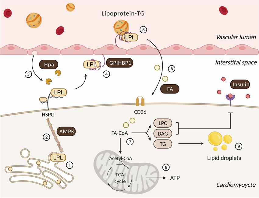
Figure
Figure 1. Regulatory
1. Regulatory processes
processes thatthat influence
influence cardiac
cardiac lipoprotein
lipoprotein lipase
lipase action
action andand
thethe utilization
utilization of delivered
of delivered FAs.FAs. Fol-
Following
lowing its synthesis and activation in cardiomyocytes (1), the actin cytoskeleton traffics LPL to myocyte
its synthesis and activation in cardiomyocytes (1), the actin cytoskeleton traffics LPL to myocyte cell surface HSPG under cell surface HSPG
under the control of AMPK (2). Onward progress is facilitated by heparanase (Hpa) secreted from the endothelial cell that
the control of AMPK (2). Onward progress is facilitated by heparanase (Hpa) secreted from the endothelial cell that cleaves
cleaves HSPG side chains to liberate LPL (3). GPIHBP1 captures this interstitial LPL and relocates it from the basolateral
HSPG side
to the chains
apical sidetoofliberate LPLcell
endothelial (3).(4).
GPIHBP1 capturesa this
At this location, interstitial
GPIHBP1 LPLand
platform and relocates
LPL enablesitlipoprotein-TG
from the basolateral to the
hydrolysis
apical
to generate FAs (5) that are delivered to the cardiomyocyte (6). In the cardiomyocyte, FAs have two major fates (7). They to
side of endothelial cell (4). At this location, a GPIHBP1 platform and LPL enables lipoprotein-TG hydrolysis
generate FAs undergo
can either (5) that are delivered to
mitochondrial the cardiomyocyte
beta-oxidation (6). In the
and oxidative cardiomyocyte,
phosphorylation FAs have
to generate ATPtwo(8)major fates (7). They
or accumulate as
lipid metabolites/droplets (9). Lipid intermediates are known to effect insulin signaling and
can either undergo mitochondrial beta-oxidation and oxidative phosphorylation to generate ATP (8) or accumulate as substrate utilization. LPC:
lysophosphatidylcholine;
lipid metabolites/droplets (9). DAG: diacylglycerol.
Lipid intermediates are known to effect insulin signaling and substrate utilization. LPC:
lysophosphatidylcholine; DAG: diacylglycerol.
Subsequent to its synthesis, LPL is secreted onto heparan sulphate proteoglycan
(HSPG) binding sites
Subsequent to itsonsynthesis,
the cardiomyocyte apical surface
LPL is secreted [9]. At this
onto heparan location,proteoglycan
sulphate positively
charged LPL is attached (by ionic interaction) to negatively charged heparan sulfate
(HSPG) binding sites on the cardiomyocyte apical surface [9]. At this location, positively (HS)
charged LPL is attached (by ionic interaction) to negatively charged heparan sulfatethe
side chains of HSPG. This is an effective arrangement, as this pool of LPL provides (HS)
heart
side with aofrapidly
chains HSPG.accessible reservoir.
This is an effectiveInarrangement,
this way, the immediate
as this pooldemand
of LPLfor FAs canthe
provides
be resolved, not by attempting to synthesize more LPL, but by simply translocating the
heart with a rapidly accessible reservoir. In this way, the immediate demand for FAs can
enzyme that has already been produced [10,11]. To reach the vascular lumen, LPL requires
be resolved, not by attempting to synthesize more LPL, but by simply translocating the
detachment from HSPG and navigation across the interstitial space, made possible by gly-
enzyme that has already been produced [10,11]. To reach the vascular lumen, LPL requires
cosylphosphatidylinositol-anchored high-density lipoprotein-binding protein 1
detachment from HSPG and navigation across the interstitial space, made possible by
(GPIHBP1) [12]. GPIHBP1 is a glycoprotein abundantly expressed in the heart, exclusively
glycosylphosphatidylinositol-anchored high-density lipoprotein-binding protein 1 (GPI-
in capillary ECs [13,14]. Intriguingly, on either side of these cells, GPIHBP1 accomplishes
HBP1) [12]. GPIHBP1 is a glycoprotein abundantly expressed in the heart, exclusively in
different functions. At its basolateral side, GPIHBP1 operates as a transporter, collecting
capillary ECs [13,14]. Intriguingly, on either side of these cells, GPIHBP1 accomplishes
LPL from the interstitial spaces surrounding myocytes and shuttling it across the ECs to
different functions. At its basolateral side, GPIHBP1 operates as a transporter, collecting
LPL from the interstitial spaces surrounding myocytes and shuttling it across the ECs to the
capillary lumen [15]. On the apical side of the Ecs, the ability of GPIHBP1 to avidly bindBiomolecules 2021, 11, 1016 3 of 11
both lipoprotein-TG and LPL allows it to serve as a platform for the lipolytic processing of
TG, which releases FAs for use by cardiomyocytes [15–17]. It should be noted that, related
to the function of LPL, its action on chylomicron-TG clearance is likely more significant
than its effect on very low-density lipoprotein (VLDL)-TG hydrolysis. Chylomicrons are
larger in size, have a lower density and a greater amount of TG. This increases the chance of
this lipoprotein particle interacting with LPL at the coronary vascular lumen [18]. An addi-
tional difference between the two lipoproteins and LPL action is that lipolysis of VLDL-TG
produces FAs that are consequently transported into cells via CD36, an FAs transporter,
whereas hydrolysis of chylomicrons generates a greater local concentration of FAs that
enter cells by passive flip-flop and/or non-CD36-mediated transport mechanisms [19].
3. Posttranslational Processes That Regulate Cardiac LPL
The cardiac muscle fulfills its energy demand by preferentially using FAs. To obtain
this substrate, the heart would prefer mechanisms to control its own delivery of FAs, rather
than depending on FAs from other organs, similar to adipose tissue. In this regard, the
heart makes use of LPL-derived FAs to provide itself with a substantial amount of energy.
Multiple regulators exist that oversee cardiac LPL action (Figure 1), and include:
3.1. AMP-Activate Protein Kinase
AMP-activated protein kinase (AMPK) is a cellular energy sensor that is activated
upon energy deprivation. Upon activation, AMPK modulates downstream effectors via
its kinase activity to augment ATP production. One mechanism by which this is achieved
is through its inhibition of acetyl-CoA carboxylase (ACC) [20]. As ACC is responsible
for the generation of malonyl-CoA, a key intermediate metabolite that blocks carnitine
palmitoyltransferase-1 (CPT-1), and thus the transport of FAs into the mitochondria, AMPK
activation facilitates the oxidation of FAs [1]. AMPK activation has also been implicated
in the delivery of FAs to cardiomyocytes through its regulation of the FAs transporter
CD36 [21]. Results from our laboratory have demonstrated a strong correlation between
the phosphorylation of AMPK and increases in coronary lumen LPL activity [22]. Specifi-
cally, by phosphorylating heat shock protein 25 (Hsp25), AMPK causes the dissociation
of Hsp25 from actin monomers, leading to actin cytoskeleton polymerization and the
transport of LPL-containing vesicles from the Golgi to the plasma membrane. In this
way, intracellular LPL is moved to the cardiomyocyte surface and eventually the vascular
lumen [3]. Given the role of AMPK as a key signaling molecule, conditions that modulate
AMPK are associated with switching on coronary LPL activity. For example, during fasting
with limited carbohydrate availability, the activation of AMPK, and thus augmented LPL,
is an adaptation that would ensure adequate cardiac energy [23]. AMPK is also activated
in response to acute hypoinsulinemia in moderately diabetic rats, possibly because im-
paired glucose utilization causes energy deficiency. In these animals, this resulted in higher
coronary LPL activity, which was responsible for changing the substrate focus of the heart
to predominantly using FAs [9]. It should be noted that severe diabetes introduces an
additional metabolic manifestation in these animals, hyperlipidemia [24], that is known to
inhibit AMPK in the heart [25]. This is followed by a corresponding reduction in coronary
LPL activity [26]. The above evidence indicates that AMPK activation contributes towards
LPL trafficking, and thus the cardiac utilization of FAs.
3.2. Heparanase
Heparanase is an endoglycosidase that is synthesized in ECs as an inactive, latent
65 kDa enzyme (L-Hpa) that undergoes cellular secretion followed by HSPG-facilitated
reuptake [27,28]. After proteolytic cleavage in lysosomes, a 50 kDa polypeptide is formed
that is at least 100-fold more active (A-Hpa) than L-Hpa [29,30]. Lysosomes store A-Hpa
until they are mobilized by different stimuli. In normal physiology, heparanase has a
function in embryonic morphogenesis, wound healing and hair growth [31]. Our lab was
the first to describe a unique responsibility of heparanase in cardiac metabolism; releasingBiomolecules 2021, 11, 1016 4 of 11
myocyte LPL for forward movement to the vascular lumen to provide the heart with
FAs [32]. In conditions of pathology, such as Type 2 diabetes, high levels of heparanase have
been reported in plasma and urine [33,34]. Using perfused hearts, we established that acute
hyperglycemia had a robust influence on heparanase secretion into the interstitial space [35].
We also discovered that incubating ECs in high glucose conditions triggered lysosomal A-
Hpa to be released into the medium [36]. In high glucose conditions, there is a redistribution
of lysosomal heparanase from a perinuclear location towards the plasma membrane of
ECs. ATP release, purinergic receptor activation, cortical actin disassembly and stress actin
formation were essential for high glucose-induced heparanase secretion [35]. These studies
fixated on the effects of high glucose on A-Hpa secretion, as we incorrectly assumed that
only the HS hydrolyzing ability of heparanase would be successful in releasing myocyte
surface-bound proteins. With additional reasoning, we realized that it would be futile
for a cell to secrete an inactive protein for subsequent reuptake and activation unless
the enzymatically inactive L-Hpa also serves a function. Intriguingly, L-Hpa has some
remarkable properties, including its ability to mediate HSPG clustering to activate p38
mitogen-activated protein kinase, Src, PI3K-Akt, and RhoA [32,37–40]. This transmembrane
signaling could then reload the cardiomyocyte surface pool of LPL that was liberated by
A-Hpa. Overall, heparanase plays an important role in EC–cardiomyocyte crosstalk to
affect LPL translocation, and thus the delivery of FAs to the heart.
3.3. GPIHBP1
GPIHBP1 is expressed exclusively in capillary ECs, while LPL is expressed in parenchy-
mal cells such as cardiomyocytes [41]. Given its previously described role in LPL translo-
cation and function, it is not surprising that GPIHBP1-deficient mice developed severe
hyperlipidemia even when fed a low-fat chow diet [15]. Similarly, patients with GPI-
HBP1 mutations have severe hypertriglyceridemia [42]. Related to its structure, GPIHBP1
has a GPI anchor, and thus can be released from the plasma membrane by cleaving off
this anchor with phosphatidylinositol-specific phospholipase C [13]. It is the N-terminal
acidic domain of GPIHBP1 that can electrostatically interact with LPL [43]. In addition to
GPIHBP10 s ability to facilitate EC LPL transport and provide a platform for lipoprotein
hydrolysis, an added function of GPIHBP1 has emerged, whereby it reduces the unfolding
of the catalytic domain of LPL by angiopoietin-like protein 4 (ANGPTL4), consequently
stabilizing LPL activity [44–46]. Evidence that GPIHBP1 expression is regulated came from
experiments investigating the effects of fasting/refeeding [47]. Fasting amplifies cardiac
GPIHBP1 expression, an effect that is reversed 6 h after refeeding [47]. In hearts from
animals with diabetes, GPIHBP1 gene and protein expression increased with exclusive EC
localization [48]. Moreover, the exposure of ECs to high glucose conditions also uncovered
a rapid increase in GPIHBP1 mRNA and protein [41,49]. Interestingly, ECs exposed to re-
combinant L-Hpa showed a substantial time-dependent increase in the GPIHBP1 gene and
protein [41]. As this effect was also duplicated by active heparanase, high glucose, together
with its secretion of heparanase, could be the unforeseen mechanism by which the EC can
increase its expression of GPIHBP1 to augment the supply of FAs following diabetes.
3.4. Fatty Acid
Multiple mechanisms have been suggested to explain the role of FAs in regulating
LPL. These include: (a) the impairment, by FAs, of LPL vesicle trafficking to the myocyte
surface through the caspase-3-mediated cleavage of protein kinase D [50]; (b) suppression,
by FAs, of EC heparanase secretion, thereby reducing the translocation of cardiomyocyte
LPL [35]; (c) displacement of luminal LPL for degradation in the liver by FAs [51]; and
(d) the direct inactivation of LPL by FAs [52]. The idea that LPL can also be inactivated
by the induction of ANGPTL4 by FAs has also been suggested [53–55]. This idea arose
from a study where ANGPTL4 deficiency caused an elevation of post-heparin plasma
LPL activity in mice [56]. ANGPTL4 is widely expressed in various tissues, including
white adipose tissue, liver, heart, and skeletal muscle [54]. In cells from these tissues, itBiomolecules 2021, 11, 1016 5 of 11
forms oligomers through disulfide linkage, and, after secretion, is cleaved at a canonical
proprotein convertase cleavage site that separates the N- and C-terminal domain [57].
The N-terminal coiled-coil domain remains oligomerized, a feature that is essential for its
stability and an LPL inhibitory effect [58]. Historically, the secreted N-terminal oligomers
were thought to act in the circulation, converting dimeric LPL at the vascular lumen into
inactive monomers [53]. More recently, a study has suggested that ANGPTL4 functions
to unfold active LPL monomers, thus reducing LPL activity [59]. This inhibitory function
of ANGPTL4 was not evident for GPIHBP1-stabilized LPL [45,60]. Circulating FAs can
increase ANGPTL4 gene transcription [61,62] and we have also reported that cardiomy-
ocytes exposed to FAs demonstrate higher ANGPTL4 expression [26]. Related to pathology,
severely diabetic animals with hyperlipidemia showed a decline in heparin-releasable
LPL activity at the vascular lumen [26]. This effect was not related to any reduction in
LPL gene expression [63] and implied that the low LPL activity is likely an upshot of
post-translational mechanisms. Strikingly, transcriptome profiling by RNA-seq revealed
that of the 1574 differentially expressed genes in these diabetic hearts, the ANGPTL4 gene
was the one with the highest fold change (~25-fold increase) [24]. At present, the location
where ANGTPL4 inactivates cardiac LPL is yet to be determined. Additionally, similar to
ANGPTL4, ANGPTL3 (in a complex with ANGPTL8) is mainly produced by the liver, and
is also known to influence LPL activity [64]. Collectively, these studies provide us with
clues to how FAs “turn off” vascular LPL, through multiple mechanisms, to avoid lipid
oversupply to the heart.
3.5. Insulin
The unique characteristic of LPL is that it is rapidly responsive to variations in circulat-
ing insulin and does so in a tissue-specific manner. Thus, in tissues such as adipose tissue
and the heart, changes in LPL activity can occur independent of alterations in mRNA [65].
Such changes would be desirable given that, during conditions such as fasting or diabetes,
wherein insulin levels are altered and the utilization of FAs is augmented, a rapid require-
ment for LPL may not be matched by the slow turnover of LPL mRNA. Related to tissue
specificity, the lowering of circulating insulin following fasting decreases LPL activity in
adipose tissue but increases it in the heart [66]. These changes in luminal LPL activity
following fasting were independent of shifts in LPL mRNA or alterations in LPL protein,
suggesting a posttranslational mechanism for this increase [23]. As a result, FAs generated
from circulating TG are diverted away from storage, towards meeting the metabolic de-
mands of cardiomyocytes (it fulfills a “gate-keeping” role by regulating the supply of FAs
to meet tissue requirements). As in perfused guinea pig hearts, newly synthesized LPL
can move from myocytes to the vascular lumen within 30 min. An increased rate of LPL
transfer could explain this effect of fasting on coronary LPL [67]. Mechanistically, because
glucose entry into the cardiomyocyte is largely dependent on insulin action, a fasting- or
diabetes-induced reduction in insulin causes a rise in the intracellular AMP-to-ATP ratio,
with a resultant activation of AMPK [2,68,69]. Once stimulated, AMPK switches off energy-
consuming processes such as protein synthesis, whereas ATP-generating mechanisms, such
as the oxidation of FAs and LPL translocation, are turned on [3].
4. The Consequences of Oscillations in LPL
As indicated, the nutritional regulation of cardiac LPL determines the delivery of FAs
to the heart in order to maintain energy demand [66]. However, oscillations in LPL have
also been linked to a disruption in cardiac metabolism, leading to pathology.
4.1. Gain-and Loss-of-Function of Cardiac LPL
The surplus provision of FAs to tissues other than the adipose tissue can trigger cellu-
lar demise [3,70–72]. Not surprisingly, then, muscle-specific LPL overexpression causes
severe myopathy, characterized by lipid oversupply, the proliferation of mitochondria
and peroxisomes, muscle fiber degeneration, excessive dilatation and impaired ventricularBiomolecules 2021, 11, 1016 6 of 11
function in the absence of vascular defects [73,74]. LPL is also known to play a role in in-
sulin signaling (Figure 1). Hence, transgenic mice with muscle-specific LPL overexpression
exhibited skeletal muscle and whole-body insulin resistance [75], and variations in the LPL
gene have been reported to play a role in determining insulin resistance in Mexican Ameri-
cans [76]. In contrast, the loss of cardiac LPL also causes heart failure [77,78]. Interestingly,
although this increases glucose use, neither this effect, nor albumin-bound FAs, could re-
place the action of LPL, and the cardiac ejection fraction decreased [78]. These experiments
in genetically modified models suggest that disturbing cardiac LPL is sufficient to induce
cardiac failure.
4.2. Fluctuations in Cardiac LPL Following Diabetes
In patients, heparin is used to displace LPL from its binding sites, enabling the
subsequent determination of its plasma activity [11,79]. The drawback with this approach
is that it measures LPL released from all tissues (skeletal muscle, adipose tissue, heart), and
hence is incapable of establishing if diabetes specifically influences cardiac LPL. Regarding
tissue-specific assessment, adipose tissue and skeletal muscle show low levels of LPL in
homogenates following diabetes [80], with virtually no information available on the cardiac
content of this enzyme. Even if tissue specificity can be resolved by using homogenates, the
estimation of cardiac LPL from patients with diabetes would be inappropriate, as it would
reflect total cardiac LPL and not the more pertinent functional pool at the coronary lumen.
Hence, data from animal studies have provided the majority of information regarding
cardiac LPL in diabetes.
Following acute insulin resistance [81,82] or moderate hypoinsulinemia and hyper-
glycemia in rats [9,26,83,84], LPL is “switched on”, causing a robust expansion of heparin-
releasable coronary LPL. We defined a model where only a fraction of the binding sites
at the luminal side of coronary EC are occupied by LPL, and hyperglycemia leads to
rapid filling of the unoccupied sites [65]. The increase in LPL at this location was imme-
diate [65,83,85] and unrelated to gene and protein expression [83,86]. Instead, it occurred
through post-translational mechanisms that increase the transfer of LPL from the under-
lying cardiomyocyte to the apical side of the ECs [87]. Transfer to the coronary lumen
requires the movement of LPL to the cardiomyocyte plasma membrane [65], controlled by
AMPK [23,88], p38 mitogen-activated protein kinase and protein kinase D [50,63,89]. The
activation of these kinases facilitates LPL vesicle formation and cytoskeletal rearrangement
for secretion onto cardiomyocyte cell-surface HSPG [63,90]. For its onward movement,
the detachment of LPL from the cardiomyocyte surface is a prerequisite and is mediated
by the cleavage of HSPG by heparanase [32,35]. In response to high glucose conditions,
ECs secrete heparanase [35,36], thereby instigating cardiomyocyte LPL release [91]. In
addition to LPL, heparanase also functions to release cardiomyocyte vascular endothelial
growth factor A (VEGFA) [41,92] and VEGFB [93], prospective partners that, by modulat-
ing both O2 delivery and inhibiting cell death, can offer safeguards when FAs are being
used disproportionately. Besides severe diabetes [9,26], a decline in LPL has also been
observed in animals infused with Intralipid [94]. As these animals exhibit elevated plasma
FAs, we concluded that the LPL-mediated delivery of FAs would be redundant in these
circumstances, and is “turned off”.
4.3. Lipid Metabolites and Diabetic Cardiomyopathy
In patients and animals with Type 1 and Type 2 diabetes, there is a reduced or low–
normal diastolic function and left-ventricular hypertrophy in the absence of vascular
defects, emblematic of diabetic cardiomyopathy [95–100]. Several factors have been as-
sociated with the development of diabetic cardiomyopathy and include an accumulation
of connective tissue and insoluble collagen, impaired sensitivity to various ligands, mi-
tochondrial dysfunction, ER stress, RAAS activation and abnormalities in proteins that
regulate intracellular Ca2+ [98,101–103]. However, with diabetes and augmentation of the
availability of FAs, either through an increase in vascular LPL or adipose tissue lipolysis,Biomolecules 2021, 11, 1016 7 of 11
the diabetic heart has a mismatch between delivery of FAs and their oxidation, leading
to myocyte lipid droplet synthesis and the accumulation of lipid metabolites (e.g., DAG,
LPC and ceramide). We and others have proposed that this mediates lipid-induced insulin
resistance, cell death and eventually diabetic cardiomyopathy [70–72,104,105]. As the
chronic management of glycemia fluctuates over the duration of diabetes, with frequent
episodes of insufficient or poor control, pathological oscillations in coronary LPL arise
during this disease to disturb cardiac metabolism and initiate heart failure.
5. Conclusions
Following diabetes, the heart shifts to the predominant use of FAs, leading to the
development of diabetic cardiomyopathy. Accordingly, treatment of this cardiac syndrome
will require us to rethink our therapeutic strategies, from a focus on controlling blood
glucose to re-establishing physiological cardiac metabolism. Understanding metabolic
disequilibrium and lipotoxicity will allow the identification of targets for a mechanism-
driven therapeutic intervention to help prevent/delay cardiovascular disease, characteristic
of diabetes [106].
Author Contributions: R.S. and B.R. contributed equally to the writing and editing of this manuscript.
Both authors have read and agreed to the published version of the manuscript.
Funding: This work was supported by operating grants from the Canadian Institutes of Health
Research [CIHR PJT-169212] and the Heart and Stroke Foundation of Canada [G-19-0026493].
Institutional Review Board Statement: Not applicable.
Informed Consent Statement: Not applicable.
Data Availability Statement: Not applicable.
Conflicts of Interest: The authors declare no conflict of interest.
References
1. Lopaschuk, G.D.; Ussher, J.R.; Folmes, C.D.; Jaswal, J.S.; Stanley, W.C. Myocardial fatty acid metabolism in health and disease.
Physiol. Rev. 2010, 90, 207–258. [CrossRef] [PubMed]
2. An, D.; Rodrigues, B. Role of changes in cardiac metabolism in development of diabetic cardiomyopathy. Am. J. Physiol. Heart
Circ. Physiol. 2006, 291, H1489–H1506. [CrossRef] [PubMed]
3. Kim, M.S.; Wang, Y.; Rodrigues, B. Lipoprotein lipase mediated fatty acid delivery and its impact in diabetic cardiomyopathy.
Biochim. Biophys. Acta 2012, 1821, 800–808. [CrossRef]
4. Camps, L.; Reina, M.; Llobera, M.; Vilaro, S.; Olivecrona, T. Lipoprotein lipase: Cellular origin and functional distribution. Am. J.
Physiol. 1990, 258, C673–C681. [CrossRef] [PubMed]
5. Blanchette-Mackie, E.J.; Masuno, H.; Dwyer, N.K.; Olivecrona, T.; Scow, R.O. Lipoprotein lipase in myocytes and capillary
endothelium of heart: Immunocytochemical study. Am. J. Physiol. 1989, 256, E818–E828. [CrossRef] [PubMed]
6. Braun, J.E.; Severson, D.L. Regulation of the synthesis, processing and translocation of lipoprotein lipase. Biochem. J. 1992, 287 Pt
2, 337–347. [CrossRef]
7. Auwerx, J.; Leroy, P.; Schoonjans, K. Lipoprotein lipase: Recent contributions from molecular biology. Crit. Rev. Clin. Lab. Sci.
1992, 29, 243–268. [CrossRef] [PubMed]
8. Beigneux, A.P.; Allan, C.M.; Sandoval, N.P.; Cho, G.W.; Heizer, P.J.; Jung, R.S.; Stanhope, K.L.; Havel, P.J.; Birrane, G.; Meiyappan,
M.; et al. Lipoprotein lipase is active as a monomer. Proc. Natl. Acad. Sci. USA 2019, 116, 6319–6328. [CrossRef] [PubMed]
9. Rodrigues, B.; Cam, M.C.; Jian, K.; Lim, F.; Sambandam, N.; Shepherd, G. Differential effects of streptozotocin-induced diabetes
on cardiac lipoprotein lipase activity. Diabetes 1997, 46, 1346–1353. [CrossRef] [PubMed]
10. Enerback, S.; Gimble, J.M. Lipoprotein lipase gene expression: Physiological regulators at the transcriptional and post-
transcriptional level. Biochim. Biophys. Acta 1993, 1169, 107–125. [CrossRef]
11. Eckel, R.H. Lipoprotein lipase. A multifunctional enzyme relevant to common metabolic diseases. N. Engl. J. Med. 1989, 320,
1060–1068. [CrossRef]
12. Allan, C.M.; Larsson, M.; Jung, R.S.; Ploug, M.; Bensadoun, A.; Beigneux, A.P.; Fong, L.G.; Young, S.G. Mobility of “HSPG-bound”
LPL explains how LPL is able to reach GPIHBP1 on capillaries. J. Lipid Res. 2017, 58, 216–225. [CrossRef]
13. Ioka, R.X.; Kang, M.J.; Kamiyama, S.; Kim, D.H.; Magoori, K.; Kamataki, A.; Ito, Y.; Takei, Y.A.; Sasaki, M.; Suzuki, T.; et al.
Expression cloning and characterization of a novel glycosylphosphatidylinositol-anchored high density lipoprotein-binding
protein, GPI-HBP1. J. Biol. Chem. 2003, 278, 7344–7349. [CrossRef] [PubMed]Biomolecules 2021, 11, 1016 8 of 11
14. Young, S.G.; Davies, B.S.; Voss, C.V.; Gin, P.; Weinstein, M.M.; Tontonoz, P.; Reue, K.; Bensadoun, A.; Fong, L.G.; Beigneux, A.P.
GPIHBP1, an endothelial cell transporter for lipoprotein lipase. J. Lipid Res. 2011, 52, 1869–1884. [CrossRef] [PubMed]
15. Davies, B.S.; Beigneux, A.P.; Barnes, R.H., 2nd; Tu, Y.; Gin, P.; Weinstein, M.M.; Nobumori, C.; Nyren, R.; Goldberg, I.; Olivecrona,
G.; et al. GPIHBP1 is responsible for the entry of lipoprotein lipase into capillaries. Cell Metab. 2010, 12, 42–52. [CrossRef]
[PubMed]
16. Weinstein, M.M.; Yin, L.; Tu, Y.; Wang, X.; Wu, X.; Castellani, L.W.; Walzem, R.L.; Lusis, A.J.; Fong, L.G.; Beigneux, A.P.; et al.
Chylomicronemia elicits atherosclerosis in mice–brief report. Arterioscler. Thromb. Vasc. Biol. 2010, 30, 20–23. [CrossRef] [PubMed]
17. Young, S.G.; Davies, B.S.; Fong, L.G.; Gin, P.; Weinstein, M.M.; Bensadoun, A.; Beigneux, A.P. GPIHBP1: An endothelial cell
molecule important for the lipolytic processing of chylomicrons. Curr. Opin. Lipidol. 2007, 18, 389–396. [CrossRef] [PubMed]
18. Goldberg, I.J.; Eckel, R.H.; McPherson, R. Triglycerides and heart disease: Still a hypothesis? Arterioscler. Thromb. Vasc. Biol. 2011,
31, 1716–1725. [CrossRef]
19. Bharadwaj, K.G.; Hiyama, Y.; Hu, Y.; Huggins, L.A.; Ramakrishnan, R.; Abumrad, N.A.; Shulman, G.I.; Blaner, W.S.; Goldberg,
I.J. Chylomicron- and VLDL-derived lipids enter the heart through different pathways: In vivo evidence for receptor- and
non-receptor-mediated fatty acid uptake. J. Biol. Chem. 2010, 285, 37976–37986. [CrossRef] [PubMed]
20. Kudo, N.; Barr, A.J.; Barr, R.L.; Desai, S.; Lopaschuk, G.D. High rates of fatty acid oxidation during reperfusion of ischemic
hearts are associated with a decrease in malonyl-CoA levels due to an increase in 5’-AMP-activated protein kinase inhibition of
acetyl-CoA carboxylase. J. Biol. Chem. 1995, 270, 17513–17520. [CrossRef] [PubMed]
21. Luiken, J.J.; Coort, S.L.; Willems, J.; Coumans, W.A.; Bonen, A.; van der Vusse, G.J.; Glatz, J.F. Contraction-induced fatty acid
translocase/CD36 translocation in rat cardiac myocytes is mediated through AMP-activated protein kinase signaling. Diabetes
2003, 52, 1627–1634. [CrossRef] [PubMed]
22. An, D.; Kewalramani, G.; Qi, D.; Pulinilkunnil, T.; Ghosh, S.; Abrahani, A.; Wambolt, R.; Allard, M.; Innis, S.M.; Rodrigues, B.
beta-Agonist stimulation produces changes in cardiac AMPK and coronary lumen LPL only during increased workload. Am. J.
Physiol. Endocrinol. Metab. 2005, 288, E1120–E1127. [CrossRef] [PubMed]
23. An, D.; Pulinilkunnil, T.; Qi, D.; Ghosh, S.; Abrahani, A.; Rodrigues, B. The metabolic “switch” AMPK regulates cardiac
heparin-releasable lipoprotein lipase. Am. J. Physiology. Endocrinol. Metab. 2005, 288, E246–E253. [CrossRef]
24. Puri, K.; Lal, N.; Shang, R.; Ghosh, S.; Flibotte, S.; Dyer, R.; Hussein, B.; Rodrigues, B. Diabetes mellitus severity and a switch
from using lipoprotein lipase to adipose-derived fatty acid results in a cardiac metabolic signature that embraces cell death. J. Am.
Heart Assoc. 2019, 8, e014022. [CrossRef] [PubMed]
25. Wu, Y.; Song, P.; Xu, J.; Zhang, M.; Zou, M.H. Activation of protein phosphatase 2A by palmitate inhibits AMP-activated protein
kinase. J. Biol. Chem. 2007, 282, 9777–9788. [CrossRef]
26. Wang, Y.; Puthanveetil, P.; Wang, F.; Kim, M.S.; Abrahani, A.; Rodrigues, B. Severity of diabetes governs vascular lipoprotein
lipase by affecting enzyme dimerization and disassembly. Diabetes 2011, 60, 2041–2050. [CrossRef] [PubMed]
27. Fairbanks, M.B.; Mildner, A.M.; Leone, J.W.; Cavey, G.S.; Mathews, W.R.; Drong, R.F.; Slightom, J.L.; Bienkowski, M.J.; Smith,
C.W.; Bannow, C.A.; et al. Processing of the human heparanase precursor and evidence that the active enzyme is a heterodimer. J.
Biol. Chem. 1999, 274, 29587–29590. [CrossRef] [PubMed]
28. Gingis-Velitski, S.; Zetser, A.; Kaplan, V.; Ben-Zaken, O.; Cohen, E.; Levy-Adam, F.; Bashenko, Y.; Flugelman, M.Y.; Vlodavsky, I.;
Ilan, N. Heparanase uptake is mediated by cell membrane heparan sulfate proteoglycans. J. Biol. Chem. 2004, 279, 44084–44092.
[CrossRef] [PubMed]
29. Pikas, D.S.; Li, J.P.; Vlodavsky, I.; Lindahl, U. Substrate specificity of heparanases from human hepatoma and platelets. J. Biol.
Chem. 1998, 273, 18770–18777. [CrossRef]
30. Abboud-Jarrous, G.; Rangini-Guetta, Z.; Aingorn, H.; Atzmon, R.; Elgavish, S.; Peretz, T.; Vlodavsky, I. Site-directed mutagenesis,
proteolytic cleavage, and activation of human proheparanase. J. Biol. Chem. 2005, 280, 13568–13575. [CrossRef] [PubMed]
31. Zcharia, E.; Metzger, S.; Chajek-Shaul, T.; Aingorn, H.; Elkin, M.; Friedmann, Y.; Weinstein, T.; Li, J.P.; Lindahl, U.; Vlodavsky, I.
Transgenic expression of mammalian heparanase uncovers physiological functions of heparan sulfate in tissue morphogenesis,
vascularization, and feeding behavior. FASEB J. 2004, 18, 252–263. [CrossRef] [PubMed]
32. Wang, Y.; Zhang, D.; Chiu, A.P.; Wan, A.; Neumaier, K.; Vlodavsky, I.; Rodrigues, B. Endothelial heparanase regulates heart
metabolism by stimulating lipoprotein lipase secretion from cardiomyocytes. Arterioscler. Thromb. Vasc. Biol. 2013, 33, 894–902.
[CrossRef] [PubMed]
33. Shafat, I.; Ilan, N.; Zoabi, S.; Vlodavsky, I.; Nakhoul, F. Heparanase levels are elevated in the urine and plasma of type 2 diabetes
patients and associate with blood glucose levels. PLoS ONE 2011, 6. [CrossRef] [PubMed]
34. Katz, A.; Van-Dijk, D.J.; Aingorn, H.; Erman, A.; Davies, M.; Darmon, D.; Hurvitz, H.; Vlodavsky, I. Involvement of human
heparanase in the pathogenesis of diabetic nephropathy. Isr. Med Assoc. J. 2002, 4, 996–1002.
35. Wang, F.; Kim, M.S.; Puthanveetil, P.; Kewalramani, G.; Deppe, S.; Ghosh, S.; Abrahani, A.; Rodrigues, B. Endothelial heparanase
secretion after acute hypoinsulinemia is regulated by glucose and fatty acid. Am. J. Physiol. Heart Circ. Physiol. 2009, 296,
H1108–H1116. [CrossRef] [PubMed]
36. Wang, F.; Wang, Y.; Kim, M.S.; Puthanveetil, P.; Ghosh, S.; Luciani, D.S.; Johnson, J.D.; Abrahani, A.; Rodrigues, B. Glucose-
induced endothelial heparanase secretion requires cortical and stress actin reorganization. Cardiovasc. Res. 2010, 87, 127–136.
[CrossRef] [PubMed]Biomolecules 2021, 11, 1016 9 of 11
37. Fux, L.; Ilan, N.; Sanderson, R.D.; Vlodavsky, I. Heparanase: Busy at the cell surface. Trends Biochem. Sci. 2009, 34, 511–519.
[CrossRef] [PubMed]
38. Gingis-Velitski, S.; Zetser, A.; Flugelman, M.Y.; Vlodavsky, I.; Ilan, N. Heparanase induces endothelial cell migration via protein
kinase B/Akt activation. J. Biol. Chem. 2004, 279, 23536–23541. [CrossRef] [PubMed]
39. Cui, H.X.; Shao, C.H.; Liu, Q.; Yu, W.J.; Fang, J.P.; Yu, W.S.; Ali, A.; Ding, K. Heparanase enhances nerve-growth-factor-induced
PC12 cell neuritogenesis via the p38 MAPK pathway. Biochem. J. 2011, 440, 273–282. [CrossRef] [PubMed]
40. Riaz, A.; Ilan, N.; Vlodavsky, I.; Li, J.P.; Johansson, S. Characterization of heparanase-induced phosphatidylinositol 3-kinase-AKT
activation and its integrin dependence. J. Biol. Chem. 2013, 288, 12366–12375. [CrossRef]
41. Chiu, A.P.; Wan, A.; Lal, N.; Zhang, D.; Wang, F.; Vlodavsky, I.; Hussein, B.; Rodrigues, B. Cardiomyocyte VEGF regulates
endothelial cell GPIHBP1 to relocate lipoprotein lipase to the coronary lumen during diabetes mellitus. Arterioscler. Thromb. Vasc.
Biol. 2016, 36, 145–155. [CrossRef] [PubMed]
42. Beigneux, A.P.; Davies, B.S.; Gin, P.; Weinstein, M.M.; Farber, E.; Qiao, X.; Peale, F.; Bunting, S.; Walzem, R.L.; Wong, J.S.; et al.
Glycosylphosphatidylinositol-anchored high-density lipoprotein-binding protein 1 plays a critical role in the lipolytic processing
of chylomicrons. Cell Metab. 2007, 5, 279–291. [CrossRef] [PubMed]
43. Gin, P.; Yin, L.; Davies, B.S.; Weinstein, M.M.; Ryan, R.O.; Bensadoun, A.; Fong, L.G.; Young, S.G.; Beigneux, A.P. The acidic
domain of GPIHBP1 is important for the binding of lipoprotein lipase and chylomicrons. J. Biol. Chem. 2008, 283, 29554–29562.
[CrossRef]
44. Kristensen, K.K.; Midtgaard, S.R.; Mysling, S.; Kovrov, O.; Hansen, L.B.; Skar-Gislinge, N.; Beigneux, A.P.; Kragelund, B.B.;
Olivecrona, G.; Young, S.G.; et al. A disordered acidic domain in GPIHBP1 harboring a sulfated tyrosine regulates lipoprotein
lipase. Proc. Natl. Acad. Sci. USA 2018, 115, E6020–E6029. [CrossRef]
45. Mysling, S.; Kristensen, K.K.; Larsson, M.; Kovrov, O.; Bensadouen, A.; Jorgensen, T.J.; Olivecrona, G.; Young, S.G.; Ploug, M.
The angiopoietin-like protein ANGPTL4 catalyzes unfolding of the hydrolase domain in lipoprotein lipase and the endothelial
membrane protein GPIHBP1 counteracts this unfolding. eLife 2016, 5. [CrossRef] [PubMed]
46. Fong, L.G.; Young, S.G.; Beigneux, A.P.; Bensadoun, A.; Oberer, M.; Jiang, H.; Ploug, M. GPIHBP1 and plasma triglyceride
metabolism. Trends Endocrinol. Metab. 2016, 27, 455–469. [CrossRef] [PubMed]
47. Kroupa, O.; Vorrsjo, E.; Stienstra, R.; Mattijssen, F.; Nilsson, S.K.; Sukonina, V.; Kersten, S.; Olivecrona, G.; Olivecrona, T. Linking
nutritional regulation of Angptl4, Gpihbp1, and Lmf1 to lipoprotein lipase activity in rodent adipose tissue. BMC Physiol. 2012,
12, 13. [CrossRef] [PubMed]
48. Pei-Ling Chiu, A.; Wang, F.; Lal, N.; Wang, Y.; Zhang, D.; Hussein, B.; Wan, A.; Vlodavsky, I.; Rodrigues, B. Endothelial cells
respond to hyperglycemia by increasing the LPL transporter GPIHBP1. Am. J. Physiol. Endocrinol. Metab. 2014, 306, E1274–E1283.
[CrossRef] [PubMed]
49. Chiu, A.P.; Bierende, D.; Lal, N.; Wang, F.; Wan, A.; Vlodavsky, I.; Hussein, B.; Rodrigues, B. Dual effects of hyperglycemia on
endothelial cells and cardiomyocytes to enhance coronary LPL activity. Am. J. Physiol. Heart Circ. Physiol. 2018, 314, H82–H94.
[CrossRef] [PubMed]
50. Kim, M.S.; Wang, F.; Puthanveetil, P.; Kewalramani, G.; Innis, S.; Marzban, L.; Steinberg, S.F.; Webber, T.D.; Kieffer, T.J.; Abrahani,
A.; et al. Cleavage of protein kinase D after acute hypoinsulinemia prevents excessive lipoprotein lipase-mediated cardiac
triglyceride accumulation. Diabetes 2009, 58, 2464–2475. [CrossRef]
51. Peterson, J.; Bihain, B.E.; Bengtsson-Olivecrona, G.; Deckelbaum, R.J.; Carpentier, Y.A.; Olivecrona, T. Fatty acid control of
lipoprotein lipase: A link between energy metabolism and lipid transport. Proc. Natl. Acad. Sci. USA 1990, 87, 909–913. [CrossRef]
52. Bengtsson, G.; Olivecrona, T. Lipoprotein lipase. Mechanism of product inhibition. Eur. J. Biochem. 1980, 106, 557–562. [CrossRef]
[PubMed]
53. Sukonina, V.; Lookene, A.; Olivecrona, T.; Olivecrona, G. Angiopoietin-like protein 4 converts lipoprotein lipase to inactive
monomers and modulates lipase activity in adipose tissue. Proc. Natl. Acad. Sci. USA 2006, 103, 17450–17455. [CrossRef]
54. Yu, X.; Burgess, S.C.; Ge, H.; Wong, K.K.; Nassem, R.H.; Garry, D.J.; Sherry, A.D.; Malloy, C.R.; Berger, J.P.; Li, C. Inhibition
of cardiac lipoprotein utilization by transgenic overexpression of Angptl4 in the heart. Proc. Natl. Acad. Sci. USA 2005, 102,
1767–1772. [CrossRef] [PubMed]
55. Lafferty, M.J.; Bradford, K.C.; Erie, D.A.; Neher, S.B. Angiopoietin-like protein 4 inhibition of lipoprotein lipase: Evidence for
reversible complex formation. J. Biol. Chem. 2013, 288, 28524–28534. [CrossRef] [PubMed]
56. Koster, A.; Chao, Y.B.; Mosior, M.; Ford, A.; Gonzalez-DeWhitt, P.A.; Hale, J.E.; Li, D.; Qiu, Y.; Fraser, C.C.; Yang, D.D.; et al.
Transgenic angiopoietin-like (angptl)4 overexpression and targeted disruption of angptl4 and angptl3: Regulation of triglyceride
metabolism. Endocrinology 2005, 146, 4943–4950. [CrossRef] [PubMed]
57. Ge, H.; Yang, G.; Huang, L.; Motola, D.L.; Pourbahrami, T.; Li, C. Oligomerization and regulated proteolytic processing of
angiopoietin-like protein 4. J. Biol. Chem. 2004, 279, 2038–2045. [CrossRef]
58. Yin, W.; Romeo, S.; Chang, S.; Grishin, N.V.; Hobbs, H.H.; Cohen, J.C. Genetic variation in ANGPTL4 provides insights into
protein processing and function. J. Biol. Chem. 2009, 284, 13213–13222. [CrossRef]
59. Kristensen, K.K.; Leth-Espensen, K.Z.; Mertens, H.D.T.; Birrane, G.; Meiyappan, M.; Olivecrona, G.; Jorgensen, T.J.D.; Young,
S.G.; Ploug, M. Unfolding of monomeric lipoprotein lipase by ANGPTL4: Insight into the regulation of plasma triglyceride
metabolism. Proc. Natl. Acad. Sci. USA 2020, 117, 4337–4346. [CrossRef] [PubMed]Biomolecules 2021, 11, 1016 10 of 11
60. Mysling, S.; Kristensen, K.K.; Larsson, M.; Beigneux, A.P.; Gardsvoll, H.; Fong, L.G.; Bensadouen, A.; Jorgensen, T.J.; Young, S.G.;
Ploug, M. The acidic domain of the endothelial membrane protein GPIHBP1 stabilizes lipoprotein lipase activity by preventing
unfolding of its catalytic domain. eLife 2016, 5, e12095. [CrossRef] [PubMed]
61. Georgiadi, A.; Lichtenstein, L.; Degenhardt, T.; Boekschoten, M.V.; van Bilsen, M.; Desvergne, B.; Muller, M.; Kersten, S. Induction
of cardiac Angptl4 by dietary fatty acids is mediated by peroxisome proliferator-activated receptor beta/delta and protects
against fatty acid-induced oxidative stress. Circ. Res. 2010, 106, 1712–1721. [CrossRef] [PubMed]
62. Staiger, H.; Haas, C.; Machann, J.; Werner, R.; Weisser, M.; Schick, F.; Machicao, F.; Stefan, N.; Fritsche, A.; Haring, H.U. Muscle-
derived angiopoietin-like protein 4 is induced by fatty acids via peroxisome proliferator-activated receptor (PPAR)-delta and is of
metabolic relevance in humans. Diabetes 2009, 58, 579–589. [CrossRef] [PubMed]
63. Kim, M.S.; Kewalramani, G.; Puthanveetil, P.; Lee, V.; Kumar, U.; An, D.; Abrahani, A.; Rodrigues, B. Acute diabetes moderates
trafficking of cardiac lipoprotein lipase through p38 mitogen-activated protein kinase-dependent actin cytoskeleton organization.
Diabetes 2008, 57, 64–76. [CrossRef]
64. Haller, J.F.; Mintah, I.J.; Shihanian, L.M.; Stevis, P.; Buckler, D.; Alexa-Braun, C.A.; Kleiner, S.; Banfi, S.; Cohen, J.C.; Hobbs,
H.H.; et al. ANGPTL8 requires ANGPTL3 to inhibit lipoprotein lipase and plasma triglyceride clearance. J. Lipid Res. 2017, 58,
1166–1173. [CrossRef]
65. Pulinilkunnil, T.; Abrahani, A.; Varghese, J.; Chan, N.; Tang, I.; Ghosh, S.; Kulpa, J.; Allard, M.; Brownsey, R.; Rodrigues, B.
Evidence for rapid "metabolic switching" through lipoprotein lipase occupation of endothelial-binding sites. J. Mol. Cell. Cardiol.
2003, 35, 1093–1103. [CrossRef]
66. Doolittle, M.H.; Ben-Zeev, O.; Elovson, J.; Martin, D.; Kirchgessner, T.G. The response of lipoprotein lipase to feeding and fasting.
Evidence for posttranslational regulation. J. Biol. Chem. 1990, 265, 4570–4577. [CrossRef]
67. Liu, G.; Olivecrona, T. Synthesis and transport of lipoprotein lipase in perfused guinea pig hearts. Am. J. Physiol. 1992, 263,
H438–H446. [CrossRef]
68. Hardie, D.G. Minireview: The AMP-activated protein kinase cascade: The key sensor of cellular energy status. Endocrinology
2003, 144, 5179–5183. [CrossRef]
69. Hardie, D.G.; Carling, D. The AMP-activated protein kinase–fuel gauge of the mammalian cell? Eur. J. Biochem. 1997, 246, 259–273.
[CrossRef]
70. Borradaile, N.M.; Schaffer, J.E. Lipotoxicity in the heart. Curr. Hypertens. Rep. 2005, 7, 412–417. [CrossRef] [PubMed]
71. van de Weijer, T.; Schrauwen-Hinderling, V.B.; Schrauwen, P. Lipotoxicity in type 2 diabetic cardiomyopathy. Cardiovasc. Res.
2011, 92, 10–18. [CrossRef] [PubMed]
72. Wende, A.R.; Symons, J.D.; Abel, E.D. Mechanisms of lipotoxicity in the cardiovascular system. Curr. Hypertens. Rep. 2012, 14,
517–531. [CrossRef] [PubMed]
73. Levak-Frank, S.; Radner, H.; Walsh, A.; Stollberger, R.; Knipping, G.; Hoefler, G.; Sattler, W.; Weinstock, P.H.; Breslow, J.L.;
Zechner, R. Muscle-specific overexpression of lipoprotein lipase causes a severe myopathy characterized by proliferation of
mitochondria and peroxisomes in transgenic mice. J. Clin. Investig. 1995, 96, 976–986. [CrossRef]
74. Yagyu, H.; Chen, G.; Yokoyama, M.; Hirata, K.; Augustus, A.; Kako, Y.; Seo, T.; Hu, Y.; Lutz, E.P.; Merkel, M.; et al. Lipoprotein
lipase (LpL) on the surface of cardiomyocytes increases lipid uptake and produces a cardiomyopathy. J. Clin. Investig. 2003, 111,
419–426. [CrossRef] [PubMed]
75. Pulawa, L.K.; Eckel, R.H. Overexpression of muscle lipoprotein lipase and insulin sensitivity. Curr. Opin. Clin. Nutr. Metab. Care
2002, 5, 569–574. [CrossRef] [PubMed]
76. Goodarzi, M.O.; Guo, X.; Taylor, K.D.; Quinones, M.J.; Saad, M.F.; Yang, H.; Hsueh, W.A.; Rotter, J.I. Lipoprotein lipase is a gene
for insulin resistance in Mexican Americans. Diabetes 2004, 53, 214–220. [CrossRef]
77. Noh, H.L.; Okajima, K.; Molkentin, J.D.; Homma, S.; Goldberg, I.J. Acute lipoprotein lipase deletion in adult mice leads to
dyslipidemia and cardiac dysfunction. Am. J. Physiol. Endocrinol. Metab. 2006, 291, E755–E760. [CrossRef]
78. Augustus, A.S.; Buchanan, J.; Park, T.S.; Hirata, K.; Noh, H.L.; Sun, J.; Homma, S.; D’Armiento, J.; Abel, E.D.; Goldberg, I.J. Loss
of lipoprotein lipase-derived fatty acids leads to increased cardiac glucose metabolism and heart dysfunction. J. Biol. Chem. 2006,
281, 8716–8723. [CrossRef]
79. Kashiwazaki, K.; Hirano, T.; Yoshino, G.; Kurokawa, M.; Tajima, H.; Adachi, M. Decreased release of lipoprotein lipase is
associated with vascular endothelial damage in NIDDM patients with microalbuminuria. Diabetes Care 1998, 21, 2016–2020.
[CrossRef]
80. Taskinen, M.R.; Nikkila, E.A. Lipoprotein lipase activity of adipose tissue and skeletal muscle in insulin-deficient human diabetes.
Relation to high-density and very-low-density lipoproteins and response to treatment. Diabetologia 1979, 17, 351–356. [CrossRef]
81. Qi, D.; Pulinilkunnil, T.; An, D.; Ghosh, S.; Abrahani, A.; Pospisilik, J.A.; Brownsey, R.; Wambolt, R.; Allard, M.; Rodrigues, B.
Single-dose dexamethasone induces whole-body insulin resistance and alters both cardiac fatty acid and carbohydrate metabolism.
Diabetes 2004, 53, 1790–1797. [CrossRef] [PubMed]
82. Kewalramani, G.; Puthanveetil, P.; Kim, M.S.; Wang, F.; Lee, V.; Hau, N.; Beheshti, E.; Ng, N.; Abrahani, A.; Rodrigues, B. Acute
dexamethasone-induced increase in cardiac lipoprotein lipase requires activation of both Akt and stress kinases. Am. J. Physiol.
Endocrinol. Metab. 2008, 295, E137–E147. [CrossRef] [PubMed]
83. Sambandam, N.; Abrahani, M.A.; St Pierre, E.; Al-Atar, O.; Cam, M.C.; Rodrigues, B. Localization of lipoprotein lipase in the
diabetic heart: Regulation by acute changes in insulin. Arterioscler. Thromb. Vasc. Biol. 1999, 19, 1526–1534. [CrossRef] [PubMed]Biomolecules 2021, 11, 1016 11 of 11
84. Sambandam, N.; Abrahani, M.A.; Craig, S.; Al-Atar, O.; Jeon, E.; Rodrigues, B. Metabolism of VLDL is increased in streptozotocin-
induced diabetic rat hearts. Am. J. Physiol. Heart Circ. Physiol. 2000, 278, H1874–H1882. [CrossRef]
85. Pulinilkunnil, T.; Qi, D.; Ghosh, S.; Cheung, C.; Yip, P.; Varghese, J.; Abrahani, A.; Brownsey, R.; Rodrigues, B. Circulating
triglyceride lipolysis facilitates lipoprotein lipase translocation from cardiomyocyte to myocardial endothelial lining. Cardiovasc.
Res. 2003, 59, 788–797. [CrossRef]
86. Pulinilkunnil, T.; An, D.; Yip, P.; Chan, N.; Qi, D.; Ghosh, S.; Abrahani, A.; Rodrigues, B. Palmitoyl lysophosphatidylcholine
mediated mobilization of LPL to the coronary luminal surface requires PKC activation. J. Mol. Cell. Cardiol. 2004, 37, 931–938.
[CrossRef] [PubMed]
87. Wan, A.; Rodrigues, B. Endothelial cell-cardiomyocyte crosstalk in diabetic cardiomyopathy. Cardiovasc. Res. 2016, 111, 172–183.
[CrossRef] [PubMed]
88. Kewalramani, G.; An, D.; Kim, M.S.; Ghosh, S.; Qi, D.; Abrahani, A.; Pulinilkunnil, T.; Sharma, V.; Wambolt, R.B.; Allard, M.F.; et al.
AMPK control of myocardial fatty acid metabolism fluctuates with the intensity of insulin-deficient diabetes. J. Mol. Cell. Cardiol.
2007, 42, 333–342. [CrossRef] [PubMed]
89. Kim, M.S.; Wang, F.; Puthanveetil, P.; Kewalramani, G.; Hosseini-Beheshti, E.; Ng, N.; Wang, Y.; Kumar, U.; Innis, S.; Proud, C.G.;
et al. Protein kinase D is a key regulator of cardiomyocyte lipoprotein lipase secretion after diabetes. Circ. Res. 2008, 103, 252–260.
[CrossRef] [PubMed]
90. Pulinilkunnil, T.; An, D.; Ghosh, S.; Qi, D.; Kewalramani, G.; Yuen, G.; Virk, N.; Abrahani, A.; Rodrigues, B. Lysophosphatidic
acid-mediated augmentation of cardiomyocyte lipoprotein lipase involves actin cytoskeleton reorganization. Am. J. Physiol. Heart
Circ. Physiol. 2005, 288, H2802–H2810. [CrossRef]
91. Wang, Y.; Chiu, A.P.; Neumaier, K.; Wang, F.; Zhang, D.; Hussein, B.; Lal, N.; Wan, A.; Liu, G.; Vlodavsky, I.; et al. Endothelial cell
heparanase taken up by cardiomyocytes regulates lipoprotein lipase transfer to the coronary lumen after diabetes. Diabetes 2014,
63, 2643–2655. [CrossRef]
92. Zhang, D.; Wan, A.; Chiu, A.P.; Wang, Y.; Wang, F.; Neumaier, K.; Lal, N.; Bround, M.J.; Johnson, J.D.; Vlodavsky, I.; et al.
Hyperglycemia-induced secretion of endothelial heparanase stimulates a vascular endothelial growth factor autocrine network in
cardiomyocytes that promotes recruitment of lipoprotein lipase. Arterioscler. Thromb. Vasc. Biol. 2013, 33, 2830–2838. [CrossRef]
[PubMed]
93. Lal, N.; Chiu, A.P.; Wang, F.; Zhang, D.; Jia, J.; Wan, A.; Vlodavsky, I.; Hussein, B.; Rodrigues, B. Loss of VEGFB and its signaling
in the diabetic heart is associated with increased cell death signaling. Am. J. Physiol. Heart Circ. Physiol. 2017, 312, H1163–H1175.
[CrossRef] [PubMed]
94. Qi, D.; Kuo, K.H.; Abrahani, A.; An, D.; Qi, Y.; Heung, J.; Kewalramani, G.; Pulinilkunnil, T.; Ghosh, S.; Innis, S.M.; et al. Acute
intralipid infusion reduces cardiac luminal lipoprotein lipase but recruits additional enzyme from cardiomyocytes. Cardiovasc.
Res. 2006, 72, 124–133. [CrossRef]
95. Regan, T.J.; Ahmed, S.; Haider, B.; Moschos, C.; Weisse, A. Diabetic cardiomyopathy: Experimental and clinical observations. N. J.
Med. 1994, 91, 776–778. [PubMed]
96. Boudina, S.; Abel, E.D. Diabetic cardiomyopathy, causes and effects. Rev. Endocr. Metab. Disord. 2010, 11, 31–39. [CrossRef]
97. Fang, Z.Y.; Prins, J.B.; Marwick, T.H. Diabetic cardiomyopathy: Evidence, mechanisms, and therapeutic implications. Endocr. Rev.
2004, 25, 543–567. [CrossRef] [PubMed]
98. Jia, G.; Hill, M.A.; Sowers, J.R. Diabetic cardiomyopathy: An update of mechanisms contributing to this clinical entity. Circ. Res.
2018, 122, 624–638. [CrossRef]
99. Bugger, H.; Abel, E.D. Rodent models of diabetic cardiomyopathy. Dis. Model. Mech. 2009, 2, 454–466. [CrossRef]
100. Severson, D.L. Diabetic cardiomyopathy: Recent evidence from mouse models of type 1 and type 2 diabetes. Can. J. Physiol.
Pharmacol. 2004, 82, 813–823. [CrossRef]
101. Shehadeh, A.; Regan, T.J. Cardiac consequences of diabetes mellitus. Clin. Cardiol. 1995, 18, 301–305. [CrossRef] [PubMed]
102. Fein, F.S.; Sonnenblick, E.H. Diabetic cardiomyopathy. Prog. Cardiovasc. Dis. 1985, 27, 255–270. [CrossRef]
103. Dhalla, N.S.; Liu, X.; Panagia, V.; Takeda, N. Subcellular remodeling and heart dysfunction in chronic diabetes. Cardiovasc. Res.
1998, 40, 239–247. [CrossRef]
104. Erion, D.M.; Shulman, G.I. Diacylglycerol-mediated insulin resistance. Nat. Med. 2010, 16, 400–402. [CrossRef]
105. Han, M.S.; Lim, Y.M.; Quan, W.; Kim, J.R.; Chung, K.W.; Kang, M.; Kim, S.; Park, S.Y.; Han, J.S.; Park, S.Y.; et al. Lysophosphatidyl-
choline as an effector of fatty acid-induced insulin resistance. J. Lipid Res. 2011, 52, 1234–1246. [CrossRef]
106. Ritchie, R.H.; Abel, E.D. Basic Mechanisms of Diabetic Heart Disease. Circ. Res. 2020, 126, 1501–1525. [CrossRef] [PubMed]You can also read