In Silico Screening of Putative Corynebacterium pseudotuberculosis Antigens and Serological Diagnosis for Caseous Lymphadenitis in Sheep by ...
←
→
Page content transcription
If your browser does not render page correctly, please read the page content below
Hindawi
Veterinary Medicine International
Volume 2021, Article ID 9931731, 14 pages
https://doi.org/10.1155/2021/9931731

Research Article
In Silico Screening of Putative Corynebacterium
pseudotuberculosis Antigens and Serological Diagnosis for
Caseous Lymphadenitis in Sheep by Enzyme-Linked
Immunosorbent Assay

          Daniela Droppa-Almeida ,1 Caroline de Santana Ferreira,1 Ioná Brito,1 Sibele Borsuk,2
          Jorge Alberto López Rodrı́guez ,3 Isabel Bezerra Lima-Verde,1
          and Francine Ferreira Padilha 1
          1
            Laboratório   de Biomateriais, Instituto de Tecnologia e Pesquisa, Universidade Tiradentes, Aracaju, Sergipe, Brazil
          2
            Laboratório   de Biotecnologia Infecto-parasitária, Centro de Desenvolvimento Tecnológico, Universidade Federal de Pelotas,
            Pelotas, Rio   Grande do Sul, Brazil
          3
            Laboratório   de Biologia Molecular, Instituto de Tecnologia e Pesquisa, Universidade Tiradentes, Aracaju, Sergipe, Brazil

          Correspondence should be addressed to Francine Ferreira Padilha; fpadilha@yahoo.com

          Received 23 March 2021; Accepted 17 July 2021; Published 31 July 2021

          Academic Editor: Carlos Gonz lez Rey

          Copyright © 2021 Daniela Droppa-Almeida et al. This is an open access article distributed under the Creative Commons
          Attribution License, which permits unrestricted use, distribution, and reproduction in any medium, provided the original work is
          properly cited.
          Corynebacterium pseudotuberculosis is the etiologic agent of Caseous Lymphadenitis (CLA), a disease leading to severe damage in
          sheep and goats farming due to the lack of serological diagnosis, treatment, and effective prophylaxis. In this context, several
          strategies in an attempt to discover new antigens to compose diagnosis assays or vaccines are fundamental. Therefore, this study
          aimed to use bioinformatics software to evaluate the critical chemical characteristics of unknown proteins of C. pseudotuberculosis
          by selecting them for heterologous expression in Escherichia coli. For this purpose, six protein sequences of ascorbate transporter
          subunit, UPF protein, MMPL family transporter, Ribonuclease, Iron ABC transporter domain-containing permease, and fimbrial
          subunit were obtained. In silico analyses were performed using amino acid sequences to access immunodominant epitopes and
          their antigenic and allergenic potential and physicochemical characterization. The expressed proteins were used as an antigen for
          serological diagnosis by ELISA. All proteins showed distinct immunodominant epitopes and potential antigenic characteristics.
          The only proteins expressed were PTS and Ribonuclease. In parallel, we expressed CP40 and all were used with ELISA antigen in
          49 CLA positive sera and 26 CLA negative sera. The proteins alone showed 100% sensitivity and 96.2%, 92.3%, and 88.5%
          specificity for rPTS, rRibonuclease, and rCP40, respectively. When proteins were combined, they showed 100% sensitivity and
          84.6%, 92.3%, 88.5%, and 92.3% specificity for rPTS/rCp40, rRibonuclease/rCP40, rPTS/rRibonuclease, and rPTS/rRibonuclease/
          rCP40, respectively. The results of this study show an excellent correlation of sensitivity and specificity with all proteins. None of
          the specificity values preclude the potential of rPTS, rRibonuclease, or rCP40 for use in ELISA diagnostic assays since the results of
          this work are superior to those of other studies on CLA diagnosis described in the literature.

1. Introduction                                                            the formation of pyogranulomas in lymph nodes and in-
                                                                           ternal organs represents its chief characteristic [1, 2]. As a
Caseous Lymphadenitis (CLA) is a chronic infectious dis-                   consequence, CLA presents two clinical presentation forms:
ease that affects small ruminants. This pathology is a con-                 the external one, which is responsible for the majority of the
sequence of infection by a Gram-positive coccobacillus                     cases and mainly affects skin and external lymph nodes, and
known as Corynebacterium pseudotuberculosis. Moreover,                     the internal one, denominated asymptomatic since it affects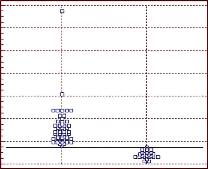
2                                                                                              Veterinary Medicine International

lymph nodes and internal organs such as lungs, kidneys,                 In addition to the design of vaccines, the in silico search
liver, and spleen [3].                                              for potentially antigenic targets and subcellular location
     Due to the vast spectrum of hosts, several cases of CLA are    among other analyses become important in the search for
present in many countries, for example, Australia, New Zea-         antigens to compose serological diagnoses. Faria et al. [20]
land, South Africa, the United States, Canada, and Brazil, and      used the bioinformatics tools to predict immunodominant
most of the cases are related to small ruminants [4, 5]. The        epitopes, approximately 50 proteins, from Leishmania
primary strategy for CLA control is based not only on vac-          infantum that obtained synthetic peptides that served as
cination but also on the adoption of complementary measures         antigens for serological diagnosis. With this, it is possible to
related to the treatment or eradication of sick animals and         realize that the bioinformatics tools can act in several as-
specialized care in routine handling [6]. Although these vac-       pects, either in the identification of proteins of interest or in
cines generate an immune response, which leads to the pro-          the design of synthetic peptides, both possible to compose
duction of specific antibodies, their efficiency is still discussed    vaccine formulation or an antigen for serological diagnosis.
[7]. Another factor that contributes to the dissemination of the        Thus, because of the number of putative proteins of
disease is the lack of diagnosis for cases of internal CLA.         C. pseudotuberculosis, ten of these proteins were selected
     The diagnosis of asymptomatic animals is an important          using bioinformatics tools, and two were expressed in E. coli
factor for the effective control of CLA, as many animals             to compose antigens for the serological diagnosis of CLA.
develop the visceral form of the disease or are still in the
initial stage of infection [8]. The current diagnosis to detect     2. Methodology
C. pseudotuberculosis is based mainly on the clinical ex-
amination of the lesions and their identification by phe-            The methodology follows the illustrative scheme as shown in
notypic and biochemical tests due to the variability of the         Figure 1.
characteristics of the genus Corynebacterium [9, 10]. Re-
cently, it has been suggested that the use of the Polymerase
                                                                    2.1. Amino Acid Sequences. The amino acid sequences of the
Chain Reaction (PCR) technique based on the 16S rRNA
                                                                    target proteins were retrieved from NCBI GenPept: ascor-
genes [11], phospholipase D (PLD), and rpoB, used together
                                                                    bate transporter subunit (PTS) (WP_058831991), UPF
[12], resulted in tests of high sensitivity, reproducibility, and
                                                                    protein    (AEP69811),      MMPL     family   transporter
diagnostic efficiency; however, they also need the initial
                                                                    (WP_013240996), Ribonuclease (WP_014300499.1), Iron
clinical examination.
                                                                    ABC transporter domain-containing permease (ATV80741),
     Because of the limited diagnostic methods available,
                                                                    and fimbrial subunit (ADK29694.1).
there is a need to use serological tests, in particular, the
ELISA technique (enzyme-linked immunosorbent assay)
that is effective in detecting all cases of CLA, asymptomatic        2.2. Subcellular Localization, Prediction of Protective Antigens,
or not. Some ELISAs have already been developed for this                 and Physicochemical Properties
purpose, using different antigens such as cell preparations
and proteins. However, the levels of sensitivity and speci-         2.2.1. Prediction of Signal Peptides and Protein Localization.
ficity are still unsatisfactory [13, 14].                            The software SignalP 4.1 available at http://www.expasy.org/
     Thus, studies are conducted regarding the importance of        proteomics was used for the verification of the signal pep-
exoproteins in the pathogen-host interaction to identify new        tides and, consequently, its location in C. pseudotuberculosis.
molecular targets to develop effective diagnostic tests with         This tool contains two types of neural networks, where it is
significant levels of sensitivity and specificity [15]. In this       possible to select sequence options with or without trans-
way, the sequencing of several genomes of                           membrane sets. All analyses performed with Gram-positive
C. pseudotuberculosis [16, 17] contributed to finding po-            bacteria used the two neural networks [21]. Then, the amino
tentially viable antigens before performing experiments to          acid sequences were evaluated by PSORTb 3.0.2 [22] to
verify them. This technique increases the speed of antigen          predict the subcellular localization of the target proteins.
identification, decreases costs, increases vaccine strategies to
obtain potential antigens for serological diagnosis. Many           2.3. Antigenic Potential. To verify the antigenicity of proteins
bioinformatics tools are available to facilitate this process,      and peptides previously predicted, VaxiJen30 v2.0 (http://
and the success of this method will depend on the accuracy          www.ddg-pharmfac.net/vaxijen/VaxiJen/VaxiJen.html) was
and prediction of antigens. According to the literature, other      employed. This software works based on independent align-
studies have been carried out to obtain antigens.                   ment prediction of protective antigens. Additionally, it was
     A study performed by Droppa-Almeida et al. [18], in            developed to allow the classification of antigens according to
which they used CP40 sequences to obtain immunodo-                  the physicochemical properties of proteins without resorting to
minant epitopes, which presented the potential to formulate         sequence alignment. Also, the server accuracy ranges from 70%
a multiepitope vaccine, showed a significant interaction of          to 89%, dependent on the target organisms [23].
peptides with essential receptors of innate immunity.
Similarly, Santana-Jorge et al. [19] verified in silico the
following virulence factors Spac, PknG, and NanH, which             2.4. Physicochemical Analysis. Different physicochemical
presented a considerable potential for the development of           properties for protein vaccines were studied, including
vaccines against CLA.                                               molecular weight (MW), isoelectric point (pI), instability
Veterinary Medicine International                                                                                                        3

                                                                                             Antigens for serological diagnosis

                          Bioinformatics
                                                    Amino acid sequences                         rPTS           rRibonuclease

                                                      Subcellular localization                          Cloning and
                                Obtaining targets                                                        expression

                                                    Antigenic         Physicochemical
                                                    potential             analysis
                                                                                                        Amplification
                                                                                                          targets
                                                                Screening                   PTS
                                                                                            UPF
                                                          B-cell and T-cell                MMPL
                                                              epitopes                  Ribonuclease
                                                                                         Iron ABC
                                                                                          Fimbrial
Figure 1: Illustrative image of the methodology used in sequence. Firstly, bioinformatics analyses were performed for the selection of
important proteins of Corynebacterium pseudotuberculosis. After analyses, regarding the signal peptide, presence of antigenicity, physi-
cochemical characterizations, and presence of immunodominant epitopes, a total of six proteins were selected and followed for PCR,
cloning, and expression. Only two of the selected proteins were expressed in E. coli.

index, in vitro and in vivo half-life and aliphatic index, and                into the pAE expression vector [27], according to the re-
the large mean of hydropathicity (GRAVY). All these                           striction sites established for each ORF (Open Reading
properties were evaluated using the ProtParam server at                       Frame), as indicated in Table 1. For this purpose, each
http://web.expasy.org/protparam [24].                                         amplification product and the vector were digested with the
                                                                              respective restriction enzymes. Then, both were ligated at
                                                                              room temperature in a solution containing 1 μl of the T4
2.5. B-Cell Epitope Prediction. Linear B-cell epitopes were                   DNA ligase enzyme (Invitrogen), 0.5 μl of the vector, 2 μl of
predicted from the target protein sequences using the fol-                    buffer, and 15.5 μl of MiliQ H2O. The products of the
lowing software: BCPreds, which is also based on machine                      constructs were transformed into E. coli TOP 10 electro-
learning methods but involves those that apply to support                     competent cells and seeded on plates containing LB medium
vector machines (http://ailab.cs. iastate.edu/bcpreds/).                      with ampicillin (100 μg/ml) and incubated for 16 h at 37°C.
Currently, BCPreds works selecting three methods of pre-                      The obtained colonies were submitted to screening by lysis
diction: (i) AAP implementation method [25]; (ii) BCPred                      with phenol-chloroform (v/v) and analyzed in 1% agarose
(EL-Manzalawy et al., 2008); (iii) FBCPred [26]. The chosen                   gel. The selected clones were cultured in LB broth with
method was FBCPred, which works by choosing peptides                          ampicillin (37°C/140 rpm for 12–16 h) before extracting the
with nine amino acid residues.                                                plasmid DNA using the Plasmid Miniprep Spin kit (GE
                                                                              Healthcare Life Sciences). Confirmation of the presence of
                                                                              each gene in the recombinant clones was carried out by
2.6. Amplification of Targets. The PCR molecular technique
                                                                              digestion with the restriction enzymes used in cloning the
amplified the genes corresponding to each protein through
                                                                              pAE vector.
specific primers designed by Vector NTI 11 software
(Invitrogen) (Table 1). The PCR amplification reaction was
performed to a final volume of 50 μL containing genomic                        2.8. Recombinant Protein Expression. Constructs with the
DNA of C. pseudotuberculosis strain 1002 (50 ng), primers                     genes of interest were transformed into E. coli BL21 Star
(10 μM), autoclaved MiliQ water (22 μL), and Mastermix                        strain by heat shock. Besides, the expression of the
(Promega) (25 μL) containing Taq DNA polymerase, dNTPs,                       recombinant proteins was induced with 1 mM Isopropyl
and MgCl2 under the following conditions: initial dena-                       β-D-1-thiogalactopyranoside (IPTG) in each culture under
turation at 94°C (5 min), followed by 30 cycles corre-                        orbital shaking (37°C/3 h). Finally, Western Blot confirmed
sponding to denaturation at 94°C (1 min), annealing at 55°C                   the expression of the recombinant proteins (36).
(1 min), extended at 72°C (1.5 min), and with a final ex-
tension of 72°C (7 min) in a thermocycler (Mastercycler
Gradient, Eppendorf, Germany). The amplicons were ana-                        2.9. Western Blot for Confirmation of Protein Expression.
lyzed on a 1% agarose gel stained with Blue Green (LGC                        Samples from the E. coli culture, which had been trans-
Biotechnology).                                                               formed by recombinant plasmids, were added to the sodium
                                                                              dodecyl sulfate (SDS) buffer and beta-mercaptoethanol.
                                                                              Then, after heating at 100°C for 10 min, the samples were
2.7. Cloning Procedures and Recombinant Proteins Expressed                    electrophoresed on 12% polyacrylamide gel. The proteins
in Escherichia coli. The gene-related amplicons were cloned                   were then electroblotted onto a nitrocellulose membrane
4                                                                                                   Veterinary Medicine International

                       Table 1: Primers for PCR and their respective restriction sites for binding to the vector pAE.
Base pairs                       Primers                                       Description                              Restriction sites
                              Forward: ptsF                    5′AGTCTGCAGAATGAACGTCCTACTTTC                                  PstI
1542 (pb)
                               Reverse: ptsR                       5′ CTAAACGTTTCACCCCAGTGGC                                HindIII
                              Forward: upfF                            5′- TAAGGATCCGCACGTGGC                               BamHI
1589 (pb)
                               Reverse: upfR                   5′-CTAGAATTCCTATCCGCCTATAGTGT                                 EcoRI
                             Forward: mmplF                         5′- CTACTGCAGTCAGGGCGTAAT                                 PstI
1618 (pb)
                             Reverse: mmplR                       5′- AGGGAATTCAAATACCTGGTAG                                 EcoRI
                          Forward: ribonucleaseF                5′- CTAGGTACCTCATGCCCACAGTATC                                 KpnI
1086 (pb)
                          Reverse: ribonucleaseR               5′ CCTAAGCTTTTATTTCTTTCTTAAAGC                               HindIII
                              Forward: ironF                       5′- ATAGGATCCTTGGTTGCATGCG                               BamHI
969 (pb)
                              Reverse: ironR                    5′-CTAGAATTCCTACCTTGGCCTTCTTG                                EcoRI
                            Forward: fimbrialF                     5′- ATAGGATCCGGCATCAAGGTAG                                BamHI
1521 (pb)
                            Reverse: fimbrialR                     5′- ATAGGTACCAACCCGCAAAGAC                                  KpnI
The underlined sequences correspond to the restriction site.

(GE Healthcare Life Sciences). The membrane was blocked                 2.12. Sheep Serum. The sheep serum sample was collected in
with 5% skimmed milk in PBS (1h/37°C) and then washed                   Nossa Senhora da Gloria city, Sergipe. 75 sheep serum
with PBS 0.05% Tween (PBS-T) (3 times for 5 min each),                  sample total were used in this study, 49 positive to CLA, with
before being incubated with the anti-6X-his monoclonal                  abscess, and these cases were confirmed with
antibody 1 : 4000 (Sigma) (1h/37°C). After three membrane               C. pseudotuberculosis with API coryne Kit and 26 negatives
washes with PBS-T, peroxidase-conjugated anti-mouse IgG                 to CLA from neonate sheep, uninfected and isolated from
antibody (Sigma), diluted in PBS-T (1 : 4000), was added.               the herd. The negative serum was used with standard to
Finally, the incubation process (1h/37°C) was performed                 evaluate the cut-off point of the ELISA test about the
before revealing it with 3,3′-diaminobenzidine (DAB) and                recombinant proteins tested [28].
H2O2.

                                                                        2.13. ELISA with Secreted Antigen from Strain 1002 C.
2.10. Recombinant Protein Purification. After centrifugation,            pseudotuberculosis. To this assay, secreted antigen from 1002
a pellet was obtained (14000g/15 min/4°C). For its solubili-            strain C. pseudotuberculosis was cultured in BHI broth for 48h
zation, it was resuspended in 40 mL of wash solution                    with standard to confirmed the sample serum positivity and
(200 mM NaH2 PO4, 500 mM NaCl, 5 mM imidazole, and                      serum negativity according to the methodology described by
8M urea pH 8.0) by adding 100 mg/mL lysozyme followed by                Seyffert et al. (2010) [28] with modifications. The immuno-
the sonication method (5 cycles of 15s, 20 kHz). In this                assay by ELISA 96-well plates, high binding, were sensitized
procedure, the solubilization solution was kept under stir-             with secreted antigens in the proportion 1 : 100 each well in
ring at 4°C for 16 h. The proteins were then filtered in a               Carbonate-Bicarbonate Buffer (0.05M, pH 9,6) and incubated
0.45 μm membrane before purification by nickel-loaded                    for 16 h at 4°C. Subsequently, the plates were washed 3 times
Sepharose column affinity chromatography (HisTrap GE
Healthcare Life Science). The protein preparation was then
                                                               ™        with PBS-T (PBS 1X, pH 7.4, 0.1% tween 20) and blocked with
                                                                        100 µL/well of 5% nonfat milk in PBS (1 h at 37°C). Then, the
dialyzed (cellulose bags, 25 nm × 16 nm, Sigma) in 1x saline            plates were washed again, and 100 µL/well of sheep serum (1 :
phosphate buffer (PBS)/0.2% urea, with two daily exchanges                50 in PBS-T) was added in duplicate in the plate. After 1 h of
(7 days/4°C) for desalting. Furthermore, protein quantifi-               incubation at 37°C and more washes, 100 µL/well of anti-
cation was performed using BCATM Protein Assay Kit                      sheep antibodies conjugated to peroxidase (Whole IgG
(Pierce Chemical Company).                                              (Sigma)) was added (1 : 10.000). After five more washes,
                                                                        50 µL/well of TMB (Bio-Rad) was added, and the plates were
                                                                        incubated for 15 min at room temperature and in the dark to
2.11. Characterization of Antigenicity of Recombinant Proteins          reveal the results. To stop the reaction, 25 µL/well of H2SO4
by Dot Blot. To verify antigenicity 5 µL of each protein                solution (4N) was added. Optical density (OD) was deter-
(rRibonuclease, rPTS e rCP40) was deposited on the ni-                  mined with an automatic photocolorimeter for ELISA
trocellulose membrane (GE Healthcare Life Sciences). After              (Microplate Reader MR-96A, Mindray) at 495 nm.
drying (15 min/room temperature), the membrane was a
block with PBS plus 5% skim milk (1 h/room temperature)
before being incubated with negative and positive sheep to              2.14. ELISA with Proteins rPTS, rRibonuclease, and rCP40.
CLA (1 : 50 in PBS-Tween 20% for 1 h/37°C). After this step,            Each recombinant protein was used individually and in the
the membrane was incubated with IgG sheep secondary                     association. According to standardization, the individual
antibody (1 : 4000 em PBS-T) conjugated with peroxidase                 assay used 100 ng of each protein and 1 : 50 of serum di-
(Sigma) (1 h/37°C). The reaction was revealed with 3,3′-                lution, in association assay were used a total of 50 ng with
diaminobenzidine (DAB) and H2O2..                                       protein mix and 1 : 100 of serum dilution.
Veterinary Medicine International                                                                                                  5

2.15. Statistical Analysis. To evaluate the specificity, sensi-     macrophages, and B lymphocytes. When this type of re-
bility, predictive value, and cut-off point, the experimental       sponse is activated, the production of IgG antibodies is
data were analyzed by Receiver Operating Characteristic            increasing, mainly of the IgG2a and IgG2b isotypes, which
(ROC) with the software MedCalc statistical (version 10.3.0).      signal a Th1 response [32]. By understanding the patho-
This analysis aims to differentiate the true positives from the     genesis of CLA, it is possible to understand the type of
true negatives to establish the cut-off point. The evaluation of    vaccines that may present possible protection to the animals.
the sensitivity and specificity of the experimental data was        The use of proteins as a vaccine antigen is increasing because
done with each protein individually and in the association.        it is an antigen whose response is dependent on T cells,
                                                                   activating both humoral and cellular response, important for
3. Results and Discussion                                          facultative intracellular pathogens.
                                                                        The use of potentiators, known as adjuvants, can aid in
3.1. Subcellular Localization, Signal Peptide, and Antigenicity.   directing the response and may direct to Th1, Th2, Th9, or
Concerning the location of the proteins, their primary             Th17 and these potentiators are crucial to the success of a
structure was first obtained before the online servers Sig-         protein antigen. This work presents six potential targets for
nalP-4.1 and PSORTb were applied (Table 2). The former             the development of vaccines proteins for CLA and with
verified that none of the sequences present a peptide signal.       important characterizations on each of them. In addition to
However, the latter showed that most proteins are present in       the use of vaccine formulations, these proteins may be useful
the cellular membrane, a site also interesting for use in          for the development of diagnostic kits for CLA since the
vaccines or as an antigen for immunodiagnosis. Membrane            diagnosis is still based on the culture and isolation of the
proteins and secreted proteins are considered potential            bacteria present in the pyogranulomas found in the animals.
vaccine targets since they represent the host-pathogen in-
terface. These proteins may interact more directly with host
molecules for cell adhesion, invasion, multiplication, and         3.3. B-Cell Epitope Prediction. After analyzing the B-cell
evasion of the immune response [29, 30].                           epitopes by the BCpreds software, a total of 18 epitopes for
     For the antigenicity profile, the Vaxijen server was used      PTS, 45 for UPF, 26 for MMPL, 14 for Ribonuclease, 9 for
and the provided z descriptors indicate the most relevant          IronABC, and 57 for Fimbrial were predicted. Immuno-
physicochemical properties for antigen recognition. The            dominant epitopes are specific regions of protein antigens
value of z has a threshold of 0.4 [23]. Given this threshold       that have binding to immunological receptors [33, 34].
and the obtained data, the antigenicity of all the proteins        Regarding B-cell epitopes, we turn to the production of
explored in this work is described in Table 2.                     antibodies, which can identify and neutralize antigens, such
                                                                   as viruses, bacteria, parasites, fungi, cancer cells, and some
                                                                   toxins, by binding to specific parts on their surface [35]. The
3.2. Physicochemical Analysis. ProtParam server was used to        paratope, a specific part of an antibody, binds to a particular
check some character molecular weight, theoretical index,          region in the antigen that is called an epitope or antigenic
amino acid composition, atomic composition, extinction co-         determinant [36, 37]. Different from T-cell epitopes, most
efficient, instability index, aliphatic index, and the large mean    functional B-cell epitopes are conformational, although
of hydropathicity (GRAVY) as described in Table 3. This            there are linear models as well [38].
parameter is the point at which the total net charge of the            Epitope databases consist of a collection of epitopes of
amino acid or protein molecule is zero. These analyzes are         pathogens such as viruses, bacteria, protozoa, and fungi. These
based on equilibrium constant points, which means that the         databases have become an increasingly used source in the
protein is stable at pH corresponding to one described in          search for antigens for application in vaccines, in the diagnosis,
Table 3. All proteins demonstrated a stable profile and the high    and as immunotherapies [39]. Given the lack of vaccines and
aliphatic index indicates protein stability at various tempera-    serological diagnosis for CLA, new target analyzes are essential,
tures. Moreover, when the GRAVY value of hydropathicity is         as well as the use of different strategies such as the use of
negative the proteins are hydrophilic; in other words, they have   synthetic peptides based on immunodominant epitopes. For
significant interaction with water molecules.                       this reason, these proteins were evaluated in silico and sub-
    C. pseudotuberculosis demonstrates interesting survival        sequently used to identify the immunodominant epitopes. The
mechanisms and can use different strategies to adapt to its         selection of the epitopes was aimed to contain nine amino acid
environment. Once it is successfully established within a          residues since the B-cell epitopes are capable of both binding
host, it can replicate within the phagocytic cells. This           antibodies and B-cell receptors (BCR) [40]. Consequently, this
pathogen will then easily block the immune system. As a            condition will lead to the transformation of B cells in antigen-
result, chronic infections can last for most, if not all, of an    specific antibody-producing plasmocytes and consequently
animal’s life [31]. Chronicity leads to the formation of           antibodies, proving to be important targets for the develop-
pyogranulomas intended to protect the animal from possible         ment of antigens for serological diagnosis.
generalized infection. Currently, bacteria are known to
bypass the pyogranuloma cell barrier to infect other parts of      4. Obtaining Recombinant Proteins
the host organism. Because it is a facultative intracellular
bacterium, the immune response against this pathogen is            4.1. Amplification of Genes. The amplification products of the
Th1 type since it will be assisted by CD4 + helper cells,          genes, which were increased from the genomic DNA of
6                                                                                                       Veterinary Medicine International

                            Table 2: Results about subcellular localization and antigenicity of the target proteins.
Proteins                                Signal peptide                               PsortB                            Vaxijen score 0.40
PTS                              No/D � 0.130 D-cutoff � 0.450                  CMP   score: 0.618                  0.4699 (Probable Antigen)
UPF0182                          No/D � 0.189 D-cutoff � 0.450                  CMP   score: 0.775                  0.4806 (Probable Antigen)
MMPL                             No/D � 0.357 D-cutoff � 0.450                  CMP   score: 0.663                  0.6489 (Probable Antigen)
Ribonuclease                     No/D � 0.137 D-cutoff � 0.450                  CMP   score: 0.484                  0.6143 (Probable Antigen)
Iron                             No/D � 0.402 D-cutoff � 0.450                  CMP   score: 0.554                  0.4981 (Probable Antigen).
Fimbrial                         No/D � 0.130 D-cutoff � 0.450                  CMP   score: 0.416                  0.7775 (Probable Antigen).
CMP: cytoplasmic membrane protein.

                           Table 3: Physicochemical properties of the target proteins estimated using ProtParam.
Physicochemical property                                  PTS        UPF            MMPL            Ribonuclease         Iron       Fimbrial
Number of amino acids                                      513        896             795               361               340          995
Molecular weight                                        52613.17   98897.67        84887.14           39313.31         34979.52    108358.72
Theoretical pI                                            8.40       5.69             6.49              9.31             10.13         4.74
Total number of negative residues                           22        101              62                27               13           148
Total number of positive residues                          24          91              59                35               26           101
Instability index (II)                                    28.17      29.18           30.25             26.58             22.14        33.35
Aliphatic index                                          121.52     89.99           114.34             105.15           130.15        76.31
Grand average of hydropathicity (GRAVY)                  0.884      −0.224          0.495              0.451             0.815       −0.474
The estimated half-life
  In vitro                                             30 hours    1.1 hours      30 hours           30 hours       30 hours       30 hours
  Yeast                                                >20 hours     3 min        >20 hours          >20 hours      >20 hours      >20 hours
  Escherichia coli                                     >10 hours   >10 hours      >10 hours          >10 hours      >10 hours      >10 hours
Negative residues: Asp + Gln; positive residues: Arg + Lys.

C. pseudotuberculosis 1002 using primers described in Table 1,                Considerable amounts of rPTS and rRibonuclease are
presented molecular sizes according to the in silico prediction.          required for further studies on the role of these proteins in
Figure 2 shows the results of PCR amplification.                           the pathogenicity and virulence of C. pseudotuberculosis.
    After the characterization of the recombinant clones, four            The use of these proteins for heterologous expression with
clones were achieved: pAE/PTS, pAE/Ribonuclease, pAE/                     Escherichia coli remains one of the most attractive uses
CPIron, and pAE/CPF-bound, as illustrated in Figure 3.                    among many systems available for protein production
    Regarding the upf and mmpl genes, although amplifi-                    [18, 42]. Thus, pts, upf, mmpl, Ribonuclease, iron, and
cation products were obtained, after repeated attempts, no                fimbrial genes were cloned into the same expression
recombinant clones were achieved. One possible explanation                vector system and individually transformed into E. coli
for the failure to obtain plasmid constructs between these                BL21 (DE3) strains. SDS-PAGE analyses show the suc-
amplicons and the pAE vector may be an amplification error                 cessful expression of the rPTS and rRibonuclease proteins
or in the synthesis of primer sequences.                                  (Figure 4). As a result, these proteins can be used to
    Upon confirmation of the genes in the pAE vector, the                  compose both recombinant vaccines and serve as an
pAE/PTS, pAE/Ribonuclease, pAE/Iron, and pAE/Fimbrial                     antigen for immunoassay since there is a lack of sero-
constructs were transformed by heat shock into the E. coli                logical diagnosis for CLA. From the present study, we
BL21 Star expression line. One colony of each recombinant                 suggest that various B-cell epitopes predicted from PTS,
clone was selected for this transformation and used for the               UPF, MMPL, Iron, Ribonuclease, and Fimbrial could be
expression of the respective proteins after induction with                used for the development of a multipeptide vaccine to
IPTG. Expression of the heterologous proteins was verified                 induce a complete immune response against
by Western Blot, presenting apparent molecular weights of                 C. pseudotuberculosis. The next step will be to experi-
approximately 55 kDa (rPTS) and 35 kDa (rRibonuclease),                   mentally evaluate these epitopes in vitro and in vivo to
corresponding to the primary amino acid sequence of the                   assess their true protective potential.
proteins encoded by the selected genes, in addition to the
six-histidines tail (Figure 4). rCP40 was used as a standard
because of its frequent expression in the laboratory.                     4.2. Antigenicity Characterization of the Recombinant Protein
    At this point, the fact that clones pAE/CPIron and pAE/               by Dot Blot. To verify antigenicity from rCP40, rRi-
Ribonuclease, when transformed in E. coli BL21 Star, did not              blonuclease, and rPTS proteins, sheep serums of positive
express the respective recombinant proteins may be related                and negative animals from CLA by Dot Blot were used.
to the presence of codons in the gene sequences of                        Figure 5 shows the protein reactivity with positive serum,
C. pseudotuberculosis that are little used by E. coli. The degree         and it did not show with negative serum. This suggests the
of frequency of some codons may be a limiting factor in the               recombinant protein preserves the antigenic potential, even
heterologous expression of proteins in expression lines [41].             after heterology expression in E. coli. The Dot Blot is a
Veterinary Medicine International                                                                                                            7

           1Kb   A   1Kb   B     1Kb   C    D      1Kb     E   1Kb       F                           rCP40/rRibonuclease /rPTS

  12000
   5000

   2000
   1650                                                                                 55 kDa
   1000
    850
                                                                                        40 kDa

                                                                                        35 kDa
Figure 2: Agarose gel electrophoresis 1% of PCR amplification
products of the ORFs of interest. 1 Kb : molecular size standard
(1 Kb Plus DNA Ladder, Invitrogen); (A) amplicon of Upf; (B)
amplicon of Mmpl; (C) amplicon of Ribonuclease; (D) amplicon of
Iron; (E) amplicon of Fimbrial; and (F) amplicon of PTS. Cloning
procedures and recombinant proteins expressed in Escherichia coli.

             1Kb     A     1Kb    B                  1Kb       C     D

                                                                             Figure 4: Confirmation of expression of the recombinant proteins
                                                                             rCP40, rRibonuclease, and rPTS, by Western Blot using anti-6Xhis.

                                           12000
                                            5000
   12000                                                                       A                     B                     C
                                           2000
    5000                                   1650
                                           1000
                                            850
    2000
    1650

    1000
     850
                                                                               A1                    B1                    C1

Figure 3: 1% agarose gel electrophoresis of the characterization of
the recombinant clones, after digestion with the respective re-
striction enzymes. 1 Kb : molecular size standard (1 Kb Plus DNA             Figure 5: Dot blot to antigenicity characterization of recombinant
Ladder, Invitrogen); (A) pAE/PTS; (B) pAE/Ribonuclease; (C)                  protein. (A) rPTS evaluate with positive serum from CLA; (A1)
pAE/CPIron; (D) pAE/CPFimbrial.                                              rPTS evaluation with negative serum from CLA; (B) rRibonuclease
                                                                             evaluation with positive serum from CLA; (B1) rRibonuclease
                                                                             evaluation with negative serum from CLA; (C) rCP40 evaluation
technique widely used to determine the antigenic property                    with positive serum from CLA; (C1) rCP40 evaluation with neg-
of recombinant protein [43].                                                 ative serum from CLA. Enzyme-Linked Immunosorbent Assay
    A total of 49 positive sera and 26 negative sera were used               (ELISA) with secreted antigens of C. pseudotuberculosis.
in indirect ELISA with secreted antigens of
C. pseudotuberculosis 1002, and this assay was done to
confirm the positivity and negativity of sheep sera. This                     positive when the cut-off showed a value of optical density
methodology was validated and applied by Seyffert et al. [28]                 (OD) 450n ≥ 0,10, referring to the mean ELISA absorbance
when carrying out a seroepidemiological study of the                         values. There are no sera below the cut-off line at 1 or above
prevalence of CLA in the State of Minas Gerais, with a                       the cut-off line at 0, so they indicate the absence of false
sensitivity of 93% and specificity of 98.5% in the detection of               negatives and positives, respectively.
antibodies against antigens secreted by the referred bacteria.                   Figure 7 indicates the results of the ELISA using the
    Figure 6 shows the frequency distribution from these                     rPTS, rRibonuclease, and rCP40 proteins individually. The
positive sera (1) and negative sera (0). The samples were                    samples were considered positive when the average
8                                                                                                               Veterinary Medicine International

                              2.0

                              1.5

                              1.0

                              0.5

                                                                                                        >10
                                                                                                        Sensibility 100.0
                              0.0                                                                       Specificity 100.0
                                                   1                     0
                                                         Sheep sera
Figure 6: Indirect ELISA evaluation with antigens secreted from C. pseudotuberculosis 1002 with 75 sheep sera through the Receiver
Operating Characteristic (ROC). (1) Positive serum samples; (0) samples of negative sera. This shows ELISA with rPTS, rRibonuclease, and
rCP40 proteins.

    1.4                                                                                       100

    1.2
                                                                                               80
    1.0

                                                                                               60
    0.8
                                                                                Sensitivity

    0.6
                                                                                               40

    0.4
                                                                                               20
    0.2
                                                                  >0.14
                                                                  Sens: 100.0
    0.0                                                           Spec: 96.2                    0
                       1                       0                                                    0      20        40        60     80   100
                                                                                                                    100-specificity
                                      (a)                                                                             (b)
    1.6                                                                                       100

    1.4
                                                                                               80
    1.2

    1.0
                                                                                               60
                                                                                Sensitivity

    0.8

    0.6                                                                                        40

    0.4
                                                                                               20
    0.2
                                                                  >0.13
                                                                  Sens: 100.0
    0.0                                                           Spec: 92.3                    0
                       1                      0                                                     0      20        40        60     80   100
                                                                                                                    100-specificity
                                      (c)                                                                             (d)
                                                         Figure 7: Continued.
Veterinary Medicine International 9
1.4 100
1.2
80
1.0
60
Sensitivity
0.8
0.6 40
0.4
20
0.2
>0.10
Sens: 100.0 0
0.0
Spec: 88.5
1 0 0 20 40 60 80 100
100-specificity
(e) (f )
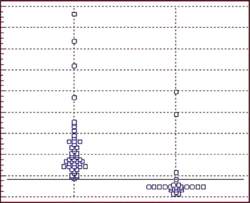
Figure 7: Indirect ELISA results with recombinant proteins (rPTS, rRibonuclease, and rCP40) as antigens, individually tested with 49
positive sera and 26 negative sera for CLA and ROC analysis of all assays. (a, b) ELISA rPTS with area under the curve (AUC): 1,000 and 95%
confidence interval (CI): 0.951–1,000; (c, d) ELISA rRibonuclease with AUC: 0.970 and 95% CI: 0.902–0.955; (e, f ) ELISA rCP40 with AUC:
0.991 and 95% CI: 0.934–0.997.
absorbance value was higher than the cut-off point calcu- Table 4: Indirect ELISA results with recombinant proteins (rPTS,
lated for each of the proteins. The ROC analysis was used to rRibonuclease, and rCP40) as antigens individually tested with 49
determine the cut-off point, sensitivity, specificity, and positive sera and 26 negative sera for CLA.
predictive value for each of the tests. The values of these cut- ELISA Sensibility (%) Specificity (%)
off points were> 0.14 for rPTS, > 0.13 for rRibonuclease, rPTS 100 96, 2
and> 0.10 for rCP40. The sensitivity obtained was 100% for rRibonuclease 100 92, 3
the three ELISAs (rPTS, rRibonuclease, and rCP40) and the rCP40 100 88, 5
specificity obtained was 96.2%, 92.3%, and 88.5% for rPTS,
rRibonuclease, and rCP40, respectively (Table 4).
On the other hand, when analyzing the results of the Table 5: Indirect ELISA results with recombinant proteins asso-
indirect ELISA tests where associations between proteins ciated and their sensibility and specificity.
were used, sensitivities of 100% were observed, but varying ELISA Sensibility (%) Specificity (%)
specificities between those associations, as can be seen in rPTS/rCP40 100 84, 6
Table 5 and Figure 8, together with their cut-offs. rRibonuclease/rCP40 100 92, 3
The rPTS, rRibonuclease, and, rCP40 proteins were rPTS/rRibonuclease 100 88, 5
individually tested and in the association were impregnated rPTS/rRibonuclease/rCP40 100 92, 3
in indirect ELISA to detect specifics antibodies of
C. pseudotuberculosis in sheep sera, to develop an effective
diagnosis for CLA in sheep. According to Bastos et al. [44], Although they have variation in terms of specificity, the
the search for tests that provide greater sensitivity in the experimental results are promising, mainly because of the
diagnosis of CLA in sheep is due to the low sensitivity in tests low sensitivities obtained, for example, by Rebouças et al.
already carried out on these animals. However, the results of [45] when developing a diagnostic ELISA to detect gamma
this work indicated that ELISAs with recombinant proteins interferon (IFN-c) as a marker of host cell-mediated im-
obtained 100% sensitivity in all tests, eliminating the munity against the pathogen in sheep, resulting in a 100%
probability of obtaining false-negative results in sheep. specificity, however a low sensitivity of 55.8%. Binns et al.
However, about specificities, the values ranged from [46] used a cell preparation for the development of an ELISA
84.6% to 96.2% (Tables 4 and 5). When comparing the values that detected antibodies against C. pseudotuberculosis in the
of ELISAs with individual proteins, it is observed that the sera of sheep, obtaining a specificity of 100% but a sensitivity
specificities of the tests with associations are lower than that of 71%. Regarding the use of C. pseudotuberculosis culture
of rPTS (96.2%), even in the combinations in which this supernatant to detect specific antibodies in sheep sera,
protein is present. It is noted that ELISAs with the associ- Rebouças et al. [14] obtained a specificity of 99% and a
ations rPTS/rRibonuclease and rPTS/rCP40 showed speci- sensitivity of 89%.
ficities of 88.5 and 84.6%, respectively, similar to that Concerning recombinant proteins, Menzies et al. [47]
observed with rCP40 in the individual test. tested, over a year, an ELISA with recombinant PLD in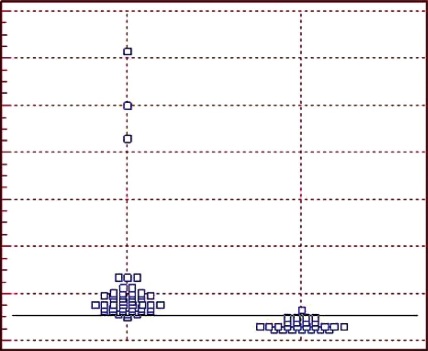
10                                                                         Veterinary Medicine International

     1.8                                                    100

     1.6

     1.4                                                     80

     1.2
                                                             60

                                              Sensitivity
     1.0

     0.8
                                                             40
     0.6

     0.4                                                     20
     0.2                        >0.16
                                Sens: 100.0
     0.0                        Spec: 84.6                    0
           1         0                                            0   20       40        60     80    100
                                                                              100-specificity
               (a)                                                             (b)
     2.0                                                    100

                                                             80
     1.5

                                                             60
                                              Sensitivity

     1.0
                                                             40

     0.5
                                                             20

                                >0.12
                                Sens: 100.0
     0.0                        Spec: 92.3                    0
           1         0                                            0   20       40        60     80    100
                                                                              100-specificity
               (c)                                                             (d)
     0.7                                                    100

     0.6
                                                             80
     0.5

                                                             60
     0.4
                                              Sensitivity

     0.3                                                     40

     0.2
                                                             20
     0.1
                                >0.10
                                Sens: 100.0
     0.0                                                      0
                                Spec: 88.5
           1         0                                            0   20       40        60     80    100
                                                                              100-specificity
               (e)                                                              (f )
                         Figure 8: Continued.
Veterinary Medicine International 11
1.2 100
1.0
80
0.8
60
Sensitivity
0.6
40
0.4
20
0.2
>0.12
Sens: 100.0 0
0.0
Spec: 92.3
1 0 0 20 40 60 80 100
100-specificity
(g) (h)
Figure 8: Result of indirect ELISA with recombinant proteins (rPTS, rRibonuclease, and rCP40) associated with each other, using 49
positive sera and 26 negative sera for CLA and ROC analysis of all ELISAs. (a, b) rPTS/rCP40 with AUC: 0.924 and 95% CI: 0.838–0.972;
(c, d) rRibonuclease/rCP40 with AUC: 0.970 and 95% CI: 0.900–0.995; (e, f ) rPTS/rRibonuclease with AUC: 0.935 and 95% CI: 0.853–0.979;
(g, h) rPTS/rRibonuclease/rCP40 with AUC: 0.931 and 95% CI: 0.847–0.977.
experimentally infected goats, with a sensitivity of 81% and compared to rRibonuclease. Finally, among the associations,
specificity of 97%. Still, regarding the rPLD protein, Sting the best results were those of the rRibonuclease/rCP40 and
et al. [48] correlated two ELISA tests (ELISA-whole cell rPTS/rRibonuclease/rCP40 tests, which obtained 92.3%
antigens and ELISA-rPLD) to evaluate the seroprevalence of specificity, matching in numbers, to rRibonuclease.
C. pseudotuberculosis in infected goats, where the first Specificity results, although inferior and, in some cases
conferred a sensitivity of 81% and specificity of 98%, while similar to those of tests with individual proteins, do not
the second sensitivity of 97% and specificity of 99%, thus compromise this approach. The associations proved to be
showing that the result obtained by the ELISA with the positive. This fact is in line with tests performed for other
recombinant protein was superior. However, according to a diseases, such as Ferra et al. [49], who used the association
study by Hoelzle et al. (2013) [15], serological tests based on between recombinant antigens successfully to diagnose
PLD as the only antigen are not able to identify all infected Toxoplasma gondii infection in horses, pigs, and sheep, with
sheep since PLD was recognized in this experiment by only sensitivity and specificity values of up to 100% depending on
70% of the positive sheep for CLA and by 100% of the the species.
positive goats. Still, at this point, Alizadeh et al. [50] developed an ELISA
Therefore, the results of this study show an excellent based on the association of two Leptospira recombinant
correlation of sensitivity and specificity (100% and 96.2%) proteins, which obtained 96.6% sensitivity and 83.2% spec-
for the rPTS test compared to other tests performed. ificity when compared to the gold standard used, is con-
However, all results are generally promising, as already sidered a useful technique for the diagnosis of leptospirosis.
mentioned above. None of the specificity values preclude the Faria et al. [20] also found a positive effect when using the
potential of rPTS, rRibonuclease, or rCP40 for use in ELISA association of Leishmania spp proteins—in diagnostic tests
diagnostic assays since the results of this work are superior to for leishmaniasis, obtaining sensitivity values of up to 75.81%
those of other studies on CLA diagnosis described in the and specificity of up to 95%, proving to be efficient.
literature. According to the ROC analysis, the negative predictive
Although Hoelzle et al. [15] have suggested that an ideal value of all tests performed was 100%, regardless of the
assay for the diagnosis of CLA is with the combination of prevalence rate of herds. This reiterates the reliability of the
two to three recombinant and immunodominant proteins of ELISAs developed in the absence of false negatives because
C. pseudotuberculosis, it should be noted that there are still the greater the sensitivity of the test, the greater its negative
no studies on the association of these proteins, this work predictive value, that is, the greater the probability of the
being the pioneer in that sense. The rCP40 test with sheep animal being healthy in the face of the negative result.
sera showed a specificity of 85.5%, but in its association with Table 6 shows the predictive values of each indirect ELISA
the rPTS protein, the value was 84.6%, having reduced the test, according to the average prevalence of 30% of CLA in
individual potential of the rPTS (which is 96, 2%) and rCP40 the herd [51], as normally occurs in Brazil.
itself. In the rRibonuclease/rCP40 test, which obtained a When analyzing the rPTS test, it showed the best positive
specificity of 92.3%, there was no variation in specificity predictive value about the other tests, showing that the12 Veterinary Medicine International
Table 6: Positive and negative predictive values according to the prevalence of CLA.
Indirect ELISA (∗ ) Prevalence of CLA (%) Positive predictive values (%) Negative predictive values (%)
rPTS 30 91, 8% 100
rRibonuclease 30 84, 7% 100
rCP40 30 78, 8% 100
rPTS/rRibonuclease 30 78, 8% 100
rPTS/rCP40 30 73, 5% 100
rRibonuclease/rCP40 30 84, 7% 100
rPTS/rRibonuclease/rCP40 30 84, 7% 100
∗
Tests performed with recombinant proteins individually and in combination.
proportion of infection among the positive animals detected Conflicts of Interest
through the test is high. This means that for every 100
positive tests, 91 sheep would be infected. The authors declare no conflicts of interest in this article.
Therefore, the test’s ability to identify true positives from
true negatives is critical to any diagnostic test. In the specific Acknowledgments
case of CLA, this ability is a crucial factor in allowing the
detection of apparently infected and asymptomatic animals, The authors appreciate the Support Research Foundation
which are a source of the spread of the infection in the herd. and Technological Innovation (Fapitec) of the state of
Thus, an efficient and rapid diagnostic test allows adequate Sergipe, Brazil, Higher Education Personnel Improvement
management of animals and the application of appropriate Coordination (CAPES), and the National Council for Sci-
prophylactic measures, considering the socio-economic entific and Technological Development (CNPq) for all fi-
importance of sheep farming for the world. nancial support.
References
5. Conclusion
[1] B. J. A. Paule, V. Azevedo, L. F. Regis et al., “Experimental
The in silico analyzes demonstrated that Corynebacterium pseudotuberculosis primary infection in
C. pseudotuberculosis has several targets with the capacity for goats: kinetics of IgG and interferon-c production, IgG avidity
the development of vaccines and serological diagnosis. It and antigen recognition by Western blotting,” Veterinary
was possible to determine six targets that presented in Immunology and Immunopathology, vol. 96, no. 3-4,
silico exciting results concerning their physicochemical pp. 129–139, 2003.
characteristics and potential allergens and antigenic. [2] S. Chakraborty, A. Kumar, R. Tiwari et al., “Advances in
Regarding immunodominant epitopes, there are numer- diagnosis of respiratory diseases of small ruminants,” Vet-
ous possibilities to compose diagnostic tests and multi- erinary Medicine International, vol. 2014, Article ID 508304,
16 pages, 2014.
epitope vaccines that could be able to differentiate
[3] A. S. Guimarães, N. Seyffert, B. L. Bastos et al., “Caseous
vaccinated animals from naturally infected animals. Of lymphadenitis in sheep flocks of the state of Minas Gerais,
the six proteins evaluated in this study, only two were Brazil: prevalence and management surveys,” Small Ruminant
expressed in E. coli, PTS, and Ribonuclease. With these Research, vol. 87, no. 1-3, pp. 86–91, 2009.
recombinant proteins obtained, ELISA assays with posi- [4] K. M. Connor, M. M. Quirie, G. Baird, and W. Donachie,
tive and negative CLA sera were performed. CP40, which “Characterization of United Kingdom isolates of Coryne-
is an endoglycosidase that has already had its antigenic bacterium pseudotuberculosis using pulsed-field gel electro-
and immunogenic potential reported in the literature, was phoresis,” Journal of Clinical Microbiology, vol. 38, no. 7,
also used in this work. These three proteins were used pp. 2633–2637, 2000.
alone and in combination, giving promising results in [5] M. Paton, S. Walker, I. Rose, and G. Watt, “Prevalence of
sensitivity and specificity. The difficulty in obtaining high caseous lymphadenitis and usage of caseous lymphadenitis
vaccines in sheep flocks,” Australian Veterinary Journal,
specificities for serological diagnosis for CLA has already
vol. 81, no. 1-2, pp. 91–95, 2003.
been reported in the literature due to the difference be- [6] R. Sting, G. Steng, and D. Spengler, “Serological studies on
tween hosts. In this work, it was evaluated only with sheep Corynebacterium pseudotuberculosis infections in goats us-
sera. With this, it is suggested new studies using these ing enzyme-linked immunosorbent assay,” Journal of Veter-
proteins in goat sera to verify the high specificity that these inary Medicine, Series B, vol. 45, no. 1–10, pp. 209–216, 1998.
antigens presented in sheep, increasing the possibility of a [7] J. I. Corrêa, A. Stocker, S. C. Trindade et al., “In vivo and in
serological diagnosis for CLA. vitro expression of five genes involved in Corynebacterium
pseudotuberculosis virulence,” AMB Express, vol. 8, no. 1,
2018.
Data Availability [8] A. F. Oreiby, “Diagnosis of caseous lymphadenitis in sheep
and goat,” Small Ruminant Research, vol. 123, no. 1,
The data from in silico and experimental analysis used to pp. 160–166, 2015.
support the findings of this study are included within the [9] F. A. Dorella, L. Carvalho Pacheco, S. C. Oliveira, A. Miyoshi,
article. and V. Azevedo, “Corynebacterium pseudotuberculosis:Veterinary Medicine International 13
microbiology, biochemical properties, pathogenesis and [24] E. Gasteiger, C. Hoogland, A. Gattiker et al., “Protein iden-
molecular studies of virulence,” Veterinary Research, vol. 37, tification and analysis tools on the ExPASy server,” The
no. 2, pp. 201–218, 2006. Proteomics Protocols Handbook, vol. 289, pp. 571–607, 2005.
[10] V. D’Afonseca, P. M. Moraes, F. A. Dorella et al., “A de- [25] K.-C. Chou and H.-B. Shen, “Signal-CF: a subsite-coupled and
scription of genes of Corynebacterium pseudotuberculosis window-fusing approach for predicting signal peptides,”
useful in diagnostics and vaccine applications,” Genetics and Biochemical and Biophysical Research Communications,
Molecular Research, vol. 7, no. 1, pp. 252–260, 2008. vol. 357, no. 3, pp. 633–640, 2007.
[11] B. Çetinkaya, M. Karahan, E. Atil, R. Kalin, T. De Baere, and [26] J. Larsen, O. Lund, and M. Nielsen, “Improved method for
M. Vaneechoutte, “Identification of Corynebacterium pseu- predicting linear B-cell epitopes,” Immunome Research, vol. 2,
dotuberculosis isolates from sheep and goats by PCR,” Vet- no. 1, p. 2, 2006.
erinary Microbiology, vol. 88, no. 1, pp. 75–83, 2002. [27] C. R. R. Ramos, P. A. E. Abreu, A. L. T. O. Nascimento, and
[12] L. G. C. Pacheco, R. R. Pena, T. L. P. Castro et al., “Multiplex P. L. Ho, “A high-copy T7 Escherichia coli expression vector
PCR assay for identification of Corynebacterium pseudotu- for the production of recombinant proteins with a minimal
berculosis from pure cultures and for rapid detection of this N-terminal His-tagged fusion peptide,” Brazilian Journal of
pathogen in clinical samples,” Journal of Medical Microbiol- Medical and Biological Research, vol. 37, no. 8, pp. 1103–1109,
ogy, vol. 56, no. 4, pp. 480–486, 2007. 2004.
[13] S. H. Binns, L. E. Green, and M. Bailey, “Postal survey of ovine [28] N. Seyffert, A. S. Guimarães, L. G. C. Pacheco et al., “High
caseous lymphadenitis in the United Kingdom between 1990 seroprevalence of caseous lymphadenitis in Brazilian goat
and 1999,” Veterinary Record, vol. 150, no. 9, pp. 263–268, herds revealed by Corynebacterium pseudotuberculosis se-
2002. creted proteins-based ELISA,” Research in Veterinary Science,
[14] M. F. Rebouças, D. Loureiro, B. L. Bastos et al., “Development vol. 88, no. 1, pp. 50–55, 2010.
of an indirect ELISA to detect Corynebacterium pseudotu- [29] A. P. Bhavsar, J. A. Guttman, and B. B. Finlay, “Manipulation
berculosis specific antibodies in sheep employing T1 strain of host-cell pathways by bacterial pathogens,” Nature,
culture supernatant as antigen,” Pesquisa Veterinária Brasi- vol. 449, no. 7164, pp. 827–834, 2007.
leira, vol. 33, no. 11, pp. 1296–1302, 2013. [30] A. M. Krachler and K. Orth, “Targeting the bacteria-host
[15] L. E. Hoelzle, T. Scherrer, J. Muntwyler, M. M. Wittenbrink, interface,” Virulence, vol. 4, no. 4, pp. 284–294, 2013.
W. Philipp, and K. Hoelzle, “Differences in the antigen [31] F. A. Dorella, A. Gala-Garcia, A. C. Pinto et al., “Progression
structures of Corynebacterium pseudotuberculosis and the of ’omics’ methodologies for understanding the pathogenicity
induced humoral immune response in sheep and goats,” of Corynebacterium pseudotuberculosis: the Brazilian expe-
Veterinary Microbiology, vol. 164, no. 3-4, pp. 359–365, 2013. rience,” Computational and Structural Biotechnology Journal,
[16] R. De Rose, J. Tennent, P. McWaters et al., “Efficacy of DNA vol. 6, no. 7, Article ID e201303013, 2013.
vaccination by different routes of immunisation in sheep,” [32] D. Droppa-Almeida, W. L. P. Vivas, K. K. O. Silva et al.,
Veterinary Immunology and Immunopathology, vol. 90, no. 1- “Recombinant CP40 from Corynebacterium pseudotuber-
2, pp. 55–63, 2002. culosis confers protection in mice after challenge with a
[17] J. T. C. Alves, A. A. O. Veras, A. L. Q. Cavalcante et al., virulent strain,” Vaccine, vol. 34, no. 8, pp. 1091–1096, 2016.
“Complete genome sequence of Corynebacterium pseudo- [33] A. Ninomiya, K. Ogasawara, K. Kajino, A. Takada, and
tuberculosis strain PA01, isolated from sheep in pará, Brazil,” H. Kida, “Intranasal administration of a synthetic peptide
Genome Announcements, vol. 4, no. 1, 15 pages, Article ID vaccine encapsulated in liposome together with an anti-CD40
e01664, 2016. antibody induces protective immunity against influenza A
[18] D. Droppa-Almeida, E. Franceschi, and F. F. Padilha, “Im- virus in mice,” Vaccine, vol. 20, no. 25-26, pp. 3123–3129,
mune-informatic analysis and design of peptide vaccine from 2002.
multiepitopes against Corynebacterium pseudotuberculosis,” [34] R. C. McComb, C.-L. Ho, K. A. Bradley, L. K. Grill, and
Bioinformatics and Biology Insights, vol. 12, Article ID M. Martchenko, “Presentation of peptides from Bacillus
117793221875533, 2018. anthracis protective antigen on Tobacco Mosaic Virus as an
[19] K. T. O. Santana-Jorge, T. M. Santos, N. R. Tartaglia et al., epitope targeted anthrax vaccine,” Vaccine, vol. 33, no. 48,
“Putative virulence factors of Corynebacterium pseudotu- pp. 6745–6751, 2015.
berculosis FRC41: vaccine potential and protein expression,” [35] A. J. J. Charles, P. Travers, M. Walport, and M. J. Shlomchik,
Microbial Cell Factories, vol. 15, no. 1, p. 83, 2016. “Antigen recognition by B-cell and T-cell receptors,”
[20] A. R. Faria, M. M. Costa, M. S. Giusta et al., “High-throughput Immunobiology: The Immune System in Health and Disease,
analysis of synthetic peptides for the immunodiagnosis of Springer, Berlin, Germany, 2001.
canine visceral leishmaniasis,” PLoS Neglected Tropical Dis- [36] J. V. Ponomarenko and M. H. V. van Regenmortel, “B-cell
eases, vol. 5, no. 9, Article ID e1310, 2011. epitope prediction BT,” Structural Bioinformatics, vol. 5, 2009.
[21] E. Drexler, C. Peterson, G. Pergamit, and S. Brand, [37] H. Ansari and G. P. Raghava, “Identification of conforma-
“Unbounding the future: the nanotechnology revolution,” tional B-cell Epitopes in an antigen from its primary se-
Precision Engineering, vol. 15, no. 4, p. 306, 1993. quence,” Immunome Research, vol. 6, no. 1, p. 6, 2010.
[22] N. Y. Yu, J. R. Wagner, M. R. Laird et al., “PSORTb 3.0: [38] W. M. Abbott, M. M. Damschroder, and D. C. Lowe, “Current
improved protein subcellular localization prediction with approaches to fine mapping of antigen-antibody interac-
refined localization subcategories and predictive capabilities tions,” Immunology, vol. 142, no. 4, pp. 526–535, 2014.
for all prokaryotes,” Bioinformatics (Oxford, England), vol. 26, [39] N. Garg, R. Singh, G. Shukla, N. Capalash, and P. Sharma,
pp. 1608–15, 2010. “Immunoprotective potential of in silico predicted Acineto-
[23] S. Saha and G. P. S. Raghava, “AlgPred: prediction of aller- bacter baumannii outer membrane nuclease, NucAb,” In-
genic proteins and mapping of IgE epitopes,” Nucleic Acids ternational Journal of Medical Microbiology, vol. 306, no. 1,
Research, vol. 34, pp. W202–W209, 2006. pp. 1–9, 2016.14 Veterinary Medicine International
[40] T. Kawai and S. Akira, “The role of pattern-recognition re-
ceptors in innate immunity: update on toll-like receptors,”
Nature Immunology, vol. 11, no. 5, pp. 373–384, 2010.
[41] S. Sahdev, S. K. Khattar, and K. S. Saini, “Production of active
eukaryotic proteins through bacterial expression systems: a
review of the existing biotechnology strategies,” Molecular
and Cellular Biochemistry, vol. 307, no. 1-2, pp. 249–264, 2007.
[42] T. Sugiki, T. Fujiwara, and C. Kojima, “Latest approaches for
efficient protein production in drug discovery,” Expert
Opinion on Drug Discovery, vol. 9, no. 10, pp. 1189–1204, 2014.
[43] L. Q. Nizoli, F. R. ConceiÇão, S. S. Silva, L. A. Dummer,
A. G. Santos Júnior, and F. P. L. Leite, “Immunogenicity and
antigenicity of the recombinant EMA-1 protein of Theileria
equi expressed in the yeast Pichia pastoris,” Revista Brasileira
de Parasitologia Veterinária, vol. 18, no. 02, pp. 1–4, 2009.
[44] B. Lopes Bastos, “Corynebacterium pseudotuberculosis: im-
munological responses in animal models and zoonotic po-
tential,” Journal of Clinical & Cellular Immunology, vol. 01,
no. S4, 2012.
[45] M. F. Rebouças, R. W. Portela, D. D. Lima et al., “Coryne-
bacterium pseudotuberculosis secreted antigen-induced
specific gamma-interferon production by peripheral blood
leukocytes: potential diagnostic marker for caseous lymph-
adenitis in sheep and goats,” Journal of Veterinary Diagnostic
Investigation, vol. 23, no. 2, pp. 213–220, 2011.
[46] S. H. Binns, L. E. Green, and M. Bailey, “Development and
validation of an ELISA to detect antibodies to Corynebac-
terium pseudotuberculosis in ovine sera,” Veterinary Mi-
crobiology, vol. 123, no. 1–3, pp. 169–179, 2007.
[47] P. I. Menzies, Y.-T. Hwang, and J. F. Prescott, “Comparison of
an interferon-c to a phospholipase D enzyme-linked im-
munosorbent assay for diagnosis of Corynebacterium pseu-
dotuberculosis infection in experimentally infected goats,”
Veterinary Microbiology, vol. 100, no. 1-2, pp. 129–137, 2004.
[48] R. Sting, B. Wagner, A. Sari-Turan et al., “Serological studies
on Corynebacterium pseudotuberculosis infections in goats in
Baden-Wuerttemberg (Germany) and seroreactions on an-
tigens used for newly developed Enzyme-Linked Immuno-
sorbent Assays (ELISA),” Berliner und Münchener
tierärztliche Wochenschrift, vol. 125, 2012.
[49] B. Ferra, L. Holec-Ga˛sior, and J. Kur, “Serodiagnosis of
Toxoplasma gondii infection in farm animals (horses, swine,
and sheep) by enzyme-linked immunosorbent assay using
chimeric antigens,” Parasitology International, vol. 64, no. 5,
pp. 288–294, 2015.
[50] S. A. Alizadeh, G. Abdolahpour, M. Pourmand, T. Naserpour,
R. Najafipour, and S. S. Eshraghi, “Evaluation of New ELISA
based on rLsa63 -rLipL32 antigens for serodiagnosis of Hu-
man Leptospirosis,” Iranian Journal of Microbiology, vol. 46,
2014.
[51] A. S. Guimarães, F. B. Carmo, M. B. Heinemann et al., “High
sero-prevalence of caseous lymphadenitis identified in
slaughterhouse samples as a consequence of deficiencies in
sheep farm management in the state of Minas Gerais, Brazil,”
BMC Veterinary Research, vol. 7, no. 1, p. 68, 2011.You can also read