Efficacy and tolerability of folate-aminopterin therapy in a rat focal model of multiple sclerosis
←
→
Page content transcription
If your browser does not render page correctly, please read the page content below
Elo et al. Journal of Neuroinflammation    (2021) 18:30
https://doi.org/10.1186/s12974-021-02073-7

 RESEARCH                                                                                                                                            Open Access

Efficacy and tolerability of folate-
aminopterin therapy in a rat focal model of
multiple sclerosis
Petri Elo1, Xiang-Guo Li1,2, Heidi Liljenbäck1,3, Maria Gardberg4, Olli Moisio1, Maxwell Miner1, Jenni Virta1,
Antti Saraste1,5, Madduri Srinivasarao6, Michael Pugh7, Philip S. Low6, Juhani Knuuti1,2,5, Sirpa Jalkanen8,
Laura Airas9, Yingjuan June Lu7 and Anne Roivainen1,3,5*

  Abstract
  Background: Activated macrophages in the experimental model of multiple sclerosis (MS) express folate receptor-β
  (FR-β), representing a promising target for the treatment of MS. Here, we both evaluated the efficacy of a novel
  folate-aminopterin construct (EC2319) in a rat focal model of multiple sclerosis (MS) and investigated the utility of
  68
     Ga-labeled 1,4,7-triazacyclononane-1,4,7-triacetic acid-conjugated folate (68Ga-FOL) for assessing inflammatory
  lesions. In addition, we investigated whether FR-β is expressed in the brain of patients with MS.
  Methods: Focal delayed-type hypersensitivity experimental autoimmune encephalomyelitis (fDTH-EAE) was induced
  in 40 Lewis rats; 20 healthy Lewis rats were used as controls. Rats were divided into six groups according to the
  duration of disease (control, acute, or chronic) and intervention (vehicle versus EC2319). 68Ga-FOL analyses,
  histology, and immunofluorescence of the brain were performed to evaluate the efficacy of subcutaneously
  administered EC2319 on lesion development. Immunofluorescence was used to assess FR-β expression in
  postmortem brain samples from 5 patients with MS and 5 healthy controls.
  Results: Immunofluorescence and histological analyses revealed significant reductions in FR-β expression (P < 0.05)
  and lesion size (P < 0.01), as well as improved inducible nitric oxide synthase/mannose receptor C type 1 ratios (P <
  0.01) in macrophages and microglia during the chronic but not acute phase of fDTH-EAE in EC2319-treated rats.
  The uptake of IV-injected 68Ga-FOL in the brain was low and did not differ between the groups, but the in vitro
  binding of 68Ga-FOL was significantly lower in EC2319-treated rats (P < 0.01). FR-β positivity was observed in
  chronically active lesions and in normal-appearing white matter in MS brain samples.
  Conclusions: EC2319 was well tolerated and attenuated inflammation and lesion development in a rat model of a
  chronic progressive form of MS. Human MS patients have FR-β-positive cells in chronically active plaques, which
  suggests that these results may have translational relevance.
  Keywords: Aminopterin, Folate receptor, Experimental autoimmune encephalomyelitis, Inflammation, Macrophages,
  Multiple sclerosis, Positron emission tomography

* Correspondence: anne.roivainen@utu.fi
1
 Turku PET Centre, University of Turku, Turku, Finland
3
 Turku Center for Disease Modeling, University of Turku, Turku, Finland
Full list of author information is available at the end of the article

                                         © The Author(s). 2021 Open Access This article is licensed under a Creative Commons Attribution 4.0 International License,
                                         which permits use, sharing, adaptation, distribution and reproduction in any medium or format, as long as you give
                                         appropriate credit to the original author(s) and the source, provide a link to the Creative Commons licence, and indicate if
                                         changes were made. The images or other third party material in this article are included in the article's Creative Commons
                                         licence, unless indicated otherwise in a credit line to the material. If material is not included in the article's Creative Commons
                                         licence and your intended use is not permitted by statutory regulation or exceeds the permitted use, you will need to obtain
                                         permission directly from the copyright holder. To view a copy of this licence, visit http://creativecommons.org/licenses/by/4.0/.
                                         The Creative Commons Public Domain Dedication waiver (http://creativecommons.org/publicdomain/zero/1.0/) applies to the
                                         data made available in this article, unless otherwise stated in a credit line to the data.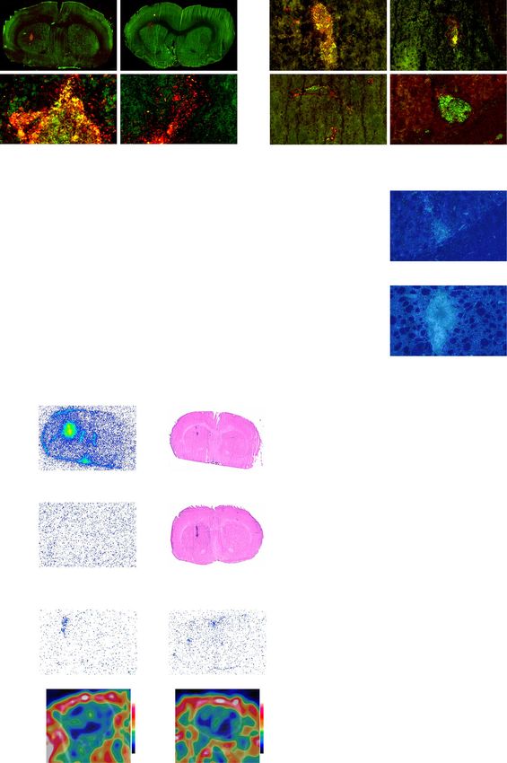
Elo et al. Journal of Neuroinflammation   (2021) 18:30                                                      Page 2 of 15

Background                                                    immune cell-mediated inflammatory response in bacillus
Most patients with a progressive form of multiple scler-      Calmette–Guérin-induced intrastriatal lesions was trig-
osis (MS) respond minimally to current disease-               gered with complete Freund’s adjuvant, resulting in an
modifying agents [1, 2], and many patients discontinue        active focal inflammatory lesion and a compromised
treatment because of undesirable side effects [3]. Thus,      blood–brain barrier during the acute phase of the in-
novel treatments are urgently needed. As activated mac-       flammation (day 14). After 3 months (day 90), the lesions
rophages in animals with experimental autoimmune en-          remodeled into well-defined chronic EAE lesions with
cephalomyelitis (EAE) [4, 5], a model of MS, express          repaired blood–brain barriers, which more accurately re-
folate receptor-β (FR-β), this represents an auspicious       sembled chronic MS in humans [15, 16].
target for treating MS.                                          The rats were randomly divided into 6 groups (n = 10/
   FRs overexpressed on cancer cells and activated mac-       group) according to health status, duration of disease,
rophages can be targeted with a novel folate-                 and intervention (Fig. 1) and were put on a folate-free
aminopterin derivative, EC2319 [6]. Folate-aminopterin        diet (5T0F:57W5; Testdiet, St. Louis, MO, USA) begin-
therapy reduces inflammation in acute myelin basic            ning 10 days before treatment. The rats in the acute
protein-induced EAE, but its effects on chronic forms         phase (day 14) groups were subcutaneously administered
are not known. There is evidence that the aminopterin         with EC2319 (750 nmol/kg of body weight/day, 400 ±
conjugate exerts antineoplastic and immunomodulatory          100 μL in saline) in the nuchal area 0, 3, 7, and 10 days
effects once it is internalized via FR binding, thereby       after lesion activation. The fDTH-EAE rats receiving
inhibiting dihydrofolate reductase and possibly suppress-     only saline and healthy Lewis rats treated with EC2319
ing immune cell proliferation and cytokine recruitment.       were used as controls. After the treatment, rats were im-
In association with central nervous system (CNS) in-          aged via PET/CT with 68Ga-FOL followed by sacrifice
flammation, a functional FR-β has been shown to be            for ex vivo gamma counting of excised tissues. The brain
present in a subpopulation of infiltrating inflammatory       cryosections were analyzed by autoradiography, hist-
macrophages identified as the cluster of differentiation      ology, and immunofluorescence. The rats in the chronic
68 (CD68)-positive subset of high major histocompatibil-      phase (day 90) groups underwent 68Ga-FOL PET/CT at
ity complex (MHC) II-expressing and high CD11b-               60 days after lesion activation as a baseline measurement
expressing cells, and folate-aminopterin therapy has sig-     prior to the initiation of the biweekly treatment with
nificantly reduced CD68-positive macrophages, inflam-         subcutaneous EC2319 (500 nmol/kg/day, 520 ± 75 μL)
mation, and demyelination [4]. Indeed, aminopterin            for 4 weeks. PET/CT imaging was repeated at day 90,
derivatives have gained interest because of their superior    and the animals were sacrificed for ex vivo analyses as
anti-inflammatory effects and improved safety profile         described above.
compared to methotrexate [7–9]. Here, we investigated            EC2319 is metabolically activated to release aminop-
the efficacy of subcutaneously administered EC2319 on         terin and an aminopterin adduct. Plasma samples ob-
lesion development during acute and chronic EAE in            tained from terminal blood samples were shipped frozen
rats. In addition to immunofluorescence and histological      to Endocyte, Inc. and stored at − 80 °C until thawing for
analyses, we used FR-targeted PET, a promising ap-            bioanalysis. EC2319, aminopterin, and the aminopterin
proach for imaging activated macrophages under inflam-        adduct were extracted from the plasma (50 μL) by solid-
matory conditions [5, 10–14]. For this, we used a 68Ga-       phase extraction. The samples were then eluted into a
labeled     1,4,7-triazacyclononane-1,4,7-triacetic  acid-    1.2-mL storage plate, evaporated, and reconstituted prior
conjugated folate (68Ga-FOL). Furthermore, we assessed        to ultra-performance liquid chromatography-tandem
the expression of FR-β in postmortem brain samples of         mass spectrometry. Incurred sample data were then gen-
MS patients to demonstrate the validity of this               erated by comparison with calibration curves from con-
approach.                                                     trol plasma samples spiked for each compound.

                                                              68
Methods                                                        Ga-FOL studies
Animals and study design                                      The precursor NOTA-folate was synthesized as previ-
Male Lewis rats (2–3 months, n = 60, 270 ± 23 g) were         ously described [13, 14]. 68Ga was obtained from a
                                                              68
purchased from Charles River (Sulzfeld, Germany). The            Ge/68Ga generator (Eckert & Ziegler Isotope Products,
rats were acclimated to the housing conditions for at         Burbank, CA, USA) by elution with 0.1 M HCl. 68Ga elu-
least 5 days prior to any experimental procedures. Food       ate (1.0 mL) was added to a mixture of 2-[4-(2-hydro-
and tap water were available ad libitum.                      xyethyl)piperazin-1-yl]ethanesulfonic acid (120 mg),
  A focal delayed-type hypersensitivity model of experi-      gentisic acid (0.3 mg), and NOTA-folate (17 μg) in
mental autoimmune encephalomyelitis (fDTH-EAE) was            120 μL TraceSELECT-grade water. The reaction mixture
established as previously described [5, 15–17]. Briefly, an   was heated at 80 °C for 10 min. After cooling to room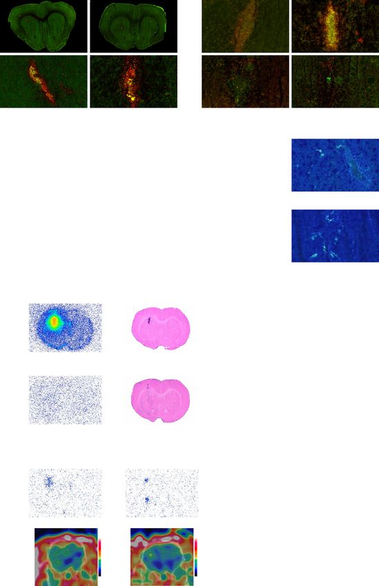
Elo et al. Journal of Neuroinflammation           (2021) 18:30                                                                             Page 3 of 15

                                                                                                     68Ga-FOL analyses
                              a                                                                         Histology
                                                                                                    Immunofluorescence

                                                                                             0 3 7 10 14              Day

                                                                                             EC2319,
                                                                                          750 nmol/kg/day
                                                                                              (n = 10)
                                                                                                Saline
                                                                            f DTH-             (n = 10)
                               Day –28                        Day 0           EAE
                              Stereotactic                Subcutaneous
                              injection of                  injection of                     0 3 7 10 14              Day
                               heat-killed                  heat-killed
                               BCG into                      TB in CFA
                                striatum                   to hind limb
                                                                                                     68Ga-FOL  analyses
                                                                                                         Histology
                                                                                                     Immunofluorescence

                                                                                                                      68Ga-FOL  analyses
                              b                                                  68Ga-FOL   in vivo analyses
                                                                                                                          Histology
                                                                                                                      Immunofluorescence

                                                                                             60     Bi-weekly for 4 weeks 90       Day

                                                                                                   EC2319, 500 nmol/kg/day
                                                                                                          (n = 10)

                                                                                                            Saline
                                                                            f DTH-                         (n = 10)
                               Day –28                        Day 0           EAE
                              Stereotactic                Subcutaneous
                              injection of                  injection of                     60     Bi-weekly for 4 weeks 90       Day
                               heat-killed                  heat-killed
                               BCG into                      TB in CFA
                                striatum                   to hind limb
                                                                                 68Ga-FOL   in vivo analyses          68Ga-FOL  analyses
                                                                                                                          Histology
                                                                                                                      Immunofluorescence

                                                                                             68Ga-FOL analyses
                              c                                                                 Histology
                                                                                            Immunofluorescence

                                                                                      0 3 7 10 14              Day

                                                                                        EC2319,
                                                                                     750 nmol/kg/day
                                                                                         (n = 10)
                                                                                         Saline
                                                                  control               (n = 10)
                                             Healthy Lewis rats
                                              (non-operated)
                                                                                      0 3 7 10 14              Day

                                                                                              68Ga-FOL analyses
                                                                                                 Histology
                                                                                             Immunofluorescence

 Fig. 1 Study protocol for fDTH-EAE rats during acute and chronic phases and for healthy Lewis rats. a fDTH-EAE rats with acute inflammation
 were administered EC2319 (750 nmol/kg/day) or saline on days 0, 3, 7, and 10 after disease activation and were used for 68Ga-FOL, histology, and
 immunofluorescence analyses on day 14. b fDTH-EAE rats with chronic inflammation were administered EC2319 (500 nmol/kg/day) or saline
 biweekly for 4 weeks after the baseline in vivo evaluation 60 days after disease activation and were sacrificed for 68Ga-FOL, histology, and
 immunofluorescence analyses on day 90. c Healthy rats were administered EC2319 (750 nmol/kg/day) or saline on days 0, 3, 7, and 10 and
 sacrificed on day 14 for further analyses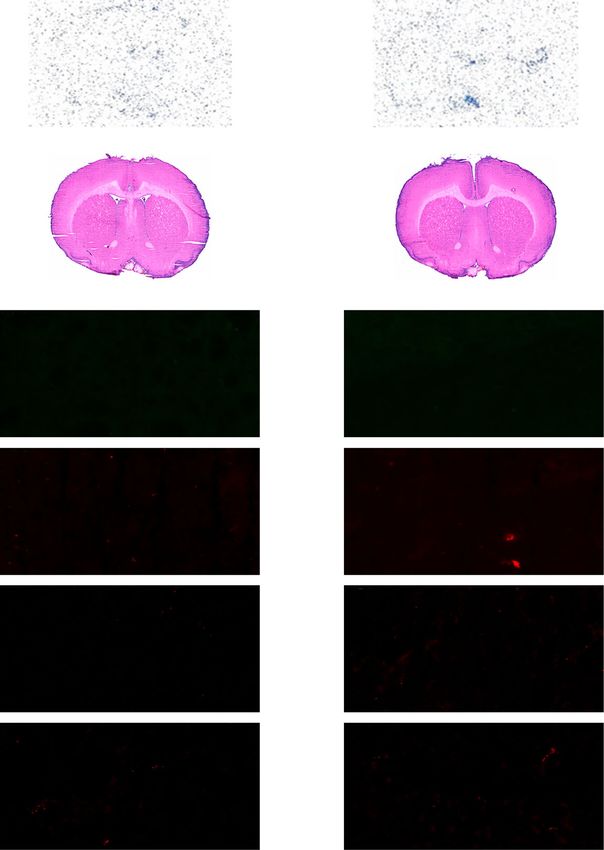
Elo et al. Journal of Neuroinflammation   (2021) 18:30                                                       Page 4 of 15

temperature, the pH was adjusted to neutral with 1 M          Then, the sections were incubated for 30 min at room
NaOH. The 68Ga-FOL was prepared in high radiochem-            temperature with primary anti-human FR-β (which also
ical purity (> 95%) with the molar activity of 20–40          recognizes rat FR-β; 1:50 dilution, m909; a kind gift from
MBq/nmol at the end of synthesis. The duration of             Professor Philip S. Low, Purdue University, West Lafa-
radiosynthesis was ~ 17 min.                                  yette, IN, USA) and anti-rat mannose receptor C type 1
   In vivo imaging was performed with an Inveon multi-        (MRC-1) (1:2000 dilution; Abcam, Cambridge, UK), or
modality small animal PET/CT device (Siemens Medical          with anti-rat inducible nitric oxide synthase (iNOS) (1:
Solutions, Knoxville, TN, USA). The rats were anesthe-        500 dilution; Abcam) antibodies for macrophages and
tized using a mixture of isoflurane and air (2.5%, 500        microglia, or with anti-human FR-β and anti-rat CD68
mL/min) on a heating pad, and a cannula was inserted          antibodies (1:1000 dilution; AbD Serotec, Hercules, CA,
into the tail vein. First, a 10-min CT was acquired for at-   USA) for macrophages/microglia. The sections were
tenuation correction and anatomical reference. Then,          then incubated with fluorochrome-labeled secondary
68
   Ga-FOL (45 ± 1 MBq, range 47–41 MBq) was injected          antibodies (1:100 dilution, anti-human Alexa Fluor 488
IV for a 30-min PET acquisition as previously described       or anti-rat Alexa Fluor 594; Invitrogen of Thermo Fisher
[5].                                                          Scientific, Waltham, MA, USA) and counterstained with
   The PET/CT data were reconstructed using a three-          hematoxylin before mounting with ProLong Gold anti-
dimensional ordered-subset expectation maximization           fade reagent (P36930; Life Technologies of Thermo
algorithm to time frames of 6 × 10 s, 3 × 20 s, 4 × 60 s, 3   Fisher Scientific). The sections were scanned using a
× 180 s, and 3 × 300 s. Representative PET/CT images          Pannoramic Midi fluorescence scanner (3D Histech,
were captured with Inveon Research Workplace v.4.1,           Budapest, Hungary) and analyzed with a Pannoramic
and the PET data were analyzed with Carimas v.2.9             viewer (3D Histech). The areas of positive staining or
(Turku PET Centre, Turku, Finland). Time frames of            loss of LFB staining were determined from three to four
17–30 min post-injection were used for quantitative PET       brain sections and averaged for each rat. The amount of
image analysis. Spherical regions of interest were defined    demyelination determined from the LFB staining was av-
in the lesion hemisphere and mirrored onto the contra-        eraged to the total area of lesion hemisphere for each rat
lateral brain hemisphere, which served as an internal ref-    and expressed as area-%. The percentage of immunopo-
erence region. Quantitative PET image analysis was            sitive FR-β signal from CD68-positive cells was deter-
complemented by defining regions of interest to the           mined from MS brain samples. The analysis was
heart left ventricle to obtain blood radioactivity concen-    performed by using automatic thresholding of positively
trations. The results are expressed as standardized up-       stained areas with the ImageJ v.1.48 software (National
take value ratios [5].                                        Institutes of Health, Bethesda, MD, USA). The iNOS/
   Immediately after imaging, the animals were sacrificed     MRC-1 staining ratio was calculated for each rat. The le-
and various tissues were excised, weighed, and measured       sion sizes were evaluated by manually defining regions
for radioactivity with a gamma counter (Triathler 3′′;        of interest on the H&E-stained sections from each rat.
Hidex, Turku, Finland). The brains were frozen and            Lymphocyte recruitment was quantified by determining
cryosectioned for digital autoradiography [5]. A subset       lymphocyte count densities from three nonoverlapping
of brain cryosections was used to evaluate the in vitro       areas within the lesion as observed with H&E staining.
binding of 68Ga-FOL [5]. The results are expressed as a       The areas were chosen according to the average density
percentage of the injected radioactivity dose per gram of     of recruited lymphocytes within the inflammatory lesion,
tissue (%ID/g), organ/blood ratio, and bound-to-free ra-      and the results are expressed as lymphocyte count dens-
tio [5].                                                      ity per millimeter squared.
   Analyses of in vivo stability and modeling of 68Ga-FOL
PET data (Logan plots) were performed as previously de-       Human tissue samples
scribed [5], with the exception that blood samples from       Human formalin-fixed paraffin-embedded tissue samples
healthy Lewis rats were withdrawn 5–30 min after 68Ga-        (n = 5 MS brain samples; n = 5 normal brain samples)
FOL injection (n = 4 per time point).                         were obtained from Auria Biobank (Turku University
                                                              Hospital, Turku, Finland). All samples had been taken at
Histology and immunofluorescence                              autopsy for histopathologic examination between 2001
Paraformaldehyde-fixed 10-μm sections were stained            and 2013. The samples presented homologous findings
with hematoxylin–eosin (H&E) and Luxol Fast Blue              within both groups. For the CD68, iNOS, and MRC-1
(LFB) with cresyl violet counterstain according to stand-     double immunofluorescence staining, the samples were
ard procedures. For double immunofluorescence stain-          stained as described above. For immunohistochemistry,
ing, the sections were first fixed with ice-cold acetone      the sections were stained as previously described for
for 3 min and washed with phosphate-buffered saline.          anti-FR-β    staining    [5].    Positivity  for   FR-β
Elo et al. Journal of Neuroinflammation (2021) 18:30 Page 5 of 15
immunohistochemistry is reported as negative, weak, treated group (Fig. 2c; 0.055 ± 0.018 [n = 9] versus 0.26
moderate, or strong on the basis of the staining ± 0.098 [n = 10], respectively, P = 0.0056). At day 90,
intensity. the chronic lesions had lower (but not statistically sig-
nificant) lymphocyte count densities in EC2319-treated
Cytokine and chemokine measurements in rat plasma than in saline-treated rats (Fig. 2c; 393.30 ± 84.66 mm2
Plasma levels of interferon γ (IFN-γ) and interleukins versus 729.80 ± 146.00 mm2, respectively, P = 0.065; n =
IL-1β, IL-4, IL-6, and IL-10 were measured in duplicates 8/group).
with Luminex assay according to the manufacturer’s in- In vitro 68Ga-FOL assays showed drastically lower
structions (MILLIPLEX MAP Rat Cytokine/Chemokine binding when the brain cryosections were first incubated
Magnetic Bead Panel, Merck Millipore, MA, USA). The with the folate-glucosamine blocking agent than in those
minimum detectable concentrations (pg/mL) for the without any blocking agent, confirming that tracer bind-
analytes were 14.6 (IFN-γ), 12.2 (IL-1β), 4.9 (IL-4), 73.2 ing is FR-specific (Fig. 2d, e). Tracer binding was signifi-
(IL-6), and 7.3 (IL-10). cantly lower in the brain sections of EC2319-treated rats
than in saline-treated rats (Fig. 2e; bound-to-free ratios,
Statistical analysis 2.47 ± 0.45 versus 4.93 ± 0.43, respectively, P = 0.0047;
All statistical analyses were performed with the Graph- n = 8/group). 68Ga-FOL revealed the inflammatory le-
Pad Prism v.7.01 software (Graph Pad Software Inc., La sions in in vivo PET/CT images and with ex vivo auto-
Jolla, CA, USA). The results are presented as means ± radiography. However, unlike the in vitro binding
SDs. Nonparametric Kruskal–Wallis tests with Mann– results, they did not show any therapeutic effect (Figs.
Whitney post hoc tests were used to compare tracer up- 2f, g, 3f, g).
take values in lesion hemispheres between the groups as According to ex vivo gamma counting, the radioactiv-
well as intragroup histological and immunofluorescence ity concentrations were significantly higher in the blood
data. Spearman’s correlation was used to analyze the re- and lower in lymph node and spleen tissues from
lationship between kinetic modeling and semiquantita- EC2319-treated rats than those in saline-treated rats
tive in vivo PET data. Two-way repeated-measures during the chronic phase of EAE (Table 1). The organ/
analysis of variance (ANOVA) was performed to com- blood ratios were markedly reduced in multiple organs,
pare cytokine levels between the groups. A result was including the adrenal glands, heart, kidneys, liver, lungs,
considered statistically significant with a P value of < lymph node, muscle, pancreas, salivary glands, skin,
0.05. skull, small intestine, spleen, and white adipose tissue
from EC2319-treated rats compared with those from
Results saline-treated rats (Table 2).
EC2319 reduces FR-β expression, lesion size, and iNOS/
MRC-1 ratio during chronic EAE
Biweekly EC2319 treatment was effective during the EC2319 is ineffective during the acute phase of focal EAE
chronic phase of fDTH-EAE. Immunofluorescence stain- During acute fDTH-EAE, no differences in FR-β or
ing revealed that FR-β expression was significantly re- CD68 immunofluorescence were observed between the
duced by 70% ± 10% in EC2319-treated rats compared EC2319- and saline-treated groups (Fig. 3a, c). With four
with saline-treated controls (Fig. 2a, c; 0.0019 ± 0.00067 therapeutic doses of EC2319 (750 nmol/kg of body
mm2 versus 0.0064 ± 0.0016 mm2, respectively, P = weight/day), there were also no differences in the iNOS/
0.017; n = 10/group). The area of CD68-positive cells MRC-1 ratio, the density of lymphocytes, lesion size, or
was smaller following treatment but did not reach statis- loss of LFB staining compared with those in saline-
tical significance (Fig. 2a, c; 0.010 ± 0.0021 mm2 [saline, treated rats (Fig. 3b, c). In vitro 68Ga-FOL binding assays
n = 8] versus 0.0053 ± 0.0011 mm2 [EC2319, n = 9], P = (Fig. 3d, e), in vivo PET/CT imaging, and ex vivo auto-
0.093). The demyelinated area assessed with LFB stain- radiography (Fig. 3f, g) similarly revealed no differences
ing tended to be lower in EC2319-treated rats compared between EC2319- and saline-treated rats. However,
to saline-treated rats in the lesion hemisphere, but the ex vivo gamma counting of excised tissues revealed
difference did not reach statistical significance (Fig. 2c; lower radioactivity in the heart, liver, muscle, pancreas,
0.012% ± 0.0096% versus 0.036% ± 0.024%, respectively, spleen, and white adipose tissues from the EC2319-
P = 0.093; n = 6/group). The iNOS/MRC-1 ratio was treated group but higher levels in the blood, brain, kid-
significantly reduced by 96% ± 2% in EC2319-treated neys, plasma, and skin samples from rats receiving only
rats (Fig. 2b, c; 15.59 ± 2.24 [saline, n = 3] versus 0.63 ± saline (Table 1). The 68Ga-FOL organ/blood ratios were
0.29 [EC2319, n = 3], P = 0.0027). Moreover, the lesion significantly lower in the adrenal glands, heart, liver,
size was reduced in EC2319-treated fDTH-EAE rats dur- lungs, lymph node, muscle, pancreas, skull, small intes-
ing the chronic phase compared with that in the saline- tine, spleen, and white adipose tissue, indicating thatElo et al. Journal of Neuroinflammation (2021) 18:30 Page 6 of 15
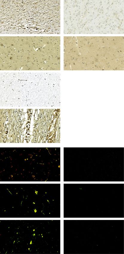
Chronic phase of fDTH-EAE, Day 90
a Saline EC2319 b Saline EC2319
FR- iNOS
Merged
FR- CD68 Merged
FR- MRC-1
Merged
c Saline EC2319
CD68 positivity area (mm2) FR-β positivity area (mm2)
0.020 30 PElo et al. Journal of Neuroinflammation (2021) 18:30 Page 7 of 15
Acute phase of fDTH-EAE, Day 14
a Saline EC2319 b Saline EC2319
FR- iNOS
Merged
FR- CD68 Merged
FR- MRC-1
Merged
c Saline EC2319
CD68 positivity area (mm2) FR-β positivity area (mm2)
0.020 30
NS EC2319
NS 2000
iNOS/MRC-1 ratio
NS
Lymphocyte count
0.015
density (mm2)
1500 20
0.010
1000
10
0.005 500
0 0 0
LFB
0.05 NS 5 Demyelination (area-%) 0.15 Saline
NS NS
Lesion area (mm2)
0.04 4
0.10
0.03 3
0.02 2
0.05
0.01 1
0 0 0
d 68
Ga-FOL e 20
PElo et al. Journal of Neuroinflammation (2021) 18:30 Page 8 of 15
Table 1 Ex vivo biodistribution (percentage of injected dose per gram of tissue) of 68Ga-FOL at 30 min post-injection in rats with
fDTH-EAE after treatment with EC2319 or saline
Organ Day 14 Day 90
EC2319 (n = 8) Saline (n = 8) P value Treatment effect EC2319 (n = 10) Saline (n = 10) P value Treatment effect
Adrenal glands 0.24 ± 0.04 0.25 ± 0.03 0.83 ↓ 0.16 ± 0.04 0.20 ± 0.05 0.05 ↓
Blood 0.03 ± 0.01 0.014 ± 0.002 0.003 ↑ 0.06 ± 0.03 0.03 ± 0.03 0.03 ↑
Brain 0.020 ± 0.004 0.011 ± 0.003 < 0.001 ↑ 0.02 ± 0.01 0.02 ± 0.01 0.46 ↑
Heart 0.12 ± 0.02 0.19 ± 0.06 0.02 ↓ 0.09 ± 0.02 0.09 ± 0.02 0.95 ↑
Kidneys 12.80 ± 2.02 6.71 ± 1.34 < 0.001 ↑ 8.21 ± 3.21 8.51 ± 1.99 0.81 ↓
Liver 0.30 ± 0.05 0.47 ± 0.13 0.007 ↓ 0.22 ± 0.09 0.23 ± 0.06 0.74 ↓
Lungs 0.14 ± 0.02 0.17 ± 0.05 0.11 ↓ 0.11 ± 0.02 0.12 ± 0.02 0.30 ↓
Lymph node 1.00 ± 0.17 0.92 ± 0.13 0.30 ↑ 0.75 ± 0.22 0.95 ± 0.22 0.05 ↓
Muscle 0.07 ± 0.01 0.09 ± 0.02 0.01 ↓ 0.05 ± 0.02 0.07 ± 0.02 0.09 ↓
Pancreas 0.26 ± 0.04 0.38 ± 0.09 0.006 ↓ 0.20 ± 0.06 0.24 ± 0.06 0.26 ↓
Plasma 0.05 ± 0.01 0.03 ± 0.02 0.05 ↑ 0.08 ± 0.03 0.06 ± 0.04 0.17 ↑
Salivary gland ND ND ND ND 0.61 ± 0.33 0.82 ± 0.20 0.11 ↓
Skin 0.26 ± 0.10 0.14 ± 0.05 0.02 ↑ 0.15 ± 0.04 0.17 ± 0.03 0.32 ↓
Skull 0.08 ± 0.02 0.09 ± 0.02 0.48 ↓ 0.06 ± 0.03 0.06 ± 0.02 0.82 ↓
Small intestine 0.44 ± 0.09 0.52 ± 0.11 0.1 ↓ 0.30 ± 0.08 0.37 ± 0.10 0.10 ↓
Spleen 0.92 ± 0.32 1.52 ± 0.38 0.004 ↓ 0.56 ± 0.31 0.91 ± 0.39 0.04 ↓
Urine 3.01 ± 2.69 2.35 ± 0.98 0.53 ↑ 7.22 ± 5.62 9.66 ± 12.70 0.59 ↓
White adipose 0.05 ± 0.02 0.07 ± 0.02 0.04 ↓ 0.031 ± 0.01 0.035 ± 0.01 0.27 ↓
Results are means ± SDs
ND not determined
Table 2 Ex vivo biodistribution (organ/blood ratio) of 68Ga-FOL at 30 min post-injection in rats with fDTH-EAE after treatment with
EC2319 or saline
Organ Day 14 Day 90
EC2319 (n = 8) Saline (n = 8) P value Treatment effect EC2319 (n = 10) Saline (n = 10) P value Treatment effect
Adrenal glands 9.55 ± 4.32 17.55 ± 2.91 < 0.001 ↓ 3.02 ± 1.65 9.89 ± 6.92 0.01 ↓
Brain 0.73 ± 0.16 0.80 ± 0.24 0.51 ↓ 0.48 ± 0.39 0.90 ± 0.58 0.08 ↓
Heart 4.94 ± 2.68 13.72 ± 5.17 0.002 ↓ 1.70 ± 0.81 4.50 ± 3.35 0.03 ↓
Kidneys 483.01 ± 135.74 480.04 ± 108.58 0.96 ↑ 168.51 ± 119.09 371.09 ± 188.35 0.01 ↓
Liver 12.03 ± 5.98 34.06 ± 10.74 < 0.001 ↓ 4.49 ± 3.6 11.82 ± 8.8 0.03 ↓
Lungs 5.57 ± 2.54 12.44 ± 3.99 0.0015 ↓ 2.14 ± 0.99 5.99 ± 4.21 0.02 ↓
Lymph node 38.28 ± 13.38 64.84 ± 6.42 < 0.001 ↓ 14.83 ± 9.51 46.01 ± 30.30 0.01 ↓
Muscle 2.82 ± 1.16 6.85 ± 1.97 < 0.001 ↓ 1.04 ± 0.58 3.47 ± 2.54 0.02 ↓
Pancreas 10.19 ± 3.83 27.53 ± 7.49 < 0.001 ↓ 3.90 ± 2.24 11.94 ± 8.63 0.02 ↓
Plasma 1.85 ± 0.78 2.17 ± 1.58 0.62 ↓ 1.43 ± 0.45 1.71 ± 0.11 0.08 ↓
Salivary gland ND ND ND ND 11.98 ± 9.84 39.39 ± 24.62 0.01 ↓
Skin 10.53 ± 8.71 10.30 ± 4.02 0.95 ↑ 2.47 ± 1.65 7.44 ± 4.34 0.01 ↓
Skull 3.23 ± 1.58 6.32 ± 1.63 0.002 ↓ 1.19 ± 0.98 2.99 ± 2.28 0.04 ↓
Small intestine 16.99 ± 7.46 37.82 ± 9.98 < 0.001 ↓ 5.71 ± 3.10 18.63 ± 14.02 0.02 ↓
Spleen 36.02 ± 17.56 107.40 ± 24.24 < 0.001 ↓ 9.68 ± 3.79 47.82 ± 39.15 0.01 ↓
Urine 102.82 ± 67.31 167.88 ± 76.67 0.09 ↓ 132.55 ± 92.40 218.37 ± 168.11 0.18 ↓
White adipose 1.93 ± 0.81 5.07 ± 1.32 < 0.001 ↓ 0.61 ± 0.36 1.53 ± 0.88 0.01 ↓
Results are expressed as mean ± SDs
ND not determinedElo et al. Journal of Neuroinflammation (2021) 18:30 Page 9 of 15
EC2319 exerts a systemic anti-inflammatory effect postmortem brain sections from MS patients and con-
(Table 2). trols. FR-β was moderately expressed in normal-
Ex vivo biodistribution results of 68Ga-FOL are pre- appearing white matter but weakly expressed or absent
sented in Table 3. In general, the biodistribution was in normal-appearing gray matter and in chronically in-
similar to that reported previously [5]; the highest up- active white matter plaques compared with normal hu-
takes were seen in the urinary bladder and FR-positive man brain tissues of corresponding anatomical areas
kidneys. The results indicate that the administration of (Fig. 4a–c). In addition, the chronic active lesions dis-
EC2319 systemically reduces the expression of FR-β and played moderate levels of FR-β at the border of chronic
thus its availability for 68Ga-FOL, thereby augmenting lesions (Fig. 4d), typically in areas known to exhibit mac-
the proportion of free 68Ga-FOL, which is detected in rophages [18]. Double immunofluorescence staining re-
circulation, lipophilic tissues, and kidneys. This view is vealed that FR-β colocalized with CD68, iNOS, and
also supported by the organ/blood ratio data (Table 3). MRC-1 in MS brain tissue samples but not in normal
The plasma cytokine measurements from fDTH-EAE brain (Fig. 4e–g), and occasionally, the blood vessels at
and healthy Lewis rats revealed no significant differences the lesion sites showed immunopositivity for FR-β.
in IFN-γ, IL-4, and IL-6 concentrations between the Quantification revealed that 62.7% ± 13.0% (n = 5) of
EC2319- and saline-treated groups (Table 4). However, CD68-positive cells in MS brain lesions were FR-β-
EC2319 treatment of healthy Lewis rats reduced the levels positive.
of IL-1β (P = 0.02) and IL-10 (P = 0.03). In addition, there
were differences in baseline levels (in saline-treated EC2319 was well tolerated in rats
groups): plasma concentrations of IFN-γ, IL-1β, IL-4, and EC2319 treatment was safe and well tolerated by the
IL-10 were significantly lower in chronic fDTH-EAE ver- rats, with no effect on body weight in any of the studied
sus healthy rats (P < 0.05), and IL-6 was significantly lower groups (Fig. 5a–c). At the time of the PET studies, the
in acute fDTH-EAE versus healthy rats (P < 0.05). plasma levels of EC2319 and its active metabolites were
under the detection limits of ultra-performance liquid
FR-β expression is increased in MS brain samples chromatography-tandem mass spectrometry (the cali-
To demonstrate the translational relevance of these find- brated ranges were 3.0–600 ng/mL for EC2319 and 0.3–
ings, we assessed the expression of FR-β using 120 ng/mL for both aminopterin and the aminopterin
Table 3 Ex vivo biodistribution of 68Ga-FOL at 30 min post-injection on day 14 in healthy Lewis rats
Organ %ID/g Organ/blood ratio
EC2319 (n = 10) Saline (n = 10) P value Treatment effect EC2319 (n = 8) Saline (n = 8) P value Treatment effect
Adrenal glands 0.31 ± 0.09 0.35 ± 0.06 0.25 ↓ 5.92 ± 1.69 10.80 ± 2.19 < 0.001 ↓
Blood 0.05 ± 0.02 0.03 ± 0.01 0.004 ↑ ND ND ND ND
Brain 0.04 ± 0.01 0.02 ± 0.003 < 0.001 ↑ 0.73 ± 0.13 0.59 ± 0.09 0.027 ↑
Heart 0.18 ± 0.08 0.20 ± 0.03 0.37 ↓ 3.35 ± 1.51 6.19 ± 0.85 < 0.001 ↓
Kidneys 13.18 ± 4.39 6.17 ± 1.13 < 0.001 ↑ 252.84 ± 84.26 170.46 ± 18.89 0.037 ↑
Liver 0.26 ± 0.07 0.47 ± 0.15 0.03 ↓ 4.89 ± 1.31 14.92 ± 4.85 < 0.001 ↓
Lungs 0.16 ± 0.03 0.22 ± 0.03 < 0.001 ↓ 3.03 ± 0.58 6.94 ± 0.79 < 0.001 ↓
Lymph node 0.94 ± 0.25 0.88 ± 0.08 0.52 ↑ 17.96 ± 4.75 27.35 ± 2.01 < 0.001 ↓
Muscle 0.11 ± 0.05 0.09 ± 0.01 0.12 ↑ 2.15 ± 0.89 2.66 ± 0.28 0.17 ↓
Pancreas 0.29 ± 0.06 0.36 ± 0.04 0.16 ↓ 5.59 ± 1.05 9.96 ± 1.42 < 0.001 ↓
Plasma 0.09 ± 0.03 0.06 ± 0.02 0.01 ↑ 1.73 ± 0.49 1.95 ± 0.61 0.89 ↓
Salivary gland 1.01 ± 0.35 1.24 ± 0.21 0.10 ↓ 19.28 ± 6.74 38.39 ± 6.48 < 0.001 ↓
Skin 0.23 ± 0.07 0.21 ± 0.08 0.69 ↑ 4.33 ± 1.32 5.99 ± 2.08 0.04 ↓
Skull 0.07 ± 0.03 0.15 ± 0.04 < 0.001 ↓ 1.32 ± 0.54 4.81 ± 1.16 < 0.001 ↓
Small intestine 0.56 ± 0.15 0.78 ± 0.10 0.002 ↓ 10.70 ± 2.87 24.30 ± 2.49 < 0.001 ↓
Spleen 0.75 ± 0.31 1.83 ± 0.37 < 0.001 ↓ 14.45 ± 5.88 55.95 ± 11.99 < 0.001 ↓
Urine 3.00 ± 2.08 2.86 ± 0.02 0.32 ↑ 57.62 ± 39.86 92.25 ± 39.95 0.13 ↓
White adipose 0.05 ± 0.02 0.03 ± 0.01 0.004 ↑ 1.04 ± 0.17 1.45 ± 0.55 0.04 ↓
Results are expressed as mean ± SDs
ND not determinedElo et al. Journal of Neuroinflammation (2021) 18:30 Page 10 of 15
Table 4 Rat plasma levels of cytokines (pg/mL)
Cytokine fDTH-EAE day 14 fDTH-EAE day 90 Healthy Lewis rats
EC2319 (n = Saline (n = 4– P EC2319 (n = 2– Saline (n = 5– P EC2319 (n = 4– Saline (n = 3– P
2) 6) value 4) 7) value 6) 7) value
IFN-γ ND 202 ± 152 0.92 216 ± 184 95 ± 77b 0.29 486 ± 507 558 ± 755b 0.81
b b
IL-1β 662 ± 614 441 ± 284 0.53 205 ± 178 203 ± 79 0.97 321 ± 167 562 ± 316 0.02
IL-4 ND 42 ± 29 0.60 28 ± 17 29 ± 22b 0.96 70 ± 70 234 ± 237b 0.09
a a
IL-6 ND 442 ± 266 0.74 ND ND ND 486 ± 386 5210 ± 4146 0.06
IL-10 780 ± 548 604 ± 290 0.58 417 ± 259 395 ± 121b 0.82 479 ± 194 700 ± 297b 0.03
Results are means ± SDs. P values are from two-way repeated-measures ANOVA
ND not determined
a
fDTH-EAE day 14 versus healthy Lewis rats P < 0.05
b
fDTH-EAE day 90 versus healthy Lewis rats P < 0.05
adduct; data not shown). In addition, healthy rats treated finding, EC2319 is therefore most likely targeting iNOS-
with EC2319 or saline showed no lesions; no immuno- positive pro-inflammatory cells rather than MRC-1-
positivity for FR-β, CD68, iNOS, or MRC-1; and no up- positive cells. This supports the view that EC2319 can
take of 68Ga-FOL in the brain (Fig. 5d). help to regulate the inflammatory processes in the CNS
that are impaired in acute and chronic EAE [26] and also
Discussion in MS [27].
In this work, we evaluated the efficacy of the novel EC2319 represents the best-in-class folate-aminopterin
folate-aminopterin conjugate EC2319 for the treatment conjugate with a similar mechanism of action as
of acute and chronic fDTH-EAE, a rat model of MS. EC0746, the first compound of this class, differing only
fDTH-EAE is a clinically relevant model for assessing le- in the linker design [8]. In a FR-dependent manner,
sion characteristics, immune cell populations, and ther- EC2319 induces cell cycle arrest (anti-proliferation),
apy responses [16, 17]. We report here, for the first time, modulates inflammatory cytokine/chemokine responses,
that EC2319 effectively reduces lesion size, FR-β expres- and demonstrates both local and systemic anti-
sion, iNOS/MRC-1 ratio, and 68Ga-FOL binding in vitro inflammatory response. In addition, EC2319 shuts down
during the chronic phase of neuroinflammation, al- a subset of inflammatory monocytes in multiple disease
though the effects were not apparent during the acute models (all part of a separate manuscript that is cur-
phase. Most intriguing, however, we found that FR-β is rently under review). Here in our study, EC2319 reduced
expressed in the brain lesions of patients with MS. the lesion size in the fDTH-EAE rat model. This conse-
In line with previous studies on inflammatory auto- quently appeared to cause the reduction of FR-β immu-
immune diseases [19, 20], we demonstrated that the FR- nopositive signal and the restoration of iNOS/MRC-1
β expressed in EAE lesions colocalized with iNOS, which equilibrium. FR-β is largely absent from other cells
is expressed by pro-inflammatory macrophages/micro- known to infiltrate the CNS in the EAE rat model, such
glia during acute and chronic phases of inflammation. as T lymphocytes [28]. This data suggest that the likely
Although EC2319 did not alter the number of iNOS- targets of EC2319 are the inflammatory CD68-positive
positive cells, it did restore the iNOS/MRC-1 equilib- cells, which infiltrate the CNS during the active phase of
rium in rats with fDTH-EAE. Similar effects were re- inflammation. In addition, these cells expressing func-
ported in EAE models treated with flavocoxid, fasudil, or tional FR-β are present in both CNS and peripheral sites
exosomes from bone marrow mesenchymal stem cells, of inflammation [28], and thus, the suppression of per-
but these agents have not been tested in clinical trials ipheral immune cells following EC2319 treatment may
[21–23]. The shift in macrophage/microglia polarization further explain the reduced lesion size observed in the
toward the iNOS-positive (M1) and away from the chronic phase of fDTH-EAE.
MRC-1-positive (M2) phenotype in relapsing EAE is Folate-conjugated therapies, such as FR-mediated anti-
known to predict inflammation severity [24]; thus, re- folates or FR-targeted immunotherapies, have shown ef-
storing the equilibrium between M1- and M2-type cells ficacy for the treatment of inflammatory conditions,
is important for recovery [24, 25]. Our data suggests that including early rheumatoid arthritis in animal models
FR-β is expressed only in a certain subpopulation of [7]. However, there is very little information about their
CD68-positive cells in fDTH-EAE. As observed during efficacy in chronic inflammatory conditions [7, 8]. Our
the chronic phase of inflammation, most of the FR-β results suggest that EC2319 may be an effective therapy
immunopositive signal appears to originate from iNOS- for patients with a chronic progressive form of MS, for
positive cells in the lesion (Fig. 2b). Based on this which there are very few effective therapies [2]. ThisElo et al. Journal of Neuroinflammation (2021) 18:30 Page 11 of 15
Multiple sclerosis Normal
a
FR-β
b
Immunohistochemistry
c
FR-β
d
e
FR- CD68
Merged
f
Immunofluorescence
FR- iNOS
Merged
g
FR- MRC-1
Merged
Fig. 4 Immunohistochemistry with postmortem sections from MS and normal human brains. a Immunohistochemistry reveals FR-β expression in
normal-appearing white matter from a patient with secondary progressive MS (left) but not in the white matter from a normal brain (right). b
Moderate FR-β expression is observed in the normal-appearing gray matter from a patient with secondary progressive MS (left) but not in a
normal brain (right). c Chronic inactive lesions display minimal or no FR-β expression. d FR-β-immunoreactive macrophages border chronic active
plaques. Immunofluorescence staining reveals that FR-β colocalizes with CD68 (e), iNOS (f), and MRC-1 (g) in normal-appearing white matter from
a patient with secondary progressive MS (left) but not in the white matter of a normal brain (right). Scale bars, 50 μm. Red arrows indicate FR-β
positivity, and white arrows indicate colocalization of FR-β and CD68, iNOS, or MRC-1
finding may potentially expand the therapeutic indica- inflammatory disorders. We show that the lesions in hu-
tions for folate-aminopterin therapies that were initially man tissue samples from patients with chronic progres-
limited to the treatment of acute peripheral sive MS have FR-β-positive cells similarly to the lesionsElo et al. Journal of Neuroinflammation (2021) 18:30 Page 12 of 15
a Acute fDTH-EAE
b Chronic fDTH-EAE
c Healthy
500 Saline 550 Saline 400 Saline
EC2319 EC2319 EC2319
500
Weight (g)
450 350
450
400 300
400
P = 0.95 P = 0.74 P = 0.79
350 350 250
0 5 10 15 60 70 80 90 0 5 10 15
Time (days) Time (days) Time (days)
d Healthy controls, day 14
Saline EC2319
autoradiography
autoradiography
Ex vivo
Ex vivo
L R L R
H&E
H&E
L R L R
FR-β
FR-β
CD68
CD68
iNOS
iNOS
MRC-1
MRC-1
Fig. 5 Body weights, digital autoradiography, histology, and immunofluorescence of healthy rats. The body weights of fDTH-EAE rats during
acute (a) and chronic (b) phases of the disease and of healthy Lewis rats (c) with EC2319 or saline treatment. d Ex vivo 68Ga-FOL
autoradiography, H&E histology, FR-β, CD68, iNOS, and MRC-1 immunofluorescence staining. Scale bars, 50 μm (high power) and 2 mm
(low power)Elo et al. Journal of Neuroinflammation (2021) 18:30 Page 13 of 15
in the fDTH-EAE animal model suggesting the transla- phase of inflammation is likely lower in the fDTH-EAE
tional potential of these findings. Brain samples from brain, limiting the anti-inflammatory efficacy of EC2319.
MS patients were obtained from late-stage chronic le- As the inflammation progresses toward the chronic
sions known to have significantly lower numbers of phase, the increase in FR-β expression facilitates EC2319
CD68-positive cells than in acute MS lesions [29]. This binding and activity. Regardless of the phase of inflam-
explains why we detected only a few FR-β-positive cells mation, however, fDTH-EAE exhibits only a focal lesion
in the lesions. Based on our data, however, the majority and hence very mild clinical symptoms during acute and
of these CD68-positive cells expressed FR-β. In addition, chronic phases of the disease limiting the capability to
we observed occasional FR-β positivity in lesion vascula- investigate EC2319 therapy effects in reducing the clin-
ture that could possibly be due to CD68-positive macro- ical severity of the disease in fDTH-EAE [15, 16]. The
phages surrounding or entering the blood vessels. It is observed therapeutic effect during the chronic phase
noteworthy that although BCG-induced chronic DTH may also be partly attributable to nontargeted effects of
lesions closely resemble those observed in the progres- EC2319 metabolites. However, like EC0746, EC2319
sive phase of MS [15], there is little evidence that this demonstrates FR-specific activity in vitro, as well as in
EAE model could mimic chronic inactive lesions typic- animal models of adjuvant arthritis, anti-glomerular
ally present in late stages of progressive MS limiting the basement membrane glomerulonephritis, and experi-
extrapolation of these findings to humans. Most import- mental autoimmune uveitis (data not shown). The ap-
antly, as EC2319 treatment reduced FR-β-positive cells parent alteration of 68Ga-FOL uptake in normal tissues
in the focal EAE lesions, further studies are needed to of both healthy rats and those with fDTH-EAE may be
determine if this would affect the long-term efficacy and an artifact of the folate-deficient diet and FR competi-
safety of folate-aminopterin therapies, which rely on FR- tion following EC2319 treatment. However, EC2319 is
mediated endocytosis of the anti-inflammatory drug also likely to have a systemic effect via FR-positive mac-
conjugate. In addition, EC2319 therapy had no signifi- rophages outside CNS that typically respond to antifo-
cant effect on cytokine levels in plasma samples col- late therapy.
lected in both acute and chronic phases of fDTH-EAE As a crude measurement of gross toxicity, the absence of
suggesting a mild systemic inflammation at the end of any effect of EC2319 on animal body weight suggests that it
the study in this model. Interestingly, both pro- was safe and well-tolerated. However, some of the rats
inflammatory cytokines (INF-γ, IL-1β) and anti- demonstrated enlarged spleens with vesicles irrespective of
inflammatory cytokines (IL-4, IL-10) were significantly their health status, disease duration, or intervention. This
lower in fDTH-EAE rats during chronic inflammation might have been a result of the folate-deficient diet used
(saline-treated) compared to healthy Lewis rats. This during the experimental protocol, as folate (B9 vitamin) de-
may refer to impaired immunoregulation during chronic ficiency can lead to mild-to-moderate megaloblastic
inflammation in fDTH-EAE, which may contribute to anemia, wherein macrocytic erythrocytes are sequestered in
triggering and sustaining inflammation. Although the the spleen as multiple small splenic lesions [30].
mechanisms of action were beyond the scope of the
current study, we recognize that brain cytokine levels Conclusions
may be more informative regarding the mode of action The results presented here demonstrate FR-β expression
than the plasma levels measured in this work. in lesions in rats with fDTH-EAE and in patients with
It is not clear why EC2319 was not effective in the MS. EC2319, a folate-aminopterin drug conjugate, ap-
acute phase of fDTH-EAE. Another folic acid- pears to be safe for use during acute and chronic fDTH-
conjugated aminopterin analog, EC0746, was highly ef- EAE. EC2319 effectively attenuated the inflammation
fective in the acute phase of myelin basic protein- and lesion burden in rats with chronic EAE, but not dur-
induced EAE, improving disease-related scores and re- ing the acute phase of inflammation. Although short-
ducing inflammation and demyelination [4]. However, term treatment with EC2319 demonstrated beneficial ef-
the discrepancy may reflect the inherent differences in fects in chronic EAE lesions, its long-term efficacy and
the disease models. In rats, myelin basic protein-induced safety remain to be determined. For the first time, we
EAE induces a severe and acute disseminated inflamma- show that the MS patients have FR-β-positive cells in
tory response throughout the brain and spinal cord with chronic active plaques, which indicates the translational
overt blood–brain barrier breakdown [4, 26], whereas relevance of these findings.
fDTH-EAE initially induces small focal inflammatory le-
sions before progressing to a chronic stage with more Abbreviations
diffuse inflammation and widespread macrophage/ %ID/g: Percentage of injected radioactivity dose per gram of tissue; 68Ga-
FOL: 68Ga-labeled 1,4,7-triazacyclononane-1,4,7-triacetic acid-conjugated fol-
microglia activation [16]. Therefore, the relative expres- ate; CD68: Cluster of differentiation 68; EAE: Experimental autoimmune
sion of FR-β on CD68-positive cells during the acute encephalomyelitis; EC2319: Folate-conjugated aminopterin; fDTH-EAE: FocalElo et al. Journal of Neuroinflammation (2021) 18:30 Page 14 of 15
delayed-type hypersensitivity model of experimental autoimmune enceph- autoimmune uveitis and autoimmune encephalomyelitis. Clin Immunol.
alomyelitis; FR-β: Folate receptor-β; H&E: Hematoxylin–eosin; iNOS: Inducible 2014;150:64–77.
nitric oxide synthase; MRC-1: Mannose receptor C type 1; MS: Multiple 5. Elo P, Li XG, Liljenbäck H, et al. Folate receptor-targeted positron emission
sclerosis tomography of experimental autoimmune encephalomyelitis. J
Neuroinflammation. 2019;16:252.
Acknowledgements 6. Vlahov IR, Leamon CP, You F, et al. Antifolate conjugates for treating
We thank Aake Honkaniemi, Erica Nyman, Marja-Riitta Kajaala, and Timo Kat- inflammation. US patent application. 2018. US20180280528A1.
telus for providing excellent technical assistance. 7. Martin-Mola E, Balsa A. Infectious complications of biologic agents. Rheum
Dis Clin N Am. 2009;35:183–99.
Authors’ contributions 8. Lu Y, Stinnette TW, Westrick E, et al. Treatment of experimental adjuvant
PE, YJL, PL, SJ, LA, and AR conceived and designed the experiments. PE, HL, arthritis with a novel folate receptor-targeted folic acid aminopterin
and JV performed the animal experiments. XGL, OM, and MM performed the conjugate. Arthritis Res Ther. 2011;13:R56.
(radio) synthesis for this study. PE, YJL, MS, and MP analyzed the data. PE, 9. Van der Heijden JW, Oerlemans R, Dijkmans BA. Folate receptor beta as a
XGL, MG, YJL, AS, MS, MP, PL, JK, SJ, LA, and AR interpreted the data. PE potential delivery route for novel folate antagonists to macrophages in the
drafted the manuscript. PE, XGL, HL, MG, YJL, AS, MS, MP, PL, JK, SJ, LA, and synovial tissue of rheumatoid arthritis patients. Arthritis Rheum. 2009;60:12–21.
AR critically revised the manuscript for important intellectual content. All 10. Schniering J, Benešová M, Brunner M, et al. 18F-AzaFol for detection of
authors read and approved the final manuscript. folate receptor-β positive macrophages in experimental interstitial lung
disease - a proof-of-concept study. Front Immunol. 2019;10:2724.
Funding 11. Chandrupatla DMSH, Jansen G, Mantel E, et al. Imaging and methotrexate
This research was financially supported by the Jane and Aatos Erkko response monitoring of systemic inflammation in arthritic rats employing
Foundation, the State Research Funding of Turku University Hospital the macrophage PET tracer [18F]fluoro-PEG-folate. Contrast Media Mol
(ERVA#13303), the Sigrid Jusélius Foundation, the Instrumentarium Imaging. 2018;2018:8092781.
Foundation, the Alfred Kordelin Foundation, the Orion Foundation, and the 12. Kularatne SA, Belanger MJ, Meng X, et al. Comparative analysis of folate
Drug Research Doctoral Programme of the University of Turku Graduate derived PET imaging agents with [18F]-2-fluoro-2-deoxy-d-glucose using a
School. rodent inflammatory paw model. Mol Pharm. 2013;10:3103–11.
13. Chen Q, Meng X, McQuade P, et al. Synthesis and preclinical evaluation of
Availability of data and materials folate-NOTA-Al18F for PET imaging of folate-receptor-positive tumors. Mol
Data supporting the conclusions of this article are presented in the Pharm. 2016;13:1520–7.
manuscript. 14. Silvola JMU, Li XG, Virta J, et al. Aluminum fluoride-18 labeled folate enables
in vivo detection of atherosclerotic plaque inflammation by positron
Ethics approval and consent to participate emission tomography. Sci Rep. 2018;8:9720.
All animal experiments were approved by the National Animal Experiment 15. Serres S, Anthony DC, Jiang Y, et al. Comparison of MRI signatures in
Board of Finland and the Regional State Administrative Agency for Southern pattern I and II multiple sclerosis models. NMR Biomed. 2009;22:1014–24.
Finland (license number: ESAVI/2979/2018) and conducted in accordance 16. Matyszak MK, Perry VH. Demyelination in the central nervous system
with the relevant European Union directive. The study of archival human following a delayed-type hypersensitivity response to bacillus Calmette-
tissues was approved by the Auria Biobank Scientific Steering Committee Guerin. Neuroscience. 1995;64:967–77.
(decision AB19-2623). According to the Finnish Biobank Act (688/2012), sep- 17. Airas L, Dickens AM, Elo P, et al. In vivo PET imaging demonstrates
arate informed consent from individual patients is not needed. diminished microglial activation after fingolimod treatment in an animal
model of multiple sclerosis. J Nucl Med. 2015;56:305–10.
Consent for publication 18. Frischer JM, Bramow S, Dal-Bianco A, et al. The relation between
Not applicable. inflammation and neurodegeneration in multiple sclerosis brains. Brain.
2009;132:1175–89.
Competing interests 19. Xia W, Hilgenbrink AR, Matteson EL, Lockwood MB, Cheng JX, Low PS. A
Michael Pugh and Yingjuan June Lu are full-time employees of Novartis. All functional folate receptor is induced during macrophage activation and can
other authors declare no potential conflicts of interest. be used to target drugs to activated macrophages. Blood. 2009;113:438–46.
20. Nagai T, Tanaka M, Tsuneyoshi Y, Matsushita K, Sunahara N, Matsuda T. In
Author details vitro and in vivo efficacy of a recombinant immunotoxin against folate
1
Turku PET Centre, University of Turku, Turku, Finland. 2Turku PET Centre, Åbo receptor beta on the activation and proliferation of rheumatoid arthritis
Akademi University, Turku, Finland. 3Turku Center for Disease Modeling, synovial cells. Arthritis Rheum. 2006;54:3126–34.
University of Turku, Turku, Finland. 4Department of Pathology, Turku 21. Kong W, Hooper KM, Ganea D. The natural dual cyclooxygenase and 5-
University Hospital and Institute of Biomedicine, University of Turku, Turku, lipoxygenase inhibitor flavocoxid is protective in EAE through effects on
Finland. 5Turku PET Centre, Turku University Hospital, Turku, Finland. Th1/Th17 differentiation and macrophage/microglia activation. Brain Behav
6
Department of Chemistry, Purdue University, West Lafayette, IN, USA. Immun. 2016;53:59–71.
7
Endocyte, Inc., now part of Novartis Institutes for Biomedical Research, West 22. Liu C, Li Y, Yu J, et al. Targeting the shift from M1 to M2 macrophages in
Lafayette, IN, USA. 8MediCity Research Laboratory, University of Turku, Turku, experimental autoimmune encephalomyelitis mice treated with Fasudil.
Finland. 9Department of Neurology, Turku University Hospital, Turku, Finland. PLoS One. 2013;8:e54841.
23. Li Z, Liu F, He X, et al. Exosomes derived from mesenchymal stem cells
Received: 12 August 2020 Accepted: 5 January 2021 attenuate inflammation and demyelination of the central nervous system in
EAE rats by regulating the polarization of microglia. Int Immunopharmacol.
2019;67:268–80.
References 24. Mikita J, Dubourdieu-Cassagno N, Deloire MS, et al. Altered M1/M2
1. Rovaris M, Confraveux C, Roberto F, Kappos L, Comi G, Filippi M. Secondary activation patterns of monocytes in severe relapsing experimental rat
progressive multiple sclerosis: current knowledge and future challenges. model of multiple sclerosis. Amelioration of clinical status by M2 activated
Lancet Neurol. 2006;5:343–54. monocyte administration. Mult Scler. 2011;17:2–15.
2. Montalban X, Hauser SL, Kappos L, et al. Ocrelizumab versus placebo in 25. Butovsky O, Landa G, Kunis G, Ziv Y, Avidan H, Greenberg N, et al. Induction
primary progressive multiple sclerosis. N Engl J Med. 2017;376:221–34. and blockage of oligodendrogenesis by differently activated microglia in an
3. Longbrake EE, Cross AH, Salter A. Efficacy and tolerability of oral versus animal model of multiple sclerosis. J Clin Invest. 2006;116:905–15.
injectable disease-modifying therapies for multiple sclerosis in clinical 26. Vilcaes AA, Degano AL, López PHH, Nores GA, Roth GA. Antibodies which
practice. Mult Scler J Exp Transl Clin. 2016;2:2055217316677868. block anti-myelin basic protein antibodies associated with development of
4. Lu Y, Wollak KN, Cross VA, et al. Folate receptor-targeted aminopterin experimental autoimmune encephalomyelitis in Wistar rats. J
therapy is highly effective and specific in experimental models of Neuroimmunol. 2005;164:31–6.Elo et al. Journal of Neuroinflammation (2021) 18:30 Page 15 of 15
27. Boven L, Van Meurs M, Van Zwam M, et al. Myelin-laden macrophages are
anti-inflammatory, consistent with foam cells in multiple sclerosis. Brain.
2006;129:517–26.
28. Lynn RC, Poussin M, Kalota A, et al. Targeting of folate receptor β on acute
myeloid leukemia blasts with chimeric antigen receptor–expressing T cells.
Blood. 2015;125:3466–76.
29. Kutzelnigg A, Lassmann H. Pathology of multiple sclerosis and related
inflammatory demyelinating diseases. Handb Clin Neurol. 2014;122:15–58.
30. Kamaya A, Weinstein S, Desser TS. Multiple lesions of the spleen: differential
diagnosis of cystic and solid lesions. Semin Ultrasound CT MR. 2006;27:389–
403.
Publisher’s Note
Springer Nature remains neutral with regard to jurisdictional claims in
published maps and institutional affiliations.You can also read