Monte Carlo analysis of the enhanced transcranial penetration using distributed near-infrared emitter array
←
→
Page content transcription
If your browser does not render page correctly, please read the page content below
Monte Carlo analysis of the enhanced
                transcranial penetration using
                distributed near-infrared emitter
                array

                Lan Yue
                Mark S. Humayun

Downloaded From: https://www.spiedigitallibrary.org/journals/Journal-of-Biomedical-Optics on 10 May 2021
Terms of Use: https://www.spiedigitallibrary.org/terms-of-use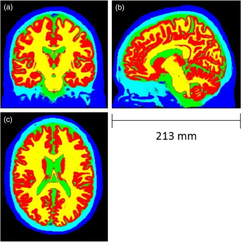
Journal of Biomedical Optics 20(8), 088001 (August 2015)

                Monte Carlo analysis of the enhanced
                transcranial penetration using distributed
                near-infrared emitter array
                Lan Yue* and Mark S. Humayun*
                University of Southern California, Institute for Biomedical Therapeutics, USC Eye Institute, 1441 Eastlake Avenue, NTT Room 4463, Los Angeles,
                California 90033, United States

                              Abstract. Transcranial near-infrared (NIR) treatment of neurological diseases has gained recent momentum.
                              However, the low NIR dose available to the brain, which shows severe scattering and absorption of the photons
                              by human tissues, largely limits its effectiveness in clinical use. Hereby, we propose to take advantage of the
                              strong scattering effect of the cranial tissues by applying an evenly distributed multiunit emitter array on the scalp
                              to enhance the cerebral photon density while maintaining each single emitter operating under the safe thermal
                              limit. By employing the Monte Carlo method, we simulated the transcranial propagation of the array emitted light
                              and demonstrated markedly enhanced intracranial photon flux as well as improved uniformity of the photon
                              distribution. These enhancements are correlated with the source location, density, and wavelength of light.
                              To the best of our knowledge, we present the first systematic analysis of the intracranial light field established
                              by the scalp-applied multisource array and reveal a strategy for the optimization of the therapeutic effects of the
                              NIR radiation. © 2015 Society of Photo-Optical Instrumentation Engineers (SPIE) [DOI: 10.1117/1.JBO.20.8.088001]

                              Keywords: Monte Carlo; near infrared; emitter array; transcranial propagation; scatter; tissues.
                              Paper 150324PR received May 19, 2015; accepted for publication Jul. 8, 2015; published online Aug. 7, 2015.

                1    Introduction                                                                  the effectiveness of the NIR treatment. Extracranially applied
                Noninvasive optical approaches for the treatment of neurologi-                     NIR light undergoes exponential decay in the magnitude of the
                cal disorders have received increasing attention in recent years.                  photon flux as it travels through the highly scattering overlying
                                                                                                   tissues, including the scalp and skull, of an adult human head.
                A number of in vitro and in vivo studies have demonstrated the
                                                                                                   Only a very small fraction of the light arrives at the cortex sur-
                therapeutic potentials of low-level near-infrared (NIR) light in
                                                                                                   face, largely limiting the NIR dose received by the brain. A
                ameliorating brain damages caused by ischemia, traumatic
                                                                                                   similar problem occurs in NIR spectroscopy and other noninva-
                injury, and neurodegeneration.1 Notably, transcranial radiation
                                                                                                   sive optical brain imaging modalities that typically reflect, for
                of an 808-nm laser NIR light was found to produce beneficial
                                                                                                   instance, oxygenation and hemodynamics of the cortex. Thera-
                outcomes in poststroke rats2 and embolized rabbits,3 though the
                                                                                                   peutically or diagnostically used low-level NIR sources fre-
                clinical effectiveness in human subjects is still under debate.4–7
                                                                                                   quently operate at an output power ranging from tens to hun-
                The use of NIR in the treatment of mild traumatic brain injury                     dreds of milliwatts with wavelengths of 600 to 1000 nm. Our
                (mTBI) has also been reported, both in animal models and in                        previous studies showed that a high-power (495 mW) 850 nm
                humans.8–13 The transcranial radiation with 870-nm NIR LED                         LED merely produced a local photon flux of 20 to 60 μW∕cm2
                cluster heads was shown to improve cognitive function, sleep,                      along the propagation axial after the light passed through tissue
                and post-traumatic stress disorder symptoms in chronic mTBI                        blocks consisting of scalp, skull, dura, and 1 to 4 cm thick
                patients.13 NIR therapies for depression, Alzheimer’s disease,                     brain.21 Substitution with an extra high-power LED emission
                and Parkinson’s disease are currently under investigation by                       is less favorable because not only does it offer rather limited
                various groups in mouse models and in clinical studies.14–16                       photon flux increment due to its incoherent and divergent
                Though the molecular mechanisms are not yet fully understood,                      nature, but also it may introduce increased thermal risks to the
                popular hypotheses link the physiological modulation of the                        radiated tissues. LEDs in direct contact with the skin or scalp are
                NIR radiation to neuron protection and cellular metabolism                         known to produce thermal effects on tissues in two forms: radi-
                arising from improved mitochondrial function. Cytochrome C                         ated and conducted heat. Despite the low-cerebral absorption of
                oxidase, the key mitochondrial component involved, has a broad                     the radiated energy,22 the conducted heat, caused by temperature
                absorption range that peaks around 830 nm.17 Upregulation of                       rise in the semiconductor junction of the NIR emitters, is not
                the neuroprotective gene products and increased neurogenesis,                      negligible. An elevation as high as 10°C in skin temperature
                for example, in lateral ventricles and the hippocampus, are also                   was reported using an 850-nm LED with a peak output of
                associated with the action of NIR radiation.1,8,18–20                              37.5 mW∕cm2 .23
                    Despite the exciting findings by these groundbreaking                              Here, we propose to use an LED array with multiple emitters
                works, limited penetration remains a major barrier in boosting                     evenly distributed on the scalp for improved transcranial pen-
                                                                                                   etration, assuming that superposition of the laterally scattered

                *Address all correspondence to: Lan Yue, E-mail: lyue@usc.edu; Mark S.
                 Humayun, E-mail: humayun@med.usc.edu                                              1083-3668/2015/$25.00 © 2015 SPIE

                Journal of Biomedical Optics                                              088001-1                                           August 2015   •   Vol. 20(8)

Downloaded From: https://www.spiedigitallibrary.org/journals/Journal-of-Biomedical-Optics on 10 May 2021
Terms of Use: https://www.spiedigitallibrary.org/terms-of-use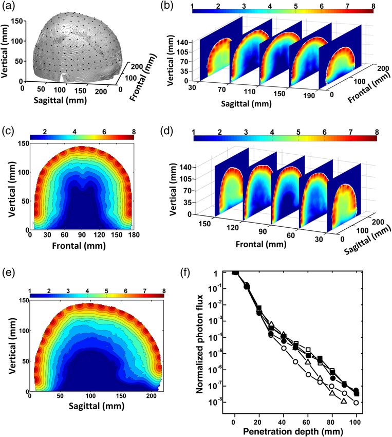
Yue and Humayun: Monte Carlo analysis of the enhanced transcranial penetration. . .

                photons emitted from multiple sources will result in increased                     (Fig. 1). All voxels were labeled by tissue types containing
                intracranial photon density. Our earlier measurement from                          the corresponding light properties: absorption coefficient, trans-
                cadaveric human tissues finds very little variation in the NIR                     port scattering coefficient, and approximate refractive index for
                penetration effectiveness through different cranial sites, for in-                 light within the simulated wavelength range. This model is
                stance, in the occipital, parietal, temporal, and frontal regions.21               described in a Cartesian coordinate system, with the center of
                This result supports the hypothesis that similarly powered emit-                   the head at (111.5, 91, 30).
                ters, when evenly spaced, are capable of producing photon dis-
                tribution of improved uniformity in the brain. With each
                individual emitter in the array operating under the safe limit, this               2.2     Light Source-Array on the Head Model
                approach could potentially achieve enhanced photon flux while                      The head model was first translated in the coordinate system
                minimizing the conducted heat to the scalp. In this study, by                      such that it is centered at the origin. The brain was treated as
                employing the Monte Carlo method to simulate the transcranial                      hemispherical in shape with its equator tilted from the x–y
                propagation of the array emitted light, we draw a comprehensive                    plane at an angle of 0.09 rad. In order to evenly distribute light
                picture of the cross-enhancement of the photon flux by multiple                    sources on the scalp, it is mathematically convenient to have the
                sources. Furthermore, a conceptual design of the proposed radi-                    brain’s equator sit on the x–y plane and henceforth consider the
                ation system is explored. To the best of our knowledge, this                       spatial arrangement of the light sources in spherical coordinates.
                study presents the first systematic analysis of the intracranial                   The coordinate frame was, therefore, rotated 0.09 rad counter-
                light field established by the scalp-applied multisource array.                    clockwise about the y-axis. The rotated z-axis now points
                                                                                                   toward the north pole of the brain hemisphere. Point sources
                2     Methods                                                                      representing the LED emitters in an array are distributed on
                2.1     Human Head Model                                                           the scalp such that the projection of all point sources on the unit
                                                                                                   sphere shares similar spacing, as quantified by the solid angle of
                The widely-used Colin27 brain template, consisting of 1 × 1 ×                      a curved quadrilateral formed between any four neighboring
                1 mm3 voxels of 256 gray levels, is adopted as the anatomical                      points. The solid angle is defined as cos θ · Δθ · Δϕ, where
                human head model in our simulation.24 The volumetric template                      θ and ϕ are conventionally defined as the elevation and the azi-
                was filtered for reduced intensity nonuniformity and then seg-                     muthal angle, respectively. Thus Δθ and cos θΔϕ represent the
                mented into five tissue types [scalp, skull, cerebrospinal fluid                   latitudinal and the longitudinal separation between given neigh-
                (CSF), gray matter, and white matter] using SPM12, a statistical                   boring sources. An exception was made for the north pole
                parametric mapping-based brain image analysis software                             source, which, as the single point at θ ¼ ðπ∕2Þ, has Δϕ undefined

                                       Fig. 1 Sections of the segmented head model [(a): frontal view at x ¼ 100 mm; (b): sagittal view at
                                       y ¼ 110 mm; (c) top view at z ¼ 91 mm]. The blue, cyan, green, red, yellow color labeled segments
                                       represent the scalp, skull, CSF, gray matter, and white matter, respectively.

                Journal of Biomedical Optics                                              088001-2                                           August 2015   •   Vol. 20(8)

Downloaded From: https://www.spiedigitallibrary.org/journals/Journal-of-Biomedical-Optics on 10 May 2021
Terms of Use: https://www.spiedigitallibrary.org/terms-of-use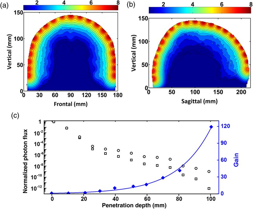
Yue and Humayun: Monte Carlo analysis of the enhanced transcranial penetration. . .

                                     Table 1 Spatial arrangement of 277 light sources on the head model and the corresponding solid angles.

                                                Increment of                          Theoretical number of          Rounded to the
                                               Azimuth in each      Solid angle         light sources per          nearest multiplication    Actual value      Actual value
                Layer      Elevation (θ)          layer (Δϕ)           (ΔΩ)               elevation layer                  of 4                 of Δϕ             of ΔΩ

                1                0                 0.157               0.025                       40.00                     40                  0.157              0.025

                2              0.157               0.159               0.025                       39.51                     40                  0.157              0.024

                3              0.314               0.165               0.025                       38.04                     36                  0.175              0.026

                4              0.471               0.176               0.025                       35.64                     36                  0.175              0.024

                5              0.628               0.194               0.025                       32.36                     32                  0.196              0.025

                6              0.785               0.222               0.025                       28.28                     28                  0.224              0.025

                7              0.942               0.267               0.025                       23.51                     24                  0.262              0.024

                8              1.100               0.346               0.025                       18.16                     20                  0.314              0.022

                9              1.257               0.508               0.025                       12.36                     12                  0.524              0.025

                10             1.414               1.004               0.025                       6.26                       8                  0.785              0.019

                11             1.571                                                                                          1

                Weighted average of actual ΔΩ                                                                                            2.451 × 10⁻²

                Standard deviation (SD) of actual ΔΩ                                                                                     1.233 × 10⁻³

                SD over average                                                                                                             5.03%

                and is, therefore, excluded from the distribution calculation. At                     tissue types at this particular wavelength were obtained from
                the maximum source density in our simulation, a total of 277                          previous studies26–28 (Table 2). The propagation direction of
                sources were arranged in a 0.05 radian elevation grid (Table 1),                      each single source is defined as the initial direction of photon
                with an average solid angle of 0.0245  0.0012 [mean                                 injection along the spherical normal toward the center of the
                standard deviation (SD)]. Reduced source densities, correspond-                       head model. A total of 109 hotons were launched from each
                ing to higher values of solid angles, were studied as well.                           light source and each individual photon was traced as it propa-
                                                                                                      gated through the medium. All photons were initially assigned
                                                                                                      with a unit weight. As a photon propagates from one scattering
                2.3     Monte Carlo Simulation                                                        event to the next, the weight is reduced by a factor of e−μa L due
                Transcranial light propagation was simulated using a Monte                            to absorption, where μa is the absorption coefficient and L is the
                Carlo approach, implemented under MATLAB® environment                                 length traveled by the photon. The change in weight is then
                MXCLab25 that describes photon migration in a complex three-                          deposited into the voxel that contains the scattering event.
                dimensional (3-D) medium. Unless otherwise stated, due to its                         The scattering angle is obtained using the probability distribu-
                wide use and excellent tissue responsiveness, we employed 850-                        tion given by the Henyey–Greenstein phase function of a ran-
                nm NIR in our simulation and the optical properties of different                      dom variable.29 The photon is subsequently migrated in this
                                                                                                      updated direction by a new scattering length that is determined
                                                                                                      with an exponential function. These steps were repeated until
                Table 2 Optical properties of the tissue types at the wavelengths of                  the photon exited the medium or has traveled an exceedingly
                850 and 690 nm.                                                                       long time in the medium. As the human head model contains
                                                                                                      spatially varying optical parameters, this algorithm checks every
                                            μa (mm−1 )                      μs0 (mm−1 )               voxel spacing for new parameters to ensure the correct calcula-
                                                                                                      tion of the weight decrease, the scattering angle, and the length.
                Tissue type            850 nm        690 nm         850 nm          690 nm            The weight of all photons dropped within each voxel was added
                                                                                                      to yield the photon flux produced by each individual light
                Scalp                  0.012          0.021           1.8             2.37            source. Furthermore, the photon flux contributed by all simu-
                Skull                  0.025          0.026           1.6             2.35
                                                                                                      lated sources was superimposed to generate a comprehensive
                                                                                                      light field map produced by multiple sources. The diffusion
                Modified CSF           0.003         0.0004           0.01            0.01            coefficient employed in this study is the reduced diffusion coef-
                                                                                                      ficient (μs0 ) that already takes account of the anisotropic scatter-
                Gray matter            0.036          0.036           0.9                 1.4         ing in the isotropic assumption. Unless otherwise stated, a
                White matter           0.014          0.014           1.1                 1.5
                                                                                                      Gaussian-beam was opted for each source to best mimic the
                                                                                                      light emission profile of an actual LED source.

                Journal of Biomedical Optics                                                    088001-3                                         August 2015    •   Vol. 20(8)

Downloaded From: https://www.spiedigitallibrary.org/journals/Journal-of-Biomedical-Optics on 10 May 2021
Terms of Use: https://www.spiedigitallibrary.org/terms-of-use
Yue and Humayun: Monte Carlo analysis of the enhanced transcranial penetration. . .
2.4 Finite Element Analysis penetration ability through various lobes. However, considering
a typically greater skull thickness in the occipital region and the
The diffusion approximation of the radio transfer equation30–32 nontrivial intersample variations in the size of the excised brain
has also been widely used to describe the light propagation in we measured, the small difference between the measured and
highly-scattering biological tissues, under the assumption that the simulated results is not surprising. Nonetheless, the compa-
the absorption coefficient is considerably smaller than the scat- rable NIR penetration effectiveness in different cerebral lobes
tering coefficient and that the light propagation is isotropic. The strongly suggests that output tuning/compensation for each indi-
diffusion equation is given by vidual source may not be required to establish a quasispherically
symmetric light field and thus is not included in our subsequent
−∇ · κðrÞ∇ΦðrÞ þ μa ðrÞΦðrÞ ¼ q0 ðrÞ; (1)
simulations.
EQ-TARGET;temp:intralink-;e001;63;668
where Φ is the photon fluence, κ is the diffusion coefficient
½3ðμa þ μs0 Þ−1 , and q0 is the isotropic source distribution at 3.2 Photon Flux Map Produced by Multiple Sources
r. The photon diffusion at the head-source interfaces were con-
strained by the surface Robin boundary condition, whereas at Figure 3 shows the results obtained from the simulation with
the tissue-tissue interfaces, fluence continuity in the normal 277 sources evenly distributed on the scalp (panel a), attaining
direction was assumed. A finite element analysis (FEA) model an average solid angle of 0.024 0.002 (mean SD). This
was constructed using COMSOL Multiphysics to estimate the high-source density produced a radially quasisymmetrical light
photon distribution in a multilayered 3-D domain that represents field that exhibited little variation at similar penetration depths
an adult human head. The geometry of the model, imported across the entire head model, as illustrated by the rainbow-
from the COMSOL Multiphysics library, was minimally modi- shaped color maps of the intracranial photon flux, in both the
fied from the original geometry provided by IEEE, IEC, and frontal (panels b and c) and the sagittal views (panels d and e).
CENELEC. The light sources, represented by 0.4-cm diameter The central dip of the penetration profile in the frontal slice shown
illumination spots, were positioned on the scalp at an average in panel c arises from the interhemispherical fissure of the brain.
interunit distance of 2 cm. A detailed description of our Under the influence of such densely packed sources, the axial
approach can be found in Ref. 21. evolvement of the photon distribution was examined at the five
previously picked sources, one located at the north pole and the
rest in the frontal, the occipital, the temporal, and the parietal
3 Results
regions, respectively. The one-dimensional representation of the
3.1 Influence on Near-Infrared Penetration by the results (panel f) reveals comparable attenuation patterns for all
Source Location five sources, despite slightly stronger decay in the occipital and the
temporal regions, as the light passed further down into the brain.
In order to compare the penetration effectiveness of the NIR In comparison with the single source results, this multisource
light that enters different cerebral lobes, the axial attenuations simulation yielded a photon flux enhancement that is progres-
of the photon flux emitted by five identical sources were exam- sively substantialized along the propagation direction (Fig. 4).
ined: four randomly placed in the frontal, occipital, temporal, Fitted by a single exponential function (τ ¼ 34.7 2.9 mm−1 ,
and parietal regions, respectively, and an additional one located R2 ¼ 0.995), the ratio of the photon flux (i.e., the gain) pro-
at the north pole, as shown in Fig. 2(a). Light emitted by these duced by a multi (circles) versus a single (squares) source along
sources exhibited similar attenuation profiles along the propa- the propagation direction of the north pole source demonstrates
gation direction, with only the occipital and temporal sources the dependence of the enhancement effect on the penetration
showing slightly more resistance to the NIR penetration, depth (black diamonds). Robust enhancement initiated around
particularly in deep brain [Fig. 2(b)]. This result resembles, 20-mm deep, approximately at the upper-mid cortex level. Beyond
yet slightly deviates from our earlier measurements on a cadav- this depth, the gain increased to about 5× at 40 mm, doubling to
eric human head that showed even greater similarity in the NIR 10× at 60 mm, and finally reached >40× at 100 mm depth. This
Fig. 2 The single source penetration profiles: (a) locations of the single sources on the scalp: frontal (1),
occipital (2), temporal (3), parietal (4), and north pole (5) source in the source coordinates; (b) penetration
profiles of the frontal (open square), occipital (open circle), temporal (open triangle), parietal (filled circle),
and north pole (filled square) source along the corresponding propagation directions.
Journal of Biomedical Optics 088001-4 August 2015 • Vol. 20(8)
Downloaded From: https://www.spiedigitallibrary.org/journals/Journal-of-Biomedical-Optics on 10 May 2021
Terms of Use: https://www.spiedigitallibrary.org/terms-of-useYue and Humayun: Monte Carlo analysis of the enhanced transcranial penetration. . .
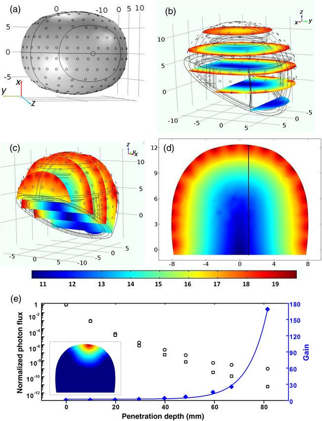
Fig. 3 Light field produced by 277 sources (850 nm, Gaussian beam). Color bars: orders of magnitude of
photon flux: (a) spatial distribution of the 277 point sources on the scalp; (b) frontal slice views of the
three-dimensional (3-D) photon flux profile; (c) two-dimensional (2-D) profile of the middle frontal slice;
(d) sagittal slice views of the 3-D photon flux profile; (e) 2-D profile of the middle sagittal slice; and (f) one-
dimensional penetration profiles along the propagation directions of the frontal (open square), occipital
(open circle), temporal (open triangle), parietal (filled circle), and north pole (filled square) source.
penetration depth-dependent enhancement results from the increased heat generation at the semiconductor junctions.
strengthened interference between sources as the examined intra- Optimal design of the radiation array calls for further under-
cranial site moves further away from the dominant source and standing of the correlation between the intersource spacing and
approaches equidistance to all sources. Changing the light source the penetration profile. Therefore, we repeated the simulations
emission profile from Gaussian to a narrowly shaped pencil beam with a series of source numbers ranging from 13 to 277, the
produced virtually identical photon flux maps in both multi spatial arrangements and the density of which were determined
(crosses) and single (triangles) source simulations, likely due to by the above-mentioned rule (see Sec. 2). At each density, we
the isotropic scattering approximation in the Monte Carlo studied the improvements in intracranial photon penetration in
model. That is, whether in form of a narrow pencil beam or a terms of enhanced flux and uniformity. Numbers of the light
divergent Gaussian beam, the incident light is rapidly scattered sources and the corresponding average solid angles are listed in
out with equal probability in all directions, resulting in a similar Table 3. The consistently low (Yue and Humayun: Monte Carlo analysis of the enhanced transcranial penetration. . .
Fig. 4 Comparison of the penetration profiles of single versus multiple
sources. Left axis (black): normalized photon flux along the propaga-
tion direction of the north pole source simulated with Gaussian
Fig. 6 Simulation of the transcranial penetration of 690 nm light: (a) 2-
(circles: multisource; squares: single source) vs. pencil beam
D representation of the photon flux color map of the middle frontal
(crosses: multisource; triangles: single source). Right axis (blue):
slice; (b) 2-D representation of the photon flux color map of the middle
ratio of the multi- versus single-source pencil-beam photon flux (i.
sagittal slice; and (c) left axis (black): photon flux decay profiles along
e., the gain) shown by the black diamonds and fitted with an exponen-
the propagation direction of the north pole source, produced by multi
tial function (solid line).
(circles) versus single (squares). Right axis (blue): gain ratio of photon
flux of multi- versus single-source at different penetration depths (solid
diamonds), fitted with an exponential function (solid line).
Table 3 Average solid angles and the SDs at different source
densities.
enhancement in the photon flux. NIR radiation penetrating
60 mm deep into a human head is, in principle, sufficient to tar-
Number of Weighted average SD of actual SD over
get the highly-convoluted cerebral cortex and a large portion of
light sources of actual ΔΩ ΔΩ average
the white matter, which are often areas of interest for the
277 2.45 × 10⁻² 1.23 × 10⁻³ 5.03% therapeutic and diagnostic use of NIR. Therefore, photon flux
enhancement up to a 60-mm depth is our main focus in deter-
229 2.99 × 10⁻² 1.67 × 10⁻³ 5.57% mining the optimal number of emitters for the array.
In addition to the magnitude enhancement of the photon flux,
181 3.82 × 10⁻² 2.61 × 10⁻³ 6.82%
improved uniformity of the radiation field was also obtained
105 6.80 × 10⁻² 4.80 × 10⁻³ 7.06% with increased light source density. This uniformity can be mea-
sured by the “relative SD” of the photon flux SDðphoton fluxÞ∕
53 1.43 × 10⁻¹ 1.17 × 10⁻² 8.16% meanðphoton fluxÞ: the lower this value is, the more uniform the
13 7.02 × 10⁻¹ 1.20 × 10⁻¹ 17.16%
radiation is. It is observed that the relative SD decays exponen-
tially with the number of light sources. Similar to the photon
Fig. 5 Dependence of the photon penetration on the source density: (a) change of photon flux enhance-
ment with penetration depth at various source densities. Penetration profiles obtained along the propa-
gation direction of the north pole point at the source number, from high to low, of 277 (stars), 229
(diamonds), 181 (down triangles), 105 (up triangles), 53 (circles), and 13 (squares); and (b) uniformity
of the photon flux (open circles) profile with the source number.
Journal of Biomedical Optics 088001-6 August 2015 • Vol. 20(8)
Downloaded From: https://www.spiedigitallibrary.org/journals/Journal-of-Biomedical-Optics on 10 May 2021
Terms of Use: https://www.spiedigitallibrary.org/terms-of-useYue and Humayun: Monte Carlo analysis of the enhanced transcranial penetration. . .
flux magnitude decay, the uniformity decay became less promi- 3.4 Influence of Reduced Wavelength
nent at the source numbers of 181 and above. Taking the system
complexity and the heat generation into account for the array To explore the multisource enhancement of the less penetrable
design, the source density needs to be contained within a rea- red light, simulation of the transcranial propagation of the 690-
sonable range. Our above analyses implicate an optimal density nm light was also conducted. Table 2 lists the optical properties
of 181 emitters, corresponding to a solid angle of ∼0.04. This of tissues at this wavelength.26 As presented in Fig. 6, switching
number was obtained under the assumption that each source is from 850 to 690 nm light led to markedly accelerated intracra-
infinitesimally small, therefore, the real application may call for nial photon flux attenuation along the propagation direction. For
a reduced source density due to the larger radiation cross-section instance, a single 690-nm source at the north pole produced a ∼1
of individual sources. to 2 orders of magnitude more exacerbated decay compared to a
Fig. 7 850-nm near-infrared penetration profiles obtained with the diffusion equation finite element analy-
sis: (a) spatial distribution of the sources on the scalp (small circles); (b) 3-D representation of the photon
flux color map of the transverse slices; (c) 3-D representation of the photon flux color map of the frontal
slices; (d) 2-D representation of the photon flux color map of the middle frontal slice. The black line shows
the direction along which the photon flux was sampled and normalized in (e); (e) comparison of the pho-
ton flux decay profile from multi (open circles) versus single (open squares) source along the propagation
direction labeled in (d). The ratios are shown by the solid diamonds and fitted with an exponential func-
tion. Inset: 2-D representation of the photon flux color map of the same slice in (d) but in the presence of
only a single source.
Journal of Biomedical Optics 088001-7 August 2015 • Vol. 20(8)
Downloaded From: https://www.spiedigitallibrary.org/journals/Journal-of-Biomedical-Optics on 10 May 2021
Terms of Use: https://www.spiedigitallibrary.org/terms-of-useYue and Humayun: Monte Carlo analysis of the enhanced transcranial penetration. . .
single 850-nm source placed at the same location [compare in the brain, as well as improved uniformity of the photon dis-
squares in Figs. 4 and 6(c)]. On the other hand, cross enhance- tribution, as revealed by the quasiradially-symmetric flux map
ment between multiple sources was augmented for the shorter and the reduced flux spatial variability. Not surprisingly, at a
wavelength, arising from the increased scattering coefficients of given wavelength, both the magnitude and the uniformity of
the tissues. For example, the gain at a 40-mm depth increased the photon flux are governed by the source density. Our study
from ∼6× for 850 nm to ∼10× for 690 nm light [compare suggests that 181 point sources evenly dispersed on the scalp,
black diamonds in Figs. 4 and 6(c)]. Similar to the results corresponding to an average solid angle of ∼0.04, may be suf-
obtained with 850 nm, spatial changes of the photon flux ficient to generate a reasonably good photon flux map. More
enhancement with penetration depth is well characterized by densely packed emitters tend to lead to only small further
a single exponential function, but with a faster increase improvements in the upper-mid part of brain, which often coin-
(τ ¼ 20.0 1.4 mm−1 , R2 ¼ 0.993). cides with the area of interest for the therapeutic NIR radiation.
Our simulation did not take the blood flow into consideration,
3.5 Diffusion Equation Approach with Finite which, assuming relatively modest variations in the blood flow
Element Method distribution in an adult head, may influence the attenuation rate
of the photon flux, but is unlikely to result in a major impact on
The diffusion approximation of the radiative transfer equation its spatial profile.
has been frequently used as an alternative approach to modeling Previous studies23 indicate that a single NIR LED introduces
photon migration in a turbid medium, though often deemed less
minimal radiation heat in biological tissues, but non-negligible
accurate than the Monte Carlo simulation. Here, we developed a
conducted heat on the skin it is in direct contact with. The Monte
diffusion equation-based FEA model in parallel to the Monte
Carlo simulation with multiple sources implies that at the maxi-
Carlo method. The FEA model also contains scalp, skull, CSF,
mum source density put forth here, photon flux enhancement
gray matter, and white matter layers, but with the fixed, instead
will not necessarily cause alerting temperature elevations in the
of SPM segmented, thickness for more convenient meshing
brain, since photon density superposition only substantialized
and less intensive computation. The tissue thicknesses were
beyond 2 cm from the scalp where the flux has already under-
set based on our earlier measurements from the cadaveric
gone a considerable decay. Additionally, blood circulation in the
samples as well as previously reported studies.21,28 The diffusion
approximation is only valid in scattering in the dominating brain will enable faster heat dissipation. Nonetheless, thermal
medium and thus does not stand for weak scatterers such as safety of the scalp-applied multiunit LED array merits further
CSF. Therefore, for this particular layer, the modified optical investigation, and attention needs to be directed to the conducted
parameters (μa ¼ 0.009 mm−1 , μs0 ¼ 0.8 mm−1 ) that account heat produced by the semiconductor junctions. Changing the
for the scattering effect of arachnoid trabeculae was employed. radiation duration, pulse width, and duty cycle may reduce the
The differences in the tissue segmentation and the optical prop- heat generation.23 In addition, optical fibers embedded in a hel-
erties between the FEA and the Monte Carlo modeling present met-like wearable can be used for light transmission, replacing
difficulty in precise quantitative comparison, but do not hinder a the direct contact of the LED array with the scalp.
qualitative verification. Coherent low-level NIR lasers have long been considered, by
Comparable to the Monte Carlo approach, FEA simulation many, superior to the noncoherent sources in terms of tissue pen-
with multiple sources yielded a rainbow-shaped quasiradially- etration efficiency. Yet the safety concern, due to its high-energy
symmetric photon flux map with small tangential variation density, as well as the bulky equipment and the hefty cost make
[Figs. 7(a)–7(d)]. Application of multiple sources also produced laser therapy difficult for home use. Our proposed approach
enhancement in the photon flux that shows its dependence on using low-cost LED arrays to provide enhanced photon flux has
the penetration depth, as illustrated in panel e. These qualitative the potential to be easily implemented for home care with
resemblances between the results obtained from both models minimal training. A recent study by Naeser et al.13 suggests
further verify the conceptual feasibility of using multiple that upregulation of the whole-brain metabolic activity may
sources for improved transcranial NIR radiation. play an important role in NIR-mediated neuroprotection against
rotenone-induced retinotoxicity. Accordingly, multiple LED
4 Conclusions emitters may serve to promote the effectiveness of NIR treat-
Transcranial NIR treatment of neurological diseases and brain ment, especially in patients with multiloci lesions, by offering
trauma has gained momentum in the past decade. However, improved uniformity of the light field that exert therapeutic
the low-NIR dose available to the brain, due to its severe scat- effects on larger surfaces of the brain.
tering and absorption of the photons by the overlying tissues, The spatial configuration of the light source array may also
largely limits its effectiveness in clinical use. Here, we propose find applications in optical diagnostics of the neurological dis-
to take advantage of the strong scattering effect of the cranial eases in the brain. Compared to the conventional single emitter–
tissues by applying an evenly distributed multiemitter array on detector pair, back scattered light contributed by multiple
the scalp to enhance the cerebral photon density while maintain- sources may carry additional spatial and temporal information
ing each single emitter operating under the safe thermal and of the brain tissues in the area(s) of interest. With the proper
electrical limits. By employing the Monte Carlo method, we computational algorithm, the signal received by the detector
first simulated NIR penetration of each single light source, can be decoded and utilized for diagnostic analysis.
respectively, located in the frontal, occipital, temporal, parietal,
and north pole regions of the human head model. The results
obtained show comparable propagation profiles in all tested Acknowledgments
regions, largely consistent with our previous measurements This work is supported by the W. M. Keck Foundation, the
from human cadaveric heads.21 Moreover, we demonstrated National Science Foundation (NSF) grant (Award No.: CBET-
that multiple sources produce markedly enhanced photon flux 1404089), the Guangdong Innovative and Entrepreneurial
Journal of Biomedical Optics 088001-8 August 2015 • Vol. 20(8)
Downloaded From: https://www.spiedigitallibrary.org/journals/Journal-of-Biomedical-Optics on 10 May 2021
Terms of Use: https://www.spiedigitallibrary.org/terms-of-useYue and Humayun: Monte Carlo analysis of the enhanced transcranial penetration. . .
Research Team Program (No. 2013S046), and Shenzhen Pea- 19. M. T. Wong-Riley et al., “Photobiomodulation directly benefits primary
cock Plan, China. neurons functionally inactivated by toxins: role of cytochrome c
oxidase,” J. Biol. Chem. 280(6), 4761–4771 (2005).
20. L. Zhang et al., “Low-power laser irradiation inhibiting Abeta25-35-
References induced PC12 cell apoptosis via PKC activation,” Cell. Physiol.
Biochem. 22(1–4), 215–222 (2008).
1. M. A. Naeser and M. R. Hamblin, “Potential for transcranial laser or 21. L. Yue et al., “Simulation and measurement of transcranial near infrared
LED therapy to treat stroke, traumatic brain injury, and neurodegener- light penetration,” Proc. SPIE 9321, 93210S (2015).
ative disease,” Photomed. Laser Surg. 29(7), 443–446 (2011). 22. Y. Ito et al., “Assessment of heating effects in skin during continuous
2. P. A. Lapchak, “Taking a light approach to treating acute ischemic wave near infrared spectroscopy,” J. Biomed. Opt. 5(4), 383–390
stroke patients: transcranial near-infrared laser therapy translational sci- (2000).
ence,” Ann. Med. 42(8), 576–586 (2010). 23. A. Bozkurt and B. Onaral, “Safety assessment of near infrared light
3. P. A. Lapchak and L. De Taboada, “Transcranial near infrared laser emitting diodes for diffuse optical measurements,” Biomed. Eng.
treatment (NILT) increases cortical adenosine-5’-triphosphate (ATP) Online 3(1), 9 (2004).
content following embolic strokes in rabbits,” Brain Res. 1306, 24. C. J. Holmes et al., “Enhancement of MR images using registration for
100–115 (2010). signal averaging,” J. Comput. Assisted Tomogr. 22(2), 324–333 (1998).
4. Y. Lampl et al., “Infrared laser therapy for ischemic stroke: a new treat- 25. Q. Fang and D. A. Boas, “Monte Carlo simulation of photon migration
ment strategy: results of the neurothera effectiveness and safety trial-1 in 3D turbid media accelerated by graphics processing units,” Opt.
(NEST-1),” Stroke 38(6), 1843–1849 (2007). Express 17(22), 20178–20180 (2009).
5. J. A. Zivin et al., “Effectiveness and safety of transcranial laser therapy 26. J. W. Barker, A. Panigrahy, and T. J. Huppert, “Accuracy of oxygen
for acute ischemic stroke,” Stroke 40(4), 1359–1364 (2009). saturation and total hemoglobin estimates in the neonatal brain using
6. W. Hacke et al., “Transcranial laser therapy in acute stroke treatment: the semi-infinite slab model for FD-NIRS data analysis,” Biomed.
results of neurothera effectiveness and safety trial 3, a phase III clinical Opt. Express 5(12), 4300–4312 (2014).
end point device trial,” Stroke 45(11), 3187–3193 (2014). 27. S. L. Jacques, “Optical properties of biological tissues: a review,” Phys.
7. P. A. Lapchak et al., “Transcranial near-infrared light therapy improves Med. Biol. 58(11), R37–R61 (2013).
motor function following embolic strokes in rabbits: an extended thera- 28. E. Okada and D. T. Delpy, “Near-infrared light propagation in an adult
peutic window study using continuous and pulse frequency delivery head model. I. Modeling of low-level scattering in the cerebrospinal
modes,” Neuroscience 148(4), 907–914 (2007). fluid layer,” Appl. Opt. 42(16), 2906–2914 (2003).
8. A. Oron et al., “Low-level laser therapy applied transcranially to mice 29. D. Boas et al., “Three dimensional Monte Carlo code for photon migra-
following traumatic brain injury significantly reduces long-term neuro- tion through complex heterogeneous media including the adult human
logical deficits,” J. Neurotrauma 24(4), 651–656 (2007). head,” Opt. Express 10(3), 159–170 (2002).
9. M. S. Moreira et al., “Effect of phototherapy with low intensity laser on 30. H. Dehghani et al., “The effects of internal refractive index variation in
local and systemic immunomodulation following focal brain damage in near-infrared optical tomography: a finite element modelling approach,”
rat,” J. Photochem. Photobiol. B 97(3), 145–151 (2009). Phys. Med. Biol. 48(16), 2713–2727 (2003).
10. W. Xuan et al., “Transcranial low-level laser therapy enhances learning, 31. M. Schweiger et al., “The finite element method for the propagation of
memory, and neuroprogenitor cells after traumatic brain injury in mice,” light in scattering media: boundary and source conditions,” Med. Phys.
J. Biomed. Opt. 19(10), 108003 (2014). 22(11 Pt 1), 1779–1792 (1995).
11. W. Xuan et al., “Low-level laser therapy for traumatic brain injury in 32. E. Okada et al., “Theoretical and experimental investigation of near-
mice increases brain derived neurotrophic factor (BDNF) and synapto- infrared light propagation in a model of the adult head,” Appl. Opt.
genesis,” J. Biophotonics 8(6), 502–511 (2014). 36(1), 21–31 (1997).
12. Y. Y. Huang et al., “Transcranial low level laser (light) therapy for
traumatic brain injury,” J. Biophotonics 5(11–12), 827–837 (2012).
Lan Yue received her bachelor’s degree in electronic engineering
13. M. A. Naeser et al., “Improved cognitive function after transcranial,
from the University of Science and Technology of China in 2006 and
light-emitting diode treatments in chronic, traumatic brain injury:
subsequently obtained her PhD in bioengineering from the University
two case reports,” Photomed. Laser Surg. 29(5), 351–358 (2011). of Illinois at Chicago in 2012. Currently, she is a postdoctoral research
14. F. Schiffer et al., “Psychological benefits 2 and 4 weeks after a single associate at the University of Southern California. Her research
treatment with near infrared light to the forehead: a pilot study of 10 focuses on biophotonic and bioelectronic therapeutics for a
patients with major depression and anxiety,” Behav. Brain Funct. 5, variety of neural disorders ranging from retinal degeneration to brain
46 (2009). trauma.
15. L. De Taboada et al., “Transcranial laser therapy attenuates amyloid-
beta peptide neuropathology in amyloid-beta protein precursor trans- Mark S. Humayun is a university professor with joint appointments in
genic mice,” J. Alzheimers Dis. 23(3), 521–535 (2011). ophthalmology and cell and neurobiology at the Keck School of Medi-
16. H. Moges et al., “Light therapy and supplementary riboflavin in the cine, and in Biomedical Engineering at the Viterbi School of Engineer-
SOD1 transgenic mouse model of familial amyotrophic lateral sclerosis ing, University of Southern California. He holds the Cornelius J. Pings
(FALS),” Lasers Surg. Med. 41(1), 52–59 (2009). Chair in biomedical sciences. He is an expert in biomedical technol-
17. J. R. Jagdeo et al., “Transcranial red and near infrared light transmission ogy and the retina, director of the University of Southern California
in a cadaveric model,” PLoS One 7(10), e47460 (2012). (USC) Institute for Biomedical Therapeutics and the Sensory Scien-
18. J. T. Eells et al., “Therapeutic photobiomodulation for methanol-induced ces Institute, and codirector of the USC Eye Institute.
retinal toxicity,” Proc. Natl. Acad. Sci. 100(6), 3439–3444 (2003).
Journal of Biomedical Optics 088001-9 August 2015 • Vol. 20(8)
Downloaded From: https://www.spiedigitallibrary.org/journals/Journal-of-Biomedical-Optics on 10 May 2021
Terms of Use: https://www.spiedigitallibrary.org/terms-of-useYou can also read