Pelargonidin ameliorates MCAO-induced cerebral ischemia/reperfusion injury in rats by the action on the Nrf2/HO-1 pathway
←
→
Page content transcription
If your browser does not render page correctly, please read the page content below

Translational Neuroscience 2021; 12: 20–31
Research Article
Kong Fu¹, Miancong Chen¹, Hua Zheng, Chuanzi Li, Fan Yang, Qian Niu*
Pelargonidin ameliorates MCAO-induced cerebral
ischemia/reperfusion injury in rats by the action
on the Nrf2/HO-1 pathway
https://doi.org/10.1515/tnsci-2021-0006 Results ‒ The results showed that pelargonidin could
received September 27, 2020; accepted December 17, 2020 effectively reduce the volume of cerebral ischemia and
Abstract improve the neurological function in MCAO rats, thereby
Background ‒ Morbidity and mortality remain high for improving memory and learning ability. With the corre-
ischemic stroke victims, and at present these patients lack sponding decreases in the expression of TNF-α, TGF-β,
effective neuroprotective agents, which improve the cure IL-6, and MDA, the level of IL-10 and SOD increased and
rate. In recent years, studies have shown that pelargonidin also promoted the nuclear metastasis of Nrf2 and the
has many biological actions. However, few studies are avail- expression of HO-1 in ischemic brain tissues.
able regarding the pelargonidin treatment of cerebral ischemia. Conclusions ‒ Our data demonstrated that pelargonidin
Methods ‒ The rat middle cerebral artery occlusion ameliorated neurological function deficits in MCAO rats,
(MCAO) model was established to investigate the neuro- and its potential mechanism of action was associated
protective effect of pelargonidin on cerebral ischemia/ with overexpression of the Nrf2/HO-1-signaling pathway.
reperfusion injury. Reperfusion was performed 2 h after This study will provide a new approach to treat cerebral
ischemia; magnetic resonance imaging (MRI) and 2, 3, 5- ischemia/reperfusion injury.
triphenyltetrazolium chloride (TTC) staining were used to Keywords: Nrf2/HO-1 pathway, MCAO, pelargonidin, stroke,
measure the volume of cerebral ischemia. Both modified cerebral ischemia/reperfusion
neurological severity scores (mNSSs) and Morris water
maze test were used to assess the neurological functions.
ELISA was applied to determine the levels of TNF-α, TGF-β,
IL-6, IL-10, MDA, and SOD. The expression of Nuclear 1 Background
factor-E2-related factor 2 (Nrf2) and heme oxygenase 1
(HO-1) protein in brain tissue was measured by immuno- Stroke is a group of diseases associated with sudden rup-
fluorescence and Western blot assays. ture of cerebral vessels or brain tissue injury caused by
blockage of blood flow to the brain and is characterized
by high morbidity, mortality, and disability rates. Accord-
1 Kong Fu and Miancong Chen contributed equally to this article. ing to epidemiological studies, approximately 80.1 mil-
lion people suffer from stroke worldwide, of which 41.1
million are female and 39 million are male; in 2016, 13.7
* Corresponding author: Qian Niu, Office of Acupuncture Clinical, million patients were newly diagnosed with stroke [1].
College of Traditional Chinese Medicine, Hainan Medical University, Atherosclerotic disease is the main causative factor for
No. 3, Xueyuan Road, Longhua District, Haikou, 571199, China, stroke. Patients with dyslipidemia and/or diabetes mellitus
e-mail: Nq020430hainmc@163.com, tel: +86-0898-66890539;
are more likely to suffer stroke [2,3]. Stroke can be classified
fax: +86-0898-66890539
Kong Fu, Hua Zheng, Chuanzi Li: Department of Radiology, The into two types: ischemic and hemorrhagic, with about
Second Affiliated Hospital, Hainan Medical University, Haikou, 60–80% of strokes being ischemic in nature [4]. The brain
570311, China functions, such as locomotor function, memory, thinking,
Miancong Chen: Department of Critical Care Medicine, The First and language, are greatly impaired after stroke [2]. Timely
Affiliated Hospital of Hainan Medical University, Haikou, 570102,
reperfusion is an effective method to treat stroke. However,
China
Fan Yang: Office of Acupuncture Clinical, College of Traditional
reperfusion could also induce additional impairments to
Chinese Medicine, Hainan Medical University, No. 3, Xueyuan Road, neurological functions [5]. Although recombinant tissue-
Longhua District, Haikou, 571199, China type plasminogen activator (r-tPA) is currently the most
Open Access. © 2021 Kong Fu et al., published by De Gruyter. This work is licensed under the Creative Commons Attribution 4.0 International
License.
Pelargonidin relieves brain ischemia/reperfusion injury via Nrf2/HO-1 pathway 21
effective way to restore blood supply in ischemic stroke, neuroprotective effect and potential mechanisms of pelar-
only about 3–5% of ischemic stroke patients are effectively gonidin on cerebral I/R injury. Our study showed that
treated due to a narrow time window for r-tPA treatment pelargonidin could effectively reduce the infarct area,
[6–8]. Patients with ischemic stroke may benefit from neuro- improve neurological functions, significantly reduce the
protective agents in the subacute phase or the late stage of level of inflammatory and oxidative factors, and promote
blood flow restoration [5]. Therefore, it is important to find the repair of neuronal cells in brain tissue after cerebral
effective neuroprotective agents that can successfully treat I/R. The neuroprotective effect of pelargonidin on cerebral
ischemic/reperfusion injury. I/R injury was associated with overexpression of the Nrf2/
The activation of inflammatory cells and increased HO-1 pathway.
pro-inflammatory factors, combined with oxidative stress
and free radical generation, induce neuron apoptosis,
axon degeneration, synaptic plasticity, and transmission
impairment [9–11]. Nuclear factor-E2-related factor 2 2 Materials and methods
(Nrf2), as an endogenous factor in brain tissue and a
member of the leucine zipper family of transcription
2.1 Animals
factors, plays an important role in reducing oxidative
stress and inflammation [12,13]. In response to cellular
oxidative damage, activated Nrf2 translocates into nucleus Male Sprague-Dawley (SD) rats (220–260 g) were pur-
and binds to the promotor regions of antioxidant response chased from Shanghai Alac Laboratory Animal Co. Ltd.
genes, thus regulating the expression of the downstream (Shanghai, China); License No.: SCXK (Shanghai) 2017-
antioxidant genes, such as enzyme heme oxygenase 1 0005 and Certificate No.: 20170005008495. All rats were
(HO-1). HO-1 and its enzymatic products possess antioxi- fed with a standard rodent diet, sterilized secondary
dant, anti-inflammatory, antiapoptotic and vasodilation ultrapure water ad libitum, housed at 22–25°C with a
actions, with concomitant improvement in the tissue humidity of 40–70% in a 12-h light–dark cycle. The ani-
microcirculation [14,15]. Recently, it has been shown mals were left to acclimatize for 7 days.
that Nrf2 activation attenuated oxidative damage induced Ethical approval: The research related to animals’ use has
by cerebral ischemic injury and that HO-1-deficient mice been complied with all the relevant national regulations
exhibited more severe brain injury [16,17]. As a result, the and institutional policies for the care and use of animals.
Nrf2/HO-1 pathway may be considered as a potential target
for neuroprotective therapy in ischemic brain injury.
Evidences have proved many drugs could protect the
neurological functions after stroke. N-Palmitoylethanolamide- 2.2 Group assignment and drug
oxazoline could decrease brain ischemic/reperfusion administration
injury via targeting the Sirtuin 1 (SIRT1) pathway [2].
Coenzyme Q10 single intravenous injection could limit A total of 60 SD rats were randomly divided into four groups
brain ischemia damage [18]. Anthocyanidins have potent (n = 15): sham, MCAO, MCAO + Pel 10 mg/kg, and MCAO +
antioxidant and anti-inflammatory effects. As a member Pel 20 mg/kg. Pelargonidin (Chengdu Herbpurify Co., Ltd.,
anthocyanidin, pelargonidin (Pel, chemical formula shown Chengdu, China; EINECS No.: 205-127-7, purity ≥98%) was
in Figure 1a) is widely distributed in vegetables and fruits, dissolved in sterile distilled water. Rats in the experimental
e.g., carrots, berries, blueberries, strawberries, and pome- groups were orally administered 10 or 20 mg/kg of pelargo-
granates [19,20]. Several studies have demonstrated that nidin per day [26,27], while the sham and MCAO groups
pelargonidin possess antioxidant [21], anti-inflammatory were given the same volume of the normal saline daily for
[22], antithrombotic [23], and antidiabetic [24]functions. In 7 days (Figure 1).
addition, pelargonidin can ameliorate memory impairment
in a rat model of Alzheimer’s disease by inhibiting glial
activation and oxidative stress [25]. However, the potential
biological activities and mechanism of pelargonidin as 2.3 MCAO model
an antioxidant and anti-inflammatory factor in cerebral
ischemia/reperfusion (I/R) injury remain unclear. The rat model of focal cerebral I/R injury was prepared
In the present study, the rat middle cerebral artery using the suture-occluded method developed by Longa.
occlusion (MCAO) model was used to investigate the The rats were anesthetized via intraperitoneal injection of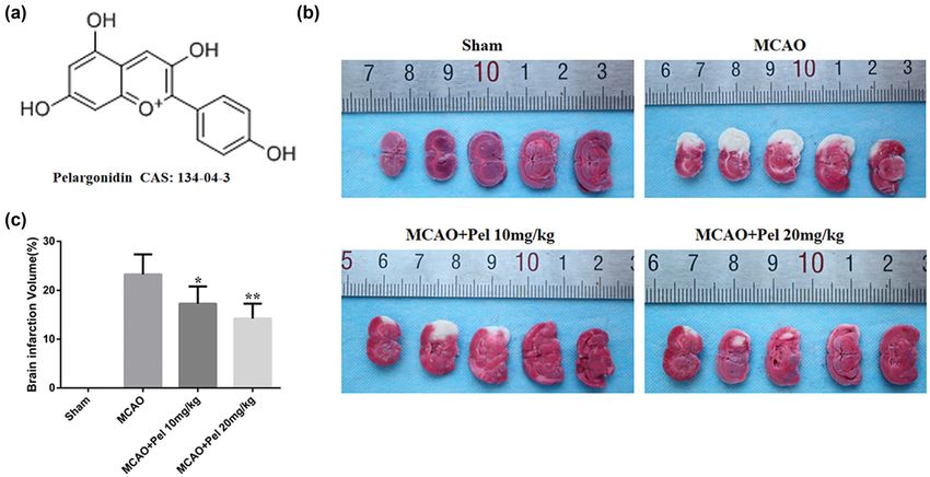
22 Kong Fu et al.
Figure 1: Flowchart of the MCAO experiments.
10% chloral hydrate (Sigma-Aldrich, CA, USA; 300 mg/ weighted imaging (DWI) were performed. An echo planar
kg) and then fixed in the supine position. The neck was imaging sequence was obtained for DWI with TR = 350 ms,
shaved and disinfected for routine skin preparation. A TE = 50 ms, b value = 1,000 s/mm2, slice thickness = 3 mm,
midline cervical incision was made to dissect the right and slice gap = 0.2 mm. The images were processed by
common carotid, external and internal carotid arteries, Functool software, and the largest slice of ischemic lesions
followed by ligation of the external carotid artery and was selected for analysis. Region of interest (ROI) with an
also its distal end. The proximal ends of the common area of 2 mm2 was placed in the lesion center and contra-
carotid and internal carotid arteries were temporarily lateral mirror location. The relative apparent diffusion
clipped. A small incision was made in the external carotid coefficient (rADC) and exponential ADC (eADC) signals
artery adjacent to the common carotid bifurcation and a of both lesion side and mirror normal side were measured.
silicon-coated suture was inserted. The clip over the rADC and reADC, the relative values of the lesion side vs
internal carotid artery was removed and the suture was the mirror normal side (rADC = ipsilateral ADC value/
gently inserted into the internal carotid artery through homologous contralateral ADC value and reADC = ipsilat-
the external carotid artery until the origin of the middle eral eADC value/homologous contralateral eADC value),
cerebral artery was occluded. The length of the suture were calculated.
was 18–20 mm. The suture was tightened and the clip
over the common carotid artery removed. Then the cer-
vical skin was sutured; 2 h after the ischemia status, the 2.5 Neurological function tests (modified
suture was removed, allowing reperfusion of the blood neurological severity score [mNSS])
supply. In the sham group, only the internal carotid artery
was dissected without any other procedures. After surgery, On day 2 after the end of treatment, the mNSS was used to
100,000 units of penicillin sodium (Sigma-Aldrich, CA, evaluate the neurological functions of MCAO rats in each
USA) were injected intramuscularly for 3 consecutive days group; mNSS was a scale with a total score of 18 and
to prevent infection. evaluates motor, sensory, reflex, and balance functions.
The function was considered normal when the mNSS was
0, and a higher mean mNSS indicated a higher severity of
2.4 Magnetic resonance image (MRI) neurological impairment [22]. The scoring details are
scanning shown in Table S1.
After treatment, the rats were examined using MRI scan-
ning (GE Discovery MR750W 3.0T Superconducting MRI
System) with a 3T experimental coil (5 cm in aperture). 2.6 2,3,5-Triphenyltetrazolium chloride
The rats were anesthetized by intraperitoneal injection of (TTC) staining
10% chloral hydrate (300 mg/kg) and fixed in the supine
position, with the head placed through the coil centrally. Rats were anesthetized by intraperitoneal injection of 10%
T2-weighted MRI scan (T2WI) and coronal diffusion- chloral hydrate (300 mg/kg) and then the thoracic cavity and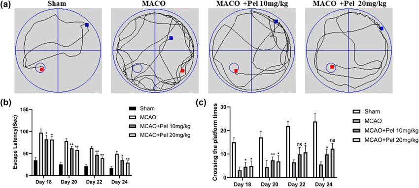
Pelargonidin relieves brain ischemia/reperfusion injury via Nrf2/HO-1 pathway 23
right auricular appendix were opened. They were then trans- Cambridge, MA, USA) at 4°C overnight, followed by
cardially perfused with PBS and then the whole brain was washing three times with PBS. Fluorescence (red light)-con-
removed and sectioned into five slices. The sections were jugated secondary antibody IgG (1:200; MultiSciences,
incubated in 2% TTC (Sigma-Aldrich, CA, USA) at 37°C for Shanghai, China) was added and incubated for 2 h at
30 min, fixed in 4% formaldehyde for 24 h, and then photo- room temperature, followed by washing three times with
graphed. Additionally, the infarct size was calculated. PBS. The slides were counterstained with DAPI (Beyotime
Biotechnology, Shanghai, China) for 10 min and photo-
graphed under a fluorescence microscope.
2.7 Morris water maze (MWM) testing
The MWM test was performed to assess the spatial learning 2.9 Western blot analysis
and memory of the rats. A round pool (120 cm diameter,
50 cm height, 30 cm depth, and water temperature 22 ± The ischemic cortex tissues were isolated and homoge-
1°C) was placed in an independent light-protected labora- nized using RIPA lysate (Beyotime Biotechnology) until
tory house and divided into four quadrants (E: East, S: no obvious lump was observed. Tissues were then cen-
South, W: West, and N: North). Each rat was trained twice trifuged at 14,000 rpm for 30 min at 4°C and the super-
daily before the MWM at 120 s/dose for 3 days at the end of natant was collected to obtain the total protein. To detect
the treatment. On day 18, 20, 22, and 24, the place naviga- the protein expression in nuclear, the Nuclear-Cytosol
tion test was performed. The platform was placed in any Extraction kit (Beyotime Biotechnology) was applied.
quadrant 2 cm under water. The adjacent and opposite The cortex tissues were homogenized with cold PBS until
quadrants of the platform were selected as water entry no obvious lump. The supernatant was collected and
points. The latency and times of crossing the platform centrifuged (4℃, 500 g × 3 min). The supernatant was
were measured during a 120-s test. The assay was per- discarded, and the separation buffer and proteinase inhi-
formed according to the instructions of the instrument bitor were added. After centrifuging (4℃, 16,000 g ×
supplier (SuperMaze MWM Experimental Analysis System, 5 min), the cytoplasmic proteins in the supernatant were
Shanghai XinRuan Information Technology Co., Ltd.). collected, while the nuclear proteins in the sediment were
also collected. The protein concentration of samples was
determined with a BCA Protein Assay kit (Beyotime
2.7.1 ELISA Biotechnology). The protein of samples were electrophor-
esed with 10% SDS-PAGE and transferred onto PVDF
Blood was collected from the abdominal inferior vena membranes (Millipore, MA, USA), The membranes were
cava. After placing at room temperature for 2 h, the blood blocked with 5% skimmed milk for 2 h and washed three
samples were centrifuged at 3,000 rpm for 10 min at 4°C times in TBST-buffered saline. Next they were incubated
to separate the serum. The levels of TNF-α, TGF-β, IL-6, with Nrf2 and HO-1 (1:1,000; Abcam) monoclonal anti-
IL-10, MDA, and SOD in rat blood serum were measured bodies at 4°C overnight. Subsequently, the membranes were
with ELISA kit (R & D Systems, Minneapolis, MN, USA). washed three times with TBST-buffered saline, incubated
with anti-IgG antibody (1:2,000; MultiSciences, Shanghai,
China) at room temperature for 1 h, and washed three times
with TBST-buffered saline. The ECL Chemiluminescent kit
2.8 Immunofluorescence (Beyotime Biotechnology) was used in a dark room for gel-
imaging acquisition and analysis. Quantity One software
Rats were anesthetized by intraperitoneal injection of was used to analyze the corresponding gray scale values
10% chloral hydrate (300 mg/kg) and decapitated to facil- of each band.
itate the removal of their brains. Brain tissues were fixed
in 4% paraformaldehyde for 72 h, embedded in paraffin,
and sectioned (4 μm slices). The sections were dehydrated
with gradient alcohol and washed 3 times with PBS. They 2.10 Data analysis
were blocked with 10% fetal bovine serum (Gibco Life
Technologies, NY, USA) for 2 h and then incubated with Data were collected from five rats separately in each
anti-Nrf2 antibody and anti-HO-1 antibody (1:100; Abcam, experiment for statistical analysis. For each experiment,24 Kong Fu et al.
the data were expressed as the mean ± SEM and were group, which exhibited enlarged infarct lesions in brain
statistically analyzed using GraphPad Prism 6.0 software. tissue. The rADC values for both the MCAO and pelargo-
Statistical differences between data were assessed using nidin-treated rats were significantly lower than those in
one-way ANOVA followed by the Newman–Keuls test. A the sham group. The rADC values in the pelargonidin
P value < 0.05 was considered to be a statistically signif- 10 mg/kg and 20 mg/kg groups were higher than those
icant finding. in the MCAO group. Although rats in both the MCAO
and pelargonidin-treatment groups had higher reADC
values than rats in the sham group, the reADC values in
both the pelargonidin 10 mg/kg group and pelargonidin
3 Results 20 mg/kg group were lower than those in the MCAO
group (Figure 3a–c).
3.1 Pelargonidin reduces the cerebral
ischemic area in MCAO rats
3.2 Pelargonidin improves neurological
MRI and TTC staining were used to measure the extent of functions in MCAO rats
cerebral infarction in MCAO rats treated with pelargo-
nidin. Figure 2b and c showed that TTC staining failed We used mNSS to assess the recovery of neurological
to reveal any infarct areas in the sham group, but there functions in the MCAO rats (a higher mNSSs indicated
was an obvious white infarct area in the MCAO group less recovery). As shown in Figure 3d, the mean mNSSs
(23.30 ± 4.10). The infarct areas in the pelargonidin in the sham group was 0, indicating normal neurological
10 mg/kg (17.29 ± 3.52) and 20 mg/kg (14.24 ± 3.04) functions. The mean mNSSs in the MCAO group and
groups were significantly reduced compared to that in pelargonidin-treatment groups increased compared
the MCAO group (P < 0.05). On MRI, the T2WI sequence with the sham group. However, both of the pelargonidin
revealed no abnormalities in the bilateral cerebral hemi- groups had a lower mean mNSSs than the MCAO group
spheres of the sham rats, but there was an abnormal (P < 0.05). Besides, the MWM test was performed to ana-
signal intensity in the MCAO group. Rats in both the lyze the spatial learning and memory capabilities of the
pelargonidin 10 mg/kg and 20 mg/kg groups had signifi- rats. A 4-day MWM test demonstrated that the latency to
cantly fewer infarct lesions compared to that in the MCAO escape was shortened for all MCAO rats (Figure 4a–c).
Figure 2: Cerebral infarction volume of rats in each group was compared. (a) Pelargonidin chemical formula and Chemical Abstracts Service
(CAS) number. (b) TTC staining of typical brains of experimental rats. (c) Cerebral ischemic volume of experimental rats in each group (n = 5).
*
P < 0.05 and **P < 0.01 compared with MCAO group.Pelargonidin relieves brain ischemia/reperfusion injury via Nrf2/HO-1 pathway 25 Figure 3: MRI detection of cerebral ischemia in each group of experimental rats. (a) Typical images of coronary angiography of T2WI scans of experimental rats in each group. (b and c) The rADC value and reADC value of experimental rats in each group (n = 5). (d) The mNSSs was used to evaluate the effect of pelargonidin on the neural function of rats in each experimental group (n = 5). *P < 0.05, **P < 0.01, and ***P < 0.001 compared with the MCAO group. However, the latency to escape in both 10 and 20 mg/kg 3.2.1 Pelargonidin reduces the levels of inflammatory the pelargonidin groups was significantly shorter than factors in the brains of MCAO rats and exerts that in the MCAO for the different 4-day tests. Similarly, antioxidative effects the rats in the pelargonidin 10 and 20 mg/kg groups crossed the platform more frequently than rats in the The pathophysiological mechanism underlying cerebral MCAO group during the different 4-day tests. These find- I/R injury is very complex. Such injuries are commonly ings suggested that pelargonidin could partially improve caused by the release of inflammatory factors and oxida- the neurological functions of I/R rats and thus enhance tive damage. Hence, ELISA was used to measure the their memory and learning capabilities. levels of TGF-β, TNF-α, IL-6, IL-10, MDA, and SOD in
26 Kong Fu et al.
Figure 4: MWM was used to detect spatial learning and memory in rats. (a) Motion trajectory diagram of MCAO rats in MWM. (b and c) The
escape latency and number of times each group crosses the platform within 2 minutes (n = 5). *P < 0.05 and **P < 0.01 compared with MCAO
group, ns: no significance.
samples of rat blood serum. Figure 5a–f showed that the 4 Discussion
levels of TGF-β, TNF-α, IL-6, and MDA in the MACO group
were higher compared to those in the sham group as well The rat MCAO-induced cerebral I/R injury rat model is
as the levels in both the pelargonidin 10 and 20 mg/kg frequently used for cerebral ischemia studies, because
groups (P < 0.05). The serum levels of IL-10 and SOD in its pathophysiological changes and gene expression
the sham group were higher than those in the MACO alterations are similar to those in humans [2]. The deter-
group as well as those in the pelargonidin 10 and mination of infarct volume and a behavioral evaluation
20 mg/kg groups (P < 0.05). The above results suggested of neurological functions are important factors for
that pelargonidin attenuated inflammatory responses measuring the effect of any treatment of cerebral
and the degree of oxidative damage in brain tissues in ischemia [28].
MCAO-induced rats. When cerebral ischemia develops at the reperfusion
stage, the compensation provided by the preexisting
nerve cells in the ischemic and hypoxic brain tissues rises
3.2.2 Pelargonidin exerts neuroprotective effects by abruptly and is accompanied by a dramatic increase in
activating the Nrf2/HO-1 pathway free radicals that mediate oxidative damage in the
affected areas [29,30]. As an end product of oxidation,
To further examine the mechanism of pelargonidin pro- MDA further aggravates any damage to cellular mem-
tecting brain tissues against MACO-induced I/R injuries, branes. Because SOD is the leading scavenger of free
the expression of Nrf2 and HO-1 proteins (components radicals, the severity of oxidative damage in a cerebral
of the Nrf2/HO-1 pathway) in the infarcted brain tissues ischemic area will depend on the balance between MDA
was detected by immunofluorescence and Western blot and SOD levels [31,32]. On the other hand, oxygen free
assays. The results revealed that the levels of nuclear radicals and other messengers that reside in the ischemic
metastasis of Nrf2 and the expression of HO-1 in the area will help to upregulate the production of adhesion
MCAO group were higher than those in the sham group molecules; this allows leukocytes to accumulate in
but lower than that in either the pelargonidin 10 mg/kg microvessels and ultimately creates a vascular obstruc-
group or the pelargonidin 20 mg/kg group (P < 0.05; tion. During this process, inflammatory factors such as
Figures 6 and 7a–d). These findings indicated that the TGF-β, TNF-α, and IL-6 may also be produced. In addi-
neuroprotection effect of pelargonidin was accompanied tion to an increased infiltration of inflammatory cells,
by the overexpression of the Nrf2/HO-1 pathway. these factors also contribute to the extracellular releasePelargonidin relieves brain ischemia/reperfusion injury via Nrf2/HO-1 pathway 27
Figure 5: Serum levels of inflammatory factors (TNF-α, TGF-β, IL-6, and IL-10) and oxidative factors (MDA and SOD) in rats of all experimental
groups were determined by ELISA (n = 5). *P < 0.05, **P < 0.01, and ***P < 0.001 compared with the MCAO group.
of numerous inflammatory mediators, which further aggra- physiological conditions, Nrf2 is predominantly retained
vate damage to the ischemic area [33,34]. in the cytoplasm by forming a complex with Keap 1.
The Nrf2/HO-1 pathway plays an important role in When exposed to external stress, such as ROS, the Nrf2
preventing oxidative stress in vitro and in vivo [35]. Under inducer reacts with Keap1 cysteine to release Nrf2 protein,
Figure 6: Immunofluorescence was used to observe the expression of Nrf2 and HO-1 in the brain tissues of rats in each group (scale bar =
100 μm).28 Kong Fu et al. Figure 7: Expression levels of Nrf2 and HO-1 in rat brain tissue. (a and b) The relative expression of Nrf2 and HO-1 in rat brain tissue was detected by immunofluorescence (n = 5). (c and d) The relative expressions of Nrf2 and HO-1 in brain tissues of rats in each group were determined by Western blot (n = 5). *P < 0.05, **P < 0.01, and ***P < 0.001 compared with MCAO group; ##P < 0.01 compared with the sham group. which subsequently translocates to the cell nucleus, Pelargonidin is an extracted nature component from where it activates Nrf2 and downstream antioxidant ripe raspberries, strawberries, and blueberries [43]. Pelar- enzymes [17]. As a transcription factor, nuclear Nrf2 gonidin has been shown to have many biological roles, induces many target gene transcription, such as HO-1, including antioxidation, anti-inflammation, antidiabetic, NADPH, GST, and thioredoxin [36]. Nrf2 could exert and antithrombotic functions [21–24]. It could protect both antioxidant and anti-inflammatory roles [37–39]. many types of cells by increasing detoxification enzymes The activation of Nrf2/HO-1 pathway exerts a neuropro- to inhibit ROS generation [21,43]. Pelargonidin could pre- tective role in Parkinson’s disease [40]. The inflamma- vent liver fibrosis via Nrf2-inhibited ROS/NLRP3/IL-1β axis tion-related pathway, NF-κB, and nucleotide-binding and inhibit inflammasome-related genes and IL-1β secre- oligomerization domain, leucine rich repeat and pyrin tion in a dose-dependent manner [44]. It has previously domain containing 3 (NLRP3) inflammasome could also been shown that pelargonidin could significantly attenuate be modulated by Nrf2 [40]. The lipid peroxidation, oxidative MDA and catalase activity in the hippocampus, decrease stress, and inflammation could be inhibited by hydroxytyr- glial fibrillary acidic protein (GFAP) levels, and thereby osol via upregulating Nrf2 in the pancreatitis-associated gut improve memory and learning functions in a rat model of injury [36]. HO-1 is an inducible rate-limiting enzyme for Alzheimer’s disease [25]. Pelargonidin has been found to heme catabolism in the microsomal enzyme system. As an inhibit transforming growth factor–beta–induced protein antioxidant enzyme targeting Nrf2, HO-1 is essential for pre- (TGFBIp)-induced human umbilical vein endothelial cell venting cerebral ischemic injuries, Parkinson’s disease, and hyperpermeability, the expression of cell adhesion mole- other neurodegenerative disorders [41,42]. cules, and the adhesion and migration of leukocytes [45].
Pelargonidin relieves brain ischemia/reperfusion injury via Nrf2/HO-1 pathway 29
It also inhibited the LPS-induced inflammatory response; provide a new perspective on the development of agents
helped to reduce the expression of TNF-α, IL-6, NF-κB, for protecting against cerebral ischemia and cerebral I/R
and other factors; and decreased mortality due to LPS- injuries.
induced endotoxemia in mice [46].
In the present study, both MRI and TTC staining Funding: This study was supported by the grants from
results revealed that pelargonidin could effectively reduce Natural Science Foundation of Hainan Province (No.
the brain infarct volume of I/R rats. Pelargonidin could 818MS061).
also enhance the performance of motor activity, sensory
skills, reflexes, and balance of MCAO rats. Further, pelar- Conflict of interest: Authors state no conflict of interest.
gonidin could also improve their memory and learning
abilities. The above results also suggested that pelargo- Data availability statement: All data generated or ana-
nidin can improve the neurological functions of MCAO lyzed during this study are included in this published
rats and may be a candidate for development as a neuro- article and its supplementary information file.
protective agent for the treatment of cerebral I/R inju-
ries. We found that pelargonidin could decrease the
levels of MDA (an oxidative factor), TNF-α, TGF-β, and
IL-6 in the serum of MCAO rats and elevate the expression
of SOD (an antioxidative factor) and IL-10 (an anti- References
inflammatory factor). Our results suggest that pelargo-
[1] Johnson CO, Nguyen M, Roth GA, Nichols E, Alam T, Abate D,
nidin can attenuate oxidative stress and inflammatory
et al. Global, regional, and national burden of stroke,
responses that occur in an MCAO-induced rat model of
1990–2016: a systematic analysis for the global burden of
I/R. disease study. Lancet Neurol. 2016;18:439–58.
Pelargonidin has been shown to reduce TPA-induced [2] Fusco R, Scuto M, Cordaro M, D’Amico R, Gugliandolo E,
methylation of the Nrf2 gene promoter region in murine Siracusa R, et al. N-palmitoylethanolamide-oxazoline protects
epidermal JB6 cells and to enhance the expression of HO- against middle cerebral artery occlusion injury in diabetic rats
by regulating the SIRT1 pathway. Int J Mol Sci. 2019;20:4845.
1 (a downstream target gene for Nrf2), and thereby helps
[3] Alloubani A, Nimer R, Samara R. Relationship between hyper-
to provide cytoprotection [43]. It has also been reported lipidemia, cardiovascular disease and stroke: a systematic
that pelargonidin upregulates the Keap1/Nrf2-signaling review. Curr Cardiol Rev. 2020;10. Epub ahead of print.
pathway and ameliorates citrinin-induced oxidative stress [4] Sun K, Fan J, Han J. Ameliorating effects of traditional Chinese
injuries in HepG2 cells [21]. Our research results show medicine preparation, Chinese materia medica and active
compounds on ischemia/reperfusion-induced cerebral micro-
that after cerebral I/R injury, the nuclear metastasis of
circulatory disturbances and neuron damage. Acta Pharm Sin
Nrf2 and expression of HO-1 increased in rat brain tissue.
B. 2015;5:8–24.
It is suggested that although cerebral I/R injury can [5] Jin YX, Silverman AJ, Vannucci SJ. Mast cells are early
promote the nuclear metastasis of Nrf2 and the expres- responders after hypoxia-ischemia in immature rat brain.
sion of HO-1, its protective effect on neurological func- Stroke. 2009;40:3107–12.
tions is also limited. When we applied pelargonidin, the [6] Vukadinović D, Schirmer SH, Vukadinović AN, Ukena C,
Scheller B, Mahfoud F, et al. Interventional closure vs. medical
nuclear metastasis of Nrf2 and expression of HO-1 sig-
therapy of patent foramen ovale for secondary prevention of
nificantly increased compared to the MCAO group, indi- stroke: updated meta-analysis. Clin Res Cardiol.
cating that pelargonidin can promote the effect of Nrf2/ 2019;108:157–66.
HO-1 to neuroprotective. [7] Kim JS. tPA helpers in the treatment of acute ischemic stroke:
are they ready for clinical use? J Stroke. 2019;21:160–74.
[8] Powers WJ, Rabinstein AA, Ackerson T, Adeoye OM,
Bambakidis NC, Becker K, et al. 2018 Guidelines for the early
management of patients with acute ischemic stroke: a guide-
5 Conclusions line for healthcare professionals from the American heart
association/American stroke association. Stroke.
The present study has demonstrated that pelargonidin 2019;49:e46–e110.
attenuated oxidative stress and inflammatory damage [9] Bramlett HM, Dietrich WD. Pathophysiology of cerebral
ischemia and brain trauma: similarities and differences.
in the cerebral I/R tissues of rats and thereby exerted
J Cereb Blood Flow Metab. 2004;24:133–50.
neuroprotective effects. Our results also showed that [10] Salman MM, Kitchen P, Woodroofe MN, Bill RM, Conner AC,
the effects of pelargonidin were achieved by inducing Heath PR, et al. Transcriptome analysis of gene expression
overexpression of the Nrf2/HO-1 pathway. These findings provides new insights into the effect of mild therapeutic30 Kong Fu et al.
hypothermia on primary human cortical astrocytes cultured [26] Roghani M, Niknam M, Jalali-Nadoushan MR, Kiasalari Z,
under hypoxia. Front Cell Neurosci. 2017;14(11):386. Khalili M, Baluchnejadmojarad T. Oral pelargonidin exerts
[11] Ginsberg MD. Adventures in the pathophysiology of brain dose-dependent neuroprotection in 6-hydroxydopamine rat
ischemia: penumbra, gene expression, neuroprotection: the model of hemi-parkinsonism. Brain Res Bull.
2002 Thomas Willis Lecture. Stroke. 2003;34:214–23. 2010;82(5–6):279–83.
[12] Wang B, Cao W, Biswal S, Doré S. Carbon monoxide-activated [27] Shen JD, Ma LG, Hu CY. Berberine up-regulates the BDNF
Nrf2 pathway leads to protection against permanent focal expression in hippocampus and attenuates corticosterone-
cerebral ischemia. Stroke. 2011;42:2605–10. induced depressive-like behavior in mice. Neurosci Lett.
[13] Kobayashi A, Ohta T, Yamamoto M. Unique function of the 2016;614:77–82.
Nrf2-Keap1 pathway in the inducible expression of antioxidant [28] Gharbawie OA, Auer RN, Whishaw IQ. Subcortical middle cer-
and detoxifying enzymes. Methods Enzymol. ebral artery ischemia abolishes the digit flexion and closing
2004;378:273–86. used for grasping in rat skilled reaching. Neuroscience.
[14] KItoh N, Wakabayashi Y, Katoh T, Ishii T, Igarashi K, Engel JD, 2006;137:1107–18.
et al. Keap1 represses nuclear activation of antioxidant [29] Stegner D, Klaus V, Nieswandt B. Platelets as modulators of
responsive elements by Nrf2 through binding to the amino- cerebral ischemia/reperfusion injury. Front Immunol.
terminal Neh2 domain. Genes Dev. 1999;13:76–86. 2019;10:2505.
[15] Wang J, Hu X, Jiang H. The Nrf-2/ARE-HO-1 axis: an important [30] Chu SF, Zhang Z, Zhang W, Zhang MJ, Gao Y, Han N, et al.
therapeutic approach for attenuating myocardial ischemia and Upregulating the expression of survivin-HBXIP complex con-
reperfusion injury-induced cardiac remodeling. Int J Cardiol. tributes to the protective role of IMM-H004 in transient global
2015;184:263–4. cerebral ischemia/reperfusion. Mol Neurobiol.
[16] Gine V, Puyal J, Clark PG, Truttmann AC. Enhancement of 2017;54:524–40.
autophagic flux after neonatal cerebral hypoxia-ischemia and [31] Wu T, Yin F, Kong H, Peng J. Germacrone attenuates cerebral
its region-specific relationship to apoptotic mechanisms. Am J ischemia/reperfusion injury in rats via antioxidative and
Pathol. 2009;175:1962–74. antiapoptotic mechanisms. J Cell Biochem. 2019;120:18901–9.
[17] Ding Y, Chen M, Wang M, Wang MM, Zhang TJ, Park JS, et al. [32] Naderi Y, Sabetkasaei M, Parvardeh S, Zanjani TM.
Neuroprotection by acetyl-11-keto-beta -boswellic acid, in Neuroprotective effect of minocycline on cognitive impair-
ischemic brain injury involves the Nrf2/HO-1 defense pathway. ments induced by transient cerebral ischemia/reperfusion
Sci Rep. 2014;4:7002. through its anti-inflammatory and anti-oxidant properties in
[18] Nikolaevna OO, Aronovna GE, Igorevna KE, Alekseevna BM, male rat. Brain Res Bull. 2017;131:207–13.
Vladimirovich GM, Gennadievich MV, et al. Intravenous [33] Xu G, Gu H, Hu B. PEG-b-(PELG-g-PLL) nanoparticles as
administration of coenzyme Q10 in acute period of cerebral TNF-α nanocarriers: potential cerebral ischemia/reperfusion
ischemia decreases mortality by reducing brain necrosis and injury therapeutic applications. Int J Nanomed.
limiting its increase within 4 days in rat stroke model. 2017;12:2243–54.
Antioxidants (Basel). 2020;7(9):E1240. [34] Yao Y, Chen L, Xiao JT, Wang CY, Jiang W, Zhang RX, et al.
[19] Noda Y, Kaneyuki T, Mori A, Packer L. Antioxidant activities of Chrysin protects against focal cerebral ischemia/reperfusion
pomegranate fruit extract and its anthocyanidins: delphinidin, injury in mice through attenuation of oxidative stress and
cyanidin, and pelargonidin. J Agric Food Chem. inflammation. Int J Mol Sci. 2014;15:20913–26.
2001;50:166–71. [35] Kowluru RA, Mishra M. Epigenetic regulation of redox sig-
[20] Giampieri F, Forbes-Hernandez TY, Gasparrini M. Strawberry naling in diabetic retinopathy: role of Nrf2. Free Radic Biol
as a health promoter: an evidence based review. Food Funct. Med. 2017;103:155–64.
2015;6:1386–98. [36] Fusco R, Cordaro M, Siracusa R, D’Amico R, Genovese T,
[21] Harath Babu GR, Anand T, Ilaiyaraja N, Khanum F, Gopalan N. Gugliandolo E, et al. Biochemical evaluation of the antioxidant
Pelargonidin modulates Keap1/Nrf2 pathway gene expression effects of hydroxytyrosol on pancreatitis-associated gut injury.
and ameliorates citrinin-induced oxidative stress in HepG2 Antioxidants (Basel). 2020;9:781.
cells. Front Pharmacol. 2017;8:868. [37] Jing X, Wei XB, Ren M, Wang LT, Zhang XM, Lou HY.
[22] Lee IC, Bae JS. Suppressive effects of pelargonidin on poly- Neuroprotective effects of tanshinone I against 6-OHDA-
phosphate-mediated vascular inflammatory responses. Arch induced oxidative stress in cellular and mouse model of
Pharm Res. 2017;40:258–67. parkinson’s disease through upregulating Nrf2. Neurochem
[23] Ku SK, Yoon EK, Lee W, Kwon S, Lee T, Bae JS. Antithrombotic Res. 2016;41:779–86.
and antiplatelet activities of pelargonidin in vivo and in vitro. [38] Zhao F, Wu T, Lau A, Jiang T, Huang Z, Wang XJ, et al. Nrf2
Arch Pharm Res. 2016;39:398–408. promotes neuronal cell differentiation. Free Radic Biol Med.
[24] Mirshekar M, Roghani M, Khalili M, Baluchnejadmojarad T, 2009;47:867–79.
Arab, Moazzen S. Chronic oral pelargonidin alleviates strep- [39] Calkins MJ, Johnson DA, Townsend JA, Vargas MR, Dowell JA,
tozotocin-induced diabetic neuropathic hyperalgesia in rat: Williamson TP, et al. The Nrf2/ARE pathway as a potential
involvement of oxidative stress. Iran Biomed J. 2010;14:33–9. therapeutic target in neurodegenerative disease. Antioxid
[25] Sohanaki H, Baluchnejadmojarad T, Nikbakht F, Roghani M. Redox Signal. 2009;11:497–508.
Pelargonidin improves memory deficit in amyloid β25-35 rat [40] Siracusa R, Scuto M, Fusco R, Trovato A, Ontario ML, Crea R,
model of Alzheimer’s disease by inhibition of glial activation, et al. Anti-inflammatory and anti-oxidant activity of Hidrox® in
cholinesterase, and oxidative stress. Biomed Pharmacother. rotenone-induced Parkinson’s disease in mice. Antioxidants
2016;83:85–91. (Basel, Switzerland). 2020;9:824.Pelargonidin relieves brain ischemia/reperfusion injury via Nrf2/HO-1 pathway 31
[41] Habtemariam S. The Nrf2/HO-1 axis as targets for flavanones: [44] Shi YS, Li XX, Li HT, Zhang Y. Pelargonidin ameliorates CCl(4)-
neuroprotection by pinocembrin, naringenin, and eriodictyol. induced liver fibrosis by suppressing the ROS-NLRP3-IL-1β
Oxid Med Cell Longev. 2019;4724920. axis via activating the Nrf2 pathway. Food Funct.
[42] Joshi G, Johnson AJ. The Nrf2-ARE pathway: a valuable thera- 2020;11:5156–65.
peutic target for the treatment of neurodegenerative diseases. [45] Jeong S, Ku SK, Baem JS. Anti-inflammatory effects of pelar-
Recent Pat CNS Drug Discov. 2013;7:218–29. gonidin on TGFBIp-induced responses. Can J Physiol
[43] Li S, Li W, Wang C. Pelargonidin reduces the TPA induced Pharmacol. 2017;95:372–81.
transformation of mouse epidermal cells -potential involve- [46] Lee BS, Lee C, Yang S. Suppressive effects of pelargonidin on
ment of Nrf2 promoter. Chem Biol Interact. lipopolysaccharide-induced inflammatory responses. Chem
2019;309:108701. Biol Interact. 2019;302:67–73.You can also read