Pretreatment with melatonin improves ovarian tissue cryopreservation for transplantation
←
→
Page content transcription
If your browser does not render page correctly, please read the page content below
Shiroma et al. Reproductive Biology and Endocrinology              (2021) 19:17
https://doi.org/10.1186/s12958-021-00705-4

 RESEARCH                                                                                                                                          Open Access

Pretreatment with melatonin improves
ovarian tissue cryopreservation for
transplantation
Marcos Eiji Shiroma1* , Luciana Lamarão Damous1, Fernanda Pereira Cotrim1, Cristiane Lima Roa1,
José Cipolla-Neto2, Russel Joseph Reiter3, Edmund Chada Baracat1 and José Maria Soares Jr1

  Abstract
  Backgroud: Melatonin has anti-inflammatory and antioxidative actions at the mitochondrial level. This indole-
  containing molecule may protect ovarian grafts during the process of cryopreservation. Therefore, we aimed to
  determine whether melatonin pretreatment improves rat ovarian graft quality.
  Methods: Twenty-six female rats were allocated to two study groups of thirteen animals each: 1) control group:
  ovaries cryopreserved using the standard protocol; and 2) melatonin group: ovaries cryopreserved in a medium
  with melatonin. Ten rats of each group were submitted to 24-h freezing, and whole ovaries autologous and
  avascular transplantation with retroperitoneal placement. After postoperative (PO) day 15, daily vaginal smears were
  obtained for estrous cycle characterization. Between PO days 30 and 35, the animals were euthanized and ovarian
  grafts were recovered for histological and immunohistochemical (Ki-67, cleaved caspase-3, TUNEL, von Willebrand
  factor, estrogen, and progesterone receptors) analyses. The ovaries of the three remaining rats from each group
  were studied immediately after thawing to assess the effects of cryopreservation. ANOVA and Tukey’s tests were
  used and the rejection level of the null hypothesis was set at 0.05 or 5% (p < 0.05).
  Results: Melatonin promoted faster restart of the estrous cycle and increased the expression of mature follicles,
  collagen type I, von Willebrand factor, Ki-67, and cleaved caspase-3 on corpora lutea and estrogen receptors in the
  ovaries as compared to control. There was a reduction in apoptosis by TUNEL on follicles, corpora lutea, and
  collagen type III.
  Conclusion: Based on the evaluated parameters, melatonin may promote the quality of ovarian grafts.
  Reproductive function enhancement should be further studied.
  Keywords: Melatonin, Ovary, Transplantation, Fertility preservation, Cryopreservation, Tissue preservation

Backgroud                                                                             new cases of cancer in women younger than 40 years of
Recent advances in the treatment of oncological diseases                              age; of these, 4000 are prepubertal children and adoles-
have resulted in significantly improved survival of young                             cents [5]. However, both chemotherapy and radiotherapy
patients with cancer [1–3], with a cure rate of over 90%                              may impair the reproductive future of these patients [1,
[4]. In the USA, yearly estimates run as high as 60,000                               3–7]. Premature ovarian failure is estimated to occur in
                                                                                      up to 68% of the women treated with alkylating agents
                                                                                      for breast cancer during menacme, 38–57% of those ag-
* Correspondence: meshiroma@gmail.com
1
 Faculdade de Medicina FMUSP, Universidade de Sao Paulo, Av. Dr. Arnaldo,             gressively treated with cytotoxic chemotherapy and
455 - Cerqueira César, São Paulo, SP CEP 01246-903, Brazil                            radiotherapy for lymphomas, and in over 90% of patients
Full list of author information is available at the end of the article

                                       © The Author(s). 2021 Open Access This article is licensed under a Creative Commons Attribution 4.0 International License,
                                       which permits use, sharing, adaptation, distribution and reproduction in any medium or format, as long as you give
                                       appropriate credit to the original author(s) and the source, provide a link to the Creative Commons licence, and indicate if
                                       changes were made. The images or other third party material in this article are included in the article's Creative Commons
                                       licence, unless indicated otherwise in a credit line to the material. If material is not included in the article's Creative Commons
                                       licence and your intended use is not permitted by statutory regulation or exceeds the permitted use, you will need to obtain
                                       permission directly from the copyright holder. To view a copy of this licence, visit http://creativecommons.org/licenses/by/4.0/.
                                       The Creative Commons Public Domain Dedication waiver (http://creativecommons.org/publicdomain/zero/1.0/) applies to the
                                       data made available in this article, unless otherwise stated in a credit line to the data.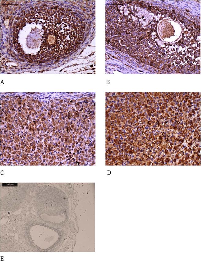
Shiroma et al. Reproductive Biology and Endocrinology   (2021) 19:17                                                    Page 2 of 12

undergoing a conditioning regimen for bone marrow                      including cancer, and also inhibits the mitochondrial
transplantation [1–4]. Radiotherapy is also known to                   apoptotic pathway as it reduces BCL2 expression and
destroy the follicle reserve [1, 4, 5]. Fertility preservation         cleaved caspase-3 activity [6, 14]. The antioxidant action
is a major cause of concern in young women.                            of melatonin is more potent than that of vitamin C or E,
   Preservation of the fertility of women is a challenge;              as even its metabolites act as free radical scavengers, in a
likewise, the offer of the best chances of maternity to pa-            process called the cascade effect [12, 13]. In addition to
tients at risk of premature, induced, or iatrogenic meno-              acting directly against free radicals, melatonin activates
pause [3, 4]. Such an offer may be made not only to                    antioxidant enzymes, such as superoxide dismutase,
patients with malignant diseases, but also to those with               glutathione peroxidase, and catalase [12–14]. The sur-
sickle cell anemia and thalassemia, those requiring a                  plus free radicals interact with lipids, proteins, and nu-
bone marrow transplant, patients with lupus, or those                  cleic acids, causing the loss of membrane integrity,
with lesions leading to bilateral oophorectomy, such as                functional and structural changes in proteins, and nu-
recurring cysts [1–4], all of which entail reproductive                cleic acid lesions [13, 15].
failure. These are further reasons for improving the                     Melatonin action is also recognized in diverse bio-
technique.                                                             logical functions, such as the circadian cycle control and
   The options for maintaining reproductive capacity in-               anticancer action and those related to the reproductive
clude oocyte, embryo, and ovarian tissue freezing [3–5].               system (ovarian activity, pregnancy, and delivery) [11,
Ovarian tissue cryopreservation is the only technique                  13] and neuroendocrinology, cardiology, and neuroim-
still deemed experimental. It allows the storage of a large            munology [15]. It is a hydrophilic and lipophilic mol-
quantity of primordial and primary follicles quickly and               ecule [11, 13, 15] and easily diffuses in various
at any phase of the menstrual cycle [1, 6]. It is the only             subcellular compartments such as membranes, cyto-
option for preserving fertility in prepubertal girls [1, 2, 4,         plasm, nucleus, and mitochondria [11–13]. There is evi-
5, 7]. In cryopreserved ovarian tissue transplantation,                dence of functional enhancement in human thawed
one of the major difficulties to surmount is the improve-              sperm quality when melatonin is applied to the cryo-
ment in the vascular bed of the receptor area [4–6],                   preservation medium [16]. Therefore, melatonin may
given the potential occurrence of ischemic lesions over                favor cell survival. However, it is not clear whether this
the time it takes for graft revascularization [1, 2, 6, 7].            effect may occur during the cryopreservation of tissue
This lesion results in fibrosis and apoptosis, which affect            fragments such as ovarian grafts. Thus, this study aimed
the follicle survival rate and graft lifespan, both of which           to evaluate rat ovarian grafts with melatonin added to
are key in fertility restoration [1, 7]. In addition, follicle         the culture medium prior to cryopreservation.
activation in the graft accelerates after transplantation,
leading to discrepancies in cell maturation in the granu-              Materials and methods
losa and oocyte development, possibly due to hypoxic                   The study was approved by the local Ethics Committee
stress and lack of anti-Mullerian hormone in the graft                 on the Use of Animals (CEUA-FMUSP 024/15). Sample
[4]. Lastly, the freezing-thawing process induces struc-               size calculation was determined using Altman Normo-
tural and morphological changes, especially in the theca               gram [17], and data from the results of a previous publi-
cell layer [4]. The main reasons for the limited quality of            cation regarding melatonin treatment and follicular
the ovarian graft are inflammatory processes and oxida-                expression of vitrified ovarian grafts [18] comparing con-
tive stress, which damage cells and mitochondria. These                trol and study groups. Sample size result was 20 animals
issues are the target of melatonin action [8].                         for 2 groups considering 80% power and statistical sig-
   Treatments such as ischemic preconditioning [9] and                 nificance of 5%. Therefore the research animal group
cell therapy with stem cells [10] have been studied by                 consisted of 26 adult female Wistar rats (Rattus norvegi-
our group, with moderate benefits. Therefore, additional               cus albinus) aged 3 months and weighing approximately
investigations are needed to improve ovarian transplant-               250 g each. The animals were kept under proper condi-
ation. One hypothesis for the impaired outcome is the                  tions of temperature and feeding as well as under a con-
presence of factors capable of interfering with tissue re-             trolled light/dark cycle of 12/12 h. Only animals with
sponse; the chief among them is a large number of free                 three regular estrous cycles were included.
radicals derived from the procedure [1]. In this experi-                 The animals were allocated to two study groups (n =
mental research scenario, new techniques have been                     13 each), namely, control and melatonin. In the control
proposed, the most prominent of which is melatonin ad-                 group, slow cryopreservation was performed according
ministration for reducing oxidative stress. Melatonin acts             to the standard protocol using the M2 culture medium,
as a free radical scavenger with vast antioxidant and                  dimethyl sulfoxide (DMSO) [19], and ethyl alcohol ve-
anti-apoptotic functions [6, 11–13]. Melatonin action di-              hicle, whereas in the melatonin group, melatonin (Sigma
minishes free radical-induced lesions in several diseases,             Aldrich, Saint Louis, MO, USA) was added to the
Shiroma et al. Reproductive Biology and Endocrinology             (2021) 19:17                                                  Page 3 of 12

Table 1 Histological analysis                                                    washed with saline (0.9% NaCl). The procedure was per-
                           Control              Melatonin            P-value     formed between 9 and 10 a.m.
Immature Follicles         10.33 ± 2.90         4.66 ± 0.88          0.13
Mature Follicles           11.00 ± 3.51         5.33 ± 1.20          0.20
                                                                                 Cryopreservation and thawing protocol
Corpora Lutea              10.00 ± 3.46         10.33 ± 4.70         0.95
                                                                                 The ovaries were placed in 1.2-mL cryotubes with 1 mL
Blood vessels              4.66 ± 1.45          2.66 ± 1.76          0.43        of 1.4 M DMSO as a cryoprotectant and the M2 medium
Histological analysis comparing the use of melatonin added to the                with or without melatonin added to the medium, de-
cryopreservation medium or not, in rat ovarian slide, freezing-thawing group
(n = 3 in each group).                                                           pending on the study group, and kept at room
                                                                                 temperature for 5 min. Slow-freezing was performed
medium at a concentration of 10− 7 M [12]. Three ani-                            using the CL-8800 temperature controller and the Cryo-
mals from each group underwent graft analysis immedi-                            genesis software, and then controlling the freezer from
ately after thawing from cryopreservation to verify the                          25 to 10 °C at 1 °C/min and to -7 °C at 0.5 °C/min, keep-
potential differences between groups induced up to the                           ing the temperature for 5 min. Next, the temperature
freezing-thawing process itself.                                                 was lowered to -55 °C at 0.5 °C/min. At this point, the
  In both groups, the ovaries underwent slow cryo-                               ovaries were transferred into liquid N2 at -196 °C and
preservation and were kept in liquid nitrogen (N2) for                           kept for 24 h [19].
24 h. Thawing took place at room temperature (25 °C).                              The cryotubes were thawed at room temperature until
The ovaries were implanted in the retroperitoneum of                             all the ice melted (15–20 min). The tissue was then
their respective donors, one on each side of the aorta,                          transferred to 5 mL of TL-HEPES at room temperature
without anastomosis, and fixed using a simple stitch                             for 10 min, while it was gently agitated to promote
with an unabsorbable thread (nylon 4–0).                                         DMSO efflux. The tissue was kept in TL-HEPES at 37 °C
                                                                                 until transplantation [19].
Estrous cycle control
At the beginning of the experiment, vaginal smears were
obtained daily, always at the same time (8–10 a.m.), to                          Collection and analysis of the material
characterize the estrous cycle using the Shorr-Harris                            The ovarian grafts were recovered and cut in half for
technique [20, 21]. Only animals with regular estrous cy-                        analysis. The rats were euthanized with a lethal dose of
cles of 4–5 days were used; diestrus was the standard                            the previously employed anesthetics. Imaging and mea-
phase for the surgical procedures (oophorectomy for                              surements were performed using a computer system
cryopreservation and euthanasia).                                                comprising a light microscope (Carl Zeiss) adapted to a
  Daily (8–10 a.m.) vaginal smear collection was re-                             high-resolution camera (Axio Cam MRC, Carl Zeiss)
sumed from postoperative (PO) days 15–30, and euthan-                            and a color video monitor. The measurements were
asia was performed as the animals entered the diestrus                           taken with an image analysis software program (Axion-
phase (PO day 30–35).                                                            Vision REL 4.6, Carl Zeiss). Counting was always per-
                                                                                 formed using four fields per animal.
Protocol for anesthesia                                                            The analyses comprised the following:
After being weighed, the animals were anesthetized with
xylazine (15 mg/kg) and ketamine (60 mg/kg) adminis-                               1) Estrous cycle
tered via intraperitoneal injection [22].
                                                                                    With the animal immobilized, the vaginal smear was
Oophorectomy protocol                                                            first obtained using a swab impregnated with saline solu-
After a median longitudinal opening of the abdomino-                             tion and then placed on a standard slide for subsequent
pelvic cavity, the ovaries were removed bilaterally and                          staining using the Shorr-Harris technique [23]. The
                                                                                 slides were subsequently analyzed using a light micro-
Table 2 Immunohistochemical analysis                                             scope at 10× and 40× magnification. The phases of the
                           Control              Melatonin            p-value     estrous cycle were determined according to the propor-
Caspase Stroma             1.61 ± 0.32          1.95 ± 0.33          0.48        tion of cells observed in the smears as follows: 1) proes-
Caspase Follicles          3.39 ± 0.75          2.37 ± 0.33          0.18        trus, the predominance of nucleated epithelial cells, 2)
                                                                                 estrus, the predominance of non-nucleated keratinized
TUNEL Stroma               0.05 ± 0.01          0.21 ± 0.09          0.05
                                                                                 cells, and 3) diestrus, an equal proportion of leukocytes
TUNEL Follicles            0.04 ± 0.01          0.07 ± 0.01          0.26
                                                                                 and nucleated keratinized epithelial cells [23].
Immunohistochemical analysis comparing the use of melatonin added to the
cryopreservation medium or not, in rat ovarian slide, freezing-thawing group
(n = 3 in each group).                                                             2) Histology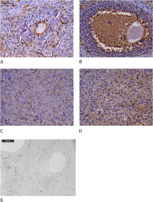
Shiroma et al. Reproductive Biology and Endocrinology               (2021) 19:17                                                          Page 4 of 12

Table 3 Histological analysis                                                         For evaluation of fibrosis, the slides were stained with
                                  Control            Melatonin         p-value     picrosirius red, and measurements were taken in eight
Immature Follicles                7.20 ± 3.23        6.00 ± 2.55       0.66        fields per animal in the ovarian stroma, with a magnifi-
Mature Follicles *                3.01 ± 0.91        8.75 ± 2.02       0.04
                                                                                   cation of 400×. The results are expressed as a percentage
                                                                                   of the positive area (unit/mm2).
Corpora Lutea                     7.86 ± 1.71        5.22 ± 1.05       0.34
                                                                                      The evaluation of the slides was conducted at our
Collagen type I*                  5.75 ± 0.52        8.52 ± 1.04       0.03        Medical Investigation Laboratory (LIM-58). For quantifi-
Collagen type III*                3.53 ± 0.28        1.49 ± 0.15       < 0.001     cation of the parameters evaluated, the images were cap-
Collagen type I and III ratio     1.62               5.71                          tured using a high-resolution camera (AxioCam-MCR,
Blood vessels                     20.13 ± 3.66       18.63 ± 3.65      0.99        Carl Zeiss) adapted to a light microscope (Axiolab, Carl
Ovarian follicle density, collagen, and blood vessels (mean ± standard             Zeiss) adjusted to the 40× objective lens [19].
deviation) comparing the use of melatonin added to the cryopreservation
medium or not, in rat ovarian slide, autologous cryopreserved grafts after the
30th day of transplantation (n = 10 in each group); *p < 0.05 t-test.
                                                                                     3) Immunohistochemistry

                                                                                     Slides with sections of the ovarian grafts were stained
  To assess follicle development, the ovarian follicles                            using immunohistochemistry to measure the von Will-
were counted and then categorized into developing folli-                           ebrand factor (AB6994, 1:100, Abcam Inc., Cambridge,
cles, regardless of their stage, and atretic follicles. The                        MA, USA), Ki67 (M724001–2, 1:100, Dako North Amer-
former was classified according to the degree of matur-                            ica Inc., Carpinteria, CA, USA), cleaved caspase-3
ation as follows: immature follicles (including primor-                            (SANT-SC-1226, 1:100, Santa Cruz Biotechnology, Santa
dial, primary, and secondary), mature follicles (with a                            Cruz, CA, USA), and TUNEL (terminal deoxynucleotidyl
single voluminous antrum), and corpora lutea. For                                  transferase (TdT)-mediated dUTP nick-end labeling,
counting purposes, the ovarian follicles comprised both                            Roche, Indianapolis, IN, USA). Hormonal receptor ex-
viable and atretic follicles as well as both normal and de-                        pression for estrogen (E1644, 1:50, Springe Bioscience
generating corpora lutea [24].                                                     Corporation, Pleasanton, CA, USA) and that for proges-
  Blood vessel count was performed in a 100-square-                                terone (AB51896, 1:30, Abcam Inc., Cambridge, MA,
micrometer area in five randomly selected fields and an-                           USA) were also studied. All samples were prepared ac-
alyzed by two independent observers.                                               cording to the manufacturer’s instructions.

  Fig. 1 Picrosirius stain with polarized light analysis of types I and III collagen comparing the use of melatonin added to the cryopreservation
  medium or not, in ovarian rat autologous cryopreserved grafts 30 days after transplantation (n = 10 in each group); a control, type I collagen,
  400×; b melatonin, type I collagen, 400×; c control, type III collagen, 400×; d melatonin, type III collagen, 400×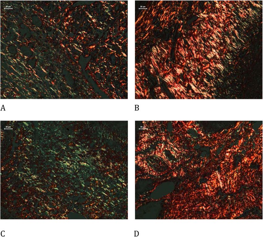
Shiroma et al. Reproductive Biology and Endocrinology             (2021) 19:17                                                               Page 5 of 12

Table 4 Immunohistochemical analysis                                               The microscopy images were obtained using a com-
                                 Control           Melatonin          p-value    puter program (Leica DM2500) and quantifications were
Cleaved caspase 3 F              11.30 ± 1.87      13.70 ± 1.79       0.38       performed using the LeicaQWin V3 program. The red-
Cleaved caspase 3 CL             5.88 ± 0.82       24.50 ± 2.06       < 0.001*   brown coloration of the cytoplasm and nucleus of granu-
TUNEL F                          0.40 ± 0.14       0.04 ± 0.02        < 0.001*   losa cells and antral follicles (for apoptosis and Ki-67) or
TUNEL CL                         1.58 ± 0.23       0.10 ± 0.03        < 0.001*   stroma (for fibrosis and expression of endothelial cells)
Ki67 F                           1.46 ± 0.29       4.09 ± 0.55        0.002*
                                                                                 was considered as positive expression and any other
                                                                                 color, as negative. Hormone receptors were evaluated in
Ki67 CL                          1.67 ± 0.33       5.27 ± 0.54        < 0.001*
                                                                                 both the stroma and follicular cells. The analysis was
Estrogen receptor F              2.41 ± 0.93       5.37 ± 0.73        0.02*
                                                                                 performed in eight different fields per animal at 400×
Estrogen receptor CL             2.25 ± 0.38       6.43 ± 0.85        < 0.001*
                                                                                 magnification, and the results are expressed as a per-
Progesterone receptor F          4.26 ± 0.53       4.65 ± 1.13        0.77
                                                                                 centage of the positive area (unit/mm2). The interpret-
Progesterone receptor CL         16.62 ± 1.07      13.47 ± 1.72       0.14       ation was performed by two independent and blinded
von Willebrand factor            1.69 ± 0.28       3.19 ± 0.38        0.003*     investigators.
Immunohistochemical analysis comparing the use of melatonin added to the
cryopreservation medium or not, in rat ovarian slide, autologous
cryopreserved grafts 30 days after transplantation (n = 10 in each group); *     Statistical analysis
p < 0.05; F Follicle, CL Corpora lutea                                           The data from each group of animals were analyzed ac-
                                                                                 cording to the type of variables. The ANOVA and

  Fig. 2 Immunohistochemical analysis of cleaved caspase 3 comparing the use of melatonin added to the cryopreservation medium or not, in
  ovarian rat autologous cryopreserved grafts 30 days after transplantation (n = 10 in each group); a control, follicle, 400×; b melatonin, follicle,
  400×; c control, corpus luteum, 400×; d melatonin, corpus luteum, 400×; e negative control, 100×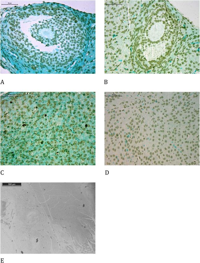
Shiroma et al. Reproductive Biology and Endocrinology (2021) 19:17 Page 6 of 12
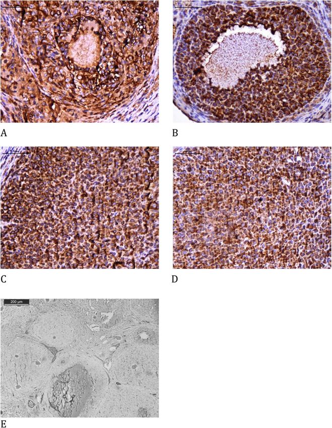
Fig. 3 Immunohistochemical analysis of TUNEL comparing the use of melatonin added to the cryopreservation medium or not, in ovarian rat
autologous cryopreserved grafts 30 days after transplantation (n = 10 in each group); a control, follicle, 400×; b melatonin, follicle, 400×; c control,
corpus luteum, 400×; d melatonin, corpus luteum, 400×; e negative control, 100×
Tukey’s tests were used, and the rejection level of the Evaluation after transplantation
null hypothesis was set at 0.05 or 5% (p < 0.05). Ten animals in the melatonin and control groups were
analyzed after autologous transplantation.
Results Estrous cycle evaluation
Evaluation after freezing-thawing For all animals in both groups, there was a
Three animals from each group were analyzed im- characterization of the estrus phase of the estrous cycle,
mediately after the freezing-thawing process to verify demonstrating ovarian hormonal activity. The melatonin
the effects of melatonin specifically on the cryo- group presented faster resumption of the estrous cycle
preservation process. Histological assessment of im- after transplantation (16.22 ± 0.50 days) compared with
mature follicles, mature follicles, corpora lutea, and the control group (20.75 ± 1.89, p = 0.0017), indicating
blood vessels showed no significant difference be- an enhancement in functional activity.
tween the control and melatonin groups (Table 1).
Immunohistochemical assessment of the freezing- Morphology and morphometry
thawing group showed no significant difference in The melatonin group had an increase in mature follicle
cleaved caspase-3 and TUNEL activities between the count compared to that in the control group, but no sig-
control and melatonin groups (Table 2). nificant difference was observed in the immature follicles
or in corpora lutea expression. With regard to immature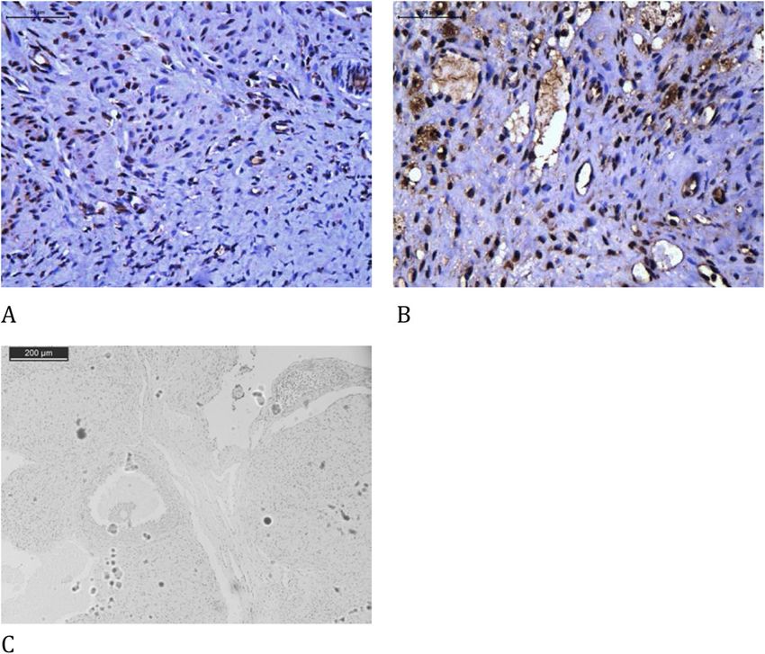
Shiroma et al. Reproductive Biology and Endocrinology (2021) 19:17 Page 7 of 12
Fig. 4 Immunohistochemical analysis of Ki-67 comparing the use of melatonin added to the cryopreservation medium or not, in ovarian rat
autologous cryopreserved grafts 30 days after transplantation (n = 10 in each group); a control, follicle, 400×; b melatonin, follicle, 400×; c control,
corpus luteum, 400×; d melatonin, corpus luteum, 400×; e negative control, 100×
follicles, we did not find any significant difference be- the cleaved caspase-3 assay on follicles and progesterone
tween the groups, even on the primordial follicular receptors on follicles or corpora lutea between both
account. groups (Table 4, Figs. 2, 3, 4, 5, 6 and 7).
Blood vessel count did not differ significantly between
the groups. The melatonin group showed enhancement Discussion
and reduction in collagen type I and type III expression, Human ovary transplantation proceeds with cortex
respectively, compared to that in the control group stripes and the present study performed whole ovary
(Table 3, Fig. 1). transplantation. Actually, previous experimental studies
regarding melatonin on rat ovarian transplantation all
Immunohistochemistry performed whole ovaries techniques [25]. Rat ovaries are
The melatonin group presented a significant increase in very small volume organs and cortex dissection would
endothelial cells (vWF), cellular proliferation (Ki67), and probably result in anatomical disruption. Besides, mela-
estrogen receptors both on the follicles and corpora tonin diffuses rapidly and easily through the different tis-
lutea, and apoptosis in the corpora lutea using the sues [11].
cleaved caspase-3 assay compared to that in the control Some studies have suggested the beneficial effects of
group. There was a significant reduction in apoptosis melatonin on ischemic-reperfusion injury [11]. In the
using the TUNEL assay on follicles and corpora lutea. ovary, there are references to two specific receptors for
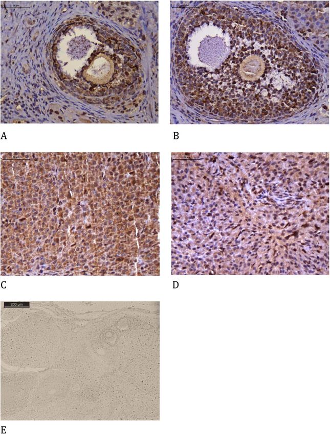
There were no significant differences in apoptosis using melatonin (MT1 and MT2) [26] of the three identifiedShiroma et al. Reproductive Biology and Endocrinology (2021) 19:17 Page 8 of 12 Fig. 5 Immunohistochemical analysis of estrogen receptor comparing the use of melatonin added to the cryopreservation medium or not, in ovarian rat autologous cryopreserved grafts 30 days after transplantation (n = 10 in each group); a control, follicle, 400×; b melatonin, follicle, 400×; c control, corpus luteum, 400×; d melatonin, corpus luteum, 400×; e negative control, 100× so far, and they act via cyclic AMP reduction [27–29]. proper functioning of oocytes and granulosa cells [11, The same receptors have been identified in human ovar- 13, 15]. Moreover, melatonin reduction also decreases ian follicles [27]. They act on ovarian function, modulat- embryo implantation [27]. These are well-known effects ing steroidogenesis, thereby notably increasing of melatonin in the reproductive system. However, the progesterone synthesis [29] and that of its receptor. effect of melatonin on free radicals and antioxidants to They also modulate the formation of corpora lutea, indi- reduce the damage to the ovarian graft is a new direc- cating the optimization of ovulatory function [28]. tion in the use of this indolamine. Our study found that Changes in serum hormone levels are related to ovula- previous melatonin treatment on the graft was beneficial tory disorders in both rats and humans [27]. Melatonin- during the cryopreservation and transplantation of rat deprived rats have a persistent anovulatory cycle, prema- ovarian graft. ture vaginal opening, ovarian hypertrophy, and increased The first study on the effect of melatonin on rat vaginal cell cornification. Moreover, melatonin replace- ovarian autotransplantation reported the indirect ben- ment reverses these effects. The hormone is found in efits of intraperitoneal administration of the substance higher concentrations in the ovarian follicle as it de- [30]. The authors described low levels of malondialde- velops, reaching maximum concentration, when com- hyde as well as high superoxide dismutase and gluta- pared to plasma in the preovulatory follicle [11, 13, 15]. thione peroxidase levels with a low ovarian necrosis The balance between free radicals and antioxidants in in melatonin-treated rats. Another study with animal ovarian follicles appears to be fundamental for the xenotransplantation treated with melatonin via the
Shiroma et al. Reproductive Biology and Endocrinology          (2021) 19:17                                                                  Page 9 of 12

 Fig. 6 Immunohistochemical analysis of progesterone receptor comparing the use of melatonin added to the cryopreservation medium or not,
 in ovarian rat autologous cryopreserved grafts 30 days after transplantation (n = 10 in each group); a control, follicle, 400×; b melatonin, follicle,
 400×; c control, corpus luteum, 400×; d melatonin, corpus luteum, 400×; e negative control, 100×

oral route and vitamin E showed improvement in                                   Histological analysis showed that the melatonin group
ovarian graft survival as evidenced by follicle count,                         presented more mature follicles than the control group,
the apoptosis index, and VEGF and PCNA expres-                                 indicating that the indolamine promoted enhancement
sion [6]. None of these previous studies investigated                          of graft function. In contrast, corpora lutea counting did
the use of melatonin in the medium used for cryo-                              not show any significant difference between groups. In
preserving ovarian grafts.                                                     rats, the estrous cycle can present corpora lutea formed
  Our results suggest that melatonin pretreatment in                           by previous cycles, thus limiting quantitative compari-
the cryopreservation medium also has beneficial ef-                            son, which may explain this result. Blood vessel quantifi-
fects on ovarian graft after transplantation: a) preco-                        cation did not differ between groups, and the melatonin
cious resumption of the estrous cycle; b) a higher                             group showed an enhancement in von Willebrand factor
number of mature follicles; c) enhancement in tissue                           expression compared to that in the control. Previous
proliferation; and d) reduction in apoptosis.                                  studies, although not involving transplantation, have also
  The animals subjected to subsequent ovarian trans-                           reported such diverse effects of melatonin on blood ves-
plantation showed that the melatonin group demon-                              sel proliferation [18, 28]. Melatonin may have a modula-
strated faster recovery of regular estrous cycles                              tory effect on blood vessels in different biological
compared to the control. This finding suggests that                            scenarios [31].
melatonin could have enabled better conditions for ovu-                          Estrogen receptor expression was significantly higher
latory and hormonal activity recovery.                                         in the melatonin group than in the control group.
Shiroma et al. Reproductive Biology and Endocrinology       (2021) 19:17                                                             Page 10 of 12

 Fig. 7 Immunohistochemical analysis of von Willebrand factor comparing the use of melatonin added to the cryopreservation medium or not, in
 ovarian rat autologous cryopreserved grafts 30 days after transplantation (n = 10 in each group); a control, 400×; b melatonin, 400×; c negative
 control, 100×

Indolamines can directly activate this hormone receptor                    group. This finding relates to the most consolidated bio-
activity or indirectly through the enhancement of follicu-                 chemical property of melatonin investigated in various
lar cell proliferation, which in turn synthesizes more es-                 tissues reported in the literature: the oxidative free rad-
trogen. This hormone is the main representative activity                   ical scavenger, which could reduce DNA degradation, in-
of ovulatory follicles [32], thereby inferring that mela-                  dependently of the apoptotic pathway. Among
tonin can enhance graft hormonal performance.                              experimental ovary transplantation studies, the first re-
   The present study is the first to assess progesterone                   search to investigate the effect of melatonin also indi-
receptors in transplanted rats and found no significant                    cated a reduction in TUNEL [32].
difference between the groups. An extended period of                          This research revealed that the melatonin group
analysis may suggest that the application of melatonin to                  showed enhancement in type I collagen and reduction in
the cryopreservation medium could enhance progester-                       type III collagen compared to that in the control. Previ-
one receptor expression.                                                   ous studies have related collagen expression to tissue re-
   Melatonin can modulate the apoptosis process [33]. In                   sponse to hypoxia. Fibroblast activity in tissue cultures
the present study, the melatonin group showed an in-                       correlates with an increase in type I collagen [35] and a
crease in cleaved caspase-3 on corpora lutea and no dif-                   reduction in type III collagen [36] with better oxygen-
ference in follicles. For follicles, melatonin increased                   ation. This result corroborates other reports of the
follicle development, which could be related to dimin-                     present study, indicating that melatonin promoted better
ished cleaved caspase-3. For corpora lutea, the natural                    functional and follicular activity in the ovarian graft. The
degradation process after ovulation might be related to                    ovulation process is indeed related to the production
the enhancement of cleaved caspase-3. This study is the                    and lysis of collagen in a dynamic process that ultimately
first to analyze cleaved caspase-3 in ovarian rat auto-                    produces ovarian follicle rupture and oocyte liberation
transplantation treated with melatonin, and further stud-                  [37].
ies might confirm how melatonin modulates cleaved
caspase-3 activity in this scenario. DNA fragmentation                     Conclusion
analysis using the TUNEL assay is the final result of cell                 Melatonin may promote a better quality of ovarian grafts
degeneration through the apoptosis cascade or not [34].                    when used to evaluate histological and protein expres-
The present study revealed that TUNEL quantification                       sion parameters. Future research can further assess the
was significantly reduced in the melatonin group, both                     potential enhancement provided by melatonin in graft
in follicles and corpora lutea, compared with the control                  reproductive performance.
Shiroma et al. Reproductive Biology and Endocrinology                (2021) 19:17                                                                         Page 11 of 12

Abbreviations                                                                        8.    Garcia S, Gimenez VMM, Maron FJM, Reiter RJ, Manucha W. Melatonin and
PO: Postoperative; DMSO: Dimethyl sulfoxide; vWF: von Willebrand factor;                   cannabinoids: mitochondrial-targeted molecules that may reduce
TUNEL: Terminal deoxynucleotidyl transferase (TdT)-mediated dUTP nick-end                  inflammaging in neurodegenerative diseases. Histol Histopathol. 2020;35:789.
labeling; MT: Receptors for melatonin; VEGF: Vascular endothelial growth             9.    Damous LL, Silva SM, Simões RS, Morello RJ, Carbonel AAF, Simões MJ, et al.
factor; PCNA: Proliferating cell nuclear antigen                                           Remote ischemic preconditioning on neovascularization and follicle viability
                                                                                           on ovary autotransplantation in rats. Transp Proc. 2008;40:861–4.
Acknowledgments                                                                      10.   Damous LL, Nakamuta JS, de Carvalho AE, Soares-Jr JM, de Jesus SM,
The authors thank Coordenação de Aperfeiçoamento de Pessoal de Nível                       Krieger JE, et al. Adipose tissue-derived stem cell therapy in rat
Superior - Brasil (CAPES) for their support.                                               cryopreserved ovarian grafts. Stem Cell Res Ther. 2015;6:57.
                                                                                     11.   Reiter RJ, Tan DX, Manchester LC, Paredes SD, Mayo JC, Sainz RM. Melatonin
                                                                                           and reproduction revisited. Biol Reprod. 2009;81:445–56.
Authors’ contributions
                                                                                     12.   Wang F, Tian XZ, Zhang L, Tan DX, Reiter RJ, Liu GS. Melatonin promotes
Marcos Eiji Shiroma, Luciana Lamarão Damous, and José Maria Soares-Jr con-
                                                                                           the in vitro development of pronuclear embryos and increases the
tributed to the study conception and design. Marcos Eiji Shiroma and Luci-
                                                                                           efficiency of blastocyst implantation in murine. J Pineal Res. 2013;55:267–74.
ana Lamarão Damous acquired data. Marcos Eiji Shiroma, Luciana Lamarão
                                                                                     13.   Cruz MHC, Leal CLV, Cruz JF, Tan DX, Reiter RJ. Essential actions of melatonin in
Damous, and José Maria Soares-Jr analyzed the data. Edmund Chada Baracat,
                                                                                           protecting the ovary from oxidative damage. Theriogenology. 2014;82:925–32.
Russel Joseph Reiter, José Cipolla-Neto, Fernanda Pereira Cotrim, and Cris-
                                                                                     14.   Ferreira CS, Maganhin CC, Simões RS, Girão MJBC, Baracat EC, Soares-Jr JM.
tiane Lima Roa contributed to revising the manuscript. All authors have read
                                                                                           Melatonina: modulador de morte celular. Rev Assoc Med Bras. 2010;56:715–8.
and approved the final manuscript.
                                                                                     15.   Tamura H, Takasaki A, Taketani T, Tanabe M, Kizuka F, Lee F, et al. The role
                                                                                           of melatonin as an antioxidant in the follicle. J Ovarian Res. 2012;5:5.
Funding                                                                              16.   Karimfar MH, Niazvand F, Haghani K, Ghafourian S, Shirazi R, Bakhtiyari S.
This study was financed in part by the Coordenação de Aperfeiçoamento de                   The protective effects of melatonin against cryopreservation-induced
Pessoal de Nível Superior- Brasil (CAPES).                                                 oxidative stress in human sperm. Int J Immunopth Ph. 2015;28:69–76.
                                                                                     17.   Altman DG. Practical statistics for medical research. London, UK: Chapman &
Availability of data and materials                                                         Hall; 1991.
The datasets used and/or analyzed during the current study are available             18.   Hemadi M, Saki G, Shokri S, Ghasemi FM. Follicular dynamics in neonate
from the corresponding author upon reasonable request.                                     vitrified ovarian grafts after host treatment with melatonin. Folia Morphol
                                                                                           (Warsz). 2011;70:18–23.
Ethics approval                                                                      19.   Guanasena KT, Lakey JRT, Villines PM, Crister ES, Crister JK. Allogeneic and
The study was approved by the University of Sao Paulo Faculty of Medicine                  xenogeneic transplantation of cryopreserved ovarian tissue to athymic mice.
Ethics Committee on the Use of Animals (CEUA-FMUSP 024/15).                                Biol Reprod. 1997;57:226–31.
                                                                                     20.   Shorr E. A new technic for staining vaginal smears: III, a single differential
                                                                                           stain. Science. 1941;94:545–6.
Consent for publication                                                              21.   Gronroos M, Kauppila O. Hormonal-cyclic changes in rats under normal
Not applicable.                                                                            conditions and under stress as revealed by vaginal smears after Shorr
                                                                                           staining. Acta Endocrinol. 1959;32:261–71.
Competing interests                                                                  22.   Smith W. Responses of laboratory animals to some injectable anaesthetics.
The authors declare that they have no competing interests.                                 Lab Anim. 1993;27:30–9.
                                                                                     23.   Marcondes FK, Bianchi FJ, Tanno AP. Determination of the estrous cycle
Author details                                                                             phases of rats: some helpful considerations. Braz J Biol. 2002;62:609–14.
1
 Faculdade de Medicina FMUSP, Universidade de Sao Paulo, Av. Dr. Arnaldo,            24.   Junqueira LC, Carneiro J. Histologia básica. 13th ed. Rio de Janeiro:
455 - Cerqueira César, São Paulo, SP CEP 01246-903, Brazil. 2Instituto de                  Guanabara Koogan; 2008.
Ciencias Biomedicas ICB, Universidade de Sao Paulo, Av. Prof. Lineu Prestes,         25.   Shiroma ME, Botelho NM, Damous LL, Baracat EC, Soares JM Jr. Melatonin
1374 - Butantã, São Paulo, SP CEP 05508-000, Brazil. 3University of Texas,                 influence in ovary transplantation: systematic review. J Ovarian Res. 2016;9:33.
Health Sciences Center, 7703 Floyd Curl Dr, San Antonio, TX 78229, USA.              26.   Soares-Jr JM, Masana MI, Ersahin C, Dubocovich ML. Functional melatonin
                                                                                           receptors in rat ovaries at various stages of the estrous cycle. J Pharmacol
Received: 6 October 2020 Accepted: 28 January 2021                                         Exp Ther. 2003;306:694–702.
                                                                                     27.   Maganhin CC, Carbonel AAF, Hatty JH, Fuchs LFP, Oliveira-Junior IS, Simões
                                                                                           MJ, et al. Efeitos da melatonina no sistema genital feminino: breve revisão.
References                                                                                 Rev Assoc Med Bras. 2008;54:267–71.
1. Demeestere I, Simon P, Emiliani S, Delbaere A, Englert Y. Orthotopic and          28.   Romeu LRG, Motta ELA, Maganhin CC, Oshima CTF, Fonseca MC, Barrueco
    heterotopic ovarian tissue transplantation. Hum Reprod Update. 2009;15:649–65.         KF, et al. Effects of melatonin on histomorphology and on the expression of
2. Abir R, Fisch B, Jessel S, Felz C, Ben-Haroush A, Orvieto R. Improving                  steroid receptors, VEGF, and PCNA in ovaries of pinealectomized female
    posttransplantation survival of human ovarian tissue by treating the host              rats. Fertil Steril. 2011;95:1379–84.
    and graft. Fertil Steril. 2011;95:1205–10.                                       29.   Teixeira CP, Simões RS, Santos MA, Calió ML, Soares-Jr JM, Simões MJ, et al.
3. Grynberg M, Poulain M, Sebag-Peyrelevade S, le Parco S, Fanchin R,                      Soybean concentrated extract counteracts oxidative stress in the uterus of
    Frydman N. Ovarian tissue and follicle transplantation as an option for                rats. Climacteric. 2014;17:402–9.
    fertility preservation. Fertil Steril. 2012;97:1260–8.                           30.   Sapmaz E, Ayar A, Celik H, Sapmaz T, Kilic N, Yasar MA. Effects of melatonin
4. Donnez J, Dolmans MM, Pellicer A, Diaz-Garcia C, Serrano MS, Schmidt KT,                and oxytetracycline in autologous intraperitoneal ovary transplantation in
    et al. Restoration of ovarian activity and pregnancy after transplantation of          rats. Neuroendocrinol Lett. 2003;24:350–4.
    cryopreserved ovarian tissue: a review of 60 cases of reimplantation. Fertil     31.   Mirza-Aghazadeh-Attari M, Reiter RJ, Rikhtegar R, Jalili J, Hajalioghli P,
    Steril. 2013;99:1503–13.                                                               Mihanfar A, et al. Melatonin: an atypical hormone with major functions in
5. Rodriguez-Wallberg KA, Oktay K. Options on fertility preservation in female             the regulation of angiogenesis. IUBMB Life. 2020;72:1560–84.
    cancer patients. Cancer Treat Rev. 2012;38:354–61.                               32.   Drummond AE, Fuller PJ. Ovarian actions of estrogen receptor-β: an update.
6. Friedman O, Orvieto R, Fisch B, Felz C, Freud E, Ben-Haroush A, et al.                  Semin Reprod Med. 2012;30:32–8.
    Possible improvements in human ovarian grafting by various host and graft        33.   Rodriguez C, Martín V, Herrera F, García-Santos G, Rodriguez-Blanco J,
    treatments. Hum Reprod. 2012;27:474–82.                                                Casado-Zapico S, et al. Mechanisms involved in the pro-apoptotic effect of
7. Labied S, Delforge Y, Munaut C, Blacher S, Colige A, Delcombel R, et al.                melatonin in cancer cells. Int J Mol Sci. 2013;14:6597–613.
    Isoform 111 of vascular endothelial growth factor (VEFG111) improves             34.   Negoescu A, Guillermet C, Lorimier P, Brambilla E, Labat-Moleur F.
    angiogenesis of ovarian tissue xenotransplantation. Transplantation. 2013;95:          Importance of DNA fragmentation in apoptosis with regard to TUNEL
    426–33.                                                                                specificity. Biomed Pharmacother. 1998;52:252–8.
Shiroma et al. Reproductive Biology and Endocrinology (2021) 19:17 Page 12 of 12
35. Hara-Saito Y, Kato H, Saito N, Shiomi A, Uenoyama A, Takagi R, et al. Distinct
differences in hypoxic responses between human oral mucosa and skin
fibroblasts in a 3D collagen matrix. In Vitro Cell Dev Biol –Animal. 2020;56:
452–79.
36. Wang X, Lin L, Chai X, Wu Y, Li Y, Liu X. Hypoxic mast cells accelerate the
proliferation, collagen accumulation and phenotypic alteration of human
lung fibroblastos. Int J Mol Med. 2020;45:175–85.
37. Lofman C, Zackrisson U, Mikuni M, Block M, Janson PO, Brannstrom M. A
method for longitudinal microscopic in vivo examinations of morphology,
vascularity, and motility in the ovary and the oviduct of the rat. J Soc
Gynecol Investig. 2002;6:379–85.
Publisher’s Note
Springer Nature remains neutral with regard to jurisdictional claims in
published maps and institutional affiliations.You can also read