Thalamocortical connectivity is associated with autism symptoms in high-functioning adults with autism and typically developing adults
←
→
Page content transcription
If your browser does not render page correctly, please read the page content below
Ayub et al. Translational Psychiatry (2021)11:93
                                    https://doi.org/10.1038/s41398-021-01221-0                                                                                                     Translational Psychiatry

                                     ARTICLE                                                                                                                                                    Open Access

                                    Thalamocortical connectivity is associated with
                                    autism symptoms in high-functioning adults with
                                    autism and typically developing adults
                                    Rafi Ayub1,2, Kevin L. Sun2,3, Ryan E. Flores2, Vicky T. Lam2, Booil Jo2, Manish Saggar2 and Lawrence K. Fung                                                                             2

                                      Abstract
                                      Alterations in sensorimotor functions are common in individuals with autism spectrum disorder (ASD). Such
                                      aberrations suggest the involvement of the thalamus due to its key role in modulating sensorimotor signaling in the
                                      cortex. Although previous research has linked atypical thalamocortical connectivity with ASD, investigations of this
                                      association in high-functioning adults with autism spectrum disorder (HFASD) are lacking. Here, for the first time, we
                                      investigated the resting-state functional connectivity of the thalamus, medial prefrontal, posterior cingulate, and left
                                      dorsolateral prefrontal cortices and its association with symptom severity in two matched cohorts of HFASD. The
                                      principal cohort consisted of 23 HFASD (mean[SD] 27.1[8.9] years, 39.1% female) and 20 age- and sex-matched
                                      typically developing controls (25.1[7.2] years, 30.0% female). The secondary cohort was a subset of the ABIDE database
                                      consisting of 58 HFASD (25.4[7.8] years, 37.9% female) and 51 typically developing controls (24.4[6.7] years, 39.2%
                                      female). Using seed-based connectivity analysis, between-group differences were revealed as hyperconnectivity in
                                      HFASD in the principal cohort between the right thalamus and bilateral precentral/postcentral gyri and between the
                  1234567890():,;
1234567890():,;
                  1234567890():,;
1234567890():,;

                                      right thalamus and the right superior parietal lobule. The former was associated with autism-spectrum quotient in a
                                      sex-specific manner, and was further validated in the secondary ABIDE cohort. Altogether, we present converging
                                      evidence for thalamocortical hyperconnectivity in HFASD that is associated with symptom severity. Our results fill an
                                      important knowledge gap regarding atypical thalamocortical connectivity in HFASD, previously only reported in
                                      younger cohorts.

                                    Introduction                                                                                        processing is hypothesized to play a pivotal role in dis-
                                      Alterations in sensorimotor processing are highly                                                 rupted cognitive functions attributed to ASD, such as
                                    common in individuals with autism spectrum disorder                                                 information processing, central coherence, perceptual
                                    (ASD)1–4. Many studies have found evidence for altered                                              integration, language development, and reading emotions
                                    auditory processing5,6, tactile processing7,8, visual pro-                                          from faces1,16,17. This relationship between sensorimotor
                                    cessing9, and motor learning10 in various age groups in                                             processing and core symptoms of ASD has been well-
                                    ASD. Such sensorimotor differences have been linked to                                              studied in younger cohorts. In older cohorts, however,
                                    core symptoms of repetitive behaviors and socio-                                                    past studies have combined adults with other age groups
                                    communicative deficits8,11–15. Additionally, sensorimotor                                            with limited behavioral and self-report data18–20. Even
                                                                                                                                        though adults with ASD overwhelmingly experience
                                                                                                                                        unusual sensory processing similarly to their younger
                                    Correspondence: Lawrence K. Fung (lkfung@stanford.edu)                                              counterparts21, the link between sensorimotor processing
                                    1
                                    2
                                     Department of Bioengineering, Stanford University, Stanford, CA, USA                               and its underlying neurophysiology in adults is poorly
                                     Department of Psychiatry and Behavioral Sciences, Stanford University,
                                                                                                                                        explored, highlighting an important clinical need. In the
                                    Stanford, CA, USA
                                    Full list of author information is available at the end of the article                              present study, we focused specifically on high-functioning
                                    These authors contributed equally: Manish Saggar, Lawrence K. Fung

                                    © The Author(s) 2021
                                                        Open Access This article is licensed under a Creative Commons Attribution 4.0 International License, which permits use, sharing, adaptation, distribution and reproduction
                                                        in any medium or format, as long as you give appropriate credit to the original author(s) and the source, provide a link to the Creative Commons license, and indicate if
                                    changes were made. The images or other third party material in this article are included in the article’s Creative Commons license, unless indicated otherwise in a credit line to the material. If
                                    material is not included in the article’s Creative Commons license and your intended use is not permitted by statutory regulation or exceeds the permitted use, you will need to obtain
                                    permission directly from the copyright holder. To view a copy of this license, visit http://creativecommons.org/licenses/by/4.0/.
Ayub et al. Translational Psychiatry (2021)11:93 Page 2 of 9 adults with ASD (HFASD) and explored the neural cor- not examine the differences in thalamocortical con- relates of sensorimotor processing and their relationships nectivity between adults and adolescents34, obscuring the with typical characteristics of autism to better inform neurobiology of thalamocortical connectivity in adults treatment. alone. Thus, the results from these combined cohort Sensorimotor processing may be related to aberrant studies are not representative of HFASD. In fact, no study inhibitory neurotransmission in ASD. Altered cortical to date has investigated thalamocortical connectivity inhibition is a widely supported theory in ASD, as evi- specifically in HFASD and its relationship with ASD denced by altered levels of glutamate22 and GABA23,24, characteristics. and sensory hyper- and hyposensitivity1. Neurodevelop- The overarching goal of this study was to examine tha- mental disorders associated with ASD, such as fragile X lamocortical FC in the less-studied ASD subpopulation of Syndrome and Tourette syndrome, also exhibit alterations high functioning adults compared to an age- and sex- of cortical inhibition and suggest an involvement of sen- matched control group and analyze its relationship with sorimotor deficits25,26. Taken together, cortical excit- clinical symptoms. We used a generalized linear model ability and sensorimotor processing appear to play an (GLM)-based approach for seed-based connectivity analysis important role in the neurobiology of ASD. and included seeds from the left and right thalami. We In addition to cortical inhibition, the thalamus has been additionally used a cortical seed-based approach for ana- shown to be associated with the neurobiology of ASD. lyzing thalamocortical FC18 and included seeds from the The thalamus is a critical functional hub for relaying medial prefrontal cortex (mPFC), posterior cingulate cortex sensory information to the cortex and modulating motor (PCC), and left dorsolateral prefrontal cortex (dlPFC). signals27. It does so by exerting fine-tuned inhibitory These seeds allowed us to examine FC in the two most control over cortico-cortical and subcortical-cortical sig- commonly studied resting state networks in ASD, default naling18,28. Treatments such as deep brain stimulation mode (DMN)35–37 and central executive (CEN)38–42. These targeting the thalamus at the anterior nucleus and the networks have shown altered connectivity with sensor- ventral intermediate nucleus are known to reduce seizures imotor regions in previous studies of adolescents with in epilepsy29 and reduce tremors in Parkinson’s disease30, ASD37 and are known to engage the thalamus as a key respectively. This inhibitory role of the thalamus in motor functional region27,43. Then, we examined brain-behavior processing and the evidence of cortical disinhibition relationships by correlating observed group differences with shown in ASD suggest that the connectivity of the tha- clinical markers of ASD. Furthermore, we explored the lamus may differ in individuals with ASD. effects of age and sex on these relationships using mediation Resting-state functional magnetic resonance imaging and moderation analyses predicting ASD symptom severity. (rsfMRI) has been a widely used tool in assessing co- Lastly, we sought to validate our results in a separate, larger fluctuations (or functional connectivity; FC) across brain cohort of HFASD from the ABIDE public database44,45. By regions in the absence of task-related cognitive studying thalamocortical connectivity in HFASD, we hope demand31,32. The task-free nature of rsfMRI has several to better understand the neural correlates of differential key benefits, including higher participant compliance and sensorimotor processing in a specific and understudied the ability to aggregate data across multiple sites, that population. enable its widespread use in investigating thalamocortical FC in individuals with ASD. An early neuroimaging study Materials and methods of children and adolescents with ASD using rsfMRI found Participants evidence of thalamocortical hyperconnectivity in limbic, Participants were of the same cohort that had been auditory, and motor regions, as well as general hypo- studied in a previous investigation by our lab24. We will connectivity with regions involved in executive function- refer to this cohort as the HFASD cohort. Twenty-eight ing and social cognition33. Another investigation that individuals with ASD and 29 TD controls were recruited. included children, adolescents, and adults from the autism Of these, 26 ASD and 23 TD individuals had completed brain imaging data exchange (ABIDE), a multisite neu- resting-state fMRI scans. Two TD individuals were roimaging database, also revealed widespread thalamic removed from this study due to an incomplete IQ hyperconnectivity with temporal, sensorimotor, and pre- assessment or not meeting the inclusion criteria for IQ. frontal cortices with limited evidence of hypoconnectivity. An additional three ASD and one TD individuals were However, these results were primarily driven by adoles- excluded due to excess motion in the scanner (see Sup- cents. Importantly, this study’s investigation of the rela- plemental Information). Thus, in total, twenty-three tionship between thalamocortical connectivity and ASD individuals with ASD (mean[SD] 27.1[8.9] years, 18-48 symptoms is inconclusive due to a lack of behavioral years; 39.1% female; full-scale IQ 103.7[16.1]) and 20 age- data18. Other studies that included adults did not use and sex-matched typically developing (25.1[7.2] years, 18- functionally distinct regions-of-interest (ROIs)19 and did 42 years; 30.0% female; full-scale IQ 111.1[14.6])
Ayub et al. Translational Psychiatry (2021)11:93                                                                                                     Page 3 of 9

Table 1 HFASD participant demographics and clinical                                  Table 2 ABIDE demographics.
assessments.
                                                                                                    ASD N = 58        TD N = 51         T-Test / χ2 Test p-value
                     ASD N = 23       TD N = 20        T-Test / χ2 Test p-value

Age                  27.13 ± 8.92     25.10 ± 7.18     0.4134
                                                                                     Age            25.40 ± 7.81      24.37 ± 6.69      0.4599
Female Sex           39.13%           30.00%           0.5309                        Female sex     37.93%            39.22%            0.8906
Right-handed         84.21%           73.68%           0.4261
                                                                                     FSIQ           108.70 ± 14.81    109.31 ± 13.82    0.8229
FSIQ                 103.70 ± 16.09   111.05 ± 14.61   0.1241
   VIQ               105.17 ± 19.27   109.65 ± 14.56   0.3919                        Values reported are mean ± SD unless otherwise noted. Bolded Welch’s T-test/χ2
                                                                                     results are P < 0.05. FSIQ = Full-scale IQ.
   NVIQ              102.13 ± 13.67   112.40 ± 13.44   0.01742
ADOS-2 – Total       10.86 ± 4.87     N/A              N/A
AQ – Total           31.13 ± 6.77     16.65 ± 8.41     0.00000041                    ABIDE participants
RAADS-R – Total      124.52 ± 37.20   54.85 ± 44.79    0.00000297                      To validate our findings in a separate cohort, we iden-
SRS-2 – Raw Total    95.04 ± 24.81    44.58 ± 31.71    0.00000249                    tified 58 ASD and 51 TD individuals from ABIDE mat-
Values reported are mean ± SD unless otherwise noted. Bolded Welch’s T-test/χ2       ched to the HFASD cohort by age, sex, and IQ (Table 2).
results are P < 0.05. FSIQ = Full-scale IQ. VIQ = Verbal IQ. NVIQ = Non-verbal IQ.   The selection process is detailed in Supplemental Infor-
ADOS-2 = Autism Diagnostic Observation Schedule, Second Edition. AQ =
Autism-Spectrum Quotient. RAADS-R = Ritvo Autism-Asperger Diagnostic Scale-          mation. Functional brain imaging data were analyzed with
Revised. SRS-2=Social Responsiveness Scale, Second Edition.                          the methods below.

individuals (Table 1) were included in the present study.                            fMRI preprocessing
Inclusion and exclusion criteria are detailed in Supple-                               Supplemental Table 2 presents the MRI acquisition
mental Table 1 and in Fung et al.24.                                                 parameters for the HFASD and ABIDE cohorts. Func-
                                                                                     tional data from both the HFASD and ABIDE cohorts
Imaging data acquisition                                                             were preprocessed identically using fMRIPrep v1.3.152, a
  High-resolution 3D T1-weighted structural images were                              pipeline that uses a combination of tools from existing
acquired using a 3T GE Signa PET/MR scanner (General                                 neuroimaging software packages.
Electric, Milwaukee, WI) using the following parameters:                               Nuisance regressors—framewise displacement (FD),
repetition time (TR) = 7.74 ms, echo time (TE) = 2.87 ms,                            average cerebrospinal fluid (CSF) signal, average white
flip angle = 12°, acquisition matrix = 256 × 256, inversion                           matter (WM) signal, and global signal—were extracted
time = 450 ms, slice thickness = 1.4 mm, in-plane reso-                              using this pipeline. In the HFASD cohort, group-mean FD
lution = 0.94 mm isotropic.                                                          was 0.162 for ASD and 0.143 for TD (two sample t-test
  Gradient echo pulse sequence was used for resting-state                            p = 0.4029). In the ABIDE cohort, group-mean FD was
functional imaging with the following parameters: TR =                               0.124 for ASD and 0.123 for TD (two sample t-test p =
2000ms, TE = 30 ms, flip angle = 80°, acquisition matrix                              0.8679). Frames that exhibited an FD of over 0.5 mm were
= 64 × 64, field of view = 24 cm, slice thickness = 4 mm,                             censored from the timeseries, as well as one before and
and interleaved slices. A fixation cross was displayed                                two after the offending frame. In the HFASD cohort,
throughout the duration of the scan. Subjects were                                   group-mean number of remaining frames out of 240 was
instructed to let their mind wander and encouraged to                                220.96 for ASD and 222.24 for TD (two sample t-test p =
keep their eyes-open and fixate on the cross while lying in                           0.8954). In the ABIDE cohort, group-mean percentage of
the scanner, promoting a state of wakeful rest. Total scan                           frames remaining was 96.25% for ASD and 97.16% for TD
time was eight minutes for each subject.                                             (two sample t-test p = 0.2806). Participants with
Ayub et al. Translational Psychiatry (2021)11:93 Page 4 of 9
radii centered at MNI coordinates (0,47,−2), (0,−49,40), and setup; group, age, and a group × age interaction term, in
(−38,52,20), respectively. These were the peak coordinates another GLM. Then, we performed the same analyses
for regions involved in the default mode and executive replacing age with sex. Significance level was set at α =
control networks53,54. The resulting time-series from the five 0.05 (two-tailed).
ROIs were placed in a first-level GLM to determine the
relationship of each seed with every other voxel in the brain. Results
Nuisance regressors were included in the GLM, which Demographics and behavioral assessments
consisted of the six rigid body head-motion parameters, According to Welch’s t-test and chi-squared test, the
CSF, WM, global signal, and their derivatives, resulting in a ASD and TD groups did not show significant differences
18-parameter model. A separate analysis without global in age, sex, and full-scale IQ (Tables 1 and 2). Because
signal regression (GSR) was performed to determine if mean non-verbal IQ (NVIQ) was identified to be sig-
results were influenced by motion artifacts. Frames censored nificantly lower in the ASD group than in the TD group in
for excessive FD were additionally included in the GLM for the HFASD cohort, NVIQ was included as a covariate in
motion scrubbing. The first-level GLM analysis was run all subsequent GLM analyses. As expected, the ASD
using FSL FEAT55. The resulting parameter estimates for group-mean score was significantly higher than that of
each participant were incorporated in a higher-level GLM, TD for AQ, RAADS-R, and SRS-2; these behavioral
which included any variables with significant group differ- assessments were identified for use in subsequent RSFC-
ences as covariates. This higher-level GLM analysis was run behavior correlations.
using FSL randomise56, a permutation-based method that
uses threshold-free cluster enhancement to correct for Thalamic hyperconnectivity in ASD
family-wise error rate in order to determine brain areas with In our seed-based correlation analysis, the GLM for the
group differences in parameter estimates between TD and right thalamus seed identified several clusters that indi-
ASD. Permutation statistical tests were deemed most cated hyperconnectivity in the ASD group (Table 3). The
appropriate for group-level analysis since the distribution of largest of these were found in the left precentral and
the difference in group means is not known and equal postcentral gyri (p = 0.007), right precentral and post-
variances were assumed. Finally, for each of these FSL- central gyri (p = 0.01), and right superior parietal lobule
identified “clusters,” its mean parameter estimate was (p = 0.012) (Fig. 1). After Bonferroni correction for five
extracted for each individual using Featquery55. The sig- seeds, only the bilateral precentral and postcentral gyri
nificance threshold for identified clusters was set to p = 0.01 retained a significant difference in mean parameter esti-
after Bonferroni correction for five seeds (α = 0.05, m = 5). mates between ASD and TD groups; the right superior
To validate our observed connectivity findings in the parietal lobule was trending towards significance (Fig. 1B).
ABIDE cohort, we ran whole-brain connectivity analysis No significant clusters were found for the left thalamus,
using a similar GLM approach as in our cohort with the mPFC, dlPFC, and PCC seeds.
five seeds of interest. To account for potential con- In post-hoc GLM analyses, age and sex did not mediate
founding effects from differences in scan acquisition or moderate the group difference in connectivity. Addi-
parameters, we included site as a covariate in our ABIDE tionally, when global signal regression was not performed
GLM analysis as one-hot encoded variables. in the seed-based correlation analysis, we did not observe
any significant clusters for the right thalamus seed.
RSFC-behavior correlates in HFASD cohort
Pearson’s correlations were performed between mean RSFC–behavior correlates
parameter estimates for each cluster identified and scores In neither the ASD group nor the TD group were sig-
on the behavioral assessments, as the relationship was nificant correlations found between behavioral assess-
observed to be linear. Analyses were run within ASD and ment scores and mean parameter estimates of the
TD groups separately. Significance was set using the identified clusters. As shown in Fig. 2A and Supplemental
Benjamini-Hochberg false discovery rate-controlling Fig. 1, within-group Pearson correlation coefficients
procedure, with level α = 0.05 (2-tailed) and the num- between AQ, RAADS-R, or SRS-2 raw total scores and
ber of tests m = 24 (4 behavioral assessments × 2 right thalamus–left precentral/postcentral gyrus con-
groups × 3 RSFC clusters found). nectivity never exceeded 0.3.
In an exploratory fashion, we looked into sex as a
Post-hoc analyses of age and sex possible complicating factor of group-wise analyses.
Post-hoc GLM analyses were used to explore the Relationships between right thalamus–left precentral/
potential effects of age and sex on group differences in postcentral gyrus RSFC and ASD symptom severity were
RSFC. ASD/TD group (coded as two binary variables) and found to differ by sex (Fig. 2B, C). In ASD males, a
age were included as independent variables in one GLM positive linear relationship approaching significance wasAyub et al. Translational Psychiatry (2021)11:93 Page 5 of 9
Table 3 Significant clusters found in seed connectivity analysis.
Contrast Anatomical region Voxels p-value Max X (MNI) Max Y (MNI) Max Z (MNI)
HFASD R thalamus, ASD > TD L precentral gyrus, L postcentral gyrus 107 0.007 −58.5 −2.5 31.5
R precentral gyrus, R postcentral gyrus 44 0.01 31.5 −16.5 41.5
R superior parietal lobule, R postcentral gyrus 33 0.012 31.5 −40.5 51.5
R precentral gyrus, R postcentral gyrus 25 0.012 55.5 −8.5 41.5
ABIDE R thalamus, ASD > TD R insula, R central operculum 75 0.024 36 6 10
R precentral gyrus, R postcentral gyrus 34 0.019 54 −6 28
R cuneus 17 0.027 8 −68 20
L cuneus 16 0.031 −12 −80 20
R cuneus, L cuneus 16 0.036 2 −74 20
R precentral gyrus, R pars opercularis 15 0.028 50 4 18
Clusters with less than 15 voxels were not included.
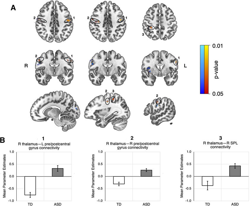
Fig. 1 Voxels that were found to exhibit hyperconnectivity with the right thalamus. Significant clusters found in the HFASD (orange) and
ABIDE (blue) cohorts are colored by p-value. A Top - axial view, middle - coronal view, bottom - sagittal view, sliced from center to right. HFASD
clusters are primarily localized in the left and right precentral gyri, left and right postcentral gyri, and right superior parietal lobule. ABIDE clusters are
primarily localized in the right precentral and postcentral gyri, right insulae, and right and left cuneus. B Bar graphs depicting group differences in
group-level GLM parameter estimates, taken as an average of all the voxels in a particular cluster for each subject. The two clusters in the right
precentral and postcentral gyri (Table 3) were averaged together as one cluster. SPL = superior parietal lobule.Ayub et al. Translational Psychiatry (2021)11:93 Page 6 of 9
Fig. 2 Correlations of left precentral/postcentral gyri cluster with AQ. Correlations by group (A), in males by group (B), and in females by group
(C). Reported r values are Pearson’s correlation coefficients.
noted between AQ total score and right thalamus–left analysis investigating the default mode, executive control,
precentral/postcentral gyrus RSFC (r = 0.48, p = 0.079). and thalamocortical networks in an age- and sex-matched
In ASD females, a negative linear relationship was found, cohort of HFASD and their typically developing coun-
though the data were too limited to reach significance terparts. We observed hyperconnectivity of the right
(r = –0.51, p = 0.16). thalamus with bilateral pre- and post-central gyri in the
To follow up on these findings, we performed a mod- ASD group, but no other seeded regions had significant
eration analysis of the ASD group to investigate whether group differences in our HFASD cohort. We further
sex moderates the effect of RSFC on behavior. We validated right thalamocortical hyperconnectivity with
included sex, RSFC, and a sex × RSFC interaction term as right precentral and postcentral gyri in a similar cohort of
independent variables of a GLM predicting AQ total HFASD in ABIDE. Taken together, these results provide
score. Significance was set at p < 0.05. Sex was found to be support for the presence of hyperconnectivity of the right
an effect modifier of the relationship between AQ and thalamus to sensorimotor regions in the cortex
RSFC (model F(3,19) = 5.059, p = 0.00962, adjusted R2 = in HFASD.
0.3563; sex × RSFC interaction p = 0.0238). The lack of group differences for seeded regions in the
Further GLM analyses of the HFASD cohort were DMN and CEN is surprising given the widespread evi-
performed to explore sex differences in the relationship dence for altered connectivity in these networks across
between other ASD behavioral measures and right age groups31,33,35,37,57. One study of male high-
thalamus–left precentral/postcentral gyrus RSFC. These functioning ASD adults found hypoconnectivity between
analyses can be found in the Supplemental Information. core DMN regions that negatively correlated with AQ36.
However, our cohort consists of both males and females,
ABIDE validation and sex is known to mediate DMN connectivity58. CEN
In the ABIDE GLM analysis, clusters were found that connectivity in autism has mixed results. Studies of ASD
had greater connectivity with the right thalamus in the children and adolescents show hypoconnectivity with
ASD group compared to the TD group. Two of these prefrontal areas33,57, but a large scale investigation of
clusters were located in the right precentral (p = 0.019) ABIDE found no group differences in connectivity in
and postcentral gyri (p = 0.028), validating the observed prefrontal areas in only adults18, corroborating our
hyperconnectivity of the same regions in our HFASD results. Sex may have also influenced our CEN results, but
cohort. The other clusters were primarily localized in the it is difficult to draw conclusions with our modest sample
insula and the cuneus (Table 3) and not in areas originally size. Further investigation into sex-mediated effects on
found in our cohort. The parameter estimates for the DMN and CEN connectivity in high-functioning ASD
largest cluster identified also did not significantly corre- adults is needed.
late with any clinical measures provided in ABIDE. The differences in connectivity from the thalamus to the
pre- and post-central gyri form a strong case for impair-
Discussion ment of sensorimotor processing in HFASD. Undoubt-
The goal of this study was to investigate thalamocortical edly, the pre- and post-central gyri are critical for the
connectivity in HFASD and how it relates to ASD execution of motor commands and perception of sensory
symptom severity. We performed seed-connectivity stimuli, respectively. One study has previously shown thatAyub et al. Translational Psychiatry (2021)11:93 Page 7 of 9 the connectivity of the thalamus to bilateral pre- and post- synchronizing tendency of excitatory thalamocortical central gyrus and the left superior parietal lobule is dis- neurons, mediated by GABAA receptors62. In fact, our rupted in ASD adolescents in response to aversive audi- earlier study on the same cohort using proton magnetic tory and tactile stimuli59. Indeed, atypical thalamic resonance spectroscopy revealed decreased thalamic connectivity to sensorimotor cortical regions is widely concentrations of GABA in ASD males compared to their replicated in studies of children and adolescents with TD counterparts24. Additionally, GABA has previously ASD18,19,33,34,57. Thus, our results demonstrate the pre- been associated with the integrity of synchronized oscil- sence of atypical thalamocortical connectivity to sensor- lations in sensory processing16. While hyperconnectivity imotor regions in HFASD and reinforce the notion that may indicate altered function in the GABAergic system in atypical thalamocortical connectivity is a neural correlate the thalamus, biophysical modeling is needed to explore of sensorimotor processing differences in ASD. the mechanisms of how thalamic inhibition can impact While the link between sensory abnormalities and tha- thalamocortical functional connectivity63. lamocortical dysconnectivity seems to be clear, the effects Our results indicate group differences in functional of age on this relationship in ASD are unclear. Our post- connectivity of the right thalamus but not the left. Similar hoc GLM analyses generally showed no significant main asymmetries have been found previously in a study of effect or interaction effect due to age. Hyperconnectivity children with ASD, which observed greater white matter between the thalami and sensorimotor regions was compromise, greater thalamic functional specialization, demonstrated in children and adolescents with ASD in and maladaptive temporo-thalamic hyperconnectivity in several recent reports18–20,33,57, consistent with our find- the right hemisphere57. To rule out the possibility of a ing in HFASD in this study. To fully understand the mediating effect of handedness, we ensured that the effects of age on thalamocortical dysconnectivity, we will proportion of individuals with ASD who were right- need to perform further investigations using a long- handed did not differ significantly from that of controls itudinal design. (Table 1). Other explanations for this asymmetry may be In addition to age, sex is known to drive considerable found through other studies of the thalamus in ASD. One variation in the clinical presentation of ASD60. study investigating serotonin levels in thalamic pathways Acknowledging our modest sample size, we explored finds asymmetries in children with ASD in serotonin potential sex differences in our observed brain-behavior synthesis in the thalamus, frontal cortex, and dentate relations and report preliminary observations (see Fig. 2B, gyrus64. Interestingly, serotonin has been previously C and Supplemental Information). We found that ASD implicated in the hyperexcitability of sensory thalamo- males exhibited a positive correlation between sensor- cortical circuits65. Thus, the asymmetry of thalamic imotor thalamocortical connectivity and AQ, while ASD hyperconnectivity with the sensorimotor cortex further females exhibited a negative correlation. Furthermore, for suggests an underlying disruption of neurotransmitter males but not females, the thalamocortical connectivity systems in the thalamus. partially mediated the ASD vs. TD group difference in There are several limitations to the present study. First, AQ. This suggests that sensorimotor function may affect the modest size of the investigated cohort limits the ASD characteristics differentially by sex in adults with generalizability of our results. Drawing conclusions from ASD. In fact, a recent meta-analysis revealed that high- the observed brain-behavior relationships with a small functioning female adults with ASD typically report dis- sample size is difficult given the heterogeneities already proportionately more sensorimotor symptoms than their present in the ASD population. Second, there is a sig- ASD male counterparts61. A potential mechanism of sex nificant group difference in NVIQ between the HFASD differences in sensorimotor processing could be differ- and TD groups. Moreover, our GLM analyses that ences in inhibitory neurotransmission. In our previous accounted for NVIQ as a covariate revealed that the study of GABA concentrations in HFASD, we observed results are not affected by between-group and within- that ASD males had a negative correlation of thalamic group variation in NVIQ. This suggests that sensorimotor GABA concentration with AQ, but ASD females had a hyperconnectivity with the thalamus and its relation to positive correlation, a similar sex-specific relationship as core behavioral abnormalities in ASD is independent of observed in the present study24. Altogether, these pre- NVIQ. Third, the observed lack of group differences in liminary results, in conjunction with the findings from connectivity without GSR may diminish the robustness of past studies, demonstrate the impact of sex on ASD our findings. However, our results are highly unlikely to neurobiology and underscore the need for investigating be contaminated by motion artifacts; GSR is known to be sex differences in studies of ASD. effective at removing spurious low-amplitude correlations The presence of thalamocortical hyperconnectivity introduced by head motion66,67, and overall head motion further suggests awry inhibitory mechanisms. Inhibitory was comparable between the ASD and TD groups. Even neurons in the thalamus are known to counter the though the inclusion of GSR in the preprocessing of
Ayub et al. Translational Psychiatry (2021)11:93                                                                                                          Page 8 of 9

resting-state fMRI is still highly contended68, it is espe-                      Publisher’s note
cially necessary for removing motion artifacts in studies of                     Springer Nature remains neutral with regard to jurisdictional claims in
                                                                                 published maps and institutional affiliations.
ASD, a population that tends towards excessive head
motion69. One study even found GSR boosted variance                              Supplementary information The online version contains supplementary
explained by whole-brain RSFC in behavioral measures,                            material available at https://doi.org/10.1038/s41398-021-01221-0.
though it is unclear if the same applies for specific con-
                                                                                 Received: 19 August 2020 Revised: 7 January 2021 Accepted: 15 January
nections as in our study70. For these reasons, GSR is                            2021
warranted. Fourth, the seed-connectivity analysis in the
present study used whole-thalamus seeds instead of
individual thalamic nuclei. Both the HFASD and ABIDE
cohorts used low-resolution functional imaging data, thus                        References
results of seed-connectivity analysis using individual tha-                       1. Marco, E. J., Hinkley, L. B. N., Hill, S. S. & Nagarajan, S. S. Sensory processing in
                                                                                      autism: a review of neurophysiologic findings. Pediatr. Res. 69, 48R–54R (2011).
lamic nuclei would have been highly susceptible to noise.                         2. Robertson, C. E. & Baron-Cohen, S. Sensory perception in autism. Nat. Rev.
Additionally, most investigations of thalamocortical con-                             Neurosci. 18, 671–684 (2017).
nectivity have used a hybrid whole-thalamus and cortical                          3. Anzulewicz, A., Sobota, K. & Delafield-Butt, J. T. Toward the autism motor
                                                                                      signature: gesture patterns during smart tablet gameplay identify children
ROI seed approach18, which we follow. Future investiga-                               with autism. Sci. Rep. 6, 1–13 (2016).
tions ought to examine the connectivity of specific tha-                           4. Cook, J. L., Blakemore, S. J. & Press, C. Atypical basic movement kinematics in
lamic nuclei to further elucidate the relationship between                            autism spectrum conditions. Brain 136, 2816–2824 (2013).
                                                                                  5. Vlaskamp, C. et al. Auditory processing in autism spectrum disorder: mismatch
thalamocortical connectivity and sensorimotor function                                negativity deficits. Autism Res. 10, 1857–1865 (2017).
in ASD. Fifth, the lack of measures for sensorimotor                              6. Linke, A. C., Jao Keehn, R. J., Pueschel, E. B., Fishman, I. & Müller, R. A. Children
symptoms could limit the extension of our conclusions to                              with ASD show links between aberrant sound processing, social symptoms,
                                                                                      and atypical auditory interhemispheric and thalamocortical functional con-
impaired sensorimotor processing in ASD. In future stu-                               nectivity. Dev. Cogn. Neurosci. 29, 117–126 (2018).
dies, we will prioritize the characterization of sensor-                          7. Mikkelsen, M., Wodka, E. L., Mostofsky, S. H. & Puts, N. A. J. Autism spectrum
imotor symptoms accordingly. Lastly, the use of only self-                            disorder in the scope of tactile processing. Dev. Cogn. Neurosci. 29, 140–150
                                                                                      (2018).
report measures may be problematic due to the sub-                                8. Cascio, C. J., Gu, C., Schauder, K. B., Key, A. P. & Yoder, P. Somatosensory event-
jectivity of reporting symptom severity.                                              related potentials and association with tactile behavioral responsiveness
  Despite these limitations, this study reports findings of                            patterns in children with ASD. Brain Topogr. 28, 895–903 (2015).
                                                                                  9. Kumar, S. L. Examining the characteristics of visuospatial information proces-
resting-state thalamocortical functional hyperconnectivity                            sing in individuals with high-functioning autism. Yale J. Biol. Med. 86, 147–156
with the sensorimotor regions in HFASD for the first                                   (2013).
time. This hyperconnectivity is associated with autistic                         10. Moraes, Í. A. Pde et al. Motor learning characterization in people with autism
                                                                                     spectrum disorder: a systematic review. Dement Neuropsychol. 11, 276–286
features across all participants. These results confirm                               (2017).
existing literature on the widespread hyperconnectivity of                       11. Perry, W., Minassian, A., Lopez, B., Maron, L. & Lincoln, A. Sensorimotor gating
the thalamus with the cortex and strongly suggest the                                deficits in adults with autism. Biol. Psychiatry 61, 482–486 (2007).
                                                                                 12. Gabriels, R. L. et al. Is there a relationship between restricted, repetitive, ste-
presence of differences in sensorimotor processing in                                reotyped behaviors and interests and abnormal sensory response in children
ASD. These differences could be indicative of disrupted                              with autism spectrum disorders? Res Autism Spectr. Disord. 2, 660–670 (2008).
neurotransmitter systems in the thalamus and can be                              13. Schulz S. E., Stevenson R. A. Sensory hypersensitivity predicts repetitive
                                                                                     behaviours in autistic and typically-developing children. Autism 2018. https://
attributed to the neurobiology of ASD independent of                                 doi.org/10.1177/1362361318774559 (2018).
typically associated cognitive differences and age. Further                      14. Boyd, B. A. et al. Sensory features and repetitive behaviors in children with
investigation into the underlying mechanisms of dis-                                 autism and developmental delays. Autism Res. 3, 78–87 (2010).
                                                                                 15. Foss-Feig, J. H., Heacock, J. L. & Cascio, C. J. Tactile responsiveness patterns and
rupted thalamocortical connectivity and how it may lead                              their association with core features in autism spectrum disorders. Res Autism
to sex-specific abnormal sensory processing is                                        Spectr. Disord. 6, 337–344 (2012).
encouraged.                                                                      16. Simon, D. M. & Wallace, M. T. Dysfunction of sensory oscillations in autism
                                                                                     spectrum disorder. Neurosci. Biobehav Rev. 68, 848–861 (2016).
                                                                                 17. Belmonte, M. K. et al. Autism as a disorder of neural information processing:
Acknowledgements                                                                     directions for research and targets for therapy. Mol. Psychiatry 9, 646–663
This work was supported by the NIMH Career Development Award (K08;                   (2004).
MH111750) to L.K.F., National Science Foundation Graduate Research               18. Woodward, N. D., Giraldo-Chica, M., Rogers, B. & Cascio, C. J. Thalamocortical
Fellowship to R.A., and NIH Director’s New Innovator Award (DP2; MH119735)           dysconnectivity in autism spectrum disorder: an analysis of the autism brain
to M.S.                                                                              imaging data exchange. Biol. Psychiatry Cogn. Neurosci. Neuroimaging 2, 76–84
                                                                                     (2017).
                                                                                 19. Cerliani, L. et al. Increased functional connectivity between subcortical and
Author details
1                                                                                    cortical resting-state networks in autism spectrum disorder. JAMA Psychiatry
 Department of Bioengineering, Stanford University, Stanford, CA, USA.
2                                                                                    72, 767–777 (2015).
 Department of Psychiatry and Behavioral Sciences, Stanford University,
                                                                                 20. Tomasi, D. & Volkow, N. D. Reduced local and increased long-range functional
Stanford, CA, USA. 3School of Medicine, Stanford University, Stanford, CA, USA
                                                                                     connectivity of the thalamus in autism spectrum disorder. Cereb. Cortex 29,
                                                                                     573–585 (2019).
Conflict of interest                                                              21. Crane, L., Goddard, L. & Pring, L. Sensory processing in adults with autism
The authors declare that they have no conflict of interest.                           spectrum disorders. Autism 13, 215–228 (2009).
Ayub et al. Translational Psychiatry (2021)11:93 Page 9 of 9
22. Brown, M. S., Singel, D., Hepburn, S. & Rojas, D. C. Increased glutamate con- 46. Lord, C., Rutter, M. & Couteur, A Le. Autism diagnostic interview-revised: a
centration in the auditory cortex of persons with autism and first-degree revised version of a diagnostic interview for caregivers of individuals with
relatives: A 1H-MRS study. Autism Res. 6, 1–10 (2013). possible pervasive developmental disorders 1. J. Autism Dev. Disord. 24, 1–27
23. Pizzarelli, R. & Cherubini, E. Alterations of GABAergic signaling in autism (1994).
spectrum disorders. Neural Plast. 2011, 1–12 (2011). 47. Lord C. et al. Autism diagnostic observation schedule, second edition. 2nd
24. Fung L. K. et al. Thalamic and prefrontal GABA concentrations but not GABAA edition. Western Psychological Services; 2012.
receptor densities are altered in high-functioning adults with autism spectrum 48. Roid G. H. Stanford Binet’s Intelligence Scales. Fifth Ed. Itasca, IL: Riverside
disorder. Mol. Psychiatry. https://doi.org/10.1038/s41380-020-0756-y (2020). Publishing; 2003.
25. Frankland, P. W. et al. Sensorimotor gating abnormalities in young males with 49. Ritvo, R. A. et al. The ritvo autism asperger diagnostic scale-revised (RAADS-R):
fragile X syndrome and Fmr1-knockout mice. Mol. Psychiatry 9, 417–425 A scale to assist the diagnosis of autism spectrum disorder in adults: An
(2004). international validation study. J. Autism Dev. Disord. 41, 1076–1089 (2011).
26. Swerdlow, N. R. et al. Tactile prepuff inhibition of startle in children with 50. Constantino J. N., Gruber C. P. Social Responsiveness Scale. Torrance, CA:
Tourette’s syndrome: In search of an ‘fMRI-friendly’ startle paradigm. Biol. Western Psychological Services; 2012.
Psychiatry 50, 578–585 (2001). 51. Baron-Cohen, S., Wheelwright, S., Skinner, R., Martin, J. & Clubley, E. The Autism-
27. Hwang, K., Bertolero, M. A., Liu, W. B. & D’Esposito, M. The human thalamus is Spectrum Quotient (AQ): Evidence from…. J. Autism Dev. Disord. 31, 5–17 (2001).
an integrative hub for functional brain networks. J. Neurosci. 37, 5594–5607 52. Esteban O. et al. FMRIPrep: a robust preprocessing pipeline for functional MRI.
(2017). BioRxiv. 2018:1–20.
28. Halassa, M. M. & Acsády, L. Thalamic inhibition: diverse sources, diverse scales. 53. Androulakis, X. M. et al. Modulation of intrinsic resting-state fMRI networks in
Trends Neurosci. 39, 680–693 (2016). women with chronic migraine. Neurology 89, 163–169 (2017).
29. Molnar, G. F. et al. Changes in motor cortex excitability with stimulation of 54. Fox, M. D. et al. The human brain is intrinsically organized into dynamic,
anterior thalamus in epilepsy. Neurology 66, 566–571 (2006). anticorrelated functional networks. Proc. Natl Acad. Sci. USA 102, 9673–9678
30. Cury, R. G. et al. Thalamic deep brain stimulation for tremor in Parkinson (2005).
disease, essential tremor, and dystonia. Neurology 89, 1416–1423 (2017). 55. Woolrich, M. W., Behrens, T. E. J., Beckmann, C. F., Jenkinson, M. & Smith, S. M.
31. Hull J. V. et al. Resting-state functional connectivity in autism spectrum dis- Multilevel linear modelling for FMRI group analysis using Bayesian inference.
orders: a review. Front Psychiatry 7, 205 (2017). Neuroimage 21, 1732–1747 (2004).
32. Mohammad-Rezazadeh, I., Frohlich, J., Loo, S. K. & Jeste, S. S. Brain connectivity 56. Winkler, A. M., Ridgway, G. R., Webster, M. A., Smith, S. M. & Nichols, T. E.
in autism spectrum disorder. Curr. Opin. Neurol. 29, 137–147 (2016). Permutation inference for the general linear model. Neuroimage 92, 381–397
33. Nair, A. et al. Regional specificity of aberrant thalamocortical connectivity in (2014).
autism. Hum. Brain Mapp. 36, 4497–4511 (2015). 57. Nair, A., Treiber, J. M., Shukla, D. K., Shih, P. & Müller, R. A. Impaired thalamo-
34. Iidaka, T., Kogata, T., Mano, Y. & Komeda, H. Thalamocortical hyperconnectivity cortical connectivity in autism spectrum disorder: a study of functional and
and amygdala-cortical hypoconnectivity in male patients with autism spec- anatomical connectivity. Brain 136, 1942–1955 (2013).
trum disorder. Front Psychiatry 10, 1–11 (2019). 58. Ypma, R. J. F. et al. Default mode hypoconnectivity underlies a sex-related
35. Padmanabhan, A., Lynch, C. J., Schaer, M. & Menon, V. The default mode autism spectrum. Biol. Psychiatry Cogn. Neurosci. Neuroimaging 1, 364–371
network in autism. Biol. Psychiatry Cogn. Neurosci. Neuroimaging 2, 476–486 (2016).
(2017). 59. Green, S. A., Hernandez, L., Bookheimer, S. Y. & Dapretto, M. Reduced mod-
36. Jung, M. et al. Default mode network in young male adults with autism ulation of thalamocortical connectivity during exposure to sensory stimuli in
spectrum disorder: relationship with autism spectrum traits. Mol. Autism 5, ASD. Autism Res 10, 801–809 (2017).
1–11 (2014). 60. Werling, D. M. & Geschwind, D. H. Sex differences in autism spectrum dis-
37. Nair, A., Jolliffe, M., Lograsso, Y. S. S. & Bearden, C. E. A review of default mode orders. Curr. Opin. Neurol. 26, 146–153 (2013).
network connectivity and its association with social cognition in adolescents 61. Moseley, R. L., Hitchiner, R. & Kirkby, J. A. Self-reported sex differences in high-
with autism spectrum disorder and early-onset psychosis. Front Psychiatry 11, functioning adults with autism: a meta-analysis. Mol. Autism 9, 1–12 (2018).
1–17 (2020). 62. Huguenard, J. R. & McCormick, D. A. Thalamic synchrony and dynamic reg-
38. Dajani, D. R. & Uddin, L. Q. Demystifying cognitive flexibility: Implications ulation of global forebrain oscillations. Trends Neurosci. 30, 350–356 (2007).
for clinical and developmental neuroscience. Trends Neurosci. 38, 63. Deco, G. et al. Whole-brain multimodal neuroimaging model using serotonin
571–578 (2015). receptor maps explains non-linear functional effects of LSD. Curr. Biol. 28,
39. Yerys, B. E. et al. Neural correlates of set-shifting in children with autism. Autism 3065–3074 (2018).
Res. 8, 386–397 (2015). 64. Chugani, D. C. et al. Altered serotonin synthesis in the dentatothalamocortical
40. Schmitz, N. et al. Neural correlates of executive function in autistic spectrum pathway in autistic boys. Ann. Neurol. 42, 666–669 (1997).
disorders. Biol. Psychiatry 59, 7–16 (2006). 65. Bauernfeind, A. L. et al. Human ecstasy use is associated with increased cortical
41. Taylor, M. J., Donner, E. J. & Pang, E. W. fMRI and MEG in the study of typical excitability: an fMRI study. Neuropsychopharmacology 36, 1127–1141 (2011).
and atypical cognitive development. Neurophysiol. Clin. Neurophysiol. 42, 66. Power, J. D. et al. Methods to detect, characterize, and remove motion artifact
19–25 (2012). in resting state fMRI. Neuroimage 84, 320–341 (2014).
42. Shafritz, K. M., Dichter, G. S., Baranek, G. T. & Belger, A. The neural circuitry 67. Power, J. D., Barnes, K. A., Snyder, A. Z., Schlaggar, B. L. & Petersen, S. E. Spurious
mediating shifts in behavioral response and cognitive set in autism. Biol. but systematic correlations in functional connectivity MRI networks arise from
Psychiatry 63, 974–980 (2008). subject motion. Neuroimage 59, 2142–2154 (2012).
43. Alves P. N. et al. An improved neuroanatomical model of the default-mode 68. Murphy, K. & Fox, M. D. Towards a consensus regarding global signal
network reconciles previous neuroimaging and neuropathological findings. regression for resting state functional connectivity MRI. Neuroimage 154,
Commun Biol. 2, 370 (2019). 169–173 (2017).
44. Di Martino, A. et al. The autism brain imaging data exchange: towards a large- 69. Cox, A. D., Virues-Ortega, J., Julio, F. & Martin, T. L. Establishing motion control
scale evaluation of the intrinsic brain architecture in autism. Mol. Psychiatry 19, in children with autism and intellectual disability: applications for anatomical
659–667 (2014). and functional MRI. J. Appl Behav. Anal. 50, 8–26 (2017).
45. Di Martino, A. et al. Enhancing studies of the connectome in autism using the 70. Li, J. et al. Global signal regression strengthens association between resting-
autism brain imaging data exchange II. Sci. Data 4, 1–15 (2017). state functional connectivity and behavior. Neuroimage 196, 126–141 (2019).You can also read