Metformin Ameliorates Oxidative Stress Induced by Diabetes Mellitus and Hepatocellular Carcinoma in Rats
←
→
Page content transcription
If your browser does not render page correctly, please read the page content below
Reports of Biochemistry & Molecular Biology
 Vol.9, No.1, Apr 2020

 Original article www.RBMB.net
[ DOI: 10.29252/rbmb.9.1.115 ]

 Metformin Ameliorates Oxidative Stress Induced
 by Diabetes Mellitus and
 Hepatocellular Carcinoma in Rats
 Maysa Ahmed Mobasher*1, 2, Hala Galal El-Tantawi3, Karim Samy El-Said4

 Abstract
Downloaded from rbmb.net at 10:06 +0430 on Thursday September 17th 2020

 Background: Several studies have found an association between Diabetes mellitus (DM) and an increased risk
 for hepatocellular carcinoma (HCC). Evidence suggests that Metformin (Met) may have a therapeutic and
 protective effect against both DM and HCC. Therefore, the aim of this study was to evaluate the antioxidant
 effect of Met against DM and HCC-induced oxidative stress in rat model.
 Methods: Forty-two male albino rats were randomly divided into six groups. Group 1 (Gp1) was the control
 group, Gp2 received an intraperitoneal (i.p.) injection with streptozotocin (STZ), Gp3 was injected i.p. with
 diethyl nitrosamine (DEN), Gp4 received an oral administration of Met, Gp5 and Gp6 received the same
 injections as Gp2 and Gp3, respectively, then received an additional injection of Met. Oxidative stress
 biomarkers, including superoxide dismutase (SOD), catalase (CAT), reduced glutathione (GSH) and
 malondialdehyde (MDA), were examined. Furthermore, biochemical parameters including liver function
 tests were assessed. Histopathological and immunohistochemical alterations of the liver were also examined.
 Results: Our results demonstrate that Gp2 and Gp3 had significant signs of liver dysfunction and had elevated
 levels of MDA and reduced levels of SOD, CAT, and GSH. Additionally, Gp2 and Gp3 showed significant
 alterations in the liver architecture shown by high PCNA and caspase-3 expression. In the Gp5 and Gp6,
 treatment with Met showed an improvement in liver function, oxidative stress biomarkers, and reduced
 histopathological changes in hepatocytes.
 Conclusions: This study offers insight into the potential for Metformin as a novel therapeutic against the
 oxidative stress induced by DM or HCC.

 Keywords: Diabetes Mellitus, Diethyl nitrosamine, Hepatocellular Carcinoma, Metformin, Streptozotocin.

 Introduction
 Diabetes mellitus (DM) is one of the most widely particular, hepatocellular carcinoma (HCC), the
 recognized chronic diseases in almost all nations malignant liver cancer represents the 5th common
 and classified as the 9th major cause of death cancer and the 3rd cause of cancer-related
 worldwide (1). DM has been strongly linked with mortality worldwide (5) in accordance with the
 the non-alcoholic fatty-liver disease (NAFLD) World Health Organization (WHO). Previous
 and many other metabolic disturbances (2). It is research has shown that obesity and its related
 worthy to mention that DM is an important risk metabolic abnormalities, particularly DM,
 factor for the development of many types of significantly, increase the danger of HCC
 malignancies (3). Cancer alone represents the development (6) due to the production of hyper-
 most significant cause of death worldwide and insulin-like growth factor 1 (IGF-1) and oxidative
 eliminates around 6 million lives each year (4). In stress (7). Diabetic patients develop several

 1: Department of Pathology, Biochemistry Division, College of Medicine, Jouf University, Sakaka, Saudi Arabia.
 2: Department of Clinical Pathology, El Ahrar Educational Hospital, Ministry of Health, Zagazig, Egypt.
 3: Zoology Department, Faculty of Science, Ain Shams University, Cairo, Egypt.
 4: Chemistry Department, Biochemistry Division, Faculty of Science, Tanta University, Egypt. 31527.
 *Corresponding author: Maysa Mobasher; Tel: 00966560574229; E-mail: mmobasher@ju.edu.sa.
 Received: 10 Jan, 2020; Accepted: 2 Feb, 2020
Ahmed Mobasher M et al

 complications including non-alcoholic fatty liver could decrease the oxidative stress state, which in
[ DOI: 10.29252/rbmb.9.1.115 ]

 that contribute in progressive liver diseases such turn ameliorates the severity of such diseases (12).
 as cirrhosis and cancer (7). Despite substantial Metformin (Met) is a first-line DM therapy,
 advancement in understanding the clinical relation which decreases the glucose production in the
 between DM and HCC, further injurious liver by increasing the body’s sensitivity to insulin
 mechanisms responsible for this cellular damage (13). Previous pharmacoepidemiologic study
 remain unknown. Indeed, this lack of knowledge, revealed that Met treatment seems to prevent
 attracts many researchers to elucidate this hepatocellular transformation, diminishing the
 relationship (8). danger of HCC development to degrees parallel to
 Simultaneously, the type and prescribed dose that of non-diabetic patients (14). The previous
 of the utilized antidiabetic drugs are also findings of De Peralta et al, (15) reported that the
 implicated in influencing the danger of HCC prevalence of HCC, as well as fibrotic and
 development (9). Thus, emerging from these inflammatory markers levels were reduced when
Downloaded from rbmb.net at 10:06 +0430 on Thursday September 17th 2020

 tremendous contributions between DM and HCC, Met treatment begun at the first signs of fibrosis.
 reliable management of DM is suggested (8). Notwithstanding, HCC burden was unchanged
 Insulin resistance in type 2 DM (T2DM) is a when Met treatment begun at the first signs of
 key pathological feature for HCC oncogenesis. It cirrhosis (15). Notably, it has been reported that
 has been reported that insulin resistance may have Met might operate to inhibit tumorigenesis
 a potential role in hepatocarcinogenesis in patients through both the insulin-dependent and insulin-
 with T2DM or prediabetes infected with hepatitis independent basic mechanisms (16). Additionally,
 B virus (HBV) (6). Reportedly, hyperinsulinemia it has been shown that Met has potent antioxidant
 upregulates the levels of insulin-like growth properties besides its therapeutic effect against
 factors (IGFs), and abnormal activation of the DM (17). Based on that, this study aimed to
 IGF/IGF-1 receptor (IGF-1R) axis plays an investigate and underlie the antioxidant potency
 important role in the development of different of Met versus the oxidative stress induced by STZ
 types of human malignancies, involving HCC and DEN in DM and HCC rats, respectively and
 (10). Therefore, these data advocated that to clarify the connection between diabetes,
 targeting insulin resistance could be an applicable oxidative stress, and cancer morbidity or cancer
 approach for repressing the incidence of obesity- prognosis. Results emerging from this work will
 related HCC. help to control the disease development and
 Furthermore, the potential role of oxidative provide an efficient therapy.
 stress in the production of tissue damage in DM
 had been discussed. In addition, oxidative stress is Materials and methods
 the primary driving force of HCC development Chemicals
 which predisposes to hepatocarcinogenesis. Streptozotocin (STZ) and diethylnitrosamine
 Under normal physiological state, a balance (DEN) were purchased from Sigma (St. Louis,
 between reactive oxygen species (ROS) MO, USA). DEN was dissolved in saline. All
 generation and oxidative defenses mechanism biochemical kits were obtained from the Bio-
 exists in all cells, which modulated by diagnostic company (Cairo-Egypt). For
 endogenous antioxidant enzymes as superoxide immunohistochemical preparation, the primary
 dismutase (SOD), catalase (CAT), and and secondary antibodies were purchased from
 glutathione peroxidase (Gpx1). During the course Dako Company (Glostrup, Denmark).
 of DM and HCC, the level of oxidant biomarkers
 such as MDA and G-S-S-G increased due to the Rats
 release of elevated levels of both reactive oxygen Forty-two male Sprague Dawley albino rats, (100
 species and nitrogen species (ROS and RNS, ± 5 g) were obtained from the National Research
 respectively) (11) together with reductions in the Center (NRC, Cairo, Egypt), housed randomly as 6
 expression of antioxidants enzymes levels. rats per cage, in 12h/12h dark/light cycle under
 Therefore, treatment with antioxidant agents standard conditions of temperature and humidity.

 116 Rep. Biochem. Mol. Biol, Vol.9, No.1, Apr 2020
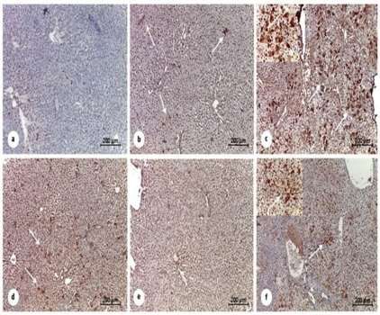
Antioxidant Effects of Metformin

 Animals were carefully observed every day and (15). For Met treatment, rats were treated by
[ DOI: 10.29252/rbmb.9.1.115 ]

 their body weights were recorded, while food gavage with 150 mg/kg every other day for 100
 consumption and water intakes were measured days (19) (Fig. 1).
 precisely every week to evaluate any signs of
 toxicity or abnormality during the experiment. Experimental design
 Animal experimentations were approved by the All rats were clinically healthy. The animals
 ethics committee at the Faculty of Science, Tanta were divided randomly into six equal groups
 University (Egypt), and were conducted ( = 7) as the following: Gp1 was served as
 according to their accepted ethical guidelines for normal untreated control rats, Gp2 was injected
 the Animal Ethics Committee (No. IACUC-SCI- with STZ, Gp3 was injected with DEN, Gp4
 TU-0084). was orally administrated with Met, Gp5 was
 injected with STZ and treated with Met and
 DM and HCC inductions Gp6 was injected with DEN and treated with
Downloaded from rbmb.net at 10:06 +0430 on Thursday September 17th 2020

 For DM induction, rats received a single Met. Met was orally administered after blood
 intraperitoneal (i.p) dose of STZ (80 mg/kg) glucose level reached up to 250 mg/dl in the
 (18). For HCC induction, rats were i.p injected DM model and at 50 days' post-DEN-injection
 with DEN (50 mg/kg) once weekly for 7 weeks in the HCC model (Fig. 1).

 Fig. 1. Experimental design. STZ: Streptozotocin (80 mg/kg once); Met: Metformin (150 mg/Kg/ each other day); DEN:
 Diethylnitrosamine (50 mg/Kg/ once per week for 7 weeks). Gp1 were normal untreated control rats, Gp2 was injected with
 STZ, Gp3 was injected with DEN, Gp4 was administered Met via gavage, Gp5 was injected with STZ and treated with Met,
 and Gp6 was injected with DEN and treated with Met.

 Samples
 At the end of the experiment, by the day 105, all relative organ weights (organ wt/b.wt × 100) of
 rats were sacrificed under ethyl ether anesthesia all rats were taken after organs being necropsied.
 and cadavers were burned in animal incinerators Blood samples were collected from arterial
 under the supervision of the Faculty of Science, blood vessels and heart chambers for
 Tanta University. Gross examinations were hematological examinations, and sera were
 performed macroscopically on all groups during buffer saline (PBS). The resulting supernatants
 sacrifice. The percentages of absolute and were used for biochemical analysis.

 Rep. Biochem. Mol. Biol, Vol.9, No.1, Apr 2020 117
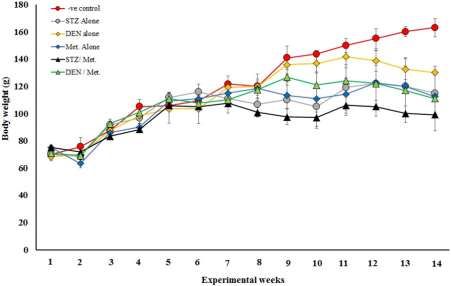
Ahmed Mobasher M et al

 Furthermore, samples from liver tissues were Immunohistochemical staining for PCNA and
[ DOI: 10.29252/rbmb.9.1.115 ]

 collected in buffered formalin for histological caspase-3 detection
 and immunohistochemical investigations. The liver tissues of Sprague Dawley albino rats
 were fixed in 10% buffered formalin for 18-
 Hematological and biochemical profiling 24hr and transferred to 70% ethanol overnight.
 Hemoglobin (Hb) levels, hematocrit (Hct%), All temperatures for processing and embedding
 platelets count, the total count of red blood cells were kept under 60 °C. The super sensitive
 (RBCs), white blood cells (WBCs) and biotin-streptavidin method for
 differential count were detected by using auto immunohistochemical localization was
 hematology analyzer (BC-3200, Mindray, performed. Negative control slides from each of
 China). Alanine aminotransferase (ALT) and the same tissues were incubated with a control
 aspartate aminotransferase (AST) were antibody. The blocking, primary, and control
 determined as described (20). Alkaline antibodies were diluted with a diluent
Downloaded from rbmb.net at 10:06 +0430 on Thursday September 17th 2020

 phosphatase (ALP), arginase activity, and total composed of 1% bovine serum albumin in I X
 protein (TP) were assessed as previously automation buffer.
 described (21-23), respectively. Albumin was
 assayed as the method described by Burtis and Statistical analysis
 Bruns (24). Total bilirubin (TB) was determined Data was presented as mean ± SD and were
 according to Walter and Gerade (25). Serum analyzed using one–way analysis of variance
 cholesterol, triglycerides, and HDL-cholesterol (ANOVA) followed by Tukey’s test and p< 0.05
 were determined using quantitative kit based on were statistically significant.
 the previously described methods (26-28),
 respectively. Low-density lipoprotein cholesterol Results
 (LDL) was calculated according to Friedewald et Treatment with Met in rats with DM
 al, (29) as follows: LDL= total cholesterol– decreased body weight and glucose levels
 HDL– VLDL. Superoxide dismutase (SOD) and The experimental design and the time course of
 catalase activities were determined as described treatments in all groups under study are shown
 (30, 31), respectively. Levels of reduced in Figures 1 & 2 shows that total body weight
 glutathione (GSH) were measured according to until week 7 was similar in all groups of rats.
 Paglia and Valentine (32). Malondialdehyde Likewise, rats receiving STZ (Gp2), Met (Gp4)
 (MDA) was determined based on methods of Li and the DEN/Met group (Gp6) showed similar
 and Chow, briefly, samples were deproteinized changes in their body weight along weeks 8 and
 and then mixed with Thiobarbituric acid (TBA) 14 of the treatment. However, at this time-
 0.67% (W/V), and the absorbance was read at period rats injected with STZ and treated with
 532 nm against a blank (33). Met (Gp5) showed a significant decrease in the
 total body weight when compared with the
 Histological staining control (Gp1).
 Tissue specimens of the liver were harvested After 3 weeks of STZ injection, glucose
 and fixed in 10% formalin. Paraffin blocks levels were increased in the diabetic group (Gp2)
 were prepared after completing the tissue up to 250 mg/dl; however, treatment with Met
 processing in different grades of alcohol and post-DM induction decreased glucose levels
 xylene. Sections (5μm) were prepared from dramatically, which returned close to the normal
 paraffin blocks using microtome, stained with level by week 8. Group of rats injected with
 hematoxylin and eosin, and observed under a DEN alone (Gp3) did not show any significant
 light microscope (Optica light microscope (B- changes in the glucose levels during the full time
 350)) to examine gross cellular damage (34). of the experiment.

 118 Rep. Biochem. Mol. Biol, Vol.9, No.1, Apr 2020
Antioxidant Effects of Metformin
[ DOI: 10.29252/rbmb.9.1.115 ]
Downloaded from rbmb.net at 10:06 +0430 on Thursday September 17th 2020

 Fig. 2. Changes in body weight in the different groups under the study. Treatment with STZ, DEN, Met, or their combinations post 14
 weeks led to differences in the body weights of rats.

 Met did not significantly alter the hematological
 parameters. blood cells (WBCs) when compared to the other
 Hematological analysis of all groups under study groups. Concomitant to this increase in WBCs, the
 showed that only the group of rats injected with percentage (%) of neutrophils and monocytes were
 DEN (Gp3) revealed an increase in the total white increased in the Gp3 (Tables 1 and 2).
 Table 1. Complete blood count of all experimental groups for 12 weeks.
 Groups Hb (g/dl) RBCs(×106/µl) Hct (%) Plt (×103/µl)
 -Ve control 12.5 ± 1.36 a,b 5.95 ± 1.54 39.3 ± 6.19 623 ± 168.9
 a,b
 STZ alone 12.26 ± 0.54 6.72 ± 0.73 37.4 ± 1.52 979.6 ± 257
 a,b
 DEN alone 12.46 ± 1.13 7.20 ± 0.81 40.86 ± 1.49 586.8 ± 23.2
 Met Alone 11.62 ± 1.97 b 6.58 ± 1.78 35.98 ± 7.29 702.2 ± 269
 a,b
 STZ/Met 13.2 ± 0.98 6.9 ± 1.26 39.1 ± 2.97 970.6 ± 157
 a
 DEN/Met 15.88 ± 1.49 8.45 ± 0.95 42.76 ± 3.25 1135.4 ± 747
 F Value 3.89 1.37 0.91 1.22
 p Value 0.025 0.303 0.508 0.359

 Table 2. Differential leucocytes count of all experimental groups for 12 weeks.
 Group WBCs(×103/µl) Neut. (%) Lymph. (%) Mono. (%)
 -Ve control 10.3 ± 3.7 b 11.6 ± 2.408 76.4 ± 6.39 11.8 ± 6.76
 STZ alone 13.4 ± 1.8 a,b
 9 ± 5.099 80.6 ±7 .50 11.8 ± 3.96
 a
 DEN alone 25.64 ± 6.8 16.4 ± 7.83 68.2 ± 11.12 17.2 ± 4.32
 b
 Met alone 10.68 ± 4.67 12 ± 8.63 73 ± 11.83 15 ± 6.96
 STZ/Met 9.48 ± 3.09 b 9.2 ± 5.40 77.6 ± 7.19 13.4 ± 2.61
 b
 DEN/Met 9.3 ± 5.52 8 ± 2.45 74.8 ± 9.18 13.4 ± 2.70
 F-Value 5.72 0.83 0.65 0.54
 p-Value 0.006 0.552 0.664 0.744

 Rep. Biochem. Mol. Biol, Vol.9, No.1, Apr 2020 119
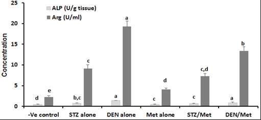
Ahmed Mobasher M et al

 Met protects against the impairment of liver
[ DOI: 10.29252/rbmb.9.1.115 ]

 functions induced by STZ and DEN
 The biochemical analysis of serum samples alone resulted in a marginal increase in the levels of
 showed that levels of ALT, AST, TB, ALP, and ALP and arginase (Fig. 3). The levels of TP and
 Arginase were increased post-DM induction by albumin decreased post-DM induction by STZ
 STZ (Gp2) (Table 3 and Fig. 3). A similar pattern (Gp2) and HCC induction by DEN injection
 of these parameters was found after DEN injection (Gp3). However, Met treatment post-STZ or DEN
 to induce HCC in rats (Gp3). Treatment with Met injections (Gp5 and Gp6) led to reduction in the
 post-STZ or DEN injections (Gp5 and Gp6) levels of ALT, AST, TB, ALP and improve the
 resulted in a decrease in ALT, AST, TB, ALP, and synthetic function of liver by increasing total
 arginase levels. However, treatment with Met protein and albumin levels (Table 3 and Fig. 3).
 Table 3. Serum alanine transaminase (ALT), aspartate transaminase (AST), total bilirubin (TB), total protein (TP) and albumin
Downloaded from rbmb.net at 10:06 +0430 on Thursday September 17th 2020

 (Alb) levels.
 Groups ALT (U/L) AST (U/L) TB (mg/dl) TP (g/dl) Alb. (g/dl)
 -Ve control 58 ± 5.5 d 135 ± 8.5 c 0.33 ± 0.03 d 5.3 ± 0.28 a 2.87 ± 0.24 a
 STZ alone 88 ± 3.06 a,b 207 ± 11.5 a 0.69 ± 0.06 a 4.12 ± 0.24 b,c 1.43 ± 0.13 b
 DEN alone 94 ± 6.91a 211 ± 9.5 a 0.61 ± 0.06 a,b 3.68 ± 0.21 c 1.56 ± 0.22 b
 Met alone 65 ± 5.01 c,d 155 ± 8.5 b,c 0.33 ± 0.04 d 4.83 ± 0.26 a 2.7 ± 0.12 a
 STZ/Met 69 ± 3.6 c,d 170 ± 7.51 b 0.43 ± 0.06 c,d 5.01 ± 0.27 a 2.46 ± 0.25 a
 DEN/Met 78 ± 5.29 b,c 167 ± 8.91 b 0.53 ± 0.05 b,c 4.73 ± 0.23 a,b 1.63 ±0.098 b
 F-Value 22.61 31.54 25.23 17.39 35.13
 0.000 0.000 0.000 0.000 0.000

 Fig. 3. ALP and arginase levels of control, DM and HCC-induced rats. The results display the mean of seven rats per group under
 the different treatment conditions. Bars represent standard deviation. Columns with different lower-case letters indicate significant
 difference between treated groups and the control at p< 0.05 (Tukey’s test).

 120 Rep. Biochem. Mol. Biol, Vol.9, No.1, Apr 2020
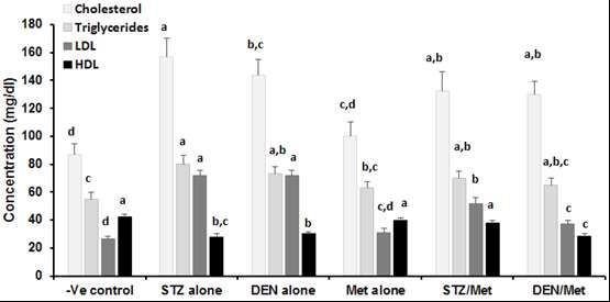
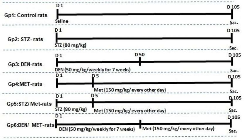
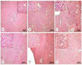
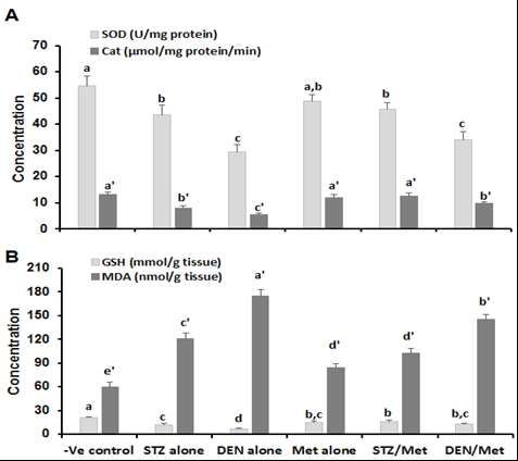
Antioxidant Effects of Metformin

 Met attenuates the elevation of lipid profile and
[ DOI: 10.29252/rbmb.9.1.115 ]

 enhances the reduction of the antioxidant
 biomarkers induced by STZ and DEN Furthermore, induction of DM with STZ or
 Cholesterol, triglycerides, and LDL levels in serum induction of HCC with DEN (Gp2 and Gp3) led
 samples from rats, which injected with STZ or to an increase in the levels of hepatic MDA
 DEN were increased while HDL levels were concomitant with a decrease in the levels of
 decreased (Gp2 and Gp3). The treatment with Met SOD, CAT, and GSH. Treating diabetic and
 post-STZ or DEN injections; however, HCC-bearing rats with Met lead to a significant
 significantly declined the levels of these parameters increase in the levels of CAT and GSH while
 when compared with STZ or DEN injected groups SOD levels were not affected. A significant
 (Fig. 4). HDL levels were slightly increased only in decrease in MDA levels (Gp5 and Gp6) when
 the STZ/Met group when compared with STZ compared to their controls (Gp2 and Gp3) (Figs.
 alone, but not changed in DEN/Met group as 5A-5B) was also noticed in these groups.
Downloaded from rbmb.net at 10:06 +0430 on Thursday September 17th 2020

 compared to DEN alone (Fig. 4).

 Fig. 4. Lipid profile of control, DM, and HCC-induced rats. The results are mean of seven rats per group under
 different treatment conditions. Bars represent standard deviation. Columns with different lower-case letters indicate
 significant difference between treated groups and the control at p< 0.05 (Tukey’s test).

 Met improved liver histology in DM and and multivacuolated cytoplasm of hepatocytes
 HCC-rats that also showed large rounded vesicular
 Light microscopic examination of control liver nuclei with prominent nucleoli together with
 sections revealed normal hepatic architecture necrotic cells and severe bleeding in the blood
 with hepatic strands radiating from the central sinusoids and portal spaces (Fig. 6C). The
 vein. In addition, the polyhedral hepatocytes Met-treated group (Gp4) recorded slight
 appeared with homogeneous acidophilic bleeding in the blood sinusoids and portal
 cytoplasm and a large central nucleus and spaces while the hepatocytes appeared intact
 anastomosing strands of hepatocytes were (Fig. 6D). STZ/Met treated group (Gp5)
 separated with blood sinusoids. Likewise, in showed some necrotic areas accompanied by
 the livers from control rats the central vein typical hepatic architecture, some vacuolated
 lined with endothelial cells (Fig. 6A). The hepatocytes and narrow blood sinusoids (Fig.
 STZ-treated group (Gp2) showed marked 6E). Notably, the liver samples of rats treated
 histological changes manifested by disrupted with DEN and Met preserved the overall liver
 lobular changes with cellular inflammation at architecture, and no evidence of major
 the narrow blood sinusoids. Marked histological injury was found. In these
 degenerative signs and characterized apoptotic samples, HCC appeared remarkably reduced,
 cells were also observed (Fig. 6B). The but apparent cellular infiltration at the blood
 analysis of DEN-treated group (Gp3) revealed sinusoids and trabeculae of large polyhedral
 marked features of grade 1 HCC characterized hepatocytes have polymorphic nuclei were still
 by multiple rounded masses of eosinophilic present (Fig. 6F).

 Rep. Biochem. Mol. Biol, Vol.9, No.1, Apr 2020 121
Ahmed Mobasher M et al
[ DOI: 10.29252/rbmb.9.1.115 ]
Downloaded from rbmb.net at 10:06 +0430 on Thursday September 17th 2020

 Fig. 5. (A) Superoxide dismutase (SOD) and Catalase, (B) Glutathione (GSH) and Malondialdehyde (MDA)
 levels as indicators for oxidative stress. The results are mean of seven rats per group under different treatment
 conditions. Bars represent standard deviation. Columns with different lower-case letters indicate a significant difference
 between treated groups and the control at p< 0.05 (Tukey’s test).

 Fig. 6. Hematoxylin & Eosin stain: photomicrographs of liver sections of rats. (A) control group (Gp1) revealing the hepatic
 strands (arrows) of the hepatic cells (HC) arising from the central vein (CV), lined by the normal endothelial cells (arrowheads),
 [X.10, 40]. (B) STZ treated group (Gp2) for 105 days showing some apoptotic cells (thin arrows) and inflammation at the
 narrowed blood sinusoids and the congestion of the central vein (arrowhead). Hepatocellular degeneration (thick arrows), [X.
 10,40]. (C) DEN treated group (Gp3) for 45 days revealing notable features of hepatocellular carcinoma HCC grade 1. The
 malignant cells consisted of multiple rounded mass of cells with a large amount of eosinophilic multivacuolated cytoplasm (small
 arrows) and large rounded vesicular nuclei with prominent nucleoli. Additionally, many necrotic sites and severe hemorrhage in
 the central vein is present [X.4,40]. (D) MET treated group (Gp4) for 105 days showing slight hemorrhage in the blood sinusoids
 and the portal spaces (arrows). Intact hepatocytes with a normal architecture of the hepatic tissue, [X. 4,40]. (E) STZ/MET treated
 group (Gp5) in which the rats were treated with STZ for 15 days followed by MET for 90 days, revealed more or less normal
 hepatic architecture (arrows) and some necrotic areas (thick arrow). The magnified region shows some narrowing blood
 sinusoids and vacuolation of few hepatocytes (thin arrow), [X. 4, 40]. (F) DEN/MET treated group (Gp6). The rats were treated
 with DEN for 45 days followed by MET for 60 days revealing apparent cellular infiltration at the vascular area (thin arrow). The
 appearance of trabeculae of large polyhedral cells (malignant hepatocytes) with polymorphic nuclei and cellular infiltrate blood
 sinusoids (thick arrow), [X. 10, 40].

 122 Rep. Biochem. Mol. Biol, Vol.9, No.1, Apr 2020
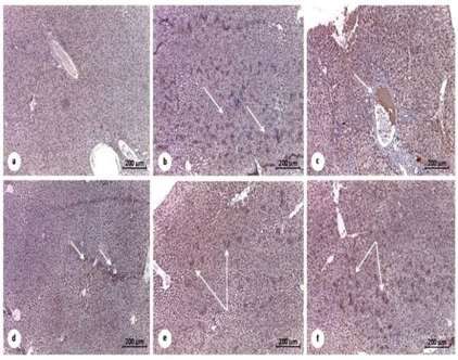
Antioxidant Effects of Metformin
[ DOI: 10.29252/rbmb.9.1.115 ]

 Met reduced the liver PCNA and caspase-3 Group of rats which received STZ followed
 immunohistochemical changes in DM and by Met completely lost the positive
 HCC-rats. immunostaining for PCNA (Fig. 7E).
 Liver sections from the control group Control liver sections revealed negative
 displayed standard lobular architecture with immunostaining for active caspase-3 (Fig.
 negative detectable immunostaining for PCNA 8A). In sections of STZ treated group (Gp2)
 (proliferating cell nuclear antibodies) (Fig. elevation of immunolabelled cells for active
 7A). Whereas DEN-treated group (Gp3) caspase-3 was markedly observed in many
 showed apparent high immunostaining for areas of the hepatic tissue (Fig. 8B). Of note,
 PCNA, particularly around the central vein DEN-treated group (Gp3) showed the most
 (Fig. 7C), treatment with STZ (Gp2) revealed abundance of immunostained apoptotic cells
Downloaded from rbmb.net at 10:06 +0430 on Thursday September 17th 2020

 very few positively stained nuclei in the that appeared surrounding the congested
 Hepatic tissue (Fig. 7B). Also, treatment with central vein (Fig. 8C). Met-treated group
 Met alone showed higher apparent scattered (Gp4) showed almost no immunostained
 positively stained nuclei compared to the cells, but revealed some apoptotic cells
 STZ-treated group (Fig. 7D). Group of rats concentrated at the portal spaces (Fig. 8D).
 which received DEN followed by Met Importantly, STZ/Met and DEN/Met treated
 treatment resulted in decreased PCNA groups (Gp5 and Gp6) showed reductions in
 immunostained cells, which were mainly immunostained active caspase-3 apoptotic
 localized in the pericentral area (Fig. 7F). cells (Figs. 8E-8F).

 Fig. 7. Photomicrographs of the immunohistochemical stain of proliferating cell nuclear antigen PCNA in the
 liver of control and treated rats. Counterstained blue nuclei of the hepatocytes are negatively expressed for PCNA in
 the control untreated group (Gp1) [Fig. (A) X40]. Many proliferating tumor cells show strong nuclear staining of PCNA
 in DEN treated group (Gp3) at many sites (thin and thick arrows) [Fig. (C) X100]. Few nuclei of hepatocytes (arrows)
 show PCNA expression in the STZ treated group (Gp2) [Fig. (B)X100] and in the STZ/MET treated group (Gp5) [Fig.
 (E) X:100. MET treated group (Gp4) showing mild PCNA expression (arrows) [Fig. (D) X100]. DEN/MET treated
 group (Gp6) shows mild expression of PCNA (arrows) and receding of the tumor cells, which become fewer and
 decreased intensity [Fig. (F) X100].

 Rep. Biochem. Mol. Biol, Vol.9, No.1, Apr 2020 123
Ahmed Mobasher M et al
[ DOI: 10.29252/rbmb.9.1.115 ]
Downloaded from rbmb.net at 10:06 +0430 on Thursday September 17th 2020

 Fig 8. Photomicrographs of immunohistochemical apoptotic cell population marker Caspase-3 in the liver of control and
 treated rats. Counterstained nuclei of the hepatocytes are negatively expressed for Caspase in the control untreated group (Gp1) [Fig
 (A) X40]. Significantly high expression of the apoptotic marker observed in STZ treated group (Gp2) at many sites (arrows) [Fig (B)
 X40]. The most expressed apoptotic cells are surrounding the congested central vein (arrow) in DEN-treated group (Gp3) [Fig (C)X40].
 MET treated group (Gp4) [Fig (D) X:40] show poor expression of Caspase -3 antibodies, but display some apoptotic cells concentrated
 at the portal space (arrows). ZET/MET treated group (Gp5) shows mild Caspase expression (arrows) [Fig (E) X40]. DEN/MET treated
 group (Gp6) shows mild expression of Caspase (arrows) as various populations become fewer and less intensity than in STZ treated
 group (Gp2) [Fig (F) X40].

 Discussion
 DM is one of the most common diseases all over the mitochondria lead to an increase in electron
 the world. Notably, several studies revealed that transport chain activity, which in turn, increases
 diabetes might lead to the initiation of HCC upon the levels of superoxide anion production and
 exposure of the tissues to the generated oxidative oxidative stress (37). Therefore, a higher levels of
 stress (35). These previous findings prompted us generated superoxide radical expected to be seen
 to design this study to address the impact of Met, in rats suffered from DM. Met acts as an
 the commonly used therapeutics to treat DM, on antioxidant agent to remove these generated free
 the oxidative status of diabetic and HCC-bearing radicals and inhibits ROS production by
 rats. Our data showed that injection with STZ or decreasing NADPH oxidase expression, a family
 DEN led to a significant no increase in body of enzymes that increase the progression of many
 weights after 14 weeks’ post-injection. Moreover, disease complications.
 the treatment with Met, post-induction of diabetes Maintaining cellular redox homeostasis is a
 or HCC did not increase total body weight. As major challenge in any biological system to
 expected, Met treatment post-STZ injection prevent lipid peroxidation, membrane damage
 reduced the levels of glucose but glucose levels and DNA mutations (38). Upon exposure to the
 were not altered upon Met treatment in either released free radicals, the lipid layer in the cell
 control or DEN-injected groups. membrane undergoing peroxidation and oxidative
 Researchers pointed out that Met not only destruction of polyunsaturated fatty acids
 reduces blood glucose levels and improves (arachidonic acid) of the cell membranes, which
 diabetics’ prognosis, but also decreases oxidative have dire consequences effects on cellular
 stress and the production of proinflammatory functions. Malondialdehyde (MDA) is one of the
 cytokines (36). The elevated glucose levels inside toxic and reactive aldehyde metabolites released

 124 Rep. Biochem. Mol. Biol, Vol.9, No.1, Apr 2020
Antioxidant Effects of Metformin

 because of this membrane destruction (38). results, which show an obvious decrease in the
[ DOI: 10.29252/rbmb.9.1.115 ]

 Nowadays, MDA is the most frequently used levels of SOD, CAT with STZ and DEN
 biomarker of oxidative stress in many health treatment. While, treating diabetic and HCC-
 conditions. Our results are in concordance with bearing rats with Met lead to a significant increase
 such a notion; show significant increases in in the levels of CAT.
 hepatic MDA concentration in diabetic rats, Our investigation showed that diabetes
 corroborating the previous work of Erejuwa et al. induction by STZ or HCC induction by DEN
 who reported that lipid peroxidation was caused an elevation in the level of liver enzymes
 significantly elevated while body weight was such as AST, ALT, ALP, arginase. Similarly, our
 reduced in diabetic rats (39). On the contrary, data revealed many histopathological alterations
 treating diabetic and HCC-bearing rats with a in hepatic tissues after treatment with STZ or
 combination of MET and STZ or DEN decreased DEN. Additionally, both the
 the hepatic MDA levels in these treated groups. immunohistochemical markers (PCNA and
Downloaded from rbmb.net at 10:06 +0430 on Thursday September 17th 2020

 These results indicate that Met display anti- caspase-3) expressions in the hepatic tissues
 peroxidative influence that could be because of its exhibited consistent confirmatory results with the
 hypoglycemic effect as mentioned before (40). biochemical and histopathological findings in all
 GSH, one of the major endogenous low treated groups. Our study demonstrated that
 molecular weight antioxidants that serve as the treatment with Met after the induction of DM or
 most abundant cellular thiol resource and provides HCC provided such kind of protection in the liver
 a buffer system to maintain cellular redox status by decreasing the levels of liver transaminases
 mainly in diabetic patients. GSH reduces H₂O₂ (ALT and AST) levels in serum. Interestingly, co-
 through glutathione peroxidase (Gpx) then being treatment of Met with STZ or DEN decreased the
 oxidized to its disulfide form (GSSG) (41). evidence of hepatic histological injury and led to a
 Decreased GSH levels were previously published remarkable reduction of HCC features in the liver
 in diabetic rats after treatment with STZ (42), tissue, although, moderate cell infiltration was
 which could suggest that the glutathione defense recorded. Despite the accumulation of a large
 system was significantly compromised. The body of data, the protective effect of Met is still
 elevated MDA levels in diabetic rats might also under study. This effect is assumed to be via
 share in the impairment of glutathione-mediated inhibition of apoptosis, improvement of
 defense mechanisms as previously reported (42). mitochondrial dysfunction, decrease in oxidative
 This was in agreement with our results, which stress, and suppression of NF-Kb (nuclear factor
 show an increase in the levels of hepatic MDA kappa-light-chain-enhancer of activated B-cells)-
 concomitant with a decrease in the levels of GSH mediated inflammation. Since Met is positively
 with STZ and DEN treatment. Our data charged, it enters into the mitochondria of
 confirmed that treating diabetic and HCC-bearing hepatocytes and inhibits ATP production, which
 rats with Met lead to a significant increase in the in turn, results in the acute inhibition of
 levels of GSH. gluconeogenesis. Also, it was previously reported
 Undoubtedly, SOD is essential in the control that Met suppresses liver inflammation by
 of oxidative stress in DM (43) and high SOD inhibiting interleukin-6-stimulated inflammatory
 activity was noticed as a response of amplified response via activation of AMP-activated protein
 superoxide anions generation, and this higher kinase (AMPK) (44). Moreover, Met may
 activity was duo to the increased turnover of promote the stabilization of phospholipid in the
 H₂O₂. Unfortunately, as a response to these cell membrane thereby preserving cell integrity,
 elevated levels, CAT, an antioxidant enzyme that and ultimately prevents the cellular escape of
 usually converts H₂O₂ to H₂O and O₂, is incapable cytoplasmic components and enzymes from the
 to counteract theses elevated levels of H₂O₂, and hepatocytes into the bloodstream. The membrane-
 was inhibited. Hence, all these effects might be stabilizing property of Met will subsequently
 the cause of diminished CAT activity noticed in prevent the necrosis of the hepatic cells (45). In
 diabetics (43). This was in agreement with our addition, an essential metabolic impact of Met in

 Rep. Biochem. Mol. Biol, Vol.9, No.1, Apr 2020 125
Ahmed Mobasher M et al

 cancer cells is the hindrance of mitochondrial reinforce the protective role of Met when used as
[ DOI: 10.29252/rbmb.9.1.115 ]

 complex I prompting an unusual increment in the a treatment against DM or HCC. Met holds a
 progression of electrons towards oxygen and great promise in the improvement of the histology
 producing ROS (45). Fu et al., (46) can interpret of hepatic tissue and improved the liver functions.
 the previous finding, who reported that the These data have important clinical implications
 metformin treatment significantly accelerates the for designing therapeutic protocols toward DM
 production of ROS and reduced mitochondrial and HCC diseases that will help to control disease
 membrane potential, causing cell death via DNA development and therapy, suggesting this drug as
 damage-induced apoptosis. Additionally, Met a novel therapeutic option against oxidative stress
 exhibits anticancer activity via ROS production induced by DM or HCC in the hepato-renal axis.
 potentially leading to apoptosis. At the molecular
 level, Met primarily functions via the activation of Acknowledgment
 the AMPK- dependent pathway to trigger cell This study was funded by a grant from Deanship
Downloaded from rbmb.net at 10:06 +0430 on Thursday September 17th 2020

 death even when the suppression of mitochondrial for scientific Research, Jouf University, Sakaka,
 complex I does not increase ROS production (47). Saudi Arabia (Grant no. 579/39).
 Taken together, the results of the present study The authors declare no conflict of interest.

 References
 1. Lam DW, LeRoith D. The worldwide diabetes 9. DeCensi A, Puntoni M, Goodwin P, Cazzaniga
 epidemic. Curr Opin Endocrinol Diabetes Obes. M, Gennari A, Bonanni B, et al. Metformin and
 2012;19(2):93-96. cancer risk in diabetic patients: a systematic review
 2. Benedict M, Zhang X. Non-alcoholic fatty liver and metaanalysis. Cancer Prev Res (Phila).
 disease: An expanded review. World J Hepatol. 2010;3(11):1451-61.
 2017;9(16):715–732. 10. Giovannucci E, Harlan DM, Archer MC,
 3. Noureddin M, Rinella ME. Nonalcoholic fatty Bergenstal RM, Gapstur SM, Habel LA, et al.
 liver disease, diabetes, obesity, and hepatocellular Diabetes and cancer: a consensus report. Diabetes
 carcinoma. Clin Liver Dis. 2015;19(2):361-79. Care. 2010;33(7):1674-85.
 4. Abdullaev FI, Rivera LR, Roitenburd BV, 11. Negre-Salvayre A, Auge N, Ayala V, Basaga
 Espinosa AJ. Pattern of childhood cancer mortality H, Boada J, Brenke R, et al. Pathological aspects of
 in Mexico. Arch Med Res. 2000;31(5):526-31. lipid peroxidation. Free Radic Res.
 5. Seeff LB, Hoofnagle JH. Epidemiology of 2010;44(10):1125-71.
 hepatocellular carcinoma in areas of low hepatitis 12. Halliwell B. Free radicals and antioxidants—
 B and hepatitis C endemicity. Oncogene. quo vadis?. Trends Pharmacol Sci.
 2006;25(27):3771-7. 2011;32(3):125-30.
 6. Huang Y, Cai X, Qiu M, Chen P, Tang H, Hu 13. Nathan DM, Buse JB, Davidson MB,
 Y, et al. Prediabetes and the risk of cancer: a meta- Ferrannini E, Holman RR, Sherwin R, et al.
 analysis. Diabetologia. 2014;57(11):2261-9. Medical management of hyperglycaemia in type 2
 7. Mantovani A, Targher G. Type 2 diabetes diabetes mellitus: a consensus algorithm for the
 mellitus and risk of hepatocellular carcinoma: initiation and adjustment of therapy: a consensus
 spotlight on nonalcoholic fatty liver disease. Ann statement from the American Diabetes Association
 Transl Med. 2017;5(13):270. and the European Association for the Study of
 8. Tan Y, Wei S, Zhang W, Yang J, Yang J, Yan Diabetes. Diabetes Care. 2009;32(1):193-203.
 L. Type 2 diabetes mellitus increases the risk of 14. Bosetti C, Franchi M, Nicotra F, Asciutto R,
 hepatocellular carcinoma in subjects with chronic Merlino L, La Vecchia C, et al. Insulin and other
 hepatitis B virus infection: a meta-analysis and antidiabetic drugs and hepatocellular carcinoma
 systematic review. Cancer Manag Res. risk: a nested case-control study based on Italian
 2019;11:705-713. healthcare utilization databases.
 Pharmacoepidemiol Drug Saf. 2015;24(7):771-8.

 126 Rep. Biochem. Mol. Biol, Vol.9, No.1, Apr 2020
Antioxidant Effects of Metformin

 15. DePeralta DK, Wei L, Ghoshal S, Schmidt 26. Allain CC, Poon LS, Chan CS, Richmond W,
[ DOI: 10.29252/rbmb.9.1.115 ]

 B, Lauwers GY, Lanuti M, et al. Metformin Fu PC. Enzymatic determination of total serum
 prevents hepatocellular carcinoma development cholesterol. Clin Chem. 1974;20(4):470-475.
 by suppressing hepatic progenitor cell activation 27. Fossati P, Prencipe L. Serum triglycerides
 in a rat model of cirrhosis. Cancer. determined colorimetrically with an enzyme that
 2016;122(8):1216-27. produces hydrogen peroxide. Clin Chem.
 16. Gallagher EJ, LeRoith D. Diabetes, cancer, 1982;28(10):2077-80.
 and metformin: connections of metabolism and 28. Burstein M, Scholnick HR, Morfin R. Rapid
 cell proliferation. Ann N Y Acad Sci. method for the isolation of lipoproteins from
 2011;1243(1):54-68. human serum by precipitation with polyanions. J
 17. Alzoubi KH, Khabour OF, Al-Azzam SI, Lipid Res. 1970;11(6):583-95.
 Tashtoush NH, Mhaidat NM. Metformin eased 29. Friedewald WT, Levy RI, Fredrickson DS.
 cognitive impairment induced by chronic L- Estimation of the concentration of low-density
Downloaded from rbmb.net at 10:06 +0430 on Thursday September 17th 2020

 methionine administration: potential role of lipoprotein cholesterol in plasma, without use of
 oxidative stress. Curr Neuropharmacol. the preparative ultracentrifuge. Clin Chem.
 2014;12(2):186-92. 1972;18(6):499-502.
 18. Brondum E, Nilsson H, Aalkjaer C. 30. Paoletti F, Mocali A. Determination of
 Functional abnormalities in isolated arteries from superoxide dismutase activity by purely chemical
 Goto-Kakizaki and streptozotocin-treated system based on NAD(P)H oxidation. Methods
 diabetic rat models. Horm Metab Res. Enzymol. 1990;186:209-20.
 2005;37(Suppl 1):56-60. 31. Aebi, H. Catalase in Vitro. Methods Enzymol.
 19. Salemi Z, Rafie E, Goodarzi MT, Ghaffari 1984;105:121-6.
 MA. Effect of Metformin, Acarbose and Their 32.Paglia DE, Valentine WN. Studies on the
 Combination on the Serum Visfatin Level in quantitative and qualitative characterization of
 Nicotinamide/Streptozocin-Induced Type 2 erythrocyte glutathione peroxidase. J Lab Clin
 Diabetic Rats. Iran Red Crescent Med J. Med. 1967;70(1):158-69.
 2016;18(3):e23814. 33. Li XY, Chow CK. An improved method for
 20. Kaplan RM. The connection between the measurement of malondialdehyde in biological
 clinical health promotion and health status: A samples. Lipids. 1994;29(1):73-75.
 critical overview. Am Psychol. 34. Bancroft JD, Stevens A. Theory and practical
 1984,39(7):755-65. of histological techniques. 4th ed. Churchill
 21. Belfield A, Goldberg DM. Revised assay for Livingstone. New York: Edinburg and
 serum phenyl phosphatase activity using 4-amino- London;1996.
 antipyrine. Enzyme. 1971;12(5):561-73. 35. Matsuzawa-Nagata N, Takamura T, Ando H,
 22. Patil NB, Somvanshi BS, Kothari RM. A Nakamura S, Kurita S, Misu H, et al. Increased
 simple and rapid high recovery protocol for the oxidative stress precedes the onset of high-fat diet-
 purification of arginase. Biotechnol Tech. induced insulin resistance and obesity. Metabolism.
 1990;4(2):133-36. 2008;57(8):1071-7.
 23. Lowry OH, Roserbrough NJ, Farr AL, Randall 36. Bułdak L, Łabuzek K, Bułdak RJ, Kozłowski
 RJ. Protein measurement with the Folin phenol M, Machnik G, Liber S, et al. Metformin affects
 reagent. J Biol Chem. 1951;193(1):265-75. macrophages’ phenotype and improves the activity
 24. Burtis C, Bruns D. Tietz fundamentals of of glutathione peroxidase, superoxide dismutase,
 clinical chemistry. 6th ed. Elsevier Health catalase and decreases malondialdehyde
 Sciences; 2007. concentration in a partially AMPK-independent
 25. Walter M, Gerade H. A colorimetric method manner in LPS-stimulated human
 for determination bilirubin in serum and plasma. monocytes/macrophages. Pharmacol Rep.
 Micro Chem J. 1970;15:231-36. 2014;66(3):418-29.

 Rep. Biochem. Mol. Biol, Vol.9, No.1, Apr 2020 127
Ahmed Mobasher M et al

 37. Wiernsperger NF. Oxidative stress as a 43. Erejuwa OO, Sulaiman SA, Wahab MS,
[ DOI: 10.29252/rbmb.9.1.115 ]

 therapeutic target in diabetes: revisiting the Salam SK, Salleh MS and Gurtu S. Comparison
 controversy. Diabetes Metab. 2003;29(6):579-85. of antioxidant effects of honey, glibenclamide,
 38. Vuppalanchi R, Juluri R, Bell LN, Ghabril M, metformin, and their combinations in the
 Kamendulis L, Klaunig JE, et al. Oxidative stress kidneys of streptozotocin-induced diabetic rats.
 in chronic liver disease: relationship between Int J Mol Sci. 2011;12(1):829-843. 44.
 peripheral and hepatic measurements. Am J Med Ahmad A, Alkreathy HM. Comparative
 Sci. 2011;342(4):314-317. biochemical and histopathological studies on the
 39. Erejuwa OO, Gurtu S, Sulaiman SA, Ab efficacy of metformin and Nigella sativa oil against
 Wahab MS, Sirajudeen KN, Salleh MS. thioacetamide-induced acute hepatorenal damage
 Hypoglycemic and antioxidant effects of honey in rats. Biomed Res. 2018;29(15):3106-16.
 supplementation in streptozotocin-induced 45. Hadi NR, Al-Amran FG, Swadi A.
 diabetic rats. Int J Vitam Nutr Res. Metformin ameliorates methotrexate-induced
Downloaded from rbmb.net at 10:06 +0430 on Thursday September 17th 2020

 2010;80(1):74-82. hepatotoxicity. J Pharmacol Pharmacother.
 40. Liu Z, Li J, Zeng Z, Liu M, Wang M. The 2012;3(3):248-253.
 antidiabetic effects of cysteinyl metformin, a newly 46. Fu YL, Zhang QH, Wang XW, He H.
 synthesized agent, in alloxan- and streptozocin- Antidiabetic drug metformin mitigates ovarian
 induced diabetic rats. Chem Biol Interact. cancer SKOV3 cell growth by triggering G2/M
 2008;173(1):68-75. cell cycle arrest and inhibition of m-
 41. Meister A, Anderson ME. Glutathione. Annu TOR/PI3K/Akt signaling pathway. Eur Rev Med
 Rev Biochem. 1983;52:711-60. Pharmacol Sci. 2017;21(5):1169-1175.
 42. Maritim AC, Sanders RA, Watkins JB. 47. Rena G, Hardie DG, Pearson ER. The
 Diabetes, oxidative stress and antioxidants: a mechanisms of action of metformin. Diabetol.
 review. J Biochem Mol Toxicol. 2003;17(1):24-38. 2017;60(9):1577-1585.

 128 Rep. Biochem. Mol. Biol, Vol.9, No.1, Apr 2020
You can also read