NOGO 66 PROMOTES Β AMYLOID PROTEIN SECRETION VIA NGR/ROCK DEPENDENT BACE1 ACTIVATION
←
→
Page content transcription
If your browser does not render page correctly, please read the page content below
MOLECULAR MEDICINE REPORTS 23: 188, 2021

                   Nogo‑66 promotes β‑amyloid protein secretion via
                      NgR/ROCK‑dependent BACE1 activation
    QING‑QING XIE1*, XIAO FENG1*, YI‑YUN HUANG2, NIAN FANG2, HUA YI3, ZI‑JIAN WANG1, QIAO‑YU CAO2,
         GUO‑FENG LOU4, JUN‑PING PAN1, YANG HU1, FANG‑CHENG LI5, QING ZHENG2 and FEI XIAO1

                               1
                               Department of Pharmacology, School of Medicine, Jinan University;
            2
             Department of Microbiology and Biochemical Pharmacy, School of Pharmacy, Jinan University, Guangzhou,
              Guangdong 510632; 3Department of Pathology, Guangzhou University of Chinese Medicine, Guangzhou,
             Guangdong 511436, P.R. China; 4Department of Clinical Molecular Biology, Akershus University Hospital,
                           University of Oslo, 1478 Lørenskog, Norway; 5Department of Neurosurgery,
                  Guangzhou Women and Children's Medical Center, Guangzhou, Guangdong 510632, P.R. China

                                      Received June 26, 2020; Accepted December 2, 2020

                                                 DOI: 10.3892/mmr.2021.11827

Abstract. The generation of β‑amyloid protein (Aβ) is consid‑      Nogo‑A may facilitate the onset and development of AD by
ered a key step in the pathogenesis of Alzheimer's disease (AD)    promoting Aβ secretion, providing information on a potential
and the regulation of its production is an important therapeutic   novel target for AD therapy.
strategy. It was hypothesized in the present study that Nogo‑A
may be involved in AD and may regulate the generation of Aβ.       Introduction
Nogo‑A is known to act as a major inhibitor of neuron regen‑
eration in the adult central nervous system. A recent study        Alzheimer's disease (AD) is the most common type of
indicated that Nogo‑A is associated with AD; however, the          dementia; the two disease‑defining pathological features
underlying effect and molecular mechanisms remain largely          of AD are senile plaques (SPs) predominantly consisting of
elusive. In the present study, the potential effects of Nogo‑A     β ‑amyloid protein (Aβ), and neurofibrillary tangles (NFTs)
on AD were investigated. ELISA was used to detect the levels       comprising p‑Tau (1,2). The excessive production and accu‑
of Aβ, enzymatic activity detection kits were used to deter‑       mulation of Aβ are essential steps in the formation of SPs. The
mine the activity of secretase enzymes in amyloid precursor        amyloid hypothesis, which states that the accumulation of Aβ
protein (APP) metabolism, and western blot analysis was used       induces neuronal loss and cognitive impairment (3), holds a
to detect the expression levels of proteins associated with        dominant position in AD pathogenesis. Aβs are a group of
the APP processing and Nogo‑A/Nogo‑66 receptor (NgR)               polypeptides containing 39‑43 amino acids produced by the
signaling pathways. The results revealed that Nogo‑66, the         proteolysis of amyloid precursor protein (APP) by β‑secretase
major inhibitory region of Nogo‑A, promoted neuronal Aβ            and γ‑secretase (4). The tau theory is another hypothesis of
secretion by increasing the activity of β‑secretase 1 via the      AD, which states that the hyperphosphorylation of tau leads
NgR/Rho‑associated coiled‑coil containing kinases pathway          to the formation of NFTs and degeneration of neurons. The
in a dose‑dependent manner. The present data suggested that        apolipoprotein (APO)E4 variant is considered a genetic
                                                                   risk factor for sporadic AD; in a previous study, converting
                                                                   APOE4 to APOE3 in brain cell types, including neurons,
                                                                   astrocytes and microglia‑like cells, was sufficient to attenuate
                                                                   numerous AD‑related pathologies (5). In addition, there are
Correspondence to: Professor Fei Xiao, Department of
                                                                   several possible novel etiologies of AD, including defective
Pharmacology, School of Medicine, Jinan University, 601 West
Huangpu Avenue, Guangzhou, Guangdong 510632, P.R. China
                                                                   mitophagy (6), lack of nicotinamide adenine dinucleotide (7)
E‑mail: xiaofei@jnu.edu.cn                                         and A β ‑induced oligodendrocyte progenitor cell senes‑
                                                                   cence (8).
Professor Qing Zheng, Department of Microbiology and Biochemical        Nogo‑A is one of three subtypes of the Nogo protein, which
Pharmacy, School of Pharmacy, Jinan University, 601 West Huangpu
                                                                   is a member of the reticulon (RTN) family; Nogo‑A consists of a
Avenue, Guangzhou, Guangdong 510632, P.R. China
E‑mail: tzhengq@jnu.edu.cn
                                                                   long N‑terminal segment and a C‑terminal reticulon homology
                                                                   domain (RHD) (9). There is a special domain between the
*
    Contributed equally                                            two hydrophobic segments of the RHD domain of Nogo‑A,
                                                                   known as Nogo‑66, which is the major inhibitory region of
Key words: Nogo‑A, Nogo‑66 receptor, β‑amyloid, β‑secretase 1,     Nogo‑A (9). Nogo‑A has been reported to be expressed by
Alzheimer's disease                                                oligodendrocytes and is considered a major component of
                                                                   myelin in the central nervous system (CNS) (10). In addition,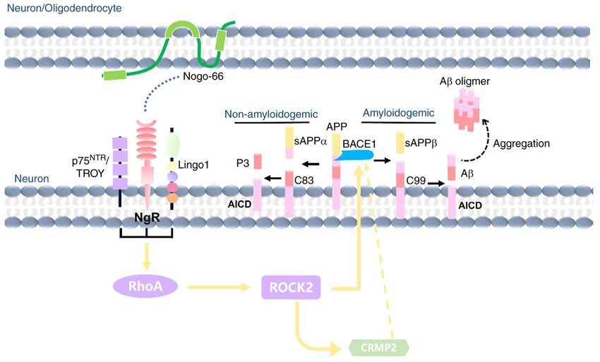
2                    XIE et al: Nogo-66 PROMOTES β-AMYLOID PROTEIN SECRETION VIA ACTIVATION OF BACE1

Nogo‑A has been identified as a key molecule that limits             of Medicine, Jinan University (Guangzhou, China; approval
axon regeneration and is considered to be a major obstacle to        no. IACUC‑20180120‑05).
nerve regeneration following injury in the adult mammalian
CNS (11). Nogo‑A interacts with the specific receptor complex        Cell culture. Cortical neurons were prepared from postnatal
on neurons to prevent neurite outgrowth; this receptor complex       rats (n=20; age, 1‑3 days; weight, 6‑8 g) (17), which were
is composed of Nogo‑66 receptor 1 (NgR1), and two comple‑            obtained from the Guangdong Medical Laboratory Animal
mentary co‑receptors p75 and Ig‑like domain‑containing               Center. Animals were housed at a standard temperature
NgR1‑interacting protein 1 (LINGO‑1) (12).                           (24±1˚C) with 60±10% humidity, diurnal conditions (lights on,
    It has been suggested that Nogo‑A may be involved in the         08:00‑20:00), and ad libitum access to food and water. Briefly,
pathology of AD. Nogo‑A is normally expressed in the hippo‑          the pups were sacrificed by cervical dislocation without
campus of healthy elderly individuals, but it is overexpressed in    anesthesia, in accordance with the procedures approved
the brain tissues of patients with AD and is associated with Aβ      by the local ethics review board, and the cerebral tissues
deposits in SPs (13). It has previously been demonstrated that       were dissociated by combined trituration and 0.25% trypsin
inhibition of the Nogo‑A/NgR pathway by Nogo‑66 antagonist           (Thermo Fisher Scientific, Inc.) at 37˚C for 30 min. The debris
peptide (NEP1‑40) attenuated amyloidogenic processing of APP         was removed and the supernatant was centrifuged at 1,000 x g
and reduced Aβ plaque deposition in APP/PS1 mice (14). It has        for 5 min at 4˚C to collect the cell pellet. Cortical neurons
also been hypothesized that Nogo‑A may alter neuronal APP            were plated at a density of 2.1x10⁵/well into 24‑well plates on
metabolism and Aβ production. Our previous study reported            poly‑L‑lysine (PLL; Merck KGaA)‑coated borosilicate glass
that Nogo‑P4, the 31‑55 amino acid of the Nogo‑66 peptide,           coverslips (16‑mm diameter; VWR International, LLC) or at
promoted Aβ secretion in cortical neurons (15). However,             a density of 1x10⁶/well in 6‑well plates coated with 100 µg/ml
whether the full‑length Nogo‑66 protein has the same effect,         PLL in neurobasal medium (Thermo Fisher Scientific, Inc.)
as well as the underlying mechanisms of Nogo‑66 in APP               containing 25 mM KCl, 2% B27 (Thermo Fisher Scientific,
regulation, are not clear. Therefore, the aim of the present study   Inc.), 1.5 mM glutamine (Thermo Fisher Scientific, Inc.) and
was to investigate whether Nogo‑66 promotes Aβ secretion in          0.01% penicillin/streptomycin (Merck KGaA).
cortical neurons, Neuro2a (N2a) cells and Sprague‑Dawley rats.           N2a mouse neuroblastoma cells stably expressing the
Furthermore, the present study explored the possible underlying      human Swedish mutant APP695 (APP695 N2a cells) (18)
molecular mechanism of Nogo‑66 on Aβ secretion, which in             were used in the present study, and were kindly provided by
turn may provide insight into possible effects and mechanisms        Dr Cuizan Cai (Macau University of Science and Technology,
of the Nogo‑A/NgR1 pathway in AD pathogenesis.                       Macao, China). The cells were maintained in Dulbecco's
                                                                     modified Eagle's medium (Thermo Fisher Scientific, Inc.)
Materials and methods                                                supplemented with 10% fetal bovine serum (Shanghai
                                                                     Shuangwei Biological Technology Co., Ltd.), 200 µg/ml G418
Reagents. Soluble Nogo‑66 protein was obtained via                   (Thermo Fisher Scientific, Inc.) and 1% penicillin���������
                                                                                                                            /��������
                                                                                                                             strepto‑
SUMO fusion in Escherichia coli from our laboratory (16).            mycin. Cells were cultured at 37˚C in a humidified atmosphere
Y‑27632 and the NEP1‑40 were obtained from Merck KGaA                containing 5% CO2 before the experiments.
and Tocris Bioscience, respectively. The Cy3‑conjugated                  Following treatment with different concentrations of
anti‑rabbit secondary antibody was obtained from Abcam (cat.         Nogo‑66 (0.25, 0.5 and 1 µM Nogo‑66) or a buffer control (PBS)
no. ab6939). Rabbit monoclonal IgG anti‑APP was purchased            for 48 h at 37˚C, cortical neurons and N2a mouse neuroblastoma
from LifeSpan BioSciences, Inc. In addition, the following           cells were cultured for 48 h. Cells were prepared in RIPA lysis
primary antibodies were obtained: Anti‑ β ‑secretase 1               buffer containing a cocktail of complete protease inhibitors
(BACE1; cat. no. 5606; Cell Signaling Technology, Inc.),             (Merck KGaA). The lysates were clarified by centrifugation at
anti‑ROCK2 (cat. no. ab125025; Abcam), anti‑phosphorylated           12,000 x g for 5 min at 4˚C to remove the insoluble components.
(p)‑collapsin response mediator protein‑2 (CRMP2; Thr514;                Following culture for 48 h, cortical neurons and N2a mouse
cat. no. 9397; Cell Signaling Technology, Inc.), anti‑CRMP2          neuroblastoma cells were treated with Y‑27632 (100 µM)
(cat. no. 35672; Cell Signaling Technology, Inc.), anti‑GAPDH        and NEP1‑40 (2 µM) for 1 h of pre‑protection at 37˚C.
(cat. no. BS72410; Bioworld Technology, Inc.) and anti‑micro‑        Subsequently, cells were treated with different concentrations
tubule‑associated protein 2 (MAP2; cat. no. 05‑346; Merck            of Nogo‑66 (1 µM) for 48 h at 37˚C. Cells were then prepared
KGaA). HRP‑conjugated goat anti‑rabbit IgG antibody (cat.            in RIPA lysis buffer containing a cocktail of complete
no. E030120‑01; Earthox Life Sciences) was used. All reagents        protease inhibitors (Merck KGaA). The lysates were clarified
and drugs used were of analytical grade.                             by centrifugation at 12,000 x g for 5 min at 4˚C to remove the
                                                                     insoluble components.
Animals. Sprague‑Dawley rats (n=24; age, 3 months; weight,
330±20 g) were provided by the Experimental Animal                   Neurite outgrowth assay. Cortical neuron cells were plated
Center of Guangdong Province. Animals were randomly                  (2.1x10⁵/well) onto 0.01% PLL‑coated coverslips in 24‑well
divided into the following groups (n=6 per group): i) Control;       culture dishes for 48 h following treatment with different
ii) low dose; iii) middle dose; and iv) high dose. Rats were         concentrations of Nogo‑66 (0.25, 0.5 and 1 µM) and a buffer
housed under standard temperature (24±1˚C) with 60±10%               control (PBS) for 48 h at 37˚C. The cells were fixed for 20 min
humidity, diurnal conditions (lights on, 08:00‑20:00), and           in 4% paraformaldehyde at room temperature and washed
ad libitum access to food and water. The present study was           with PBS. After rinsing several times with PBS, the cells were
approved by the Animal Research Committee of the School              blocked and permeabilized for 30 min at room temperature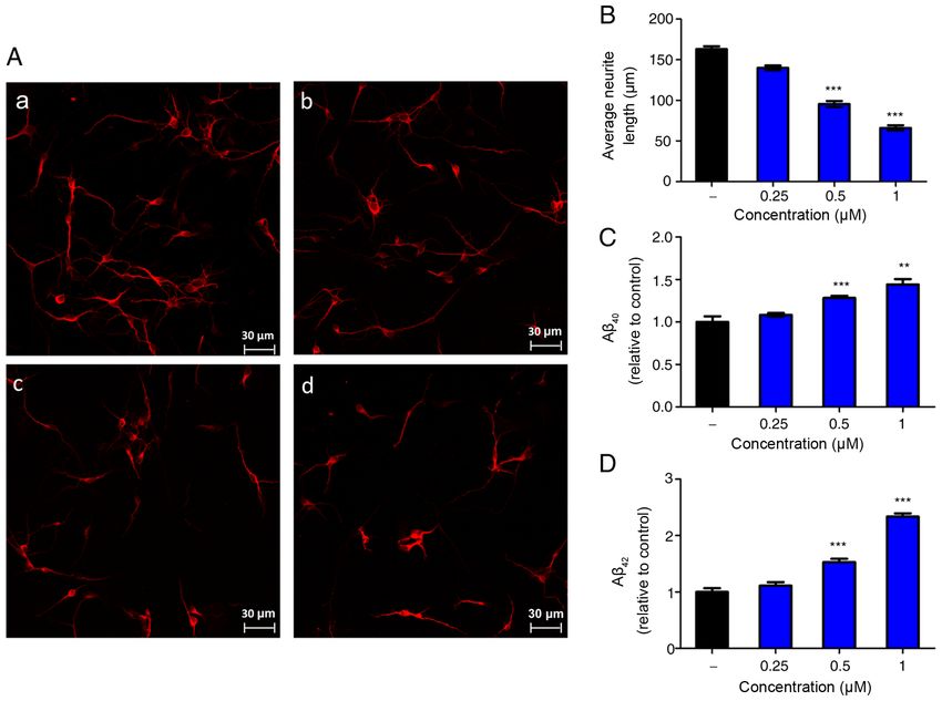
MOLECULAR MEDICINE REPORTS 23: 188, 2021                                                  3

with PBS containing 5% goat serum (Gibco; Thermo Fisher                  Abcam), anti‑CRMP2 (1:1,000; Cell Signaling Technology,
Scientific, Inc.), 1% (w/v) BSA (GenView Scientific, Inc.) and           Inc.), anti‑p‑CRMP2 Thr 509/514 (1:1,000; Cell Signaling
0.05% Triton X‑100. The cells were then rinsed and incubated             Technology, Inc.) and anti‑GAPDH (1:10,000; Bioworld
with anti‑MAP2 antibody (1:500; Merck KGaA) at 4˚C over‑                 Technology, Inc.). The membranes were then incubated with
night. The cells were then rinsed again and incubated for 1 h            HRP‑conjugated goat anti‑rabbit IgG antibody (1:2,000;
with goat anti‑rabbit Cy3 secondary antibody (1:1,000; cat.              cat. no. E030120‑01; Earthox Life Sciences) for 1 h at room
no. ab6939; Abcam) at room temperature. All slides were visu‑            temperature. The blots were detected using an ECL detection
alized under a laser scanning confocal microscope (LSM 700;              kit (Tanon Science and Technology Co., Ltd.), according to the
Carl Zeiss AG). Each group was assessed in triplicate and the            manufacturer's instructions. The results were analyzed using
experiments were repeated four times. The average length                 Quantity‑One software (version 4.6.6; Bio‑Rad Laboratories,
of each neuron was measured with Image‑Pro Plus software                 Inc.) to determine the relative ratio. The hippocampal tissue
(version 6.0; Media Cybernetics, Inc.). Neurites were measured           was prepared and analyzed in the same way.
only if they were >20 µm in length. A total of 100 neurons were
measured from each group in each experiment. These counts                ELISA for Aβ40 and Aβ42. The total levels of Aβ40 and Aβ42 in
were averaged and the SD values were calculated using Graph              the culture medium of cultured neurons and the hippocampal
Pad Prism v5.02 (GraphPad Software, Inc.). Mean values                   homogenate of rats were detected using ELISA kits (cat.
were obtained by averaging values from the measurement of                nos. 294‑62501 and 292‑64501; Wako Chemicals GmbH),
~100 neurons per well in three separate wells per group.                 according to the manufacturer's instructions. For human or rat
                                                                         Aβ40 and Aβ42 ELISA, the monoclonal BAN50 antibody, which
I n t ra ‑ h i p p o c a m p a l i nj e c t i o n of Nogo ‑ 6 6. Ma le   specifically detected the N‑terminal portion of human Aβ1‑16,
Sprague‑Dawley rats (age, 12 weeks; weight, 330±20 g;                    was used as the capturing antibody, and the monoclonal BA27
Guangdong Medical Laboratory Animal Center) were anes‑                   and BC05 antibodies, which detect the C‑terminal portion
thetized via an intravenous injection of 30 mg/kg pentobarbital          of Aβ1‑40 and Aβ1‑42, respectively, were used as detector anti‑
sodium (Merck KGaA) and were placed in a stereotaxic frame.              bodies. The amount of Aβ was calculated by comparing these
Unilateral injections of 2.5 µl physiological saline, or 0.5, 1 and      absorbance values with the control solutions.
2 mg/kg Nogo‑66, were administered at a rate of 0.5 µl/min
into the left and right hippocampus (3.0 mm posterior to the             Enzymatic activity assay. The β‑Secretase Activity Assay Kit
bregma, 1.8 mm lateral to the midline and 2.6 mm below the               (cat. no. 565785; Merck KGaA) was used to measure the BACE1
dura mater). Following the intra‑hippocampal injection, the              activity of cells and hippocampal tissue according to manufac‑
scalp was sutured and sterilized. The rats were kept warm for            turer's protocol. Protein lysates (20‑30 µl; 5‑7 mg/ml) were used
2 h before being returned to their cages. After 4 days, the rats         in vitro assays. All fluorescent values were detected on a plate
were euthanized by CO2 asphyxiation; the filling rate of CO2             reader with excitation (Ex) at 320 nm and emission (Em) at
was 20% chamber volume per minute. Subsequently, hippo‑                  405 nm (BioTek China). The ADAM10 Activity Detection kit
campal tissues from the rats were rapidly harvested and stored           (AS‑72226; AnaSpec) was used to measure ADAM10 activity
at ‑80˚C until further processing for biochemical analysis.              in cells and hippocampal tissue according to the manufac‑
      After 4 days, hippocampal tissue lysates were extracted            turer's protocol. Samples (50 µl) were used for in vitro assays.
for biochemical assays. Hippocampal tissues were dissected               Fluorescence intensity was measured at Ex/Em=490/520 nm
and homogenized in RIPA buffer (Beyotime Institute of                    after incubating the reaction at 37˚C for 30 min in the dark.
Biotechnology) containing a cocktail of complete protease
inhibitors (Merck KGaA). The lysate was centrifuged at                   Statistical analysis. All experiments were repeated 3‑4 times.
12,000 x g for 15 min at 4˚C. Protein concentrations were deter‑         All values are expressed as the mean ± SD. Analyses were
mined using a BCA assay kit (Thermo Fisher Scientific, Inc.).            performed using SPSS 16.0 statistical software (SPSS, Inc.).
                                                                         Comparison of parameters among more than two groups was
Western blotting. The cortical neurons and APP‑overexpression            made by one‑way ANOVA followed by the post hoc Tukey
(induced by the human Swedish mutant APP695) N2a cells                   test. P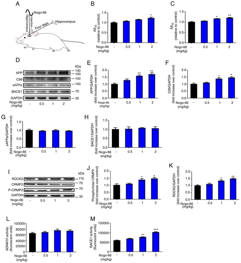
4                        XIE et al: Nogo-66 PROMOTES β-AMYLOID PROTEIN SECRETION VIA ACTIVATION OF BACE1

Figure 1. Nogo‑66 inhibits neurite outgrowth and promotes Aβ secretion in primary cortical neurons. (A) Immune‑fluorescence micrographs of microtu‑
bule‑associated protein 2 in cultured neurons treated with different concentrations of Nogo‑66. (a) PBS (vehicle control); (b) 0.25 µM Nogo‑66; (c) 0.5 µM
Nogo‑66; (d) 1 µM Nogo‑66. Scale bar, 30 µm. (B) Semi‑quantification of neurite outgrowth in (A). Data are presented as the mean ± SD (n=8). ***P
MOLECULAR MEDICINE REPORTS 23: 188, 2021                                                                5

Figure 2. NEP1‑40 or Y‑27632 overcome Nogo‑66‑induced production of Aβ40 and Aβ42 in cortical neurons and N2a cells. NEP1‑40 and Y‑27632 decreased
the levels of (A) Aβ40 and (C) Aβ42 in cortical neurons. NEP1‑40 and Y‑27632 decreased the levels of (B) Aβ40 and (D) Aβ42 in N2a cells. Data are presented
as the mean ± SD (n=6). ###P
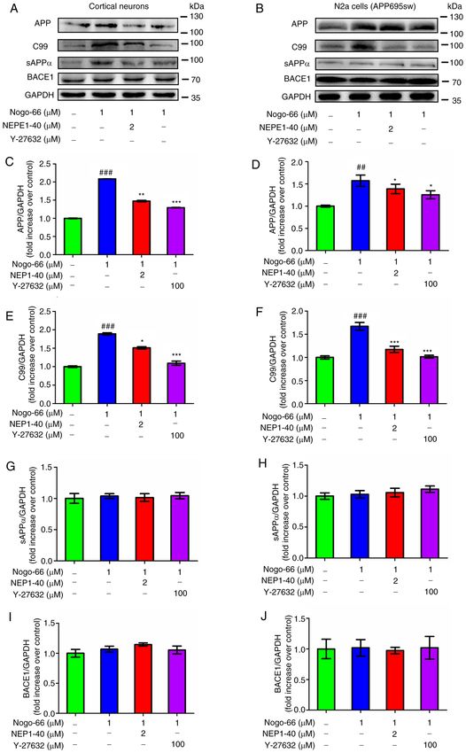
6                        XIE et al: Nogo-66 PROMOTES β-AMYLOID PROTEIN SECRETION VIA ACTIVATION OF BACE1

Figure 3. Effects of Nogo‑66, NEP1‑40 and Y‑27632 on the protein expression levels of APP, C99, sAPPα and BACE1 in (A, C, E, G and I) primary cortical
neurons and (B, D, F, H and J) N2a cells. Data are presented as the mean ± SD from three biological repeats. ##P
MOLECULAR MEDICINE REPORTS 23: 188, 2021                                                                7

Figure 4. Nogo‑66 inhibits neurite outgrowth and induces A β production by regulating ROCK2 protein expression and CRMP2 phosphorylation.
(A, C and E) Cortical neurons were treated with various concentrations of Nogo‑66. Western blot analysis results showing the protein expression levels
of ROCK2, p‑CRMP2 and CRMP2. Equal protein loading was confirmed using GAPDH. Data are presented as the mean OD values ± SD of triplicate
samples. **P
8                        XIE et al: Nogo-66 PROMOTES β-AMYLOID PROTEIN SECRETION VIA ACTIVATION OF BACE1

Figure 6. Effects of Nogo‑66 on the expression levels and enzymatic activity of proteins involved in the Aβ pathway in the hippocampus of Sprague‑Dawley rats.
(A) Schematic diagram of treatment of rats with Nogo‑66. Nogo‑66 induced (B) Aβ40 and (C) Aβ42 production in a dose‑dependent manner in the brains of rats.
Data are presented as the mean ± SD (n=6). *P
MOLECULAR MEDICINE REPORTS 23: 188, 2021                                                           9

Figure 7. Proposed schematic working model: Nogo‑66 promotes A β secretion through the NgR/ROCK/BACE1 pathway. A β, β ‑amyloid protein;
ROCK, Rho‑associated coiled‑coil containing kinase; BACE1, β‑secretase 1; APP, amyloid precursor protein; sAPP, secreted APP; NgR, Nogo‑66 receptor.

compared with those in the control groups (Fig. 6B and C).                  Nogo‑A is considered a major obstacle to neurological regen‑
The results were consistent with those obtained in vitro.                   eration following traumatic injury in mammals (19). Nogo‑66
    The protein expression levels of ROCK2 and p‑CRMP2                      is a 66‑amino acid residue in the extracellular domain of
were increased in the hippocampus of rats exposed to                        Nogo‑A, which inhibits neurite outgrowth by binding to the
Nogo‑66 in a dose‑dependent manner. High (2 mg/kg) and                      NgR1 in the CNS (20). In vitro, Nogo‑66 was previously
medium (1 mg/kg) doses of Nogo‑66 significantly increased                   shown to inhibit neurite outgrowth in a variety of nerve cells,
the expression of phosphorylated CRMP2 and ROCK2 in                         such as dorsal root ganglion cells (21,22), PC12 cells (21) and
the hippocampus of rats (Fig. 6I‑K). A low dose (0.5 mg/kg)                 cerebellar granule cells (23). In the present study, Nogo‑66
of Nogo‑66 also increased the protein expression levels of                  was revealed to inhibit neurite outgrowth of cortical neurons
ROCK2 and p‑CRMP2 in the hippocampus, but without a                         in a dose‑dependent manner.
significant effect (Fig. 6I‑K). These trends in the hippocampus                 Previous studies have reported that Nogo‑A and its recep‑
were consistent with those observed in vitro, indicating that               tors are involved in the regulation of synaptic plasticity, as
Nogo‑66 may promote the secretion of Aβ by increasing the                   well as the metabolism of Aβ, suggesting that it may serve
expression of APP and activating NgR1 downstream signal                     an important role in AD. In a previous study, the levels of
proteins, such as ROCK2 and p‑CRMP2.                                        Aβ deposition were decreased in NgR2‑knockout mice (24).
    The activity of BACE1 in the hippocampus of rats                        However, whereas NgR1 has been shown to bind to APP (24),
was increased in a dose‑dependent manner in response to                     contrasting effects of this interaction have also been reported;
Nogo‑66. High (2 mg/kg) and medium (1 mg/kg) doses                          Park et al (25) observed decreased Aβ production following
of Nogo‑66 significantly increased BACE1 activity in the                    NgR1‑overexpression in neuroblastoma in vitro. These previous
hippocampus of rats (Fig. 6M). A low dose (0.5 mg/kg) of                    studies have suggested a strong linkage between Nogo‑A and
Nogo‑66 also increased the enzymatic activity of in the                     Aβ; however, the exact roles need to be elucidated. The excess
hippocampus; however, this was not significant. In addi‑                    production of Aβ is considered the initiating factor of AD,
tion, Nogo‑66 treatment had a slight effect on ADAM10                       which is generated from the hydrolysis of APP via BACE‑1
enzymatic activity, but there was no significant differ‑                    and γ‑secretase. NgR can cause parallel changes in sAPPα
ence (Fig. 6L). The effects of Nogo‑66 on APP metabolic                     and Aβ, indicating that NgR blocks APP hydrolysis (26). In
enzymatic activity in the hippocampus were consistent                       contrast to these findings, Zhou et al (24) reported that the
with those observed in vitro, and indicated that Nogo‑66                    increased interaction between NgR and APP reduced the
mainly promoted the cleavage of the APP protein through                     surface expression of APP and favored the processing of APP
the BACE1 pathway by increasing the activity of BACE1,                      via the BACE1 pathway.
thereby increasing the secretion of Aβ.                                         The Nogo‑A protein, also termed RTN4A, is a member
                                                                            of the RTN family. RTNs are characterized by the highly
Discussion                                                                  conserved C‑terminal RHD, which is composed of two puta‑
                                                                            tive transmembrane domains separated by one hydrophilic
Nogo‑A is a member of the Nogo family found in the myelin                   loop, plus a hydrophilic tail. Notably, all four human RTN
sheath of the CNS, which inhibits axonal regeneration.                      proteins appear to be involved in the pathogenesis of AD.
10 XIE et al: Nogo-66 PROMOTES β-AMYLOID PROTEIN SECRETION VIA ACTIVATION OF BACE1
He et al (27) discovered that BACE1 co‑immunoprecipitated bound to NgR, and potentially exerted its regulatory effects
with RTN1, RTN2, RTN3 and RTN4, and revealed that RTN on Aβ levels by increasing the expression of ROCK2 and
family members are binding partners of BACE1. Furthermore, promoting the catalytic action of BACE1, which may then
increased RTN3 expression caused a dose‑dependent reduc‑ facilitate amyloidogenic metabolism of APP, thus resulting in
tion in A β, whereas suppressing the expression of RTN3 increased Aβ42 secretion.
by RNA interference increased the secretion of A β, thus In the in vivo experiment conducted in the present study,
suggesting that RTN3 may block access of BACE1 to APP, Nogo‑66 promoted the levels of Aβ40 and Aβ42 in rat tissues.
reduce the cleavage of this protein and inhibit the production Nogo‑66 also enhanced the expression levels of proteins asso‑
of Aβ. RTN4‑B/C has also been reported to interact with ciated with the APP processing pathway and activated BACE1
BACE1 and inhibit its ability to produce Aβ (28). Certain in rats, which may eventually lead to increased secretion of
studies have reported that Nogo‑A promotes the pathogenesis Aβ. Subsequently, western blot analysis was used to explore
of AD. Masliah et al (29) demonstrated that deleting Nogo‑A the expression levels of ROCK2 and p‑CRMP2 in the hippo‑
ameliorated learning and memory deficits of APP‑transgenic campus; it was revealed that Nogo‑66 increased the expression
mice in the Morris water maze at an early/intermediate stage levels of ROCK2 and p‑CRMP2 in vivo. The results regarding
of the disease, thus suggesting that Nogo‑A may influence the effects of Nogo‑66 on the rat hippocampus and related
the metabolism of APP; however, the detailed effect and mechanism were consistent with the results obtained for
mechanism are not clear. In the present study, ELISA results neurons in vitro.
revealed that Nogo‑66 promoted the levels of Aβ40 and Aβ42 In conclusion, although Nogo‑A has an important role in
in primary cortical neurons in a dose‑dependent manner. AD pathology, its effects and mechanisms are not clear. The
The present results showed that increased Nogo‑66 increased present study demonstrated that Nogo‑66 inhibited neurite
production of Aβ, which is the opposite effect to other RTNs. outgrowth, and increased the levels of Aβ 42 and Aβ 40 in a
Notably, Nogo‑A may be considered a very different protein dose‑dependent manner. The underlying mechanism may
compared with others from the RTN family, as it not only has be as follows (Fig. 7): Nogo‑66 increases Aβ generation by
the unique function of inhibiting neurite outgrowth but it may activating BACE1 and increasing APP expression, which
also increase Aβ. is associated with activation of NgR1 and its downstream
When cortical neurons were treated with NEP1‑40 in the signaling molecule ROCK2, as well as the phosphorylation of
present study, the levels of Aβ 40 and Aβ 42 were decreased. CRMP2. Therefore, Nogo‑A may be a precipitating factor that
After treatment with Nogo‑66, the protein expression levels causes the onset and development of AD; however, the exact
of APP and activation of BACE1 were increased in cortical mechanism still requires further exploration. From the view‑
neurons and N2a cells, indicating that Nogo‑66 simultane‑ point of drug therapeutic targets, NgR and ROCK2 appear to
ously increased the levels of APP and activated BACE1, which be potentially useful and effective therapeutic targets for AD
may eventually lead to an increase in the secretion of Aβ. treatment, and targeting them may promote neurite outgrowth
The Rho/ROCK signaling pathway was activated when and inhibit the neuronal production of Aβ.
Nogo‑A interacts with NgR1 (30). The downstream event
included activation of RhoA‑GTP, ROCK, MAP and CRMP2, Acknowledgements
which eventually exerted effects on the cytoskeleton associated
with neurite growth. The results of western blotting revealed The authors would like to thank Dr Fei Fang (Department of
that higher ROCK2 expression and increased p‑CRMP2 Clinical Molecular Biology, Akershus University Hospital,
expression was induced by Nogo‑66. It was therefore suggested University of Oslo, Norway) for critical discussion and assis‑
that Nogo‑66‑stimulated ROCK2‑induced regulation of tance in the preparation of the manuscript.
CRMP2 phosphorylation may contribute to neurite outgrowth
inhibition. Funding
The present results also revealed that the ROCK inhibitor
Y‑27632 suppressed Nogo‑66‑stimulated A β 42 and A β 40 The study was funded by the National Natural Science
secretion. These findings indicated that Y‑27632 may reduce Foundation of China (grant nos. 81202519, 81873739, 81572497
Aβ42 by inhibiting the activation of ROCK and Nogo‑66 may and 81703011), the Science and Technology Program of
increase Aβ 42 secretion by activating the ROCK pathway. Guangzhou Province (grant no. 201607010216) and the
Subsequently, western blot analysis was performed to explore Natural Science Foundation of Guangdong Province (grant
whether Nogo‑66 stimulated A β secretion by regulating nos. 2019A1515010936, 2014A030313362 and 2017A030313487).
ROCK2 expression. It was revealed that Nogo‑66 significantly
increased the expression levels of ROCK2 in cortical neurons Availability of data and materials
and N2a cells. A previous study used a variety of non‑steroidal
anti‑inflammatory drugs to demonstrate that the activation The datasets used and/or analyzed during the current study are
of Rho/ROCK2 led to a large production of Aβ 42, whereas available from the corresponding author on reasonable request.
the inhibition of ROCK2 activity reduced the APP amyloid
enzymatic degradation pathway and the levels of Aβ42 in the Authors' contributions
brain (31). Another study confirmed that inhibition of RhoA
or ROCK2 knockdown could interfere with the β‑cleavage of FX, QZ, HY and FCL were involved in revising the manuscript
APP, and significantly reduced the levels of brain Aβ in AD critically for important intellectual content, making substantial
model mice (32). It was therefore hypothesized that Nogo‑66 contributions to conception and design, and giving final approvalMOLECULAR MEDICINE REPORTS 23: 188, 2021 11
of the version to be published. QQX participated in the entire 14. Fang Y, Yao L, Li C, Wang J, Wang J, Chen S, Zhou XF and Liao H:
The blockage of the Nogo/NgR signal pathway in microglia alle‑
process, drafting of the article and data analysis. XF, YYH, NF, viates the formation of Aβ plaques and tau phosphorylation in APP/
QYC, GFL, JPP, YH and ZJW fed the animals, performed the PS1 transgenic mice. J Neuroinflammation 13: 56, 2016.
experiments and collected samples from the rats. All authors 15. Xiao F, Lin LF, Cheng X, Gao Q and Luo HM: Nogo‑66 receptor
activation inhibits neurite outgrowth and increases β‑amyloid
read and approved the final version of the manuscript. protein secretion of cortical neurons. Mol Med Rep 5: 619‑624, 2012.
16. Dai X, Sun Z, Liang R, Li Y, Luo H, Huang Y, Chen M, Su Z and
Ethics approval and consent to participate Xiao F: Recombinant Nogo‑66 via soluble expression with SUMO
fusion in Escherichia coli inhibits neurite outgrowth in vitro. Appl
Microbiol Biotechnol 99: 5997‑6007, 2015.
All animal experiments were conducted according to the Jinan 17. zur Nedden S, Doney AS and Frenguelli BG: Modulation of intra‑
University ethical guidelines and were approved by the Animal cellular ATP determines adenosine release and functional outcome
in response to metabolic stress in rat hippocampal slices and
Research Committee of the School of Medicine of Jinan cerebellar granule cells. J Neurochem 128: 111‑124, 2014.
University. 18. Gao R, Wang Y, Pan Q, Huang G, Li N, Mou J and Wang D:
Fuzhisan, a chinese herbal medicine, suppresses beta‑secretase gene
transcription via upregulation of SIRT1 expression in N2a‑APP695
Patient consent for publication cells. Int J Clin Exp Med 8: 7231‑7240, 2015.
19. Jin SG, Ryu HH, Li SY, Li CH, Lim SH, Jang WY and Jung S:
Not applicable. Nogo‑A inhibits the migration and invasion of human malignant
glioma U87MG cells. Oncol Rep 35: 3395‑3402, 2016.
20. Yan J, Zhou X, Guo JJ, Mao L, Wang YJ, Sun J, Sun LX, Zhang LY,
Competing interests Zhou XF and Liao H: Nogo‑66 inhibits adhesion and migration
of microglia via GTPase Rho pathway in vitro. J Neurochem 120:
721‑731, 2012.
The authors declare that they have no competing interests. 21. GrandPré T, Nakamura F, Vartanian T and Strittmatter SM:
Identification of the Nogo inhibitor of axon regeneration as a
References Reticulon protein. Nature 403: 439‑444, 2000.
22. Chen MS, Huber AB, van der Haar ME, Frank M, Schnell L,
Spillmann AA, Christ F and Schwab ME: Nogo‑A is a myelin‑asso‑
1. Kerr JS, Adriaanse BA, Greig NH, Mattson MP, Cader MZ, Bohr VA ciated neurite outgrowth inhibitor and an antigen for monoclonal
and Fang EF: Mitophagy and Alzheimer's disease: Cellular and antibody IN‑1. Nature 403: 434‑439, 2000.
molecular mechanisms. Trends Neurosci 40: 151‑166, 2017. 23. Niederöst B, Oertle T, Fritsche J, McKinney RA and Bandtlow CE:
2. Stephenson‑Jones M, Yu K, Ahrens S, Tucciarone JM, Nogo‑A and myelin‑associated glycoprotein mediate neurite
van Huijstee AN, Mejia LA, Penzo MA, Tai LH, Wilbrecht L growth inhibition by antagonistic regulation of RhoA and Rac1.
and Li B: A basal ganglia circuit for evaluating action outcomes. J Neurosci 22: 10368‑10376, 2002.
Nature 539: 289‑293, 2016. 24. Zhou X, Hu X, He W, Tang X, Shi Q, Zhang Z and Yan R: Interaction
3. Pereira JB, Janelidze S, Ossenkoppele R, Kvartsberg H, between amyloid precursor protein and Nogo receptors regulates
Brinkmalm A, Mattsson‑Carlgren N, Stomrud E, Smith R, amyloid deposition. FASEB J 25: 3146‑3156, 2011.
Zetterberg H, Blennow K, et al: Untangling the association of 25. Park JH, Gimbel DA, GrandPre T, Lee JK, Kim JE, Li W, Lee DH
amyloid‑β and tau with synaptic and axonal loss in Alzheimer's and Strittmatter SM: Alzheimer precursor protein interaction with
disease. Brain awaa395, 2020. the Nogo‑66 receptor reduces amyloid‑beta plaque deposition. J
4. Hardy J and Selkoe DJ: The amyloid hypothesis of Alzheimer's Neurosci 26: 1386‑1395, 2006.
disease: progress and problems on the road to therapeutics. 26. Jiang R, Wu XF, Wang B, Guan RX, Lv LM, Li AP, Lei L, Ma Y,
Science 297: 353‑356, 2002. Li N, Li QF, et al: Reduction of NgR in perforant path decreases
5. Lin Y‑T, Seo J, Gao F, Feldman HM, Wen HL, Penney J, Cam HP, amyloid‑β peptide production and ameliorates synaptic and cognitive
Gjoneska E, Raja WK, Cheng J, et al: APOE4 causes widespread deficits in APP/PS1 mice. Alzheimers Res Ther 12: 47, 2020.
molecular and cellular alterations associated with Alzheimer's 27. He W, Lu Y, Qahwash I, Hu X‑Y, Chang A and Yan R: Reticulon
disease phenotypes in human iPSC‑derived brain cell types. family members modulate BACE1 activity and amyloid‑β peptide
Neuron 98: 1141-1154.e7, 2018. generation. Nat Med 10: 959‑965, 2004.
6. Fang EF, Hou Y, Palikaras K, Adriaanse BA, Kerr JS, Yang B, 28. Murayama KS, Kametani F, Saito S, Kume H, Akiyama H and
Lautrup S, Hasan‑Olive MM, Caponio D, Dan X, et al: Mitophagy Araki W: Reticulons RTN3 and RTN4‑B/C interact with BACE1 and
inhibits amyloid‑β and tau pathology and reverses cognitive deficits inhibit its ability to produce amyloid β‑protein. Eur J Neurosci 24:
in models of Alzheimer's disease. Nat Neurosci 22: 401‑412, 2019. 1237‑1244, 2006.
7. Lautrup S, Sinclair DA, Mattson MP and Fang EF: NAD+ in brain 29. Masliah E, Xie F, Dayan S, Rockenstein E, Mante M, Adame A,
aging and neurodegenerative disorders. Cell Metab 30: 630‑655, Patrick CM, Chan AF and Zheng B: Genetic deletion of Nogo/Rtn4
2019. ameliorates behavioral and neuropathological outcomes in amyloid
8. Zhang P, Kishimoto Y, Grammatikakis I, Gottimukkala K, precursor protein transgenic mice. Neuroscience 169: 488‑494,
Cutler RG, Zhang S, Abdelmohsen K, Bohr VA, Misra Sen J, 2010.
Gorospe M, et al: Senolytic therapy alleviates Aβ‑associated oligo‑ 30. Xu W, Xiao P, Fan S, Chen Y, Huang W, Chen X, Liu G, Dang C,
dendrocyte progenitor cell senescence and cognitive deficits in an Zeng J and Xing S: Blockade of Nogo‑A/Nogo‑66 receptor 1
Alzheimer's disease model. Nat Neurosci 22: 719‑728, 2019. (NgR1) inhibits autophagic activation and prevents secondary
9. Oertle T, van der Haar ME, Bandtlow CE, Robeva A, Burfeind P, neuronal damage in the thalamus after focal cerebral infarction in
Buss A, Huber AB, Simonen M, Schnell L, Brösamle C, et al: hypertensive rats. Neuroscience 431: 103‑114, 2020.
Nogo‑A inhibits neurite outgrowth and cell spreading with three 31. Zhou Y, Su Y, Li B, Liu F, Ryder JW, Wu X, Gonzalez‑DeWhitt PA,
discrete regions. J Neurosci 23: 5393‑5406, 2003. Gelfanova V, Hale JE, May PC, et al: Nonsteroidal anti‑inflam‑
10. Huebner EA and Strittmatter SM: Axon regeneration in the matory drugs can lower amyloidogenic Abeta42 by inhibiting Rho.
peripheral and central nervous systems. Results Probl Cell Differ 48: Science 302: 1215‑1217, 2003.
339-351, 2009. 32. Herskowitz JH, Feng Y, Mattheyses AL, Hales CM,
11. Grandpré T and Strittmatter SM: Nogo: A molecular determinant Higginbotham LA, Duong DM, Montine TJ, Troncoso JC,
of axonal growth and regeneration. Neuroscientist 7: 377‑386, 2001. Thambisetty M, Seyfried NT, et al: Pharmacologic inhibition of
12. Mosyak L, Wood A, Dwyer B, Buddha M, Johnson M, Aulabaugh A, ROCK2 suppresses amyloid‑β production in an Alzheimer's disease
Zhong X, Presman E, Benard S, Kelleher K, et al: The structure of mouse model. J Neurosci 33: 19086‑19098, 2013.
the Lingo‑1 ectodomain, a module implicated in central nervous
system repair inhibition. J Biol Chem 281: 36378‑36390, 2006. This work is licensed under a Creative Commons
13. Gil V, Nicolas O, Mingorance A, Ureña JM, Tang BL, Hirata T, Attribution-NonCommercial-NoDerivatives 4.0
Sáez‑Valero J, Ferrer I, Soriano E and del Río JA: Nogo‑A expression
International (CC BY-NC-ND 4.0) License.
in the human hippocampus in normal aging and in Alzheimer
disease. J Neuropathol Exp Neurol 65: 433‑444, 2006.You can also read