Sea anemones (Cnidaria, Actiniaria) of Singapore: redescription and taxonomy of Phymanthus pinnulatus Martens in Klunzinger, 1877 - ZooKeys
←
→
Page content transcription
If your browser does not render page correctly, please read the page content below
A peer-reviewed open-access journal
ZooKeys 840: 1–20 (2019)
    Sea anemones (Cnidaria, Actiniaria) of Singapore: redescription and taxonomy of Phymanthus... 1
doi: 10.3897/zookeys.840.31390                            RESEARCH ARTICLE
http://zookeys.pensoft.net                                                                                     Launched to accelerate biodiversity research

    Sea anemones (Cnidaria, Actiniaria) of Singapore:
  redescription and taxonomy of Phymanthus pinnulatus
               Martens in Klunzinger, 1877

                      Nicholas Wei Liang Yap1,2, Ria Tan3, Clara Lei Xin Yong1,
                                Koh Siang Tan2, Danwei Huang1,2

1 Reef Ecology Laboratory, Department of Biological Sciences, National University of Singapore, Block S3
Level 4, Science Drive 4, Republic of Singapore 2 St. John’s Island National Marine Laboratory, Tropical
Marine Science Institute, National University of Singapore, 18 Kent Ridge Road, Republic of Singapore 3 c/o
Lee Kong Chian Natural History Museum, National University of Singapore, 2 Conservatory Drive, Republic
of Singapore

Corresponding author: Nicholas Wei Liang Yap (cosmogony84@gmail.com)

Academic editor: James Reimer | Received 16 December 2018 | Accepted 15 March 2019 | Published 17 April 2019

                                   http://zoobank.org/0E7E9691-1F87-4C47-B482-F4E15052A987

Citation: Yap NWL, Tan R, Yong CLX, Tan KS, Huang D (2019) Sea anemones (Cnidaria, Actiniaria) of Singapore:
redescription and taxonomy of Phymanthus pinnulatus Martens in Klunzinger, 1877. ZooKeys 840: 1–20. https://doi.
org/10.3897/zookeys.840.31390

Abstract
Despite the ubiquity of sea anemones (Cnidaria: Actiniaria) in tropical ecosystems, our understanding of
their biodiversity and taxonomy is limited. Here we re-establish the identity of an intertidal zooxanthellate
species, Phymanthus pinnulatus Martens in Klunzinger, 1877. Originally described from a single preserved
specimen in the Berlin Museum by CB Klunzinger, his brief footnote lacked crucial details to positively
identify the species. Our redescription is based on more than 50 living individuals of P. pinnulatus
collected from its type locality, Singapore. These were examined and compared with type materials of
the species and its congeners. Specimens of P. pinnulatus differ from syntypes of species described as
Phymanthus levis Kwietniewski, 1898 from Indonesia, as well as Phymanthus sansibaricus Carlgren, 1900
and Phymanthus strandesi Carlgren, 1900, both described from East Africa. Phymanthus pinnulatus was
encountered on the lower intertidal, among coral rubble and between rocky crevices. It is vibrantly coloured
and has 96 marginal tentacles with branching outgrowths along each, resulting in a ‘frilly’ appearance. The
anemone has a flat expanded oral disc, with discal tentacles that are inconspicuous and reduced, unlike
syntypes of its congeners. Details of its live appearance, musculature, and cnidom are also provided for the first
time. Overall, types of cnidae and capsule sizes differ from other known species of Phymanthus documented

Copyright Nicholas Wei Liang Yap et al. This is an open access article distributed under the terms of the Creative Commons Attribution License
(CC BY 4.0), which permits unrestricted use, distribution, and reproduction in any medium, provided the original author and source are credited.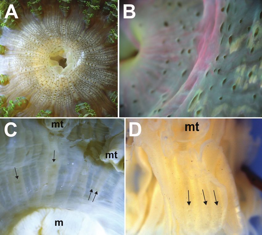
2                       Nicholas Wei Liang Yap et al. / ZooKeys 840: 1–20 (2019)

elsewhere. It is inferred that P. pinnulatus has a wide distribution that extends eastwards from Singapore,
as far as Ambon and the Torres Straits. Some individuals reported as Phymanthus muscosus Haddon and
Shackleton, 1893 and Phymanthus buitendijki Pax, 1924 are probably P. pinnulatus. This morphological
analysis provides new insights into the characters used to delimit P. pinnulatus, clarifies its geographical
distribution, and contributes to an ongoing revision of the genus Phymanthus.

Keywords
Actinoidea, Indo-Pacific, intertidal, Southeast Asia, zooxanthellae

Introduction

Sea anemones (Cnidaria, Actiniaria) are ecologically successful invertebrates found
in many tropical marine ecosystems. In spite of their ubiquity, few from the Indo-
Pacific region have been subjected to rigorous taxonomic studies, and the identities of
many species remain poorly defined (den Hartog 1997; Fautin et al. 2009). Among
them are members of the zooxanthellate family Phymanthidae, which comprise two
genera: Heteranthus and Phymanthus (Fautin 2013; 2016). Within the latter genus,
Phymanthus pinnulatus [= Phymanthus pinnulatum] Martens in Klunzinger, 1877, was
first described based on a single preserved specimen collected by Eduard von Martens
from Singapore (Haddon 1898), and housed in the Berlin Museum (now Museum
für Naturkunde). Its appearance was briefly described in a footnote by Klunzinger
(1877: 87) stating “… wo statt der Wärzchen beim Lebenden (nach der Zeichnung
von Martens) deutliche und mehrfach gefiederte Seitenstäbchen am Hauptstamm
sitzen,” alluding to the presence of suckers [=verrucae] along the animal’s body, and
ramified tentacles. Klunzinger’s (1877) footnote also makes mention to a drawing of
the anemone by Martens. However, we were not able to locate it, nor does it appear in
Martens’ comprehensive reports of biodiversity from his expedition in Southeast Asia
(see Martens 1867, 1875).
     Klunzinger’s footnote (1877) provided no further details or illustrations to sup-
port his description. Since then, the taxonomic validity of P. pinnulatus’ appearance
has remained equivocal, with no illustrations or taxonomic work published thereafter
to ascertain the identity of the species. Here we provide for the first time in over a cen-
tury since Klunzinger’s (1877) description, details of P. pinnulatus’ external and internal
structure (i.e., retractor and sphincter musculature), an inventory of cnidae [= cnidom],
and notes on its habitat and distribution. We also provide field photographs of the living
animal. These data are now convention in contemporary actinian taxonomic accounts.
     We encountered P. pinnulatus at the lower intertidal zone, where its lower column
was buried in sand or wedged between crevices of silt covered rocks and/or coral rub-
ble. These anemones were very conspicuous and common in the northern and south-
ern shores of Singapore. They were also easily recognizable in the field because of the
frilly and colourful appearance of its marginal tentacles.
     Prior to this study, 25 anemones which are well known taxonomically were iden-
tified from Singapore (see Fautin et al. 2009, 2015; Yap et al. 2014; Fautin and Tan
Sea anemones (Cnidaria, Actiniaria) of Singapore: redescription and taxonomy of Phymanthus... 3

2016). Unlike these species, disagreements still persist for the diagnosis of Phymanthus
and species attributed to this genus (Pax 1924; Carlgren 1949; Gonzalez-Muñoz et al.
2015), so clearly a taxonomical revision is overdue. Many members of Phymanthus are
poorly defined. The objective of this study is to provide a comprehensive morphologi-
cal characterization of P. pinnulatus so that its identity would be unambiguous. For a
common tropical sea anemone, data are lacking on much of its biology, ecology, and
biogeography. Our redescription opens up opportunities for further research on this
common intertidal species in a wide range of disciplines.

Materials and methods
All anemones we report here were collected from Singapore over 16 years: between
2002 and 2018. Some animals were observed in situ and photographed; others were
brought back to the laboratory for further study. Collected anemones were kept alive
for at least one week. Details on their behaviour and appearance of the living ani-
mal were noted. Thereafter, the whole animal was fixed in 10% formalin. Internal
morphology was examined in dissected specimens. Musculature of the anemones was
visualized from 8-µm-thick histological sections stained with haematoxylin and eosin
(Humason 1967).
    Unfired cnidae capsules were examined from tissues of the marginal tentacle tip,
protuberances, discal tentacles, marginal projections, mid-column, actinopharynx, and
mesenterial filaments. Cnidae were viewed at 1000 × magnification. We also examined
discharged capsules from living specimens, to confirm identities of cnidae encountered
(see Yap et al. 2014). Cnidae taxonomy follows Mariscal (1974).
    We examined the holotype of P. pinnulatum, kept as two separate lots—one at the
Museum für Naturkunde Berlin (ZMB) and the other at Naturhistoriska Riksmuseet,
Stockholm, Sweden (NRS) (see Fautin 2016). Voucher specimens of individuals col-
lected from Singapore by KW England and FB Steiner between 1960s and 1980s,
deposited at Natural History Museum (known also as the British Museum of Natural
History; BMNH) and California Academy of Sciences, Department of Invertebrate
Zoology and Geology (CASIZ) respectively were also studied.
    To further establish the identity of P. pinnulatus and to delineate the
species, available and accessible type material of its congeners were examined:
Phymanthus buitendijki Pax, 1924 present at the Rijksmuseum van Natuurlijke
Historie and Naturalis (RMNH; now the Naturalis Biodiversity Center);
Phymanthus levis Kwietniewski, 1898, present at both ZMB and NRS;
Phymanthus muscosus Haddon & Shackleton 1893, kept at the University Museum of
Zoology, Cambridge University, United Kingdom (MZC); Phymanthus sansibaricus
Carlgren, 1900 and Phymanthus strandesi Carlgren, 1900, both in the invertebrate
collection at Zoologisches Museum Hamburg (ZMH).
    We relied on published descriptions of Phymanthus crucifer (Le Sueur, 1817),
Phymanthus loligo (Hemprich & Ehrenberg in Ehrenberg, 1834), Phymanthus pulcher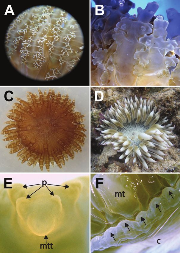
4                   Nicholas Wei Liang Yap et al. / ZooKeys 840: 1–20 (2019)

(Andrès, 1883) and Phymanthus rhizophorae (Mitchell, 1890) to obtain details of their
appearance and morphological traits. The types of P. pulcher and P. rhizophorae could
not be located (Fautin 2013, 2016).
     While syntypes of both P. crucifer and P. loligo do exist and are present at the Mu-
seum of Zoology, Lund University (MZL), these are in the form of microscope slides of
the anemones’ mesenteries and musculature. The slides alone are not useful for defin-
ing species boundaries of members in Phymanthus. Furthermore, we did not study the
syntype of Phymanthus coeruleus (Quoy & Gaimard, 1833) because specimens present
in the lot are of two different anemone species (Fautin 2013).
     Because uncertainty still lies with distinguishing the two genera of Phymanthidae
(i.e., Phymanthus and Heteranthus; see González-Muñoz et al. 2015), to verify
that P. pinnulatus is morphologically distinct from members of Heteranthus, we
examined the syntype of Heteranthus verruculatus Klunzinger, 1877 and holotype
of Heteranthus insignis Carlgren, 1943, kept also at Museum für Naturkunde Berlin
(ZMB) and Naturhistoriska Riksmuseet, Stockholm, Sweden (NRS) respectively.
     All new P. pinnulatus voucher specimens collected from Singapore for this study
since 2002 were deposited in the Zoological Reference Collection, Lee Kong Chian
Natural History Museum, National University of Singapore (ZRC).

Taxonomic Account
Family Phymanthidae Andres, 1883
Genus Phymanthus Milne-Edwards, 1857

Phymanthus pinnulatus Martens in Klunzinger, 1877
Figs. 2–7

Phymanthus pinnulatum Martens in Klunzinger, 1877: 87 (original description).
Phymanthus pinnulatum: Haddon 1898: 496; Carlgren 1949: 75.
Phymanthus pinnulatus: Fautin 2016: 346.

Occurrence and materials collected in Singapore (Fig. 1). (* – observed alive; bold
– morphotypes with smooth marginal tentacles or reduced protuberances):
    Berlayer Creek (ZRC.CNI.1343 x4*), Big Sister’s Island (ZRC.CNI.0982 x1;
ZRC.CNI.1103 x1*; ZRC.CNI.1163 x1*; ZRC.CNI.1045 x1*; ZRC.CNI.1347 x4*),
Changi East Beaches (ZRC.CNI.1084 x1*; ZRC.CNI.1106 x1*), Chek Jawa (photo-
graphed but not collected), Cyrene Reef (ZRC.CNI.1089 x1*; ZRC.CNI.1112 x2*;
ZRC.CNI.1145 x1*; ZRC.CNI.1342 x4*), East Coast Park Beaches (ZRC.CNI.1039
x1*; ZRC.CNI.1046 x1*; ZRC.CNI.1110 x1*), Kusu Island (ZRC.CNI.1162 x1*),
Pulau Hantu (ZRC.CNI.0015 x1; BMNH1995.1006 x1; CASIZ161242 x1), Pulau
Jong (BMNH1996.355 x1), Pulau Sekudu (ZRC.CNI.0738 x1), Pulau Semakau
(ZRC.CNI.1031 x1*; ZRC.CNI.0318 x1; ZRC.CNI.0321 x1; ZRC.CNI.0322 x1;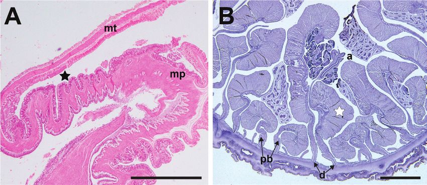
Sea anemones (Cnidaria, Actiniaria) of Singapore: redescription and taxonomy of Phymanthus... 5

Figure 1. Map of Singapore where specimens of Phymanthus pinnulatus were collected for this study:
1, Berlayer Creek (1°15'56"N; 103°48'25"E); 2, Big Sisters’ Island (Pulau Subar Laut) (1°12'50"N;
103°50'05"E); 3, Changi East Beaches (1°18'45"N; 104°00'31"E); 4, Chek Jawa (1°24'25"N; 103°59'23"E);
5, Cyrene Reef (Terumbu Pandan) (1°15'28"N; 103°45'19"E); 6, East Coast Park Beaches (1°17'36"N;
103°53'46"E); 7, Kusu Island (Pulau Tembakul) (1°13'25"N; 103°51'39"E); 8, Pulau Hantu (1°13'35"N;
103°45'03"E); 9, Pulau Jong (1°12'54"N; 103°47'12"E); 10, Pulau Sekudu (1°24'19"N; 103°59'17"E);
11, Pulau Semakau (1°11'58"N; 103°45'31"E); 12, Pulau Tekukor (1°13'51"N; 103°50'18"E); 13, Sen-
tosa (Tanjong Rimau) (1°14'47"N; 103°49'56"E); 14, St John’s Island (1°13'17"N; 103°50'55"E); 15,
Tanah Merah (1°18'45"N; 103°59'34"E); 16, Terumbu Bemban (1°12'36"N; 103°44'27"E); 17, Ter-
umbu Pempang Tengah (1°13'33"N; 103°43'50"E); 18, Terumbu Raya (1°12'46"N; 103°45'09"E); 19,
Terumbu Semakau (1°12'46"N; 103°46'07"E).

ZRC.CNI.0639 x1; ZRC.CNI.1098 x1*; ZRC.CNI.1361 x1*), Pulau Tekukor (ZRC.
CNI.0993 x3*, of these only one has reduced protuberances; BMNH1996.313
x1; ZRC.CNI.1306 x1*), Sentosa (Tanjong Rimau) (ZRC.CNI.1345 x4*), St John’s
Island (ZRC.CNI.0467 x1), Tanah Merah (photographed but not collected), Ter-
umbu Bemban (ZRC.CNI.1223 x1*), Terumbu Pempang Tengah (ZRC.CNI.1028
x1*; ZRC.CNI.1029 x1*), Terumbu Raya (ZRC.CNI.1111 x1*), Terumbu Semakau
(ZRC.CNI.0493 x1).
    Type material examined. Holotype, ZNB Cni 1324, collected by E. von Mar-
tens. A single specimen, 60 mm in length, flaccid, cut longitudinally, a slice of the
distalmost margin and part of the proximal end missing, though a little of the pedal
6                      Nicholas Wei Liang Yap et al. / ZooKeys 840: 1–20 (2019)

Figure 2. Holotype of Phymanthus pinnulatum Martens in Klunzinger, 1877 A entire specimen present
at the Museum für Naturkunde, Berlin (ZMB Cni 1324), Germany B three pieces of the holotype re-
moved from the Berlin specimen now at Naturhistoriska Riksmuseet (NRS76), Stockholm, Sweden. Ab-
breviations: m, mesenteries. mt, marginal tentacles. mf, mesenterial filaments. mp, marginal projection.
o, oral disc. pd, pedal disc. Photographs by NWL Yap.
Sea anemones (Cnidaria, Actiniaria) of Singapore: redescription and taxonomy of Phymanthus... 7

Figure 3. Living specimens of Phymanthus pinnulatus, external morphology of oral end A expanded
individual of green “banded” colour morph, in situ. Photograph by R Tan B an expanded slaty-green
“plain” coloured individual, with extensive branching of its protuberances, in situ. Photograph by R Tan
C a third colour morph with brilliant electric blue marginal tentacles, in situ. Photograph by NWL Yap
D a partially contracted individual in situ, with its oral end protruding from the substratum; note longi-
tudinal rows of verrucae along intermesenterial spaces, extending proximally from the oral end towards
mid column, in situ. Photograph by R Tan.

disc remains, cream-coloured entirely (Fig. 2A); NRS76 consists of three pieces origi-
nating from ZMB Cni 1324 (see Fautin 2016), all pieces cream-coloured in preserva-
tive: a piece of the distalmost end with oral disc and marginal tentacles present, 11
mm in length; a piece of mesentery, fertile, 9 mm wide; a 30 mm longitudinal strip
of the column (Fig. 2B).
     Natural history. Usually encountered during low tide, with upper portion ex-
posed, oral disc and marginal tentacles expanded (Fig. 3A, B, C). Sediment and small
shell fragments may adhere to verrucae (Fig. 3D). Lower body usually deep in crevices
8 Nicholas Wei Liang Yap et al. / ZooKeys 840: 1–20 (2019) Figure 4. Marginal tentacle and marginal projection appearance of Phymanthus pinnulatus A close-up of ramified protuberances of a living specimen. Photograph by NWL Yap B close-up of protuberances from a fixed specimen (ZRC.CNI.1345); note that finer details of protuberance branching are lost in preserved/fixed specimens, branching of protuberances now appear as knobs. Photograph by NWL Yap C a morphotype of that lacks ramified protuberances. Note that this individual (ZRC.CNI.1029) is atypi- cal as it has lesser marginal tentacles, by which are also octo-ramously arranged. Photograph by NWL Yap. D a “smooth” tentacle morphotype, in situ. Photograph by R Tan E close-up of a marginal tentacle tip from a fixed specimen (ZRC.CNI.1342). Note absence of perforation at tip F close-up of a row of mar- ginal projections of a fixed specimen (ZRC.CNI.1342). Note perforations (arrowed). Abbreviations: mt, marginal tentacles; mtt, marginal tentacle tip; p, protuberances. Photographs by NWL Yap.
Sea anemones (Cnidaria, Actiniaria) of Singapore: redescription and taxonomy of Phymanthus... 9
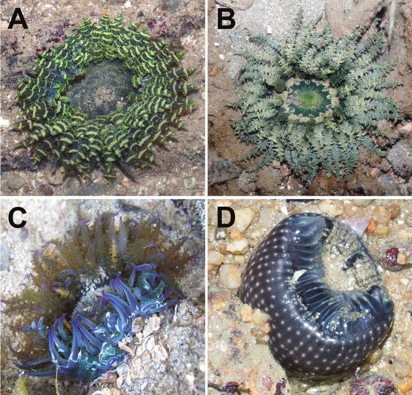
Figure 5. Detail of the oral discs of Phymanthus pinnulatus, external morphology A top view of discal
tentacles present on a live individual, arranged as radial rows between intermesenterial spaces, extending
from the mouth towards the region of the marginal tentacles (ZRC.CNI.1361) B side view of discal
tentacles of a living specimen; note the low and reduced elevation of tentacles (ZRC.CNI.1046) C faint
and reduced radial rows of discal tentacles (arrowed) present on a recent, formalin-fixed specimen (ZRC.
CNI.1345) D very reduced and barely noticeable remnants of discal tentacles (arrowed) present on the
holotype (NRS76); note that this specimen was preserved before 1877. Abbreviations: m, mesenteries;
mt, marginal tentacles. Photographs by NWL Yap.
or buried in sand or coral rubble. Pedal disc attached to buried rock, fragments of shell
or coral rubble. Retracts quickly and deeply into substratum when disturbed, pulling
in marginal tentacles completely. Animal typically found singly, with multiple indi-
viduals separated by a short distance (> 20 cm), although clusters up to four have been
observed. Zooxanthellate.
Marginal tentacles. 96 in total; one individual with 98 (ZRC.CNI.1342). All
of similar length, equal to oral disc radius or longer (Figs. 3A, B). Arranged hexam-
erously in four cycles but octamerously in one individual (ZRC.CNI.1029). Cycle
closest to margin exocoelic; innermost cycles endocoelic. One per endo-/exocoel.10 Nicholas Wei Liang Yap et al. / ZooKeys 840: 1–20 (2019)
Figure 6. Internal musculature of Phymanthus pinnulatus A longitudinal section of ZRC.CNI.0321 at
the margin, showing lack of marginal sphincter muscle. Fosse is indicated by a black star B cross-section
of ZRC.CNI.0993. Note the presence of well-developed retractor muscles (white star), and oocytes. Ab-
breviations: a, actinopharynx; d, directives; mp, marginal projection; mt, marginal tentacle; pb, parietol-
basilar muscle. Scale bar: 10 mm. Photographs by NWL Yap.
Ramified protuberances occur laterally along both sides, symmetrical, alternating
between large and small knobs (Fig. 4A). In life, branching appears extensive; when
preserved, appears as low knobs (Figs. 4A, B, respectively). Extent of branching may
vary; some individuals have protuberances as slight bumps while in others the entire
length is smooth (Figs. 4C, D). Tip narrow and blunt, without perforation (Fig. 4E);
base wide. Colour variable, from greenish-brown, slatey-grey to blue with golden
tinge (Figs. 3A, B, C); tip with green, purple, or pink cast. In fixed individuals, tenta-
cles cream-coloured to greenish, translucent. Protuberances cream-coloured to gold
on oral side; usually with a white line adjoining opposite protuberances (Fig. 4A).
Column. Colour variable, from tan to translucent white. Distalmost end dark-
brown. May appear whitish or cream-coloured in life, or with a light green tinge in
preserved specimens. Distalmost end flared outwards when animal is expanded; mid-
section uniform diameter; pedal end may spread outwards when animal is attached
to a surface. Diameter of distalmost end greater than pedal disc. Marginal projec-
tions present along margin of distalmost end; may be inflated, perforated (Fig. 4F),
with a central white dot. Dot not visible in preserved specimens. Longitudinal rows
of adherent verrucae present, extending proximally to mid-section (Fig. 3D). In life,
shell fragments or substratum particles may be attached to verrucae. Verrucae outline
eye-shaped, as low white bumps, middle depressed, diameterSea anemones (Cnidaria, Actiniaria) of Singapore: redescription and taxonomy of Phymanthus... 11
Figure 7. Cnidae of Phymanthus pinnulatus A spirocyst B basitrich C spirocyst D basitrich E spirocyst
F basitrich G basitrich H basitrich I basitrich J microbasic p-mastigophore K small basitrich L large basi-
trich M microbasic p-mastigophore. Scale bar: 10 μm. Photographs by NWL Yap.
Oral disc. Outline round, flat when fully expanded; diameter 40 mm or greater.
Colour in life grey to dark brown, with white markings flanking outwards; in fixed
specimens, cream-coloured to translucent white. Discal tentacles present, arranged in
radial rows extending from mouth to marginal tentacles, both endo- and exocoelic,
numerous in a row (Fig. 5A). Discal tentacles outline: slim oval, as low bumps (Fig.
5B), some with middle sunken in, dependent on state of expansion. Discal tentacles
dark-brown or grey in life. In preserved individuals, these are very inconspicuous, seen
as horizontally radiating short grey dashes (Fig. 5C), may be very faint, or not seen at
all, depending on state and age of specimen (e.g. Figs. 5C, D). Wall thin; dark lines
corresponding to mesenterial insertions visible through wall, extends from the mouth
to margin. Central mouth oval and flat, area around it may be translucent. Two sipho-
noglyphs, symmetrical. In life, these may be white with pinkish streaks. Preserved,
siphonoglyphs appear cream-coloured.
Pedal disc. Oval, flat, same colour as proximal section of column. Thin-walled,
mesenterial insertions appear as radiating white lines. Strongly adherent; readily at-
taches to surfaces to follow contour of substratum.
Internal morphology. Actinopharynx longitudinally pleated, extends proximally
until mid-column. Oral and marginal stomata present. Mesenteries contain zooxan-
thellae, arranged in three orders. All 12 pairs of highest order complete, fertile and
with filaments; two of these directives, each attached to a siphonoglyph. Mesenteries
of second order incomplete, but all fertile with filaments, 12 pairs. In one individual
(ZRC.CNI.0467) nine pairs of imperfect mesenteries were present in the second order.
Twenty-four pairs of mesenteries small, without filaments and retractor pennon make
up third order. All mesenteries, except smallest, extend to the proximal end. Sphincter12 Nicholas Wei Liang Yap et al. / ZooKeys 840: 1–20 (2019)
Table 1. Cnidom of Phymanthus pinnulatus Martens in Klunzinger, 1877. Measurements in μm; size
outliers of single capsules are presented in values within parentheses. Abbreviations: N = the number of
specimens having that type of cnidae to total specimens examined; n = total number of capsules measured
for each type. Letters in parentheses following cnidae type refer to its illustration in Fig. 7.
Tissue Cnidae Phymanthus pinnulatus Martens in Klunzinger,
1877
Range length × range width N n
Marginal tentacles Spirocysts (A) 13.5–26.5(27.5) × 2.0–4.2 9/9 91
Basitrichs (B) 14.7–25.7(26.5) × 2.0–3.7(4.7) 9/9 93
Protuberances Spirocysts (C) (10.0)13.0–24.0 × 2.0–4.0 9/9 90
Basitrichs (D) 11.0–21.0 × 2.0–3.0 9/9 100
Discal tentacles Spirocysts (E) 14.0–26.0 × 2.0–4.5(5.0) 9/9 90
Basitrichs (F) (8.0)10.0–19.0 × 2.0–3.0 9/9 90
Marginal pseudoacrorhagi Basitrichs (G) 12.0–17.0 × 2.0–3.0 9/9 100
Column Basitrichs (H) 13.0–23.1(25.0) × 2.0–3.7(4.2) 9/9 100
Actinopharynx Basitrichs (I) 11.5–25.5 × 2.0–4.5 9/9 100
Microbasic p-mastigophores (J) 14.7–25.7 × 4.0–7.9 9/9 88
Mesenterial filaments Small basitrichs (K) 10.0–23.1 × 2.0–3.7 9/9 92
Large basitrichs (L) (26.3)27.0–38.0 × (2.6)3.0–4.7 9/9 91
Microbasic p-mastigophores (M) 15.8–25.0 × (2.5)3.5–5.5(6.3) 9/9 92
muscle absent (Fig. 6A). Retractor muscles: strong, diffuse to diffuse circumscript.
Parietobasilar muscle extends away from mesentery, as a reduced pennon (Fig. 6B),
poorly developed. No internal broods encountered.
Cnidom.Spirocysts, basitrichs, microbasic p-mastigophores (Table 1). Cnidae il-
lustrated in Fig. 7. No cnidom data yielded from holotype (i.e., ZMB Cni 1324 and
NRS 76), cnidae present damaged with crystalline appearance.
Distribution. Singapore (Klunzinger 1877; this study), Indonesia (pers. obs.; see
discussion below) and Northern Australia (see discussion below).
Remarks. Of the 53 specimens collected in this study and those examined in situ,
we encountered five individuals having reduced protuberances. These morphotypes
were only encountered along the southern Singapore shores.
While Klunzinger (1877) makes no mention of the etymology of the name, nor
does it appear in Martens’ (1867, 1875) reports, the original species name is ren-
dered as pinnulatum, made up of both a noun (pinnula = small wing) and a neuter,
adjective forming suffix (-tum), thereby making it an adjective in the nominative
singular (ICZN Article 11.9.1). Therein, the original spelling of the species name
is incorrect. The species name, being an adjective in a genitive case, according to
Article 31.2 of the International Code of Zoological Nomenclature (International
Commission on Zoological Nomenclature 1999), must agree in gender with the ge-
nus. The gender of Phymanthus is masculine, therefore the species name is pinnulatus
(see also Fautin 2016).Sea anemones (Cnidaria, Actiniaria) of Singapore: redescription and taxonomy of Phymanthus... 13
Discussion
Discal tentacles
A feature that unites pieces of holotype (i.e., ZMB Cni 1324 and NRS76), vouchers
and fresh specimens of Phymanthus pinnulatus (N = 53), is the appearance of discal
tentacles. For all individuals examined here, this feature is inconspicuous, reduced
and oval; occurring as faint and dark horizontal dashes radiating outward from the
mouth in preserved specimens (Figs. 5A and C) and in older preserved specimens, less
obvious (Figs. 5D). There is no mention of discal tentacles whatsoever in Klunzinger’s
(1877) description, and it is likely he described what was most obvious from the
Berlin specimen. Regardless of anemone size or location where it was collected, shape
and size of discal tentacles were consistent in its form for all materials we examined.
The inconspicuous and reduced form of discal tentacles in P. pinnulatus is
distinct from its congeners depicted in primary scientific literature, and of type
materials we studied. Those of P. crucifer, P. loligo, and P. rhizophorae are illustrated
to be conspicuous and papilliform (see P. crucifer: Durden 1900: pl 10, fig 1;
P. loligo: Klunzinger 1877: pl. 6, fig. 7, pl. 7, fig. 3 and Carlgren 1900: pl. 2, fig. 3;
P. rhizophorae: Mitchell 1890, pl. 36, fig. 5). Discal tentacles present on syntypes of
P. sansibaricus (ZMH C2620 and ZMH C2627) and P. strandesi (ZMH C2585) are
also conspicuous and papilliform, like those depicted for P. loligo. Among syntypes
of P. levis (ZMB.CNI.3811, NRS5557), we found that discal tentacles of this species
are very different: they resemble marginal tentacles stunted in growth, unlike those
of P. pinnulatus and P. loligo.
On P. muscosus found nearby (i.e., Indonesia and northern Australia), Kwiet-
niewski (1898: 420) wrote “Sonst erscheint diese Partie der Mundscheibe ganz glatt,
und nur nach sehr sorgfältiger Prüfung fand ich auf mehreren Sectoren der Mund-
scheibe Reihen von runden, äusserst geringen Erhebungen, welche als die Rudi-
mente der scheibenständigen Tentakel zu deuten sind,” referring to slight bumps
as ‘rudiments of disc-like tentacles,’ visible after ‘careful examination’, and an ap-
parent overall smoothness to the area around the mouth. Moreover, illustrations in
Kwietniewski’s report (1898: pl. 29, figs. 57, 58) show P. muscosus with a smooth
oral disc, without discal tentacles. Similarly, Pax (1924) makes no mention of any
discal tentacles found on the oral disc of P. buitendijki. Both Kwietniewski (1898)
and Pax (1924), did not examine the holotype of P. pinnulatus; neither did workers
before and after them (e.g. Haddon and Shackleton 1893; Haddon 1898; Stephen-
son 1922; Carlgren 1949, 1950). Given the close geographical proximities between
these three reported species, it is possible that some individuals described by them
as P. muscosus and P. buitendijki are in fact P. pinnulatus instead. Having examined
the syntypes of P. muscosus collected by Dr AC Haddon, and P. buitendijki that Pax
(1924) had examined, we found at least one resembling P. pinnulatus within the lots
(i.e., MZC.I.33745 and RMNH.COEL.3876).14 Nicholas Wei Liang Yap et al. / ZooKeys 840: 1–20 (2019)
Discal tentacle form appeared to be consistent for all P. pinnulatus type and vouch-
er materials examined here. We propose that this trait could be a stable character to
help infer and define species boundaries for members of Phymanthus. As of writing,
many members of this genus remain poorly described; whether the discal tentacle form
is truly a useful trait to define species boundaries among members of Phymanthus war-
rants further study.
Cnidom
In this study, we report upon the cnidom of P. pinnulatus (Table 1, Fig. 7) for the first
time. Although cnidom data are a necessary component in modern actinian taxonomic
descriptions (Fautin 1988), none have been reported in the original description.
All three cnidae types (i.e., spirocysts, basitrichs and microbasic p-mastigophores)
were found in tissues of P. pinnulatus examined for this study; these agree with the
diagnosis of the genus Phymanthus (Carlgren 1949: 74). Because the use of cnidom
data in anemone systematics only became routine after the 1940s (Fautin 1988),
many published descriptions of other Phymanthus species we reviewed did not have
any cnidae type or capsule size data for comparison (e.g. Haddon and Shackleton
1893; Kwietniewski 1898; Pax 1924) as these descriptions were published before the
1940s. Among reports that had cnidae size data, we found those of P. crucifer from
the Gulf of Mexico (see Gonzalez-Muñoz et al. 2015), P. pulcher from the Aegean Sea
(see Chintiroglou and den Hartog 1995) and Phymanthus muscosus from the Great
Barrier Reef (Carlgren 1940, 1950) to be sufficiently detailed and therefore useful
for comparison. Cnidae sizes and types of P. pinnulatus were consistently different
from P. crucifer and P. pulcher. Basitrichs in the mesenterial filaments of P. pinnulatus
were much longer than those found in P. crucifer (basitrich length: P. pinnulatus =
27.0–38.0 μm; P. crucifer = 24.0–25.0 μm, see Gonzalez-Muñoz et al. 2015: fig.
3). Also, small basitrichs like those depicted in the actinopharynx of P. crucifer
(see Gonzalez-Muñoz et al. 2015: fig. 3) were absent in P. pinnulatus. In tissues of
P. pulcher, microbasic b-mastigophores were found (Chintiroglou and den Hartog
1995), but we did not encounter any in tissues of P. pinnulatus. Moreover, basitrichs
in the marginal tentacles and mesenterial filaments of P. pinnulatus are larger than
those in P. pulcher. The cnidom data of P. muscosus (shown in Carlgren 1940, 1950)
largely agreed with those of P. pinnulatus – we hypothesize that individuals identified
by Carlgren (1950) as P. muscosus are likely to be P. pinnulatus too. From all syntypes
of P. levis examined in this study, we found microbasic p-mastigophores present
in the tissues of marginal projection and column. This cnida was absent from the
same tissues of P. pinnulatus. Overall, we found a difference of both cnidae type and
capsule sizes among P. pinnulatus and its congeners. While cnidae type and size alone
cannot distinguish species (Fautin 1988), when used together with other traits that
are consistent (i.e., discal tentacle appearance) it appears that this feature can be useful
in differentiating Phymanthus species.Sea anemones (Cnidaria, Actiniaria) of Singapore: redescription and taxonomy of Phymanthus... 15 Morphotypes Intraspecific morphotypes of Phymanthus anemones have been widely documented in primary scientific literature, with reports focused on P. crucifer’s variable appearance of protuberances (see Duerden 1900; González-Muñoz et al. 2015). In some individuals these are reduced knobs; in others they are entirely absent. Uncertainty persists concern- ing the usefulness of this character in distinguishing congeners or even members of Phy- manthidae (i.e., between Phymanthus and Heteranthus; see Gonzalez-Muñoz et al. 2015). Among P. pinnulatus individuals collected and examined here, we encountered some specimens exhibiting this variation. Morphotypes with absent or reduced protuberances were typically encountered from the south of Singapore, although this was confined to a small number of individuals (5 out of 53) that was collected over 16 years. Like the study on P. crucifer by Gonzalez-Muñoz et al. (2015), we did not find an ecological cause for this. Furthermore, Gonzalez-Muñoz et al. (2015) and Brugler et al. (2018) found little genetic differentiation among P. crucifer morphotypes examined. While we did not have the opportunity to test for any genetic differences among P. pinnulatus morphotypes, we hypothesize that there is little or no variation among them. On a population basis we con- clude that all morphotypes examined in this study must be of a single species, similar to observations and interpretations of Duerden (1900), Gonzalez-Muñoz et al. (2015) and Brugler et al. (2018) on P. crucifer. We conclude that this variation may not be extensive; overall the appearance of ramified protuberances is a useful character to distinguish mem- bers of the Phymanthidae at genus-level. Conversely, this trait is not diagnostic of Phy- manthus species due to its variable appearance; here we infer that the discal tentacle form and cnidom are more useful and consistent for differentiating members of the genus. Biology Little is known about the biology and ecology of Phymanthus anemones. On reproduc- tion, Jennison (1981) found brooded juveniles within P. crucifer. These were encoun- tered in individuals he had collected during the months of “December, February, and May” (Jennison 1981: 1717). In specimens of P. pinnulatus dissected for this study, no brooded juveniles were encountered. Most individuals studied here were collected at different times of the year, spanning more than 40 years; we hypothesize that it is unlikely that internal brooding occurs among P. pinnulatus. In observing individuals kept alive in the aquaria before fixation, we did not observe any evidence for asexual reproduction, as with Jennison (1981) and Lin et al. (2001). Morphological comparisons with Heteranthus As stated on the onset, the family Phymanthidae consists of two valid and extant genera, Phymanthus and Heteranthus (Fautin 2013; 2016). Originally, members of
16                  Nicholas Wei Liang Yap et al. / ZooKeys 840: 1–20 (2019)

Heteranthus were classified as a separate family, Heteranthidae (see Carlgren, 1900).
In studying a single specimen from Vietnam (i.e., H. insignis), Carlgren (1943) placed
both genera together in a single family, though he was not explicit on details for his
rationale, remarking (Carlgren 1943: 30), “… we now know the organization of the
genus better, I think that it can be brought together with Phymanthus in a family.
Both genera are closely related to each other.” Based on his monograph published
thereafter, we infer that Carlgren grouped these two genera together due to members
of Phymanthus and Heteranthus having discal and marginal tentacles (Carlgren 1949).
Largely, he distinguished these two genera largely on the presence of protuberances on
the marginal tentacles (see Carlgren 1940: 74).
     However, in his diagnosis of Heteranthus, Carlgren (1949: 75) further differenti-
ates Heteranthus from Phymanthus stating that members of the former have, “… large
verrucae, which at the margin are small and more numerous and overhang the fosse.”
We examined the syntype of H. verruculatus (ZNB Cni 1852) and holotype of H.
insignis (NRS4076) and found this character to be present: verrucae resembling con-
spicuous warts densely cover each marginal projection of Heteranthus specimens, that
extend out into the fosse. These were absent on the marginal projections of all type
and voucher specimens of Phymanthus anemones we have examined for this study (e.g.
see Fig. 4F). This character clearly distinguishes the two Phymanthidae genera, despite
both having discal and marginal tentacles.

Conclusions
In Table 2, we summarise differences in discal tentacle appearance, cnidom, and type
localities for eight Phymanthus species. These are based on details from prior publi-
cations and our own observations of type materials, if present. Of those for which
we have examined type materials, eight were comprised only of one anemone species
within the lot. An updated, detailed taxonomic account for the other Phymanthus spe-
cies will be discussed in a separate manuscript.
      Fautin (2013, 2016) listed eleven valid species of Phymanthus worldwide. Despite
the comprehensive redescription of P. pinnulatus attempted here, taxonomic confusion
still exists for nearly all remaining Phymanthus species. Apart from P. crucifer, species
boundaries defining remaining members are unclear. In reviewing published descrip-
tions of other Phymanthus species, apart from P. crucifer, we found that much of the
confusion is exacerbated by a lack of thoroughness in examining type and voucher speci-
mens. Our study addresses this limitation for P. pinnulatus, but other members of the ge-
nus require similar treatment along with a formal revision of the family Phymanthidae.

Acknowledgements
The persistent and productive efforts of the “Anemone Army,” from 2002, led to many of
the specimens listed in this study. We thank those involved for their continued assistance
Sea anemones (Cnidaria, Actiniaria) of Singapore: redescription and taxonomy of Phymanthus... 17
Table 2. Comparison of discal tentacle form, cnidom, and type localities of eight Phymanthus species.
The symbol “×” indicates that the trait was found; “?” indicates that the trait was not examined in detail
when the animal was first described and thereafter; a blank denotes that the trait was not observed from
the species at all. Abbreviations: a, actinopharynx; c, column; pt, protuberances; dt, discal tentacles; mf,
mesenterial filaments; mp, marginal projection; mt, marginal tentacles.
Morphological traits Phymanthus species
pinnulatus levis strandesi sansibaricus crucifer loligo pulcher rhizophorae
Papilliform × × × × ? ×
Discal tentacle
Conspicuous × × × × × ? ×
form
Reduced × ?
As a stunted tentacle × ?
a
Basitrichs × × × × × ? × ?
c × × × × × ? × ?
pt × × × × × ? ? ?
dt × × × × × ? ? ?
mf × × × × × ? × ?
mp × × × × × ? ? ?
mt × × × × × ? × ?
Microbasic a × × × × × ? ?
p-masti- c × ? ?
gophores
pt ? ? ?
dt ? ? ?
mf × × × × × ? × ?
mp × ? ? ?
Cnidae
mt ? ?
Microbasic a ? ?
b-masti- c ? ?
gophores
pt ? ? ?
dt ? ? ?
mf ? × ?
mp ? ? ?
mt ? ?
Spirocysts a ? × ?
c ? ?
pt × × × × × ? ?
dt × × × × × ? ?
mf ? ?
mp ? ?
mt × × × × × ? × ?
Type locality Singapore Indonesia East Africa Caribbean Red Sea Mediterranean Indonesia
References Klunzinger Kwietniewski Carlgren 1900 Andrès Andrès Andrès 1883; Mitchell
1877; This 1898 1883; 1883; Chintiroglou 1890;
study Duerden Carlgren and den Haddon
1900; 1900 Hartog 1995 1898
González-
Muñoz et
al 2015
over many years, especially during very early morning fieldtrips. Permissions granted to
examine types and voucher specimens were crucial to the success of this study. For this,
NWL Yap thanks the personnel from each of the following institutions: (BMNH) Ms
Miranda Lowe and Mr Andrew Cabrinovic; (CASIZ) Ms Christina Piotrowski and Ms
Elizabeth Kools; (MZC) Mr Matthew Lowe and Dr Richard Preece; (MZL) Ms Maria
Mostadius; (NRS) Dr Lena Gustavsson, Ms Emma Wahlberg and Ms Emily Dock Åk-
erman; (RMNH) Dr Bert W Hoeksema and Ms Karen Van Dorp; (ZMB) Drs Lüter18 Nicholas Wei Liang Yap et al. / ZooKeys 840: 1–20 (2019)
Carsten and Birger Neuhaus; (ZMH): Dr Andreas Schmidt-Rhasea, Ms Helma Roggen-
buck and Mr Jan Raeker; (ZRC): Ms Iffah Iesa and Mr Chua Keng Soon, and the staff
of Tropical Marine Science Institute, including Ms Gan Bin Qi, Mr Ahmed Aliyar and
Mr Jackson Chan for logistical support at St John’s Island National Marine Laboratory.
From the International Commission on Zoological Nomenclature, we thank secretary
Dr Gwynne S Lim, for clarifying doubts on nomenclature and associated Articles. Also,
we thank the anonymous reviewer whose comments greatly improved this manuscript;
we are most appreciative. Finally, special thanks to Prof Daphne Fautin; we are grateful
to the many years of advice and encouragement over emails that led to the completion of
this study. Any errors of interpretations present here are of the first author, YNWL. This
research is supported by the National Research Foundation, Prime Minister’s Office, Sin-
gapore under its Marine Science R&D Programme (MSRDP-P03 and MSRDP-P38).
References
Andrès A (1883) Le Attinie (Monografia). Coi Tipi der Salviucci, Roma, 460 pp.
Brugler MR, González-Muñoz RE, Tessler M, Rodríguez E (2018) An EPIC journey to locate
single-copy nuclear markers in sea anemones. Zoologica Scripta 47(6): 756–776. https://
doi.org/10.1111/zsc.12309
Carlgren O (1900) Ostafrikanische Actinien. Gesammelt von Herrn Dr F Stuhlmann 1888
und 1889. Mittheilungen aus dem Naturhistorischen Museum 17: 21–144.
Carlgren O (1940) A contribution to the knowledge of the structure and distribution of the
cnidae in the Anthozoa. Kungliga Fysiografiska Sällskapets Handlingar 56(9): 1–24.
Carlgren O (1949) A survey of the Ptychodactiaria, Corallimorpharia and Actiniaria. Kungliga
Svenska Vetenskaps Akademiens Handlingar 1(1): 1–121.
Carlgren O (1950) Actinia and Corallimorpharia. Scientific Reports of the Great Barrier Reef
Expedition 1928/29 5(7): 427–457.
Chintiroglou C, den Hartog JC (1995) Additional records of Actiniaria (Anthozoa) from
Greece. Zoologische Mededelingen Leiden 69: 353–364.
Duerden JE (1900) Jamaican Actiniaria. Part II. Stichodactylinæ and Zoantheae. Scientific
Transactions of the Royal Dublin Society 7: 133–208.
Ehrenberg CG (1834) Beiträge zur physiologischen Systematik derselben. Abhandlungen der
Königlichen Akademie der Wissenschaften zu Berlin 1: 225–380.
Fautin DG (1988) Importance of nematocysts to actinian taxonomy. In: Hessinger DA, Len-
hoff HM (Eds) The Biology of Nematocysts. Academic Press Inc, San Diego and other
cities, 487–500. https://doi.org/10.1016/B978-0-12-345320-4.50030-4
Fautin DG (2013) Hexacorallians of the World. http://geoportal.kgs.ku.edu/hexacoral/anem-
one2/index.cfm
Fautin DG (2016) Catalog to families, genera, and species of orders Actiniaria and Coral-
limorpharia (Cnidaria: Anthozoa). Zootaxa 4145(1): 1–449. https://doi.org/10.11646/
zootaxa.4145.1.1Sea anemones (Cnidaria, Actiniaria) of Singapore: redescription and taxonomy of Phymanthus... 19
Fautin DG, Tan R (2016) Sea anemones (Cnidaria: Actiniaria) of Singapore: redescriptions
of Paracondylactis singaporensis (England, 1987) and P. hertwigi (Wassilieff, 1908). Raffles
Bulletin of Zoology Supplement 34: 170–177.
Fautin DG, Tan SH, Tan R (2009) Sea anemones (Cnidaria: Actiniaria) of Singapore: abun-
dant and well-known shallow-water species. Raffles Bulletin of Zoology Supplement 22:
121–143.
Fautin DG, Tan R, Yap WLN, Tan SH, Crowther A, Goodwill R, Sanpanich K, Tay YC (2015)
Sea anemones (Cnidaria: Actiniaria) of Singapore: shallow-water species known also from
the Indian subcontinent. Raffles Bulletin of Zoology Supplement 31: 44–59
González-Muñoz R, Simões N, Mascaró M, Tello-Musi JL, Brugler MR, Rodríguez E (2015)
Morphological and molecular variability of the sea anemones Phymanthus crucifer (Cnidar-
ia, Anthozoa, Actiniaria, Actinoidea). Journal of the Marine Biological Association of the
United Kingdom 95(1): 69–79 https://doi.org/10.1017/S0025315414000988
Haddon AC (1898) The Actiniaria of Torres Straits. Scientific Transactions of the Royal Dublin
Society series 2, 6: 393–520.
Haddon AC, Shackleton AM (1893). Description of some new species of Actiniaria from Tor-
res Straits. Scientific Transactions of the Royal Dublin Society 8: 116–131.
den Hartog JC (1997) Box 8–2: The sea anemone fauna of Indonesian coral reefs.In: Tomascik
T, Mah AJ, Nontji A, Moosa MK (Eds) The Ecology of the Indonesian Seas Part 1. Periplus
Editions, Republic of Singapore, 351–370.
Humason GL (1967) Animal tissue techniques. Second edition. WH Freeman & Company,
San Francisco, 569 pp.
ICZN (1999) International Code of Zoological Nomenclature (4th edn). International Trust for
Zoological Nomenclature, London, 306 pp.
Jennison BL (1981) Reproduction in three species of sea anemones from Key West, Florida.
Canadian Journal of Zoology 59: 1708–1719. https://doi.org/10.1139/z81-235
Kwietniewski CR (1898) Actiniaria von Ambon und Thursday Island. In: Zoologische Forschun-
greisen in Australien und dem Malayischen Archipelago von Richard Semon. Volume 5
[Systematik, tiergeographie, anatomie wirbelloser tiere]. Gustav Fischer, Jena, 385–430
Klunzinger CB (1877) Die Korallthiere des rothen Meeres,1: Die Alcyonarien und Malacoder-
men. Verlag der Gutmann’schen Buchhandlung, Berlin, 98 pp. [8 pls]
La Sueur CA (1817) Observations on several species of the genus Actinia; illustrated by figures.
Journal of the Academy of Sciences of Philadelphia 1: 149–154, 169–189.
Lin MD, Chen CA, Fang LS (2001) Distribution and sexual reproduction of a seagrass-bed-in-
habitiing Actinarian, Phymanthus strandesi (Cnidaria: Anthozoa: Phymanthidae), at Hsiao-
Liuchiu Island, Taiwan. Zoological Studies 40(3): 254–261.
Mariscal RN (1974) Nematocysts. In: Muscatine L, Lenhoff HM (Eds) Coelenterate Biol-
ogy: Reviews and New Perspectives. Academic Press, Inc, New York, 129–178. https://doi.
org/10.1016/B978-0-12-512150-7.50008-6
von Martens E (1867) Die preussiche expedition nach Ost-Asien: nach amtlichen quellen,
Zoologischer Theil, 2 die landschnecken. Verlag Der Königlichen Geheimen Ober-Hofbu-
chdruckerei, Berlin, 469 pp. [22 pls]20 Nicholas Wei Liang Yap et al. / ZooKeys 840: 1–20 (2019)
von Martens E (1875) Die preussiche expedition nach Ost-Asien: nach amtlichen quellen, Zo-
ologischer Theil, 1, allgemeines un wirbelthiere. Verlag Der Königlichen Geheimen Ober-
Hofbuchdruckerei, Berlin, 427 pp. [15 pls]
Milne-Edwards H (1857) Histoire Naturelle des Coralliaires ou Polypes Proprement Dits. Vol.
1. Librairie Encylopédique de Roret, Paris, 326 pp.
Mitchell PC (1890) Thelaceros rhizophoræ, n. g. et sp., an actinian from Celebes. Quarterly
Journal of Microscopical Science 30: 551–563.
Pax F (1924) Anthozoen des Leidener Museums. Zoologische Mededelingen Leiden 8: 1–17.
Quoy JRC, Gaimard P (1833) Voyage de Découvertes de l’ Astrolabe Exécuté par Ordre du
Roi, Pendant les Années 1826–1827–1828–1829, Sous le Commandement de M J Du-
mont D’Urville, volumen 4. J. Tastu, Paris, 390 pp.
Stephenson TA (1922) On the classification of Actiniaria. Part III – Definitions connected
with the forms dealt with in Part II. Quarterly Journal of Microscopic Science 66(262):
247–319.
Yap WLN, Fautin DG, Ramos DA, Tan R (2014) Sea anemones of Singapore: Synpeachia te-
masek new genus, new species, and redescription of Metapeachia tropica (Cnidaria: Actini-
aria: Haloclavidae). Proceedings of the Biological Society of Washington 127(3): 439–454.
https://doi.org/10.2988/0006-324X-127.3.439You can also read