The only complete articulated early Miocene chameleon skull (Rusinga island, Kenya) suggests an African origin for Madagascar's endemic chameleons
←
→
Page content transcription
If your browser does not render page correctly, please read the page content below
www.nature.com/scientificreports

            OPEN           The only complete articulated early
                           Miocene chameleon skull (Rusinga
                           Island, Kenya) suggests an African
                           origin for Madagascar’s endemic
                           chameleons
                           Andrej Čerňanský1*, Anthony Herrel2, Job M. Kibii3, Christopher V. Anderson4, Renaud Boistel5
                           & Thomas Lehmann6
                           We here present the first detailed study of the specimen KNM-RU 18340 from Rusinga Island (Kenya),
                           the only known complete early Miocene chameleon skull, using micro-CT. This specimen represents one
                           of the oldest chameleon fossils ever recovered. For the first time, the skull bone internal surfaces, their
                           sutures, and elements contained inside the rocky matrix are observed. Our morphological comparisons
                           and phylogenetic analyses place this specimen confidently in the genus Calumma and a new species,
                           Calumma benovskyi sp. nov., is erected for it. Since all species of this genus are endemic to Madagascar,
                           this fossil uniquely demonstrates the existence of Calumma on continental Africa in the past. Our results
                           challenge the long-held view that chameleons originated on Madagascar and dispersed over water to
                           Africa, and provide a strong evidence of an African origin for some Malagasy lineages. The Oligocene–
                           early Miocene dispersal to Madagascar, using oceanic currents that favoured eastward dispersal at that
                           time, is a highly supported scenario matching the suggested dispersal of lemurs to this island. This is
                           consistent with a previously suggested hypothesis based on molecular data.

                           The Chamaeleonidae is an unusual family of lizards including extant species from Africa, Madagascar, the Middle
                           East, southern India, Sri Lanka, and the Mediterranean region of Europe. It is a highly characteristic and morpho-
                           logically specialized clade of acrodont iguanians. This clade is mainly composed of arboreal forms, but includes
                           a ground-dwelling desert form –the Namaqua chameleon1,2. About half of the accepted species of chameleons
                           occur in Madagascar. This island has therefore been suggested to be a centre of diversity of the clade from where
                           it likely radiated via oceanic dispersal3. Later, in contrast, the molecular phylogeny of Tolley et al.4 suggested that
                           the family most probably originated in Africa, with two separate oceanic dispersal events to Madagascar during
                           the Palaeocene and the Oligocene, when prevailing oceanic currents would have favoured eastward dispersal.
                           The fossil record of these animals, the only form of direct evidence regarding the early evolution and palaeobio-
                           geography of these animals, is unfortunately scant. Thus, a key element for resolving this conundrum is lacking.
                                Molecular data suggest a Cretaceous origin4, but the oldest known fossil record of crown members only dates
                           back to the early Miocene (MN3)5,6. Chamaeleonid fossils are mostly reported from the Miocene of Europe6–12
                           and are also known from Africa13,14 and potentially India15. Further African fossils have been documented from
                           early Pliocene deposits16,17. The vast majority of the fossil record is represented by isolated elements, mostly

                           1
                             Department of Ecology, Laboratory of Evolutionary Biology, Faculty of Natural Sciences, Comenius University in
                           Bratislava, 84215, Bratislava, Mlynská dolina, Slovakia. 2Département Adaptation du Vivant, UMR 7179C.N.R.S/
                           Muséum National d’Histoire Naturelle, 55 rue Buffon, 75005, Paris, France. 3Earth Sciences Department, National
                           Museums of Kenya, Nairobi, Kipande Road P.O. BOX, 40658– 00100, Nairobi, Kenya. 4University of South Dakota,
                           Department of Biology, 414 E. Clark Street – UCL 191, Vermillion, South Dakota, USA. 5Muséum National d’Histoire
                           Naturelle, UMR 7179C.N.R.S/M.N.H.N., Bâtiment d’Anatomie Comparée, 55 rue Buffon, CP 55, 75005, Paris,
                           France. 6Senckenberg Research Institute and Natural History Museum Frankfurt, Department of Messel Research
                           and Mammalogy, Senckenberganlage 25, 60325, Frankfurt, am Main, Germany. *email: cernansky.paleontology@
                           gmail.com

Scientific Reports |   (2020) 10:109 | https://doi.org/10.1038/s41598-019-57014-5                                                               1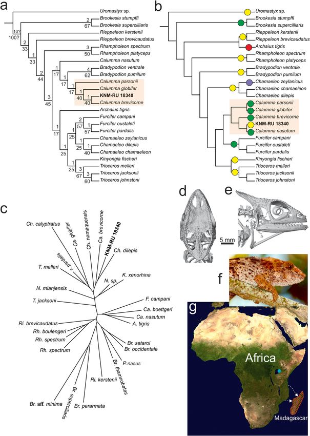
www.nature.com/scientificreports/                                                                www.nature.com/scientificreports

                           jaw fragments, which renders taxonomical identification to the genus or species level often difficult or impos-
                           sible. Only two notable exceptions exist - Chamaeleo intermedius, based on a natural calcite cast described by
                           Hillenius13 from the middle Miocene of Fort Ternan in Kenya; and Ch. andrusovi from the early (and potentially
                           middle) Miocene of Europe, based on well-documented isolated cranial elements described by Čerňanský6.
                               The fossil locality of Rusinga Island (early Miocene, Lake Victoria, Kenya) is famous for its numerous fossil
                           mammals18 (for geology see Supplementary Data 1), but this locality also yielded numerous reptile specimens
                           such as varanids, crocodiles19,20, as well as the almost complete, and remarkably three-dimensionally preserved,
                           skull of a fossil chamaeleonid (KNM-RU 18340; National Museums of Kenya). The skull is not fully freed from
                           the rock, as matrix fills the entire internal region and covers many bone elements (Fig. 1). The specimen has been
                           only preliminary reported by Rieppel et al.14. These authors suggested that it may be an early representative of the
                           genus Rhampholeon, but their report was based on a cast of the specimen and photographs of the original only.
                           Importantly, this specimen represents the only known complete fossil chameleon skull from the early Miocene.
                           Thus, detailed knowledge on the morphology of this fossil African chameleon and its taxonomy is crucial to our
                           understanding of the evolution of this lizard clade and for understanding African paleobiogeography and possible
                           dispersal events.
                               The aims of this paper are: 1) to use high-resolution X-ray microcomputed tomography (μCT) to virtually pre-
                           pare all skeletal elements of KNM-RU 18340; 2) to discover potentially hidden bones embedded in the sediment
                           not visible from the outside; and 3) on the basis the obtained new anatomical data to evaluate the possible taxo-
                           nomic position of KNM-RU 18340 and shed light on scenarios for the origin and diversification of chameleons.

                           Systematic Palaeontology
                           Squamata Oppel, 181121
                              Chamaeleonidae Gray, 182522
                              Calumma Gray, 186523
                              Calumma benovskyi sp. nov.

                           Etymology. After Count Móric Beňovský (also spelled Benyovszky). He was born in Slovakia (Vrbové, 9.
                           20. 1746) and died at the age of 39 in Madagascar (5. 23. 1786). He is known as an important traveler and adven-
                           turer - the first European to sail in the North Pacific Ocean (seven years before James Cook) and the first person
                           who explored Saint Lawrence Island. He traveled to Madagascar, where local tribal chiefs elected him as a “king”
                           (ampansacabe) of Madagascar in 1776. His story parallels the history of Calumma - born abroad, he reached the
                           island by sailing across the ocean.

                           Holotype. KNM-RU 18340 (field number RU 1916'87; National Museums of Kenya, Nairobi): skull, mandible
                           and three cervical vertebrae in connection (Figs. 1, 2 and Supplementary Figs. 1–13).

                           Type locality. Rusinga Island (Lake Victoria, Kenya, Africa), site R107.
                           Age.     Early Miocene, base of the Hiwegi Formation.

                           Diagnosis.       A species of Calumma distinguishable from other species by the posterior corners of the frontal,
                           forming well-developed posterolateral processes. Besides this feature, this taxon is characterized by the unique
                           combination of the following characters: (1) nasal-frontal contact present; (2) prefrontal fenestra present and
                           connected to external naris; (3) prefrontal inclined laterally in dorsal view; (4) contact between prefrontal and
                           postorbitofrontal, excluding the frontal from the orbit; (5) orbital margin medially concave rather than straight in
                           dorsal view; (6) single line of protuberances on the external surface of jugal; (7) hour-glass shaped parietal, bear-
                           ing sculpture arrangement roughly resembling the Greek letter Ψ (psi); (8) well-developed and rather regularly
                           distributed sculpture formed by protuberances on the dorsal surface of the frontal; (9) frontal completely pierced
                           by the parapineal foramen; (10) posterodorsal process of postorbitofrontal reaching above the orbit, inducing an
                           horizontal upper temporal fenestra, dorsally open, and not visible in lateral view; (11) posterodorsal process of
                           squamosal more or less horizontal rather than vertical; and (12) the maximum width of the skull in dorsal view
                           located at the level of the frontal-parietal contact rather than posterior to it.

                           General description and comparison. In dorsal view, the skull presents an ovoid shape, with its widest dimension
                           being at the level of the frontal-parietal contact (Fig. 2A,B). In lateral aspect, the skull is triangular and gradually
                           tapers anteriorly (Fig. 2C). The orbits are large, their anteroposterior length forms 1/3 of the complete anteropos-
                           terior length of the skull. The casque is flat and weakly elevated posteriorly, without a dorsally convex parietal. For
                           detailed description and ilustration of each virtually isolated element, see Supplementary Data 1. The specimen
                           KNM-RU 18340 shares the following features mainly with members of extant Calumma (see also Supplementary
                           Figs. 14–16):

                           (1) The contact of the premaxilla with the frontal. This is present in Calumma globifer, Ca. boettgeri and Ca.
                               gubei, but not in Ca. parsonii, Ca. brevicorne, Ca. nasutum, Ca. fallax, and Ca. juliae. It is present in Fur-
                               cifer pardalis and occasionally in F. oustaleti (but not in F. bifidus and F. campani)24,25. Is is also present in
                               Rhampholeon and Rieppeleon, but not in Bradypodion, Chamaeleo, Kinyongia, Trioceros and Brookesia. The
                               premaxilla-frontal contact is absent in the outgroup and character optimization in Mesquite supports this
                               character state as being the condition at the basal node of the Chamaeleonidae clade. Character optimiza-
                               tion in Mesquite shows that the contact of these two bones evolved independently in Rieppeleon, Rhamp-
                               holeon and some members of Calumma. Among Calumma, the absence of the contact is at the basal node
                               of the clade. So its presence can be regarded as derived among of this clade.

Scientific Reports |   (2020) 10:109 | https://doi.org/10.1038/s41598-019-57014-5                                                                2
www.nature.com/scientificreports/ www.nature.com/scientificreports
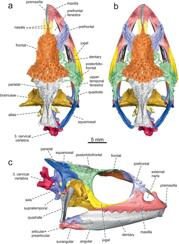
Figure 1. Calumma benovskyi sp. nov., the holotype KNM-RU 18340 from the lower Miocene of Kenya in
(a,f) dorsal; (b,g) ventral; (c,h) anterior; (d,i) right; and (e,j) left views. These 3D models of the specimen were
obtained by µCT and show the specimen in its original preservation condition (with a sedimentary matrix) on
the upper side (a–e), whereas the lower side (f–j) shows the specimen with the sediment virtually removed by
digital means.
(2) The paired nasals do not contact one another and are separated by a strut of bone formed by the premaxilla
and the frontal. The majority of the chameleons have paired nasals, whereas Bradypodion and Brookesia
often have a single nasal24.
Scientific Reports | (2020) 10:109 | https://doi.org/10.1038/s41598-019-57014-5 3www.nature.com/scientificreports/ www.nature.com/scientificreports
Figure 2. Calumma benovskyi sp. nov., the holotype KNM-RU 18340 from the lower Miocene of Kenya with
virtually segmented bones from the dorsal and right side (best preserved elements) in (a) dorsal view; (b) the
reconstruction of the complete specimen in dorsal view restored by mirror imaging; and (c) virtually segmented
bones in right lateral view.
(3) The prefrontal fenestra is connected to the external naris (contra Rieppel et al.14). This character state can
be observed in some extant chameleons, such as Calumma nasutum, Ca. fallax, Ca. vatosoa, Ca. guibei (but
not in Ca. globifer, Ca. parsonii, Ca. brevicorne, Ca. boettgeri and Ca. gehringi), but also in Rhampholeon,
Archaius and Trioceros melleri24–26. This connection is absent in e.g., Brookesia, Rieppeleon, Nadzikambia,
Bradypodion, Chamaeleo and Furcifer. As for Calumma, character optimization in Mesquite supports
absence of the connection as being the condition at the basal node of the clade, whereas its presence is
derived.
Scientific Reports | (2020) 10:109 | https://doi.org/10.1038/s41598-019-57014-5 4www.nature.com/scientificreports/ www.nature.com/scientificreports
(4) The single line of protuberances on the postorbital process of the jugal. Two lines are present e.g., Calumma
ambreense, Ca. parsonii and in the European early Miocene Chamaeleo andrusovi6, whereas one line is
present in Ca. brevicorne.
(5) The suborbital process of the jugal in lateral aspect; only a dorsal narrow portion of this process is ex-
posed dorsal to maxilla. This is present in Calumma, Chamaeleo, Trioceros, Kinyongia, Rhampholeon and
Brookesia. It is largerly exposed in Rieppeleon and Bradypodion, where, moreover, the process often bears
a sculpture formed by protuberances. It is also largerly exposed in Uromastyx (outgroup). However, the
suborbital process in lateral view is covered by the maxilla in Archaius and many Furcifer species (not in,
e.g, F. lateralis, where a narrow portion is exposed)24.
(6) The absence of jugal - squamosal contact. This contact is absent in Calumma (except for Ca. parsonii),
Brookesia, Rieppeleon and Bradypodion, whereas the contact is usually present in members of the genera
Chamaeleo, Nadzikambia, Archaius, Kinyongia, and some members of Furcifer (not in e.g., F. campani)
and Trioceros24. The character optimization in Mesquite supports the absence of jugal - squamosal contact
as being the condition at the basal node of the clade formed by all chameleons. Character optimization
in Mesquite shows that presence of this contact is the condition at the basal node of the clade [Cha-
maeleo + Trioceros + Kinyongia + Furcifer + Calumma], whereas the contact of these bones in Archaius
evolved independently. The absence of this contact in members of Calumma is regarded as a reversal.
(7) The exclusion of the frontal fom the orbital margin, due to the prefrontal-postorbitofrontal contact. The
prefrontal and postorbitofrontal meet in many chameleons, such as Calumma globifer, Ca. ambreense and
Ca. brevicorne (not in Ca. nasutum, Ca. fallax, Ca. boettgeri, Ca. linotum, Ca. vatosoa, Ca. guibei and Ca.
gehringi)24–27, Chamaeleo, Trioceros and it can vary among Furcifer - present in F. pardalis and F. lateralis, but
not in F. campani and F. bifidus. Conversely, these two elements do not meet in many small chamaeleonids
like Brookesia, Rhampholeon and Rieppeleon24, but also in Bradypodion thamnobates, Bra. setaroi and Na-
dzikambia mlanjensis17. The character optimization in Mesquite supports the prefrontal - postorbitofrontal
contact as being the condition at the basal node of the clade formed by [Chamaeleo + Trioceros + Kinyon-
gia + Furcifer + Calumma]. Its absence in some members of this clade can be regarded as a reversal.
(8) The orbital margin, formed by the prefrontal and postorbitofrontal, is medially concave rather than straight
in dorsal view. This varies among members of Calumma: while the margin is concave in Ca. nasutum, Ca.
fallax, Ca. guibei and Ca. juliae, it is straight in Ca. globifer, Ca. ambreense, Ca. parsonii, Ca. brevicorne and
Ca. boettgeri. Besides Calumma, a concave margin occurs in Rhampholeon and some Furcifer (F. bifidus
and F. campani, but not in F. pardalis and F. oustaleti). It is straight in Brookesia, Palleon, Rieppeleon, Archai-
us, Bradypodion, Trioceros, Kinyongia, Chamaeleo and Nadzikambia17,24,27–30.
(9) The sculpture formed by protuberances is well-developed and rather regularly distributed on the dorsal
surface of the frontal. Among Calumma, an ornamentation formed by protuberances in the central region
of the frontal is present in Ca. globifer, Ca. ambreense, Ca. parsonii, Ca. brevicorne and Ca. lefona, but not
in Ca. nasutum or Ca. beottgeri27,29,30. Except for Calumma, this can be observed in Brookesia, Rieppeleon
and Bradypodion28,29, but also in some large males of Trioceros jacksonii. Character optimization in Mes-
quite evaluated its presence in members of Calumma in two equally parsimonious ways: as the condition at
the basal node in this lineage with an additional reversal in Ca. nasutum and Ca. boettgeri, or as represent-
ing a derived condition in Ca. globifer + Ca. parsonii and Ca. brevicorne.
(10) The hour-glass shaped parietal (mid-constriction present) with a triangular wide posterior portion, which
does not form a posteriorly narrowing terminus (laterally compressed crest). The parietal is flat and does
not form a dorsally elevated casque. This can be observed in members of the extant Calumma (note that
the additional widening of the posterior portion is usually less developed or absent in Ca. nasutum).
Athough a flat parietal is present even in Bradypodion, Rhampholeon and Rieppeleon, it gradually nar-
rows posteriorly, being triangular rather than hour-glass shaped. The posterior portion is usually laterally
compressed, forming a crest, in Archaius, Chamaeleo, Furcifer, Nadzikambia, Kinyongia and Trioceros.
Brookesia species and Palleon can be distinguished by a presence of the supratemporal processes of the pa-
rietal15,17,24,27–29. Character optimization in Mesquite evaluated the change of laterally compressed parietal
in two equally parsimonious ways: the laterally compresed parietal as being the condition at the basal node
of the [Chamaeleo + Trioceros + Kinyongia + Furcifer + Calumma] lineage, showing its additional reversal
in Calumma, or the lateral compresion as representing an independent derived character in Chamaeleo,
Furcifer and Trioceros. Character optimization in Mesquite evaluated the change of additional widening of
the posterior portion of the parietal (hour-glass shape) as well as the absence of a dorsally elevated casque
as being the conditions at the basal node of the Calumma clade.
(11) The posterodorsal process of the postorbitofrontal reaches above the orbit and thus, the upper temporal
fenestra is horizontal, dorsally open, being not visible in lateral view. Among Calumma, this is present
in Ca. globifer, Ca. parsonii, Ca. ambreense and Ca. brevicorne, but not in Ca. nasutum, Ca. gallus, Ca.
boetgeri, Ca. uetzi, Ca. lefona, Ca. juliae, Ca. guibei and Ca. gehringi24,25,30. The upper temporal fenestra is
well visible laterally in many chamaeleonid genera, e.g. Brookesia, Rieppeleon, Rhampholeon, Bradypodion,
Furcifer and Chamaeleo24,28,29. The polarity of this character in Mesquite is unresolved. However, the upper
temporal fenestra being horizontal, dorsally open, not visible in lateral view appears to be consistently
present in the lineage [Ca. parsonii + Ca. globifer + Ca. ambreense] sister to all other species of Calumma4.
(12) The presence of a well-developed supratemporal. The supratemporal is absent in Brookesia and
Rieppeleon24.
(13) The absence of a lacrimal. The lacrimal is absent in Calumma (except for Ca. parsonii), Rhampholeon,
Bradypodion and Brookesia, but present in Chamaeleo, Kinyongia and Trioceros24. Character optimization
in Mesquite evaluated this change in two equally parsimonious ways: as the condition at the basal node
in the [Chamaeleo + Trioceros + Kinyongia + Furcifer + Calumma] lineage with an additional reversal in
Scientific Reports | (2020) 10:109 | https://doi.org/10.1038/s41598-019-57014-5 5www.nature.com/scientificreports/ www.nature.com/scientificreports
Calumma + Furcifer, or as representing independent derivations of the presence of lacrimal in Chamaeleo
and Kinyongia + Trioceros. As for the outgroup, the lacrimal is absent in Uromastyx, but present in Leiolep-
is24. The presence of lacrimal can be regarded as a plesiomorphic state among lizards31.
(14) The partial fusion of the vomers (at least in the posterior portion). Rieppel and Crumly24 noted that the
vomer in chamaeleons may be represented by: (a) a single fused bone; (b) an element divided anteriorly but
fused posteriorly; or (c) two separate bones. Most extant chamaeleons possess a vomer that is only partly
divided or fused into a single element. Paired vomers are present e.g., in Archaius.
(15) The broad anteriormost region of the palate formed by the vomers and the anterior portions of the
vomerine processes of the palatine. It is rather narrow in e.g., Rhampoleon spectrum, Furcifer pardalis and
Chamaeleo calyptratus, but wide in Calumma globifer.
(16) The dentary tooth row ends far anterior to the dorsal process of the coronoid24.
(17) The presence of an angular24.
(18) The wide skull (its width is more than 50% of the maximum anteroposterior length). It is present in the
outgroup (Uromastyx) and character optimization in Mesquite supports wide skull as being the condition
at the basal node of the clade formed by all chameleons. Character optimization in Mesquite shows that
narrow, laterally compressed skull is the condition at the basal node of the clade [Chamaeleo + Triocer-
os + Kinyongia + Furcifer + Calumma], whereas the condition in Calumma is regarded as a reversal.
(19) The absence of synapophyses on the axis. They are absent in Calumma, but also in Furcifer and Chamaeleo.
Their presence is documented in Rhampholeon and Brookesia32.
Phylogenetic placement of KNM-RU 18340. In all analyses (see methods), despite the different topol-
ogies within the Chamaeleoninae, specimen KNM-RU 18340 is consistently recovered as a member of the
Calumma clade (Fig. 3a,b).
1. In analysis 1, a New Technology (NT) search in TNT produced a single most parsimonious tree. The posi-
tion of KNM-RU 18340 within Calumma is recovered as sister to Ca. brevicorne (Bremer value 1, relative
Bremer 40), whereas Ca. globifer (Bremer value 1, relative Bremer 40) and Ca. parsonii (Bremer value 1,
relative Bremer 17) are sister to this clade (Fig. 3a). Remarkably, Ca. nasutum is separated from the other
Calumma species and appears as the sister taxon of the clade Bradypodion + Calumma (the remaining
species used in the analysis). According to Hillenius33, the skull of Ca. nasutum is very similar to that of
Rhampholeon. As was already stated by Rieppel and Crumly24, Ca. nasutum lacks numerous apomorphies,
possibly due to its small size.
2. In analysis 2, a traditional heuristic Traditional search in T.N.T. produced a single parsimonious tree. The
topology of the examined taxa is identical to that recovered from the NT search (Fig. 3a); KNM-RU 18340
is nested within the genus Calumma (except for Ca. nasutum still recovered sister to Bradypodion and
other Calumma species), as sister to Ca. brevicorne.
3. In the third analysis, the molecular phylogeny of Tolley et al.4 was used to constrain the ingroup relation-
ships. The analysis produces a single tree, in which KNM-RU 18340 is again nested within Calumma, as
sister to Ca. nasutum (Fig. 3b).
Geometric morphometric analyses. Both the principal component analysis and the neighbour joining
tree performed on the 3D landmarks showed that KNM-RU 18340 is morphologically most similar to a group of
species including two Chamaeleo (Ch. namaquensis and Ch. dilepis) and two Calumma species (Ca. brevicorne
and Ca. globifer) providing further support for the phylogenetic analysis which unambiguously places the fossil
within the Calumma group (Fig. 3c).
Discussion
Taxonomic allocation of KNM-RU 18340. Rieppel et al.14 only wrote a preliminary report on the
KNM-RU 18340 specimen as their study was based only on a cast and on photographs of the original material.
These authors suggested that this fossil may potentionally be an early representative of the genus Rhampholeon,
although as acknowledged by them, no living species of the genus Rhampholeon shows a fully separated prefrontal
fenestra (called “prefrontal fontanelle” in their report) - they claimed (erroneously as we know now) the complete
separation of external nares from the prefrontal fenestra in KNM-RU 18340.
Thanks to micro-computed tomography (μCT), we were able to identify all skull bones and observe their
internal sides as well as their sutures. Moreover, the micro-CT revealed elements that are hidden by the rocky
matrix and could not be studied before, such as an almost complete braincase with stapes, the palate region, a
ceratohyal and the first three cervical vertebrae (Fig. 2, Supplementary Data 1).
The morphological comparisons are in agreement with the results of the phylogenetic analyses that specimen
KNM-RU 18340 can be distinguished from all extant genera, except from Calumma. Overall the morphology of
the fossil skull most closely resembles that seen in the extant Ca. brevicorne (see Fig. 3d–f), especially the shape of
the parietal, the arrangement of protuberances on the parietal, the dorsal opening of the supratemporal fenestra
(not visible in lateral aspect; this is also present in Ca. parsonii, Ca. globifer and Ca. ambreense), the presence
of the prefrontal-postorbitofrontal contact over the orbit and the maximum width of the skull being located at
the level of the frontal-parietal contact in dorsal view (the maximum width in Ca. parsonii, Ca. globifer and Ca.
ambreense is posterior to this region, in the mid-section of upper temporal fenestra). But there are also some dif-
ferences, e.g. the absence of the premaxilla-frontal contact, the separation of the external naris from the prefrontal
fenestra, and the straight orbital margin in dorsal view in the extant species (see above).
Scientific Reports | (2020) 10:109 | https://doi.org/10.1038/s41598-019-57014-5 6www.nature.com/scientificreports/ www.nature.com/scientificreports
Figure 3. Phylogenetic position of KNM-RU 18340 (Calumma benovskyi sp. nov.) within Chamaeleonidae.
(a) single parsimonious tree recovered by TNT using NT (New Technology) search (with ratchet) and 1000
iterations, showing Bremer (above node)/relative Bremer (below node) values at nodes recovered by TNT; (b)
tree recovered by TNT using constraint based on Tolley et al.4, circles in (b) indicate distribution of taxa: yellow
African, green Madagascar, red Seychelles and purple India. (c) Neighbour joining tree illustrating the phenetic
similarity between the skull of the chameleons included in the analyses. The tree was constructed using the PC
axes representing 99.6% of the overall variance. The analysis shows how the fossil is morphologically similar
to a group of species including Chamaeleo dilepis, Calumma brevicorne, Ch. namaquensis, Ca. globifer, Ch.
calyptratus, Furcifer pardalis and Trioceros melleri. The skull of extant Calumma brevicorne in (d) dorsal and (e)
lateral views, and (f) a living representative of this species (photo by C.V.A). (g) Distribution of the Calumma
species. Blue dot indicates a location of the early Miocene member Ca. benovskyi; red lines indicate the
distribution of extant species endemic to Madagascar. White arrows indicate potential eastward dispersal routes
(the map has been modified from the NASA satellite orthographic map which is in the public domain).
Scientific Reports | (2020) 10:109 | https://doi.org/10.1038/s41598-019-57014-5 7www.nature.com/scientificreports/ www.nature.com/scientificreports
The preserved elements of KNM-RU 18340 possess a unique combination of character states (see Diagnosis),
and a new taxon name is therefore erected: Calumma benovskyi sp. nov. There is also one unique character: well
defined and laterally expanded triangular posterolateral processes of the frontal. Although in some species of
Calumma (e.g., Ca. nasutum and Ca. vatosoa)24,26 the posterolateral corners of the frontal expand laterally, they do
not form a well-defined triangular processes as present in the early Miocene Ca. benovskyi sp. nov. The processes
are not developed in Brookesia and Palleon, but are well-expanded laterally in the outgroup taxon Uromastyx.
Biogeographic history of malagasy chameleons. Our results show that KNM-RU 18340 represents
the first evidence of a member of the Calumma lineage on continental Africa. The extant species of this genus
(together with the Brookesiinae, see; Fig. 3b here) are endemic to the island of Madagascar (Furcifer is distributed
on Madagascar, but there are two species on the Comoros as well)4,34, which harbours an exceptional biodiversity.
The separation of this island from Africa occurred already in the early Cretaceous35. Although the presence of a
Malagasy lineage in continental Africa during the early Miocene might appear as a surprise, similar patterns have
been observed for Madagascar’s endemic terrestrial extant mammals. Based on phylogenetic analyses, tenrecs36,37,
euplerid carnivores36,38 and nesomyine rodents36 appear to be monophyletic taxa, whose sister group is found in
Africa. They each result from a single colonization event and subsequent radiation within the island39. Likewise,
recent palaeontological discoveries suggest that two lemur lineages likely dispersed from Africa to Madagascar
across the Mozambique Channel independently, and thus have an African origin36,40. The dispersal window for
these four endemic clades is estimated to span from the Oligocene to the early Miocene36,40. It is also worth noting
that some other squamate lineages such as pythons and varanids did not manage to follow this dispersal route.
The origin of chameleons?. Raxworthy et al.3 suggested that chameleons originated on Madagascar and
dispersed over water at least three times to Africa, and once each to the Seychelles, to the Comoros archipelago
(where they occupy two islands) and to Reunion. In contrast, Tolley et al.4 suggested that the family originated in
Africa, with two separate oceanic dispersal events to Madagascar during the Palaeocene (Brookesiinae lineage)
and the Oligocene, when prevailing oceanic currents would have favoured eastward dispersal. The early Miocene
Calumma benovskyi sp. nov. supports an African continental origin for Malagasy chamaeleonine lineages, at least
for the Calumma clade. The oceanic dispersal using currents that favoured eastward dispersal appears to be the
most probable scenario (Fig. 3g). The existence of such eastward currents from the African shore to Madagascar
between the Palaeocene and the early Miocene is well documented41,42. Lizards such as chameleons might have
used floating islands—rafts of trees (this is especially plausible for arboreal lizards) —to cross such distances43.
Rafting has been suggested for anole lizards migrating around the Caribbean44 or for mabuyid skink Trachylepis
atlantica on the island of Fernando de Noronha (Brazil), where the ancestors of this species are believed to have
rafted from Africa, across the Atlantic, during the last 9 million years45; Trachylepis is otherwise distributed in
Africa and Madagascar46.
The African origin of Calumma is also supported by the distribution of members forming a clade [Chama
eleo + Trioceros + Kinyongia + Furcifer + Calumma]; where only the latter two taxa are found in Madagascar4.
According to Tolley et al.4, Calumma and Furcifer are sister taxa and the split of these two lineages occured in the
Eocene. But within these genera, species-level divergence occured during the Oligocene and Miocene. However,
if the split is older than the documented early Miocene occurrence of the extinct continental species Ca. benovskyi
sp. nov., this might suggest that the last common ancestor of these two lineages most likely lived on the continent.
This then suggests a continental origin for both, Calumma and Furcifer.
Although the Miocene Calumma benovskyi sp. nov. represents the oldest known member of this genus and
shares some characters with the basal node of this clade, based on our current knowledge we can assume that Ca.
beniovskyi most likely does not form an ancestral lineage for the Calumma clade. It is recovered as sister to Ca.
brevicorne rather than in a basal position to all Calumma. Moreover, molecular data suggest that the ancestral
lineage should be older than the early Miocene4; yet, this remains to be verified with fossil data of an older age. For
now the diversity and morphological disparity of the Oligocene and early Miocene Calumma in Africa remains
unknown. However, the assignment of KNM-RU 18340 to the genus Calumma and its presence in the early
Miocene of Kenya demonstrates that the Calumma lineage was present in Africa at a time when oceanic currents
supported eastward dispersal41,42. This is consistent with the previously suggested hypothesis of Tolley et al.4
based on molecular data.
Is the alternative scenario possible?. The above scenario challenges a dispersal of the genus Calumma
from Madagascar to Africa through oceanic dispersal, as previously suggested by Raxworthy et al.3. According
to several authors (e.g., Ali and Huber41, Samonds et al.42), the prevailing oceanic currents at that time sup-
port dispersal from Africa to Madagascar, and not the other way around. According to McCall47, some areas
of the Mozambique Channel were dry land during the middle Eocene - early Miocene. Even if animals used
several intervening islands in a stepping-stone chain, eastward currents would have hindered their westward
dispersal. Moreover, chameleons are relatively poor swimmers rendering the westward route scenario even less
probable. Ali and Huber41 also pointed out that shortly after the early Miocene, the currents between Africa and
Madagascar turned in the opposite direction (i.e. westwards, toward Africa), like in present-day surface-water
circulation. From the middle Miocene onwards, currents would thus have hindered journey to Madagascar for
any non-volant and non-swimming taxa, and could have supported the development of insular endemism for
terrestrial animals there.
Hence, the “out of Madagascar” dispersal scenario would have required the existence of fully terrestrial land
bridge between the late Oligocene and the early Miocene. Of note is that the separation of Africa and Madagascar
occurred already during the Mesozoic35. But such a bridge would have enabled any taxon, even large animals
Scientific Reports | (2020) 10:109 | https://doi.org/10.1038/s41598-019-57014-5 8www.nature.com/scientificreports/ www.nature.com/scientificreports
(e.g., elephants), to disperse. This scenario would neither explain the quasi absence of large-bodied terrestrial
mammals, nor the limited number of mammal families that live on the island today.
Methods
Material. The skull KNM-RU 18340 (field number RU 1916'87) is housed at the National Museums of Kenya,
Nairobi. The specimen comes from the early Miocene locality Rusinga Island and is remarkably complete and
three-dimensionally well-preserved. Its maximum anteroposterior length (from the snout to the end of parietal)
is 32.1 mm. The specimen has not been fully prepared from the rock and this matrix still fills the whole internal
region and hides many bony elements.
X-ray microtomography and three-dimensional visualization. We µCT scanned this skull at the
University of Witswatersrand (South Africa), using a Nikon Metrology XTH 225/320 LC Xray microtomograph,
with an acceleration tension of 100 kV and an intensity of 50 µA. As much as 2000 radiographs were acquired with
an exposure time of 4 s and an averaging of 1. The resolution was 18.83 µm. The micro-CT data were analyzed
using VG Studio Max 3.2 and Avizo 8.1. The right side of the skull is in better condition and more completely
preserved than the left side. For this reason, we focused our reconstruction efforts on the right side, and supple-
mented certain areas with elements from the left side, when better preserved. For the reconstruction of the whole
skull in dorsal view, missing areas were restored by mirror imaging.
Phylogenetic analysis. A morphological data matrix (see Supplementary Data 2) was developed based on
characters taken primarily from Rieppel and Crumly24 (see Supplementary Data 1). Nineteen characters of rele-
vance for chameleons were added. The matrix comprises 43 characters scored for 23 extant chameleon ingroup
taxa (Archaius tigris, Bradypodion ventrale, Bra. pumilum, Brookesia superciliaris, Bro. stumpffi, Calumma globifer,
Ca. parsonii, Ca. brevicorne, Ca. nasutum, Chamaeleo zeylanicus, Ch. chamaeleon, Ch. dilepis, Furcifer campani, F.
oustaleti, F. pardalis, Kinyongia fischeri, Rhampholeon spectrum, Rh. platyceps, Rieppeleon kerstenii, Ri. brevicau-
datus, Trioceros melleri, T. johnstoni and T. jacksonii), in addition to the extinct taxon represented by the Kenyan
material described here. Uromastyx was used as the outgroup. The principal goal of this analysis is to understand
the relationship of the Kenyan early Miocene taxon among Chamaeleonidae. The data matrix was analysed using
maximum parsimony as an optimality criterion in the program TNT and the NT (New Technology) search (with
ratchet) with 1000 iterations48. All characters were treated as unordered and were equally weighted. Support was
estimated through Bremer support indices49. Mesquite v.2.7550 was used to visualize all trees. In the third analysis
(see above), the molecular phylogeny of Tolley et al.4 was used to constrain the ingroup relationships. The com-
mand used was: [force = ((1 2) (((14 (3 4))) ((((5 6) ((8 9) ((17 (16 15)) ((((18 (19 20))((12 13) (10 11)))((7 ((23 (21
22)))); constrain=;]. KNM-RU 18340 was the only taxon excluded from the constraint tree, leaving it free to float.
Geometric morphometric analyses. We ran a 3D geometric morphometric analysis on 27 species of cha-
meleons in addition to the fossil. Extant species covered all genera with most genera represented by two or three
species. Specimens were micro CT-scanned and surfaces were cleaned in Geomagic Studio and imported in the
‘Landmark’ software51. Forty-three landmarks (see Supplementary Data 1 - Supplementary Tab. 1) were taken on
the right side of each skull to describe the overall shape (Supplementary Data 1 - Supplementary Fig. 17). A gen-
eral Procrustes analysis (GPA)52 was run and was followed by a principal component analysis (PCA) performed
on the Procrustes residuals. We constructed a neighbor-joining tree on the PC axes that jointly explained 99.6%
of the variance to evaluate phenotypic proximity between the fossil and the extant genera. All analyses were per-
formed in R (v. 2.15.3; R Core Team 2016) using the libraries RMORPH53, MASS54, APE55 and ADE456.
Character reconstruction. Mesquite v.2.7550 was used for optimizations and ancestral state determinations
using parsimony (see Supplementary Data 1 - Supplementary Fig. 18). The Mesquite tree is based on Tolley et al.4.
KNM-RU 18340 is excluded here due to its uncertain topology within Calumma. In contrast, Ca. boettgeri is
added to increase taxon sampling in regards of Calumma.
Data availability
Digital surface models of the figured fossil specimen KNM-RU 18340 are available on the Dryad Digital
Repository (https://doi.org/10.5061/dryad.msbcc2fts). The following items are available in this collection:
KNM-RU 18340, holotype: 3D printable surface file in STL format.
Received: 14 November 2019; Accepted: 13 December 2019;
Published: xx xx xxxx
References
1. Estes, R. Sauria terrestria, Amphisbaenia. Handbuch der Paläoherpetologie, Part 10A. (Gustav Fischer Verlag, Stuttgart, 1983).
2. Glaw, F. Taxonomic checklist of chameleons (Squamata: Chamaeleonidae). Vertebr Zool 65, 167–246 (2015).
3. Raxworthy, C. J., Forstner, M. R. J. & Nussbaum, R. A. Chameleon radiation by oceanic dispersal. Nature 415, 784–787 (2002).
4. Tolley, K. A., Townsend, T. M. & Vences, M. Large scale phylogeny of chameleons suggests African origins and rapid Eocene
radiation. Proc. R. Soc. B. 280, 20130184 (2013).
5. Fejfar, O. & Schleich, H. H. Ein Chamäleon fund aus dem unteren Orleanium des Braunkohlen-Tagebaus Merkur-Nord
(Nordböhmen). Cour. Forsch-Inst. Senckenberg 173, 167–173 (1994).
6. Čerňanský, A. A revision of chamaeleonids from the Lower Miocene of the Czech Republic with description of a new species of
Chamaeleo (Squamata, Chamaeleonidae). Geobios 43, 605–613 (2010).
7. Moody, S. & Roček, Z. Chamaeleo caroliquarti (Chamaeleonidae Sauria): a new species from the Lower Miocene of central Europe.
Věst. Ústř. Úst. geol. 55, 85–92 (1980).
8. Schleich, H. H. Die mittelmiozäne Fossil-Lagerstätte Sandelhausen 13. Chamaeleo bavaricus sp. nov., ein neuer Nachweis aus dem
Jungtertiär Süddeutschlands. Mitt. Bayer. Staatssamml. Paläontol. Hist. Geol. 23, 77–81 (1983).
Scientific Reports | (2020) 10:109 | https://doi.org/10.1038/s41598-019-57014-5 9www.nature.com/scientificreports/ www.nature.com/scientificreports
9. Schleich, H. H. Neue Reptilienfunde aus dem Tertiär Deutschlands 2. Chamaeleo pfeili sp. nov. Von der untermiozänen
Fossilfundstelle Rauscheröd/Niederbayern (Reptilia, Sauria Chamaeleonidae). Mitt. Bayer. Staatssamml. Paläontol. Hist. Geol. 24,
97–103 (1984).
10. Čerňanský, A. A revision of the chameleon species Chamaeleo pfeili Schleich (Squamata; Chamaeleonidae) with description of anew
material of chamaeleonids from the Miocene deposits of southern Germany. Bull. Geosci. 86, 275–282 (2011).
11. Čerňanský, A., Szyndlar, Z. & Mörs, T. Fossil squamate faunas from the Neogene of Hambach (northwestern Germany). Palaeobio.
Palaeoenv. 96, 329–354 (2017).
12. Georgalis, G. L., Villa, A. & Delfino, M. First description of a fossil chamaeleonid from Greece and its relevance for the European
biogeographic history of the group. Sci. Nat. 103, 12, https://doi.org/10.1007/s00114-016-1336-5 (2016).
13. Hillenius, D. Notes on chameleons IV. A new chameleon, from the Miocene of Fort Ternan, Kenya (Chamaeleonidae, Reptilia).
Beaufortia 28, 9–15 (1978).
14. Rieppel, O., Walker, A. & Odhiambo, I. A preliminary report on a fossil Chamaeleonine (Reptilia: Chamaeleoninae) skull from the
Miocene of Kenya. J. Herpetol. 26, 77–80 (1992).
15. Sankhyan, A. R. & Čerňanský, A. A first possible chameleon from the late Miocene of India (the hominoid site of Haritalyangar): a
tentative evidence for an Asian dispersal of chameleons. Sci. Nat. 103, 94 (2016).
16. Tolley, K. A., Burger, M., Turner, A. A. & Matthee, C. A. Biogeographic patterns and phylogeography of dwarf chameleons
(Bradypodion) in an African biodiversity hotspot. Mol. Ecol. 15, 781–793 (2006).
17. Dollion, A.Y. et al. Morphometric analysis of chameleon fossil fragments from the Early Pliocene of South Africa: a new piece of the
chamaeleonid history. Sci. Nat. 102(2) (2015).
18. Werdelin, L. & Sanders, W. J. Cenozoic Mammals of Africa. (University of California Press, Berkeley, California, 2010).
19. Clos, L. M. A new species of Varanus (Reptilia:Sauria) from the Miocene of Kenya. J. Vertebr. Paleont. 15, 254–267 (1995).
20. Conrad, J. L. et al. New specimens of ‘Crocodylus’ pigotti (Crocodylidae) from Rusinga Island, Kenya, and generic reallocation of
the species. J. Vertebr. Paleont. 33, 629–646 (2013).
21. Oppel, M. Die ordnungen, familien und gattungen der reptilien als prodromeiner naturgeschichte derselben. (München: Joseph
Lindauer Verlag, 1811).
22. Gray, J. E. A Synopsis of the genera of reptiles and amphibia, with a description of some new species. Ann. Philos. 10, 193–217
(1825).
23. Gray, J. E. Revision of the genera and species of Chamaeleonidae, with the description of some new species. Ann. Mag. Nat. Hist. 15,
340–354 (1865).
24. Rieppel, O. & Crumly, C. Paedomorphosis and skull structurein Malagasy chamaeleons (Reptilia: Chamaeleoninae). J. Zool. 243,
351–380 (1997).
25. Prötzel, D., Vences, M., Scherz, M. D., Vieites, D. R. & Glaw, F. Splitting and lumping: An integrative taxonomic assessment of
Malagasy chameleons in the Calumma guibei complex results in the new species C. gehringi sp. nov. Vertebr. Zool. 67, 231–249
(2017).
26. Prötzel, D., Ruthensteiner, B. & Glaw, F. No longer single! Description of female Calumma vatosoa (Squamata, Chamaeleonidae)
including a review of the species and its systematic position. Zoosyst. Evol. 92, 13–21 (2016).
27. Prötzel, D., Ruthensteiner, B., Scherz, M. D. & Glaw, F. Systematic revision of the Malagasy chameleons Calumma boettgeri and C.
linotum (Squamata: Chamaeleonidae). Zootaxa 4048, 211–231 (2015).
28. Rieppel, O. The phylogenetic relationships within the Chamaeleonidae, with comments on some aspects of cladistic analysis. Zool.
J. Linn. Soc. 89, 41–62 (1987).
29. Hillenius, D. The skull of Chamaeleo nasutus adds more information to the relationship of Chamaeleo with Rhampholeon and
Brookesia (Chamaeleonidae, Reptilia). Bijdragen tot de Dierkunde 58, 7–11 (1988).
30. Prötzel, D. et al. Endangered beauties: micro-CT cranial osteology, molecular genetics and external morphology reveal three new
species of chameleons in the Calumma boettgeri complex (Squamata: Chamaeleonidae). Zool. J. Linn. Soc. 184, 471–498 (2018).
31. Estes, R., de Queiroz, K. & Gauthier, J. A. Phylogenetic relationships within squamata. (Eds. Estes, R. & Pregill, G. K.) Phylogenetic
relationships of the lizard families (Stanford: Stanford University Press, 119–281, 1988).
32. Čerňanský, A. et al. The atlas-axis complex in chamaeleonids (Squamata: Chamaeleonidae), with description of a new anatomical
structure of the skull. Anat. Rec. 297, 369–396 (2014).
33. Hillenius, D. The relationship of Brookesia, Rhampholeon and Chamaeleo (Chamaeleonidae, Reptilia). Bijdr. Dierkd. 56, 29–38
(1986).
34. Townsend, T. M., Tolley, K. A., Glaw, F., Böhme, W. & Vences, M. Eastward from Africa: palaeocurrent-mediated chameleon
dispersal to the Seychelles islands. Biol. Lett. 7, 225–228 (2011).
35. Rabinowitz, P., Coffin, M. & Falvey, D. The separation of Madagascar and Africa. Science 220, 67–69 (1983).
36. Poux, C. et al. Asynchronous colonization of Madagascar by the four endemic clades of primates, tenrecs, carnivores, and rodents as
inferred from nucleargenes. Syst. Biol. 54, 719–730 (2005).
37. Everson, K. M., Soarimalala, V., Goodman, S. M. & Olson, L. E. Multiple loci and complete taxonomic sampling resolve the
phylogeny and biogeographichistory of tenrecs (Mammalia: Tenrecidae) and reveal higher speciation ratesin Madagascar’s humid
forests. Syst. Biol. 65, 890–909 (2016).
38. Yoder, A. D. et al. Single origin of Malagasy Carnivora from an African ancestor. Nature 421, 734–737 (2003).
39. Yoder, A. D. & Nowak, M. D. Has Vicariance or Dispersal Been the Predominant Biogeographic Force in Madagascar? Only Time.
Will Tell. Annu. Rev. Ecol. Evol. Syst. 37, 405–431 (2006).
40. Gunnell, G. F. et al. Fossil lemurs from Egypt and Kenya suggest an African origin for Madagascar’s aye-aye. Nat. Commun. 9, 3193,
https://doi.org/10.1038/s41467-018-05648-w (2018).
41. Ali, J. R. & Huber, M. Mammalian biodiversity on Madagascar controlled by ocean currents. Nature 463, 653–657 (2010).
42. Samonds, K. E. et al. Spatial and temporal arrival patterns of Madagascar’s vertebrate fauna explained by distance, ocean currents,
and ancestor type. Proc. Natl Acad. Sci. USA 109, 5352–5357 (2012).
43. Houle, A. Floating islands: a mode of long distance dispersal for small and medium-sized terrestrial vertebrates. Divers. Distrib. 4,
201–216 (1998).
44. Losos, J. B. Lizards in an evolutionary tree: ecology and adaptive radiation of anoles. (University of California Press, Oakland,
California, 2009).
45. Carranza, S. & Arnold, E. N. Investigating the origin of transoceanic distributions: mtDNA shows Mabuya lizards (Reptilia,
Scincidae) crossed the Atlantic twice. Syst. Biodivers. 1, 275–282 (2003).
46. Mausfeld, P. et al. Phylogenetic affinities of Mabuya atlantica Schmidt, 1945, endemic to the Atlantic Ocean archipelago of Fernando
de Noronha (Brazil): necessity of partitioning the genus Mabuya Fitzinger, 1826 (Scincidae: Lygosominae). Zool. Anz. 241, 281–293
(2002).
47. McCall, R. A. Implications of recent geological investigations of the Mozambique Channel for the mammalian colonization of
Madagascar. Proc Biol Sci 264, 663–665 (1997).
48. Goloboff, P. A., Farri, S. J. S. & Nixon, K. C. T. N. T. a free program for phylogenetic analysis. Cladistics 24, 774–786 (2008).
49. Bremer, K. Branch support and tree stability. Cladistics 10, 295–304 (1994).
50. Maddison, W. P. & Maddison, D. R. Mesquite: a modular system for evolutionary analysis. Version 2.75. (2011). Available at: http://
mesquiteproject.org/.
Scientific Reports | (2020) 10:109 | https://doi.org/10.1038/s41598-019-57014-5 10www.nature.com/scientificreports/ www.nature.com/scientificreports
51. Wiley, D. F. et al. Evolutionary morphing. Proceedings of IEEE Visualization 2005 (2005).
52. Rohlf, F. J. & Slice, D. E. Extensions of the procrustes method for the optimal superimposition of landmarks. Syst Zool 39, 40–59
(1990).
53. Baylac, M. Rmoprh: a geometric and multivariate morphometrics library. (2013). Available from the author: baylac@mnhn.fr.
54. Venables, W. N. & Ripley, B. D. Modern applied statistics with R. Fourth edition. (Springer, 2002. ISBN 0-387-95457-0).
55. Paradis, E. Analysis of Phylogenetics and Evolution with R, Second edn. (Springer, New York, NY, USA, 2012).
56. Dray, S., Dufour, A. B. & Chessel, D. The ade4 package-II: two-table and K-table methods. R News 7, 47–52 (2007).
Acknowledgements
For access to the specimen KNM-RU 18340, we thank Dr. Fredrick Manthi (National Museums of Kenya,
Nairobi). We are indebted to Dr. Kudakwashe Jakata (University of the Witwatersrand, Johannesburg, South
Africa) for his help with the µCT. We are also indebted to J. D. Daza (Sam Houston State University, TX, USA)
for his help with Bremer value processing in TNT and to the Willi Hennig Society for making the TNT program
freely available. A.H. thanks Anne-Claire Fabre for help with the morphometric analysis. We thank NASA for
the public domain regarding the map in Figure 3g. We acknowledge Dr. Georgios Georgalis (Dipartimento di
Scienze della Terra, Università degli Studi di Torino, Italy), and an anonymous reviewer for their comments and
revisions on the manuscript. A.Č. acknowledges financial support from the Scientific Grant Agency of Ministry
of Education of Slovak Republic and Slovak Academy of Sciences, Grant Nr. 1/0209/18.
Author contributions
A.Č. and T.L. conceived the study. A.Č. virtually segmented individual elements of the skull, made descriptions
and performed the phylogenetical analyses. J.M.K. and T.L. scanned the fossil skull and collected the data on
geology. A.H. performed GM analyses. A.Č., A.H., R.B. and C.V.A. made 3D models of extant chameleons. A.Č.,
A.H., C.V.A. and T.L. wrote the paper.
Competing interests
The authors declare no competing interests.
Additional information
Supplementary information is available for this paper at https://doi.org/10.1038/s41598-019-57014-5.
Correspondence and requests for materials should be addressed to A.Č.
Reprints and permissions information is available at www.nature.com/reprints.
Publisher’s note Springer Nature remains neutral with regard to jurisdictional claims in published maps and
institutional affiliations.
Open Access This article is licensed under a Creative Commons Attribution 4.0 International
License, which permits use, sharing, adaptation, distribution and reproduction in any medium or
format, as long as you give appropriate credit to the original author(s) and the source, provide a link to the Cre-
ative Commons license, and indicate if changes were made. The images or other third party material in this
article are included in the article’s Creative Commons license, unless indicated otherwise in a credit line to the
material. If material is not included in the article’s Creative Commons license and your intended use is not per-
mitted by statutory regulation or exceeds the permitted use, you will need to obtain permission directly from the
copyright holder. To view a copy of this license, visit http://creativecommons.org/licenses/by/4.0/.
© The Author(s) 2020
Scientific Reports | (2020) 10:109 | https://doi.org/10.1038/s41598-019-57014-5 11You can also read