PCV cap proteins fused with calreticulin expressed into polymers in Escherichia coli with high immunogenicity in mice - BMC Veterinary Research
←
→
Page content transcription
If your browser does not render page correctly, please read the page content below
Liu et al. BMC Veterinary Research   (2020) 16:313
https://doi.org/10.1186/s12917-020-02527-9

 RESEARCH ARTICLE                                                                                                                                  Open Access

PCV cap proteins fused with calreticulin
expressed into polymers in Escherichia coli
with high immunogenicity in mice
Chang Liu1†, Yunchao Liu1†, Hua Feng1, Baolei Zhao2, Yumei Chen3, Huimin Huang2, Pan Wang1,
Ruiguang Deng1 and Gaiping Zhang1,2,3*

  Abstract
  Background: Porcine circovirus type 2 (PCV2) is the main causative agent of porcine circovirus diseases (PCVDs)
  which causes huge yearly economic losses in the swine industry. Capsid protein (Cap) is the major structural
  protein of PCV2 that can induce a protective immune response. Therefore, developing a novel and safe subunit
  vaccine against PCV2 infection is needed.
  Results: In this study, the Cap gene was bound to the truncated calreticulin (CRT) (120–250 aa/120–308 aa) at the
  N/C terminal, and then the CRT-Cap fusion genes were expressed in Escherichia coli (E.coli). The size-exclusion
  chromatography and dynamic light scattering (DLS) data showed that the purified recombinant CRT-Cap fusion
  protein (rP5F) existed in the form of polymers. Immunization with rP5F stimulated high levels of PCV2 specific
  antibody and neutralization antibody in mice, which were almost identical to those induced by the commercial
  subunit and inactivated vaccines. The lymphocyte proliferation and cytokine secretion were also detected in rP5F
  immunized mice. According to the results of PCV2-challenge experiment, the virus loads significantly decreased in
  mice immunized with rP5F. The data obtained in the current study revealed that rP5F had the potential to be a
  subunit vaccine candidate against PCV2 in the future.
  Conclusions: We have successfully expressed Cap-CRT fusion proteins in E.coli and optimized rP5F could form into
  immunogenic polymers. Mice immunized with rP5F efficiently induced humoral and part of cellular immune
  responses and decreased the virus content against PCV2-challenge, which suggested that rF5P could be a potential
  subunit vaccine candidate.
  Keywords: Porcine circovirus type 2, CRT-cap fusion protein, Escherichia coli, Polymers, Immunogenicity

* Correspondence: zhanggaip@126.com
†
 Chang Liu and Yunchao Liu contributed equally to this work.
1
 Key Laboratory of Animal Immunology of the Ministry of Agriculture, Henan
Provincial Key Laboratory of Animal Immunology, Henan Academy of
Agricultural Sciences, Zhengzhou 450002, Henan, China
2
 College of Animal Science and Veterinary Medicine, Henan Agricultural
University, Zhengzhou 450002, Henan, China
Full list of author information is available at the end of the article

                                       © The Author(s). 2020 Open Access This article is licensed under a Creative Commons Attribution 4.0 International License,
                                       which permits use, sharing, adaptation, distribution and reproduction in any medium or format, as long as you give
                                       appropriate credit to the original author(s) and the source, provide a link to the Creative Commons licence, and indicate if
                                       changes were made. The images or other third party material in this article are included in the article's Creative Commons
                                       licence, unless indicated otherwise in a credit line to the material. If material is not included in the article's Creative Commons
                                       licence and your intended use is not permitted by statutory regulation or exceeds the permitted use, you will need to obtain
                                       permission directly from the copyright holder. To view a copy of this licence, visit http://creativecommons.org/licenses/by/4.0/.
                                       The Creative Commons Public Domain Dedication waiver (http://creativecommons.org/publicdomain/zero/1.0/) applies to the
                                       data made available in this article, unless otherwise stated in a credit line to the data.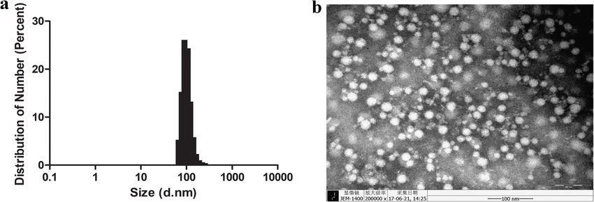
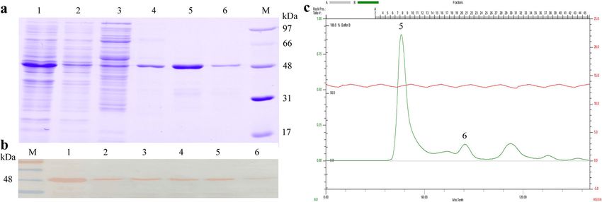
Liu et al. BMC Veterinary Research   (2020) 16:313                                                                Page 2 of 10

Background                                                        protein folding and polymerization [17, 18]. Recombin-
Porcine circovirus (PCV) is a circular single-stranded            ant truncated CRT in polymers, as compared with
DNA virus belonging to the family Circoviridae [1].               monomers, can induce higher level of immune response
There are three major genotypes of PCV, namely PCV1,              [18]. Furthermore, CRT fused foreign proteins also
PCV2, and PCV3. PCV1 is nonpathogenic [2], and PCV2               formed into polymers and showed excellent immunogen-
is associated with several diseases, collectively named as        icity of the foreign proteins [19]. In the present study,
porcine circovirus associated disease (PCVAD), which              high-yield Cap-CRT fusion protein was expressed in
causes reproductive failure and huge economic losses all          E.coli, and the recombinant protein, rP5F could form into
over the world [3]. PCV3 is a recently identified circo-          immunogenic polymers. Mice immunized with rP5F effi-
virus that induces cardiac pathology and multi-systemic           ciently mounted humoral and cellular immune responses,
inflammation [4]. In April 2019, a new circovirus with a          and decrease the infection rate against PCV2-challenge,
distinct relationship to other circoviruses was found in          suggesting that rF5P could be a potential subunit vaccine
Hunan Province, China and designated as PCV4 (doi:                candidate.
https://doi.org/10.1111/TBED.13446). At present, at
least five commercial vaccines have been licensed, in-            Result
cluding the Circovac® vaccine (Merial), Ingelvac Circo-           Expression of cap-CRT fusion proteins and purification of
FLEX® (Boehringer Ingelheim), Circumvent® (Intervet/              rF5P
Merck), Porcilis® PCV (Schering-Plough/Merck), as well            The Cap-CRT fusion proteins (rP4C, rC4P, rP5F and
as Fostera™ PCV (Pfizer Animal Health Inc.) [5]. It has           rF5P) were successfully expressed in E. coli, whereas all
been reported that all the commercial vaccines were able          of them led to inclusion bodies (IBs) at 37 °C (Fig. 1b)
to reduce clinical symptoms and improve reproduction              (Supplementary Material Original Fig. 1b). SDS-PAGE
to some extent in PCV2 positive farms, while they failed          indicated that only rF5P achieving soluble expression
to eradicate this virus from farms [6, 7]. As PCV2 infec-         with a molecular mass of 48 kDa at low temperature
tion may initiate immunosuppression and also cause                (25 °C) for 16 h (Fig. 2a lane 1). After rF5P was purified
subsequent failure of the immune response in pigs [8],            by Ni-NTA affinity chromatography, its quality was
thus, a more effective vaccine should be developed to             nearly 0.5 mg/mL with a purity of about 90% (Fig. 2a
prevent PCV2 infections in swine herds. The BALB/c                lane 4) (Supplementary Material Original Fig. 2a).
mouse is one of the animal models, as it has a clear                The purified rF5P by Ni-NTA was eluted from the
background and frees from external interference, it is            Superdex 200 pg (26/60) gel filtration column. The target
the most extensively used in PCV2 inactivated or sub-             protein was presented as the first and highest peak, which
unit vaccine researches [9, 10].                                  beyond the detection limit of the column, suggesting that
  The genome of PCV2 consists of two major open read-             rF5P could form high-molecular-weight polymers (Fig. 2c
ing frames (ORFs): ORF1 and ORF2. ORF1 encodes two                lane 5). Besides, enrichment was also detected after elu-
viral replication-associated proteins, Rep and Rep’ [11];         tion from the column as the quality of rF5P was about
ORF2 encodes a capsid protein (Cap), which is the pri-            0.65 mg/mL (Fig. 2a, c lane 5). The results of Western Blot
mary immunogenic protein of PCV2. Cap contains critical           suggested that rF5P reacted specifically with anti-His
epitopes for inducing a protective immune response, so it         mAbs (Fig. 2b) (Supplementary Material Original Fig. 2b).
has been used as the target for vaccine development [12].         The third peak also recognized anti-His mAbs, revealing
The Cap protein has been expressed in multiple protein            that only a small fraction of rF5P might exist in the form
expression systems (e.g., insects, mammalian, yeast, and E.       of monomer (Fig. 2b, c lane 6).
coli cells) [13, 14] in vitro, whereas only baculovirus insect
expression system generates two commercially available            Characterization of rF5P
PCV2 vaccines [6]. However, low yield and high cost still         To examine the morphology of high-molecular-weight
exist for large scale preparation. Compared with insect ex-       polymers, the purified rF5P was analyzed under a TEM.
pression, Escherichia coli (E.coli) is an efficient prokaryotic   The observed result revealed that rF5P was assembled into
expression system as many significant benefits in terms of        a spheroidal particle with a diameter of 30 nm, whereas
low cost, ease-of-use and scale preparation.                      the size distribution of the particles was not exactly the
  Generally, immunogenic protein with high-molecular-             same, as shown in Fig. 3a, suggesting that there might be
weight can induce stronger immune response than low-              some incompletely assembled protein fragments. The DLS
molecular-weight protein [15]. Calreticulin (CRT) is a            result indicated that the average hydrodynamic diameter
highly conserved endoplasmic reticulum luminal Ca2+-              of rF5P was about 100 nm (Fig. 3b). The sizes of rF5P par-
binding protein and found to be involved in cellular pro-         ticles observed using the two methods were not consist-
cesses (e.g., calcium storage and chaperone function)             ent, probably attributed to the hydration radius detected
[16]. Numerous studies primarily focused on its roles in          by DLS was larger than the theoretical or real value.
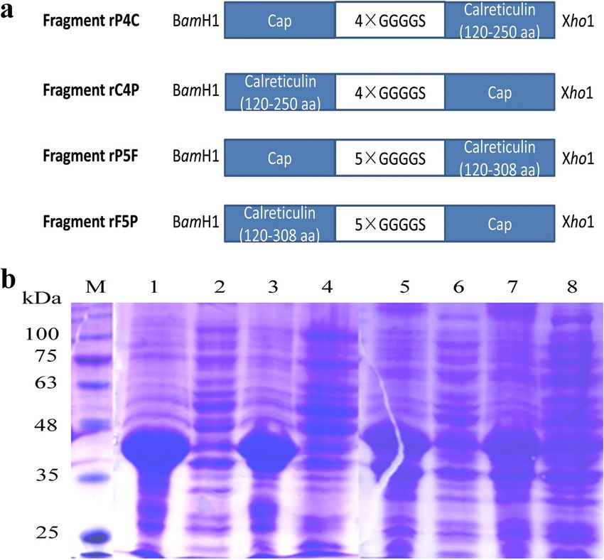
Liu et al. BMC Veterinary Research      (2020) 16:313                                                                                        Page 3 of 10

 Fig. 1 The schematic structure (a) and SDS-PAGE (b) of four recombinant Cap-CRT fusion proteins. a Fragments were used for constructing the
 recombinant proteins. Blue squares represent completely cap of PCV2 and truncated calreticulin. GGGGS in grey are linkers between cap and
 calreticulin. Each fragment is encoded by BamHI and XhoI, respectively. b Solubility of rP4C, rC4P, rP5F and rF5P induced by IPTG at 37 °C. M:
 protein ladder; Lane 1,3,5,7: precipitate of pET-28a-rP4C/rC4P/rV5P/rF5P; Lane 2,4,6,8: supernatant of pET-28a-rP4C/rC4P/rV5P/rF5P

 Fig. 2 Purification and identification of rF5P. SDS-PAGE (a), Western-Blot (b) and Size-exclusion chromatography (c) of rF5P. M: protein ladder;
 Lane 1: lysate of rF5P; Lane 2: supernatant after settling the Ni-NTA resin by gravity; Lane 3: supernatant after washing resin; Lane 4: fraction after
 eluting (purified rF5P); Lane 5: the first peak of flow through by Superdex 200 pg (enriched rF5P); Lane 6: the third peak
Liu et al. BMC Veterinary Research (2020) 16:313 Page 4 of 10
Fig. 3 Characterization of rF5P. a Negative staining electron microscopy of rF5P, bar size, 100 nm. b Dynamic light scattering result of rF5P
The results of the antigenic analysis showed that rF5P significant difference between them. No antibody was
could recognize clinical positive serum and anti-PCV2 produced in the PBS group before the challenge, and the
mAbs 6A4, which indicated that rF5P had the similar antibody level increased immediately at 7 days after chal-
characters as intact particle (Fig. 4). Compared with clin- lenge and reached peak at 14 days.
ical positive serum, the mAbs 6A4 showed a relative Whether the antibodies generated by immunized mice
weaker ability to recognize rF5P (Fig. 4b). However, the could neutralize the virus, NA was adopted to further
rF5P exhibited a high background interference of clinical detect the PCV2-specific humoral immune response.
negative serum, which probably associated with the The results indicated that all immune groups produced
complexity of the field sample (Fig. 4a). neutralizing antibodies except the PBS group, which
were consistent with the results of indirect ELISA. The
NA titers of rF5PH groups were higher compared with
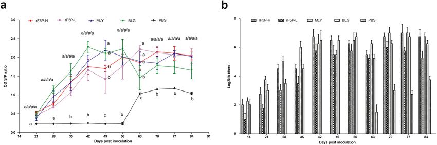
PCV2-specific humoral immune response those of MLY and BLG at 42 and 49 dpi (Fig. 5b). After
Indirect ELISA was performed to evaluate PCV2-specific the challenge, NA titers in the PBS group increased rap-
humoral immune response induced by rF5P in mice. idly and reached 1:16 at 4 weeks. Besides, the NA level
Figure 5a shows that compared with the PBS group, in other immune groups decreased at 63 dpi (1 week
PCV2-specific antibodies appeared at 21 dpi in all after challenge); it returned to the level of pre-challenge
groups and increased with the advancement of the im- at 70 dpi and remained unchanged until the completion
mune process. The antibody levels of MLY and BLG of the test.
groups were overall higher than those of rF5PH and
rF5PL groups before virus challenging, but the contrary
phenomenon happened after that. During the entire im- Lymphocyte proliferative response and cytokine assay
mune process, the levels of rF5PH group were higher Three mice in each group were sacrificed to isolate
than those of the rF5PL group, whereas there was no lymphocyte for lymphocyte proliferation and cytokine
Fig. 4 Antigenic characterizations of rF5P using swine clinical positive sera (a), anti-PCV2 mAbs 6A4 (b) and anti-His mAbs (c) by ELISA, and the
results are expressed as mean OD value ± SEM, the statistical significance differences between each group was analyzed by two-way ANOVA
statistical analysis, *P < 0.05, **P < 0.01, ***P < 0.001, ns represented not significantLiu et al. BMC Veterinary Research (2020) 16:313 Page 5 of 10 Fig. 5 Detection of PCV2-specific immune responses in mice. Groups of mice (n = 6) were immunized with 30 μg and 15 μg of rF5P, commercial inactivated Circovac® vaccine (Merial), subunit vaccine Ingelvac CircoFLEX® (Boehringer Ingelheim) and PBS in injection, Blood samples were collected for PCV2-speicific IgG titers(a) and virus neutralization antibody (b). Titers of antibodies are expressed as mean ± SEM. Different letters (a, b, and c) indicate statistically significant difference (P < 0.05) among groups quantification through PCV2 strain DF-1 stimulation. Quantification of PCV2 in tissues The lymphocyte proliferative responses were detected PCV2 DNA extracted from different tissues of all experi- in all immunized groups aside from the mock group. mental groups post-challenge was quantified using real- The SIs of rF5PH, MLY and BLG groups were signifi- time fluorescent quantitative PCR. Figure 7 suggested cantly higher than that of the PBS group (P < 0.01), that excepted kidneys, the PBS group showed a signifi- and there was no significance between the four im- cantly higher viral load than the other groups. The munized groups (P > 0.05) (Fig. 6f). The results sug- amounts of virus in the spleens and lungs of the immu- gested that cytokine levels were slightly higher in all nized groups were lower than that in the PBS group the immune groups than the mock group, whereas (P < 0.05), and it showed no difference between the im- there was no regular correlations and significant dif- munized groups (Fig. 7c, d). The rF5P groups exhibited ference in the values (Fig. 6a-e). the highest viral loads in the livers (Fig. 7b), but the Fig. 6 Analysis of cytokines secreted by lymphocyte of mice. Lymphocytes isolated from the spleen of mice at 56 dpi were stimulated with PCV2 strain DF-1 for 72 h, the supernatants were collected to detect the concentrations of cytokine of IFN-γ (a), IL-10 (b), IL-18 (c), TNF-ɑ (d), GM-CSF (e) by ELISA method and the T-lymphocyte proliferation (f) . Date are shown as mean ± SEM, statistical differences between each group was measured by one-way ANOVA, *P < 0.05, **P < 0.01, ***P < 0.001, ns represented not significant
Liu et al. BMC Veterinary Research    (2020) 16:313                                                                                  Page 6 of 10

 Fig. 7 Protection from PCV2 strain DF-1 challenge in mice. All of the mice were challenged with 100 μL of 106.5 TCID50/mL of the PCV2 strain
 DF-1 at 56 dpi and examined for 28 days. Spleens were isolated and the genomes were extracted to measure the content of PCV2 using
 quantitative real-time PCR. Date are shown as mean ± SEM, statistical differences between each group was measured by one-way ANOVA, *P <
 0.05, **P < 0.01, ***P < 0.001, ns represented not significant

lowest in the hearts (Fig. 7a). There was no difference                   synthesis and lack of post translational modification.
among all groups in the kidneys (Fig. 7e). All the results                CRT has been shown to be able to self-assemble effect-
revealed that mice immunized with rF5P could effect-                      ively and acts as a chaperone to help dissolve and form
ively reduce viral loads in organs against the PCV2                       the correct structure [21]. Three fourths design of Cap-
challenge.                                                                CRT fusion proteins formed into IBs, only the rF5P
                                                                          transformed into soluble macromolecular particles
Discussion                                                                in vitro by optimizing the expression conditions. How-
PCV2, an agent of PCVDs, acts as a vital economical viral                 ever, the observations of TEM and DLS revealed that the
pathogen affecting the global swine industry. Vaccination                 particle radius was not the same, probably attributed to
has been demonstrated as a feasible means to control                      the DLS of hydrated radius larger than the theoretical or
PCVAD. In this study, the Cap-CRT fusion proteins which                   actual size. Besides, compared with other VLPs reports,
could form into high immunogenic polymers were first                      the rF5P did not form VLPs.
produced in E. coli. Though there are multiple protein ex-                   The BALB/c mouse is one of the animal models, as it
pression systems for protein expression in vitro, each sys-               has a clear background and frees from external interfer-
tem exhibits features and advantages, it also has                         ence, it is the most extensively used in PCV2 inactivated
limitations such as low yield and high cost which hinder                  or subunit vaccine researches [9, 10]. Though mice may
the development of the recombinant protein into a truly                   not be an ideal animal model to resemble PCV2 infection
useful vaccine. Meanwhile, E. coli prokaryotic expression                 as observed for pigs, PCV2 can infect and replicate in
system has been extensively adopted for recombinant pro-                  some mouse strains including BALB/c mouse when used
tein production in laboratories and industry for its simpli-              with the appropriate inoculating dose and administered
city, rapid growth rate and relatively low cost.                          route. In the present study, the BALB/c mouse model was
  Studies on Cap proteins focus on their abilities to self-               used to assess the immunogenicity and protective capabil-
assemble into virus-like particles (VLPs) and thus exert                  ities of an experimental vaccine based on the recombinant
immune effects as an entire virus, which also prove that                  Cap-CRT fusion protein expressed in E.coli.
large molecular particles have stronger immune effects                       The Cap-CRT fusion protein (rF5P) induced production
than monomer proteins [20]. However, the expression of                    of PCV2-specific ELISA antibodies and neutralization
recombinant proteins in E.coli often results in insoluble                 antibody against PCV2 were detected, the PCV2-specific
and/or nonfunctional IBs which may due to rapid                           ELISA antibodies were positively correlated with the
Liu et al. BMC Veterinary Research (2020) 16:313 Page 7 of 10
neutralization antibodies, which was consistent as de- and utilized for virus neutralization assay (NA) and ex-
scribed in Zhu [22]. The specific antibody levels of protein perimental challenge.
groups were lower than those of commercial vaccine
groups before virus challenge, but it went opposite after Experimental animals
the virus challenge. The neutralizing antibody levels of Thirty female BALB/c mice of 4 weeks old weighing 14-18
protein groups were slightly lower than commercial vac- g were chosen arbitrarily and purchased from the Experi-
cine groups during the immune process. Both the protein mental Animal Center of Zhengzhou University. The ex-
and the commercial vaccine groups induced only part of perimental mice were randomly separated into five groups
the cellular immune response. Under the stimulation of and given 5 days to acclimate the housing environmental
PCV2, T lymphocytes proliferated significantly, whereas conditions (temperature: 22 ± 3 °C, humidity: 55 ± 15%,
various cytokines were irregularly secreted. PCV2 infec- lighting: 12 h light/dark cycle). The mice were allowed free
tions mainly induce fetal and neonatal mortality, and the access to clean water and food. The animal experiments
level of viruses in the tissues of PCV2 infected mice is a were carried out according to the Animal Experiment
good indicator of the antiviral effects of any vaccine, and it Committee of Henan Academy of Agricultural Sciences
primarily occurred in the lymphoid tissue and spleen [23]. (Approval number SYXK 2014–0007). All animals received
After the challenge test, the viral loads in the spleens of humane care in compliance with good animal practice ac-
mice in the protein and commercial inactivated vaccine cording to the animal ethics procedures and guidelines of
group were significantly lower than those in the mock China. All sections of this report adhere to the ARRIVE
group. It was also slightly effective in the organs of the Guidelines for reporting animal research [24].
heart, liver and lung, which was not completely consistent
with Wang’s research, PCV2 mainly deposited in the lungs
[9]. The humoral immune response showed no significant Plasmid construction
difference between protein groups and commercial vac- As shown in Fig. 1a, complete Cap gene of PCV2 (Gen-
cine groups. Overall, the humoral and cellular immune Bank Accession No. AY686763) was fused with the trun-
levels of rF5P groups were similar to the two types of cated calreticulin (120–308 aa/120–250 aa) (GenBank
commercial vaccine groups, and the aggregate perform- Accession No. EU639407) at N/C terminal using 4 ×
ance of rF5P was closer to BLG subunit vaccine. Our re- GGGGS or 5 × GGGGS linker. All these four recombin-
sults clearly verified that the Cap-CRT fusion protein ant fragments, named rP4C/rC4P/rP5F/rF5P, were syn-
(rF5P) elicited humoral and part of cell mediated immune thesized after codon optimization by Genscript. All the
responses comparable to commercial inactivated and sub- plasmids were inserted into pET-28a in BamHI and
unit vaccines, and protected mice against epidemic PCV2 XhoI sites and then transformed into E. coli BL21 (DE3)
strain DF-1 challenge. competent cells, respectively.
Conclusions Protein expression and purification
To sum up, this paper first describes that the PCV2 Cap All the positive clones were cultured in Luria-Bertani
protein fused with truncated calreticulin (rF5P) could be (LB) medium containing 50 mg/L kanamycin and in-
soluble expressed into immunogenically polymers in E. duced for protein expression with 0.1 mM IPTG at 37 °C
coli. Vaccination of mice elicited humoral and part of for 6 h. The parameters of protein expression were opti-
cellular immune responses comparable to the commer- mized according to IPTG concentrations (0.1 mM, 0.2
cial inactivated and subunit vaccines, and significantly mM), induction temperature and time (18 °C for 24 h,
reduced the viral loads in tissues subsequent to a viral 25 °C for 16 h). Protein expression was verified by so-
challenge. Besides, the immune effect of Cap-CRT fusion dium dodecyl sulfate polyacrylamide gel electrophoresis
protein requires further verifications in pigs as the nat- (SDS-PAGE). The optimal harvest cells were suspended
ural hosts of PCV2. The rF5P can potentially develop a in lysis buffer (50 mM PB, 150 mM NaCl, 5% (w/v) Gly-
subunit vaccine against PCV2 infection. cerol, 5% (w/v) Triton X-100, 2 mM EDTA, 2 mM DTT,
pH 7.0) and then lysed by sonication (99 cycles of 2 s
Methods On/5 s Off, amp 25%)). After centrifugation, the precipi-
Cells and virus tation was removed and the supernatant of rF5P was
PK-15 cells (ATCC™ CCL-33) were cultured in Dulbecco’s purified by Ni-NTA affinity chromatography. After
Modified Eagle Medium (DMEM; Gibco) containing 10% washing the Ni-NTA column (Invitrogen, USA) with
fetal bovine serum (FBS, HyClone), 100 IU/mL Penicillin wash buffer (50 mM PB, 150 mM NaCl, 30 mM imid-
and 100 mg/L Streptomycin (InvivoGen, France) at 37 °C azole, pH 7.0), rF5P was eluted with elution buffer (50
in a 5% CO2 atmosphere. PCV2 strain DF-1 (GenBank mM PB, 150 mM NaCl, 250 mM imidazole, pH 7.0). Pro-
Accession Number: JN119255) was grown in PK-15 cells tein fractions were analyzed by SDS-PAGE.Liu et al. BMC Veterinary Research (2020) 16:313 Page 8 of 10
The purified rF5P was enriched and analyzed by size- Neutralization assay
exclusion chromatography with Superdex 200 prep The abilities of all serum samples to neutralize the PCV2 strain
grade (pg) (26/60) gel filtration column (GE Healthcare, DF-1 were assessed using virus NA. In brief, 50 μL sera pre-
USA). The samples were eluted using lysis buffer at a treated at 56 °C for 30 min were diluted in a serial two-fold way
flow rate of 1 mL/min and detected at 280 nm wave- from 1:2 to 1:1024 and mixed with an equal volume of virus
length. The collected fractions were identified by SDS- (400 TCID50) at 37 °C for 1 h. The serum-virus complex was
PAGE and Western Blot and then quantified using BCA transferred into confluent PK-15 cells in each well and then in-
Protein Assay Kit (TIANGEN, China). cubated at 37 °C for 72 h. Since no visible cytopathic effect was
verified, immunoperoxidase monolayer assay (IPMA) was per-
Characterization of rF5P formed to ascertain the presence of the virus [10]. Virus
The purified rF5P was observed under a transmission neutralization titer was expressed as the highest dilution as
electron microscopy (TEM) using the negative staining log2NA in which no higher than 80% reduction of virus replica-
method and dynamic light scattering (DLS) according to tion was detected as compared with the virus control.
the previous study [25].
Spleen lymphocyte proliferation assay
Antigenicity analysis of rF5P Spleens of mice from each group were removed at 56 days
Indirect enzyme-linked immunosorbent assay (ELISA) post inoculation (dpi). The spleen lymphocytes were iso-
was performed to test the antigenicity of rF5P with lated by Lydroxypropylmethyl Cellulose (Solarbio, China)
swine clinical positive/negative serum and mouse anti- and then resuspended in RPMI 1640 medium containing
PCV2 monoclonal antibodies (mAbs) 6A4 (Abcam, 10% FBS. Lymphocyte proliferation assay was performed by
USA). The ELISA procedure was operated as routine. cell counting kit-8 assay (Beyotime Biotechnology, China)
as previously described [26]. T lymphocyte proliferation
Vaccination and challenge in mice was represented as the stimulation index (SI), the ratio of
Thirty female BALB/c mice were randomly divided into the mean reading of stimulated wells to unstimulated ones.
5 groups (n = 6). The mice were inoculated subcutane-
ously with 30 μg and 15 μg of rF5P as Group rF5PH and Analysis of cytokine production by activated lymphocytes
Group rF5PL, respectively; 50 μL of commercial inacti- The supernatants from the spleen lymphocytes
vated Circovac® vaccine (Merial), subunit vaccine Ingel- employed in the proliferation assay were removed and
vac CircoFLEX® (Boehringer Ingelheim) and PBS were adopted to analyze cytokines. The assays were per-
classified as positive and negative controls, named as formed using commercially available mice IFN-γ, IL-10,
Group MLY, BLG and PBS, respectively. IL-18, TNF-ɑ and GM-CSF ELISA kits (USCN Life Sci-
The rF5P was diluted in 50 μL of PBS and then emulsi- ence, China) following the manufacturer’s instructions.
fied with 50 μL of Complete Freund’s adjuvant for the first
immunization, and subsequently with 50 μL of Incomplete Determination of PCV2 in tissue
Freund’s adjuvant for booster at an interval of 4 weeks. At PCV2 DNA from different organs (heart, liver, spleen,
56 days after the first immunization, 3 mice from each lung and kidney) of all groups at 28 days post-challenge
group were sacrificed for both lymphocyte proliferation was quantified by real-time fluorescent quantitative PCR
assay and cytokine production. In order to reduce the pain as previously described [27]. The viral load was calcu-
of mice to the greatest extent, cervical dislocation was lated according to the standard curve plotting Ct values
chosen to kill them. It is the fastest method to make the against different dilutions of a standard plasmid.
spinal cord and brain spinal cord disconnected, so that the
central nervous system instantly lost control of the whole Statistical analyses
body, which is in line with the requirements of animal GraphPad Prism version 5.00 (USA) analysis of variance
welfare. The rest alive mice received 100 μL of 106.5 (ANOVA) was performed. The data is expressed as the
(TCID50)/mL PCV2 strain DF-1, and they were monitored mean ± SEM. Statistical significance was found by two-way
for the following 28 days. Next, the mice were sacrificed or one-way ANOVA at*P < 0.05, **P < 0.01, ***P < 0.001; ns
for PCV2 content in different organs. Blood samples were represents no statistical significance. All the experimenters
collected from the tail veins each week. were not blinded to any stage of the experiment.
Antibody response in mice Supplementary information
The serum samples taken at each point post immunization Supplementary information accompanies this paper at https://doi.org/10.
were monitored for specific antibodies using Porcine circo- 1186/s12917-020-02527-9.
virus type 2 ELISA antibody test kit (KeQian, China). Oper-
Additional file 1.
ation steps followed the manufacturer’s instructions.Liu et al. BMC Veterinary Research (2020) 16:313 Page 9 of 10
Additional file 2. 3. Opriessnig T, Meng XJ, Halbur PG. Porcine circovirus type 2 associated
disease: update on current terminology, clinical manifestations,
Additional file 3. pathogenesis, diagnosis, and intervention strategies. J Vet Diagn Investig.
2007;19(6):591–615.
Abbreviations 4. Phan TG, Giannitti F, Rossow S, Marthaler D, Knutson TP, Li L, Deng X,
PCV2: Porcine circovirus type 2; PCVDs: Porcine circovirus diseases; PCVA Resende T, Vannucci F, Delwart E. Detection of a novel circovirus PCV3 in
D: Porcine circovirus associated diseases; Cap: Capsid protein; ORFs: Major pigs with cardiac and multi-systemic inflammation. Virol J. 2016;13(1):184.
open reading frames; CRT: Calreticulin; E.coli: Escherichia coli; IBs: Inclusion 5. Chae C. Commercial porcine circovirus type 2 vaccines: efficacy and clinical
bodies; SDS-PAGE: Sodium dodecyl sulfate-polyacrylamide gel electrophor- application. Vet J. 2012;194(2):151–7.
esis; ELISA: Indirect enzyme-linked immunosorbent assay; DLS: Dynamic light 6. Afghah Z, Webb B, Meng XJ, Ramamoorthy S. Ten years of PCV2 vaccines
scattering; TEM: Transmission electron microscopy; MAbs: Monoclonal and vaccination: is eradication a possibility? Vet Microbiol. 2017;206:21–8.
antibodies; IPMA: Immunoperoxidase monolayer assay; NA: Neutralization 7. Feng H, Blanco G, Segalés J, Sibila M. Can porcine circovirus type 2 (PCV2)
assay; Dpi: Days post inoculation; SI: Stimulation index; RT-PCR: Real-time infection be eradicated by mass vaccination? Vet Microbiol. 2014.
PCR; IL: Interleukin; TNF: Tumor necrosis factor; IFN: Interferon; GM- 8. Meng XJ. Porcine circovirus type 2 (PCV2): pathogenesis and interaction
CSF: Granulocyte-macrophage colony stimulating factor with the immune system. Annu Rev Anim Biosci. 2013;1:43–64.
9. Wang YP, Liu D, Guo LJ, Tang QH, Wei YW, Wu HL, Liu JB, Li SB, Huang LP,
Acknowledgments Liu CM. Enhanced protective immune response to PCV2 subunit vaccine by
We thank Dr. Yinbiao Wang at Xinxiang Medical College for English writing co-administration of recombinant porcine IFN-gamma in mice. Vaccine.
help. We thank Dr. Shujun Chai at Henan Academy of Agricultural Sciences 2013;31(5):833–8.
for providing high-level quality of animal care. We thank Dr. Hongying Chen, 10. Liu C, Liu Y, Chen H, Feng H, Chen Y, Wang Y, Wang J, Liu D, Deng R,
and Mr. Guanpeng Guo for their assistance in obtaining the PCV2 isolates Zhang G. Genetic and immunogenicity analysis of porcine circovirus type 2
DF-1 from the Henan Agricultural University. strains isolated in Central China. Arch Virol. 2018;163(4):937–46.
11. Cheung AK. Identification of the essential and non-essential transcription
Authors’ contributions units for protein synthesis, DNA replication and infectious virus production
CL, YL, HF, RD and GZ designed the study, participated in all tests and of porcine circovirus type 1. Arch Virol. 2004;149(5):975–88.
drafted the manuscript. CL, BZ, PW and YC participated in collecting and 12. Nawagitgul P, Morozov I, Bolin SR, Harms PA, Sorden SD, Paul PS. Open
testing samples. CL, YL, HF, HH and GZ analyzed the data and revised the reading frame 2 of porcine circovirus type 2 encodes a major capsid
manuscript. All authors reviewed the results and approved the final version protein. J Gen Virol. 2000;81(Pt 9):2281–7.
of the manuscript. 13. Lin HX, Ma Z, Fan HJ, Lu CP. Construction and immunogenicity of
recombinant swinepox virus expressing capsid protein of PCV2. Vaccine.
Funding 2012;30(44):6307–13.
We are grateful to the financial support from three fund projects. National 14. Li PC, Qiao XW, Zheng QS, Hou JB. Immunogenicity and immunoprotection
Key Research and Development Program of China (2017YFD0501103) of porcine circovirus type 2 (PCV2) cap protein displayed by Lactococcus
supported the design of study and data collection and writing the lactis. Vaccine. 2016;34(5):696–702.
manuscript; Key Scientific and Technological Research Projects in Henan 15. Xu F, Wang Y, Tao T, Song D, Liu X. Calreticulin attenuated microwave
Province (182102110087) and Scientific and Technological Projects for radiation-induced human microvascular endothelial cell injury through
Overseas Students (22991803) supported analysis, interpretation of data. promoting actin acetylation and polymerization. Cell Stress Chaperones.
2017;22(1):87–97.
Availability of data and materials 16. Michalak M, Corbett EF, Mesaeli N, Nakamura K, Opas M. Calreticulin: one
The datasets used and/or analyzed during the current study are available protein, one gene, many functions. Biochem J. 1999;344(Pt 2):281–92.
from the corresponding author on reasonable request. 17. Hong C, Qiu X, Li Y, Huang Q, Zhong Z, Zhang Y, Liu X, Sun L, Lv P, Gao
XM. Functional analysis of recombinant calreticulin fragment 39-272:
Ethics approval and consent to participate implications for immunobiological activities of calreticulin in health and
The animal experiments were carried out and approved by the Animal disease. J Immunol. 2010;185(8):4561–9.
Experiment Committee of Henan Academy of Agricultural Sciences 18. Huang SH, Zhao LX, Hong C, Duo CC, Guo BN, Zhang LJ, Gong Z, Xiong SD,
(Approval number SYXK 2014–0007). All animals received humane care in Gong FY, Gao XM. Self-oligomerization is essential for enhanced
compliance with good animal practice according to the animal ethics immunological activities of soluble recombinant calreticulin. PLoS One.
procedures and guidelines of China. 2013;8(6):e64951.
19. Qiu X, Hong C, Li Y, Bao W, Gao XM. Calreticulin as a hydrophilic chimeric
Consent for publication molecular adjuvant enhances IgG responses to the spike protein of severe
Not applicable. acute respiratory syndrome coronavirus. Microbiol Immunol. 2012;56(8):554–61.
20. Xi X, Mo X, Xiao Y, Yin B, Lv C, Wang Y, Sun Z, Yang Q, Yao Y, Xuan Y, et al.
Competing interests Production of Escherichia coli-based virus-like particle vaccine against
The authors declare that they have no competing interests. porcine circovirus type 2 challenge in piglets: structure characterization and
protective efficacy validation. J Biotechnol. 2016;223:8–12.
Author details 21. Shiraishi N, Inai Y, Hirano Y, Ihara Y. Calreticulin inhibits prion protein PrP-
1
Key Laboratory of Animal Immunology of the Ministry of Agriculture, Henan (23-98) aggregation in vitro. Biosci Biotechnol Biochem. 2011;75(8):1625–7.
Provincial Key Laboratory of Animal Immunology, Henan Academy of 22. Zhu X, Liu J, Bai J, Liu P, Zhang T, Jiang P, Wang X. Baculovirus expression of the N-
Agricultural Sciences, Zhengzhou 450002, Henan, China. 2College of Animal terminus of porcine heat shock protein Gp96 improves the immunogenicity of
Science and Veterinary Medicine, Henan Agricultural University, Zhengzhou recombinant PCV2 capsid protein. J Virol Methods. 2016;230:36–44.
450002, Henan, China. 3School of Life Sciences, Zhengzhou University, 23. Becskei Z, Aleksic-Kovacevic S, Rusvai M, Balka G, Jakab C, Petrovic T,
Zhengzhou 450001, China. Knezevic M. Distribution of porcine circovirus 2 cap antigen in the lymphoid
tissue of pigs affected by postweaning multisystemic wasting syndrome.
Received: 5 August 2019 Accepted: 18 August 2020 Acta Vet Hung. 2010;58(4):483–98.
24. Kilkenny C, Browne WJ, Cuthill IC, Emerson M, Altman DG. Improving
bioscience research reporting: the ARRIVE guidelines for reporting animal
References research. J Pharmacol Pharmacother. 2010;1(2):94–9.
1. Allan GM, Ellis JA. Porcine circoviruses: a review. J Vet Diagn Investig. 2000; 25. Ding P, Zhang T, Li Y, Teng M, Sun Y, Liu X, Chai S, Zhou E, Jin Q, Zhang G.
12(1):3–14. Nanoparticle orientationally displayed antigen epitopes improve
2. Tischer I, Mields W, Wolff D, Vagt M, Griem W. Studies on epidemiology and neutralizing antibody level in a model of porcine circovirus type 2. Int J
pathogenicity of porcine circovirus. Arch Virol. 1986;91(3–4):271–6. Nanomedicine. 2017;12:5239–54.Liu et al. BMC Veterinary Research (2020) 16:313 Page 10 of 10
26. Ji P, Liu Y, Chen Y, Wang A, Jiang D, Zhao B, Wang J, Chai S, Zhou E, Zhang
G. Porcine parvovirus capsid protein expressed in Escherichia coli self-
assembles into virus-like particles with high immunogenicity in mice and
Guinea pigs. Antivir Res. 2017;139:146–52.
27. Li J, Shi JL, Wu XY, Cong XY, Xu SJ, Yuan XY, Wu JQ, Sun WB, Du YJ, Peng Z,
et al. Differentiation of PCV1 and PCV2 by a multiplex real-time PCR assay.
Vet Rec. 2013;173(14):346.
Publisher’s Note
Springer Nature remains neutral with regard to jurisdictional claims in
published maps and institutional affiliations.You can also read