REVIEW ARTICLE The spectrum of pathological findings of tonsils in children: A clinicopathological review
←
→
Page content transcription
If your browser does not render page correctly, please read the page content below
Malaysian J Pathol 2018; 40(1) : 11 – 26

REVIEW ARTICLE
The spectrum of pathological findings of tonsils in children:
A clinicopathological review
Geok Chin TAN*,** MBBS, PhD, Melissa STALLING* DO, Sura AL-RAWABDEH* MD, Basil M Kahwash***
MD, Razan F ALKHOURY**** MD and Samir B KAHWASH* MD

*Department of Pathology and Laboratory Medicine, Nationwide Children’s Hospital and the Ohio
State University, Columbus OH 43205, USA, **Department of Pathology, National University of
Malaysia, 56000 Kuala Lumpur, Malaysia, ***Department of Internal Medicine, Indiana University,
Indiana, USA and ****Department of Pediatrics, the Ohio State University and Nationwide Children’s
Hospital, Columbus OH 43205, USA

Abstract

Tonsillectomy is among the most commonly performed operations in children. Although follicular
lymphoid hyperplasia is usually the main and only pathologic finding at microscopic examination,
a variety of other rare but important pathologic changes may be encountered. This review aims to
provide an inclusive practical resource and reference for both training and practising pathologists.
It discusses the spectrum of pathologic findings, including both neoplastic and non-neoplastic
conditions and provides illustrative images.

Keywords: Tonsil, tonsillectomy, children, pathology

INTRODUCTION                                                              deep crypts that penetrate each tonsil (Fig. 1A).
                                                                          Tonsillar tissue also includes minor mucous
In an ideal system, removed tonsils – as all                              salivary glands along with a rich network of
surgically removed specimens - would benefit                              blood and lymphatic vessels (Fig. 1 B & C).
from pathologic examination. However,                                     The lingual and pharyngeal tonsils consist of
institutions are increasingly limiting or                                 loose submucosal collections of similar lymphoid
eliminating routine microscopic examination                               tissue; however, no crypts are present. The
of tonsils as a cost saving measure. Aside from                           overlying epithelium of the pharyngeal tonsils
losing the opportunity to document findings that                          may be of respiratory or non-keratinising
may be unexpected, other downsides of this                                squamous epithelium type. The lingual tonsils
trend include depriving practicing pathologists                           are covered by non-keratinising squamous
as well as trainees in the field of pathology of                          epithelium.
a valuable experience and diagnostic skill in                                 In the palatine tonsils, the squamous
tonsillar pathology. In this article, we discuss the                      epithelium becomes thinner and loosely textured
spectrum of common and uncommon pathologic                                in crypts.1 Ultra-structural studies have shown
findings of tonsils with illustrative images.                             small holes in the epithelium of crypts,2 perhaps
                                                                          to facilitate maximal infiltration of lymphocytes
1. Anatomy and Histology of Tonsils                                       in the context of an inflammatory reaction
The upper respiratory tract contains a significant                        (Fig. 1D). The tonsillar lymphoid follicles
amount of lymphoid tissue, most of which is                               are similar to those in lymph nodes, with the
arranged in the Waldeyer’s ring. The latter                               observation that marginal zones are usually
consists of the palatine tonsils, lingual tonsils                         obscured except in florid follicular hyperplasia.
and pharyngeal tonsils (also referred to as the                           The lymphoid cells infiltrating the epithelium of
adenoids). The normal palatine tonsils consist                            the crypts appear to be of marginal zone B-cell
of lymphoid tissue arranged in follicles, covered                         type, while most of those in surface epithelium
by squamous epithelium which extends to line                              are of T-cell type.1

Address for correspondence: Samir B. Kahwash, MD; Department of Pathology and Laboratory Medicine, Nationwide Children’s Hospital and the Ohio
State University, Columbus OH 43205, USA. Tel: 614-722-5427; Fax: 614-722-2899. Email: samir.kahwash@nationwidechildrens.org

                                                                                                                                         11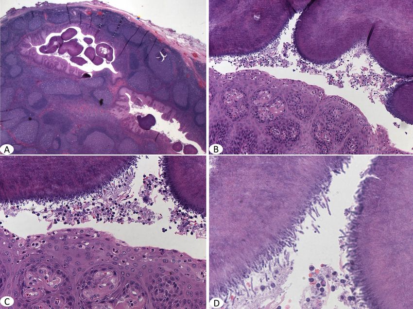
Malaysian J Pathol April 2018
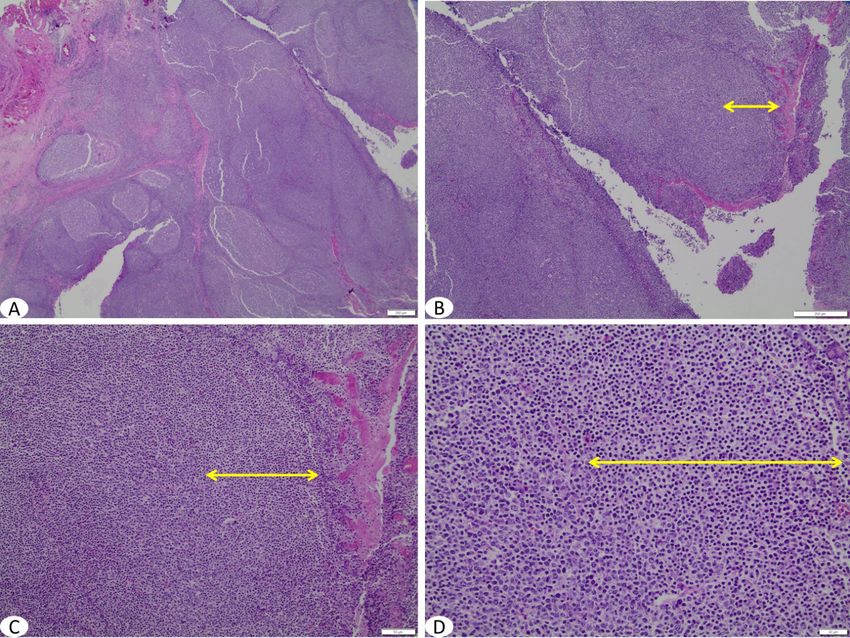
FIG. 1: Normal palatine tonsil (A), demonstrating lymphoid follicles, blood vessels, lymphatic vessels & minor
salivary glands (B&C) and superficial inflammation in tonsillar crypt with neutrophils surrounding a
droplet of bacteria containing saliva (D).
2. Clinical and Surgical Aspects 5- Peritonsillar abscess in children with other
indications for adenotonsillectomy.
Tonsillectomy is among the most commonly
6- Halitosis.
performed operations in children, with more than
7- Chronic tonsillitis unresponsive to
half a million adenotonsillectomy procedures
antimicrobials.
performed annually in the United States.3 The two
8- Tumour or haemorrhage of tonsils.
most common indications are throat infections
9- Paediatric autoimmune neuropsychiatric
and sleep-related breathing disorders. 3-5
disorder associated with streptococci.
The absolute indications for tonsillectomy
10- Chronic group-A streptococcus carrier status.
include obstruction of the nasopharyngeal
or oropharyngeal airways, interference with In the United States, the number of tonsillectomies
swallowing, clinical suspicion of malignant has declined progressively since the 1970s.6,7
tumour of the tonsils, and uncontrollable Reports suggested that the decline was mainly
haemorrhage from tonsillar blood vessels.6 The in tonsillectomies performed for infectious
American Academy of Otolaryngology-Head and indications, while the number of tonsillectomies
Neck Surgery clinical practice guidelines list the performed for obstructive indications may
following indications for adenotonsillectomy in have actually increased.6,8,9 Most of these
children.3,4 operations are performed as ambulatory, same-
day procedures.6,7 Tonsillectomy is performed
1- Sleep disordered breathing.
infrequently in children younger than 3 years
2- Recurrent throat infections.
of age. Contraindications for tonsillectomy
3- Dysphagia or voice quality changes related
include 3 general categories: velopharyngeal
to enlarged tonsils.
insufficiency, haematologic disorders (anaemia
4- Periodic fever, aphthous stomatitis,
and disorders of haemostasis), and active local
pharyngitis, and cervical adenitis.
infection.6
12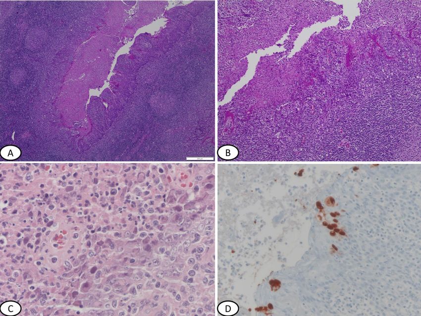
PATHOLOGY OF TONSIL IN CHILDREN

3. Pathologic Examination                              routine microscopic examination. This trend is
                                                       driven mainly by cost rationing.
Examination protocols
The approach to pathologic examination varies
                                                       4. Common Pathologic Findings
from one health care system to another, and may
be different from one hospital to the other in the     4.1 Follicular lymphoid hyperplasia (FLH)
same system. Many hospitals perform histologic         Similar to pathologic findings in enlarged
examination routinely on all specimens. Some           paediatric lymph nodes, the most common
use age as a criterion to identify the need for        finding in removed tonsils is reactive follicular
histologic study. There are published studies that     hyperplasia. Areas of active, mixed and chronic
attempted to include risk factors for malignancy       inflammation may be seen.
to assist in making decisions about whether to
perform histologic evaluation,10 while some            4.2 Active inflammation and peritonsillar
suggested clinical suspicion and specimen              abscesses
asymmetry as required criteria for performing          Areas of active and chronic inflammation are
a thorough histological examination.11                 common and there may be areas of superficial
                                                       ulceration. Peritonsillar abscesses, while rare
Advantages of routine microscopic examination          and usually unilateral, are the most common
In an ideal system, all surgically removed             complication of acute tonsillitis. In these cases,
specimens would benefit from pathologic                the acute infection of the tonsillar crypts may
examination. The advantages for routine                extend beyond the tonsillar capsule to involve
pathologic examination include detection of            the peritonsillar space, between the tonsil and
incidental findings and providing tissue material      the superior pharyngeal constrictor muscle
for research, teaching, and other academic             (Fig. 2). If epithelial inflammation is extensive
activities. Increasingly, institutions are limiting,   and ulcerations of squamous epithelium are
or eliminating, certain specimens (including           prominent, the possibility of viral infections
tonsils) from the list of those that qualify for       such as Epstein–Barr virus (EBV) or adenovirus

FIG. 2: An abscess deep at the base of a tonsil (A&B; scale bar = 200µm) and higher magnification showing
        neutrophilic infiltrates separating muscle fibers (C&D; scale bar- C = 50µm).

                                                                                                     13
Malaysian J Pathol                                                                                April 2018

should be kept in mind.                                  trauma.14 More common are simple keratin filled
                                                         cysts which vary in size from a few millimeters
4.3 Actinomyces                                          to more than 2 cm (Fig. 4).
Actinomyces are gram positive, anaerobic,
commensal bacteria, found in oral cavity,                5. Uncommon Pathologic Findings
colon and vagina in humans. The incidence of
                                                         5.1 Non-neoplastic
Actinomyces of the palatine tonsil can be as
                                                         5.1.1 Viral infections
high as 40%, as reported in a recent study.12 It
                                                         Bacterial and viral infections can cause tonsillitis.
has been suggested that actinomycosis infection
                                                         Streptococcal strains are the most common
of the tonsil may indicate an aetiologic role
                                                         bacteria, while common viral causes of tonsillitis
for this organism in tonsillar and adenoidal
                                                         include adenoviruses, Epstein-Barr virus, human
hypertrophy.13 On H&E section, the presence of
                                                         papilloma virus, influenza and parainfluenza
actinomycosis can be recognized as aggregates of
                                                         viruses, and others such as enteroviruses, herpes
filamentous basophilic microorganisms arranged
                                                         simplex viruses. Microscopic examination of
in a radial spoke-like fashion, the so called
                                                         tonsils in most viral and bacterial infections
“ray-fungus” appearance of an Actinomyces
                                                         shows hypertrophied and hyperplastic lymphoid
colony (Fig. 3).
                                                         follicles with excessive development of the
                                                         germinative clear center as a reaction to the
4.4 Keratin filled cysts
                                                         presence of antigens.
Squamous lined cysts are commonly found in the
head and neck region. Dermoid and epidermoid
                                                         Certain viruses show specific histologic findings
cysts can rarely occur in the palatine tonsil. They
                                                         as follows:
arise from the epithelium that has been trapped
                                                         Adenovirus
in deeper tissue during embryonic period or from
                                                         Viral intranuclear and intracytoplasmic
abnormal inclusion of cells during surgery or
                                                         inclusions with positive immunohistochemical

FIG. 3: Aggregates of Actinomyces bacteria in tonsillar pits (A&B) and higher magnification shows the filamen-
        tous bacteria arranged in radial spoke-like fashion (C&D).

14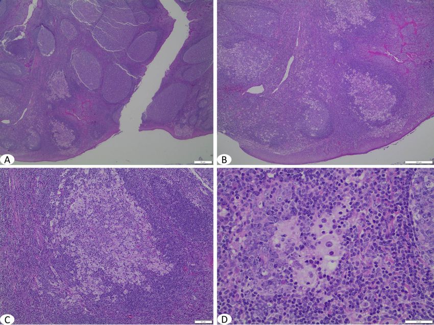
PATHOLOGY OF TONSIL IN CHILDREN

                                                         EBV
                                                         Palatine tonsils of children can be colonised
                                                         by EBV and this virus may be involved in the
                                                         pathogenesis of recurrent tonsillitis and tonsillar
                                                         hypertrophy. The microscopic appearence
                                                         of tonsils infected with EBV is similar to
                                                         histologic findings of lymph nodes in infectious
                                                         mononucleosis, which range from non-specific
                                                         follicular hyperplasia to proliferation of large
                                                         immunoblastic cells of varying severity. Plasma
                                                         cells and plasmacytoid cells are also admixed
                                                         with immunoblasts and lymphocytes, giving
                                                         a polymorphous appearance. Sometimes the
FIG. 4: A keratin cyst lined by squamous epithelium      immunoblasts form clusters, or even sheets,
        and filled with keratin material                 partially effacing the lymph node, which may
                                                         be confused with lymphoma (Fig. 6).
staining specific for adenovirus are noted. The
intranuclear inclusions during late infection are        HPV
surrounded by a clear halo, which may obstruct           There is little data about tonsillar HPV infection
visualisation of the nuclear membrane, resulting         in the literature. In a study, the rate of HPV
in a smudged appearance. These “smudged”                 infection in non-neoplastic tonsils was 8.5% and
cells are classically seen in adenovirus infection.      in neoplastic tonsils was 51%.16 HPV infection
Adenovirus may persist in tonsils and adenoid            of the tonsils can show similar viral cytopathic
as a latent infection15 (Fig. 5).                        effects seen in HPV infection of other body

FIG. 5: Active inflammation and epithelial hyperplasia of tonsilar pit due to adenovirus infection (A&B) and
        higher magnification shows the characteristic glassy nuclear inclusions in H&E (C) and that was stained
        with adenovirus immunoperoxidase stain (D). Scale bar = 200µm.

                                                                                                           15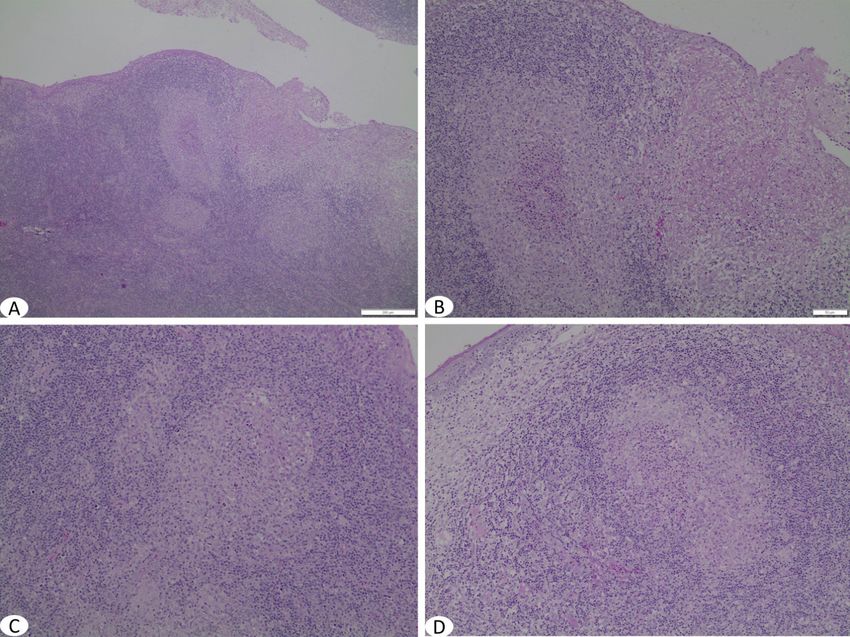
Malaysian J Pathol                                                                                April 2018

FIG. 6: Tonsil shows inflammation, superficial ulceration and expanded interfollicular T-cell zone typical of
        EBV infection (A&B; scale bar = 200µm) and at higher magnification there is mixed inflammation with
        atypical lymphocytes (C; scale bar = 20µm). Reactive atypical lymphoid cells may show binucleation or
        multinucleation with prominent nuclei that may be misconstrued as Reed-Sternberg cells (D; scale bar =
        20µm) (yellow arrow).

sites (skin and cervix) in the form of extensive         performed in the case of any tonsillar granuloma
papillomatosis, hyperkeratosis, koilocytosis and         to exclude this possibility.
prominent keratohayline globules (Fig. 7).
                                                         Cat-scratch disease
5.1.2 Granulomatous inflammation                         Cat-scratch disease (CSD) is a zoonotic infection
Granulomatous inflammation of the tonsils                caused by Bartonella henselae occurring in about
and adenoids is uncommon.17 The underlying               24000 patients annually in the United States.
disorders may be local or systemic. Tuberculosis,        It typically presents with fever and regional
sarcoidosis, Crohn’s disease, fungal infection           lymphadenopathy and is considered as one of the
and histoplasmosis have been reported to cause           most common causes of chronic lymphadenitis in
granulomas in the tonsils.18-21 Granulomatous            paediatric population. It generally has a benign
inflammation of the tonsil may occur secondary           and self-limited course, though this is not always
to foreign body material (like food particles) or        the case. Rare forms of extra-nodal involvement
ruptured inclusion cyst, and sometimes represents        may occur such as involvement of the tonsils,
an exaggerated immune response to chronic                the initial clinical features were indistinguishable
tonsillitis. However, a careful work-up must be          from those of acute bacterial tonsillitis with
done to exclude any underlying systemic causes.          jugulodigastric lymphadenopathy.22
                                                            Histologic examination may show focal areas
Tuberculosis                                             of necrotising granulomatous inflammation,
Primary tonsillar tuberculosis is rare and               with extensive neutrophilic infiltrates seen in
seen mostly in areas with high incidence of              center of well developed granulomas. Early non-
tuberculosis. Ziehl-Neelsen stain should be              necrotising granulomas and multinucleated giant

16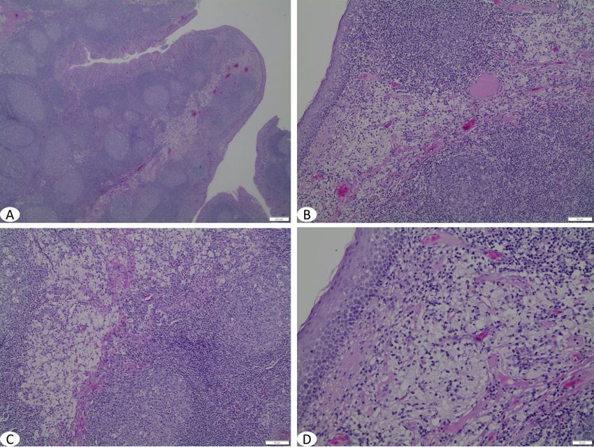
PATHOLOGY OF TONSIL IN CHILDREN

FIG. 7: Papillomatosis and hyperkeratosis of HPV infection of a tonsil (A, B and C). Higher magnification shows
        the characteristic koilocytosis of the squamous epithelium (D).

cells may also be seen (Fig. 8). The causative           variable. About 25% of cases have involvement
organisms are difficult to find even when                of extranodal sites, especially the upper
Warthin-Starry stain is used; however additional         respiratory tract, salivary glands, orbit, testis
ancillary techniques such as polymerase chain            and skin. Lesions at other sites may appear
reaction (PCR) may be used for diagnosis23 and           before the appearance of lymph nodes. Patients
are now available for paraffin embedded tissue.          with extensive lung and liver involvement may
                                                         not have an innocent course. There may be
Crohn’s disease                                          osseous involvement in paediatric age group.
A non-caseating epithelioid granuloma in the             Other rare sites included solitary involvement
tonsil is a rare extraintestinal manifestation of        of talus, triquetrum, sclera, conjunctiva, thymus,
Crohn’s disease. The prevalence rate of oral             pleura, nasopharynx, genitourinary and central
involvement in Crohn’s disease quoted in the             nervous system.25
literature ranges between 0.5 to 80.0%.18,24 In              It is a disease of unknown aetiology and the
addition to granulomatous inflammation, oral             histological characteristics of the histiocytic cells
involvement can manifest as mucosal ulceration           are pale granular cytoplasm and round nuclei
in different anatomic sites and the involvement          with distinct red central nucleoli. These cells
may precede intestinal manifestation (Fig. 9).           also exhibit emperipolesis and are positive for
                                                         S-100 and negative for CD1a (Fig. 10). 26-28
5.1.3 Rosai Dorfman disease                                  An isolated form of Rosai Dorfman disease
Sinus histiocytosis with massive lymphade-               involving the tonsils with no associated
nopathy (Rosai Dorfman disease) is an                    lymphadenopathy or other organ involvement
uncommon benign condition characterised                  may be encountered.29 Our literature search
clinically by massive enlargement of cervical            revealed seven reported cases of tonsillar
lymph nodes and histologically by lymphatic              involvement, combining those that included
sinus dilatation due to histiocytic proliferation        lymph node involvement in addition to cases of
and mixed inflammation. The clinical course is           isolated tonsilar involvement of Rosai Dorfman

                                                                                                           17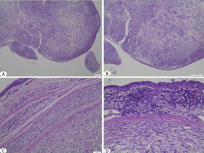
Malaysian J Pathol                                                                                  April 2018

FIG. 8: Sections of a tonsil with cat-scratch infection demonstrating necrotising granulomatous inflammation
        (A&B; scale bar- A = 200µm, B = 50µm) with higher magnification showing neutrophils in the center of
        some granulomas (C&D).

FIG. 9: Non-necrotising epithelioid granulomata in the tonsil of a child with Crohn’s disease. (A&B, scale bar =
        50µm; D, scale bar = 200µm)

18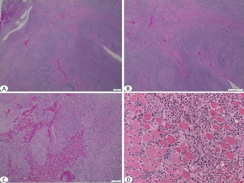
PATHOLOGY OF TONSIL IN CHILDREN

FIG. 10: Rosai-Dorfman disease of tonsil showing infiltrates of large histiocytic cells, mainly in germinal centers
         of lymphoid follicles (A, B & C). Higher magnification shows that individual histiocytes possess round
         centrally located nuclei, prominent nucleoli and exhibit abundant cytoplasm that contains lymphocytes
         or other inflammatory cells. (A&C, scale bar = 50µm, B, scale bar = 200µm, D, scale bar = 20µm)

disease. It involved a wide range of age, from              results in extensive accumulation of cholesterol
4 to 79 years old. Of the 5 cases which the age             esters in macrophages in peripheral tissues.
was recorded, three belong to paediatric age                Clinically, the pathognomonic finding, apart
group. The male to female ratio is 5:2.28-32                from an abnormal plasma lipoprotein profile,
    Rosai-Dorfman’s disease usually follows a               is the presence of large, hyperplastic, bright
self-limiting course that spontaneously resolves.           orange-yellow coloured tonsils and adenoids.33, 34
However, a minority of cases may have immune                   Microscopically, the tonsils and adenoids
dysfunction or may potentially relapse, hence               demonstrate a prominent accumulation of pale-
it is important to diagnose and follow up these             staining, foamy histiocytes that contain lipid
cases.                                                      droplets and occasional crystalline material. The
                                                            foamy histiocytes aggregate in clusters, often in
5.1.4 Storage diseases                                      the parafollicular areas.34 Although there is no
Tangier disease                                             treatment for Tangier disease, this is an important
Tangier disease is a rare autosomal recessive               diagnosis to recognise, as these patients have a
lipid metabolism disorder caused by a mutation              high risk of premature coronary artery disease.34, 35
in the ABCA1 gene, located in chromosome
9q31. The ABCA1 gene encodes an ATP-binding                 Lipid storage disease
cassette transporter that facilitates phospholipid          Lysosomal storage disease is a broad term used
and cholesterol transport. The mutation gives               to describe a heterogeneous group of more
rise to a defect in cellular cholesterol removal,           than fifty inherited disorders that arise due to
resulting in severe deficiency of plasma levels             deficiencies or defects in lysosomal enzymes
of high density lipoproteins and apolipoprotein             and other essential proteins that leads to the
A-1.33 A defect in cellular cholesterol removal             accumulation of undigested substrates. This

                                                                                                               19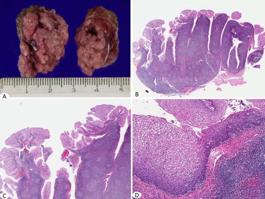
Malaysian J Pathol                                                                                       April 2018

results in cellular dysfunction and a variety                5.1.5 Neoplastic
of clinical manifestations. The diseases are                 Tonsils are common site for various neoplastic
generally classified by the specific accumulated             lesions, both benign and malignant.
substrate.36,37
   Lipid storage diseases are a subgroup of                  Squamous papilloma
disorders with a lysosomal hydrolase deficiency              Pedunculated squamous papillomas may arise
that causes lysosomal accumulation of specific               from the soft palate, tonsil, or the epiglottis. Male
sphingolipid substrates. Examples include                    to female ratio is 1:1.5 and the mean age was 33
Gaucher disease, Niemann-Pick disease, Wolman                years.42 Study showed an established connection
disease and cholesterol ester storage disease.               between HPV and the development of squamous
Abnormal storage of substrate can be found in                papilloma.43 It commonly arises from the tonsillar
many tissues including the reticuloendothelial               pillar, although it may arise from the surface of
system. Certain lipid storage diseases, including            the tonsil itself. The stratified squamous lining
mucopolysaccharidoses, have been reported to                 layer in this area gives rise to this lesion. This
present clinically as tonsillar hypertrophy.38,39            lesion is purely an incidental finding usually,
   Histologic examination of affected tissues,               and rarely requires treatment. Histologically,
including the tonsil, demonstrate aggregates                 tonsillar squamous papillomas are composed of
and/or sheets of macrophages with round,                     well differentiated benign-appearing squamous
uniform lipid inclusions that fill and distend               epithelial papillae on thin fibrovascular stalks.
the cytoplasm giving them a foamy appearance                 The majority of these papillomas usually lack
(Fig. 11). Although non-specific, identification             the viral associated koilocytic changes.
and awareness of these histologic features can
help support further biochemical and molecular               Lymphomas
testing that can lead to earlier diagnosis of certain        Primary extranodal non-Hodgkin’s lymphomas
storage diseases.40,41                                       are the most common malignant tumours of
                                                             the head and neck in the paediatric population.

FIG. 11: Clusters of foamy histiocytes at the parafollicular and interfollicular regions of the tonsil of a child with
         a lipid storage disease (A, B, C &D). (Scale bar: A=200µm, B&C=50µm, D=20µm)

20
PATHOLOGY OF TONSIL IN CHILDREN
Primary tonsillar lymphoma comprises < 1% lymphoma as the commonest type, occurring in
of head and neck cancer with non-Hodgkin 27 of the 51 cases of tonsillar lymphoma.46
lymphoma (NHL) being the most common type. Although the exact aetiology remains unclear,
Non-Hodgkin’s lymphoma of the oral cavity and a number of predisposing factors have been
oropharynx accounts for 13% of all primary identified, including human immunodeficiency
extranodal NHL with approximately 70% of these virus and Epstein-Barr infection.47
occurring in the tonsils.44 The palatine tonsil is
the most frequently involved site followed by Burkitt lymphoma
palate, gingiva and tongue. Most lymphomas Burkitt lymphoma is an aggressive form of
found in the palatine tonsils are of B-cell type. non-Hodgkin B-cell lymphoma mostly seen
A review of the literature showed that a in childhood. There are three subtypes of
majority of tonsillar lymphomas in the paediatric Burkitt lymphoma: endemic (found in Africa),
population were Burkitt lymphoma, with few sporadic (found in North America and Europe)
cases of diffuse large B-cell lymphoma, follicular and immunodeficiency related. The endemic
lymphoma and T-cell lymphoma (see Table 1). (African) subtype of Burkitt lymphoma is
However in the adult population, diffuse large mostly seen between ages 5-7 and more than
B-cell lymphoma is the most common type. 50% of cases involve the maxilla or mandible.
Mohammadianpanah et al. found that in his The sporadic (non-African) form of the disease
series of 14 cases of NHL diagnosed over 10 may be seen in adults as well as children around
years, that diffuse large B-cell lymphoma is 10-12 years of age. The sporadic form of the
the most common subtype, followed by small disease presents with abdominal, medullary
cell lymphoma, immunoblastic lymphoma, and or lymphatic involvement rather then head
anaplastic large cell lymphoma.45 Similarly, and neck involvement. Only 5% of the cases
Makepeace et al. also found diffuse large B-cell involve Waldeyer’s ring as an initial presentation.
TABLE 1. Reported cases of tonsillar lymphoma in paediatric population
Authors No of Age Gender Types of lymphoma
patients (years)
Amit et al. 2012 [53] 1 6 Male Follicular lymphoma
Banthia et al. 2003 [54] - - - Burkitt lymphoma
Berkowitz et al. 1999 [55] 7 2 to 9 - NHL
Booth et al. 2013 [56] 3 5 - Burkitt lymphoma
14 - Follicular lymphoma
15 - DLBCL
Cianci et al. 2013 [57] 1 7 Male Burkitt lymphoma
Dolev et al. 2008 [58] 6 Paediatric - -
Garavello et al. 2004 [59] 2 Paediatric - NHL
García-Ortega et al. 1999 [60] - - - Burkitt lymphoma
Guimarães et al. 2012 [44] 2 5 Female DLBCL
11 Female DLBCL
Kraus et al. 1990 [61] 1 10 Male Burkitt lymphoma
Meirelles et al. 1998 [62] 2 Paediatric - Burkitt lymphoma
Poulsen 1982 [63] - Paediatric - Burkitt lymphoma
Tewfik et al. 1996 [64] - Paediatric - Burkitt lymphoma
van Lierop et al. 2007 [65] - Paediatric - T-cell lymphoma
Williams et al. 2003 [11] 1 2 Male Burkitt lymphoma
Zeglaoui et al. 2009 [66] 1 4 Male NK/ T-cell lymphoma
21Malaysian J Pathol April 2018
Palatine tonsil involvement is reported in 2.9% The histological hallmark features of MALT
of head and neck Burkitt lymphoma.48 are small atypical lymphoid cells with irregular
The histologic features of tonsillar involvement nuclear membranes infiltrating lymphoid follicles
are similar to those in lymph nodes, showing a and/or expanding marginal zones and infiltrating
monotonous lymphoid proliferation composed mucosa (Fig. 13). Immunophenotypically, MALT
of medium sized cells with round nuclei, finely lymphomas have been reported to express
clumped chromatin and multiple small basophilic CD21, CD35, CD43, CD20 and bcl-2 protein,
nucleoli. The cytoplasm is deeply basophilic and with absence of markers such as CD10, CD5,
usually contains lipid vacuoles. The tumour has and CD23 antigen, and the presence of light
an extremely high proliferation activity as evident chain restriction. There is no specific marker
by Ki-67 immunohistochemical Staining. A for MALT.51,52
‘starry sky’ pattern is present, which is imparted
by numerous benign macrophages that ingested Non-haematopoietic small blue cell tumours
apoptotic tumour cells (Fig. 12). The tonsillar region can be the primary site for
non-haematopoietic tumours of childhood, most
Mucosa associated Lymphoid Tissue Lymphoma notably rhabdomyosarcoma.67, 68 Radiologically,
(MALT) it may mimic peritonsillar cellulitis or abscess.69
The gastrointestinal tract is the most frequent Histologically, it is composed of sheets of
extra nodal location for lymphoma of mucosa- spindled, moderate to poorly differentiated
associated lymphoid tissue (MALT). MALT cells with eosinophilic eccentric cytoplasm. A
arising from the tonsil is rare. However if condensed layer of tumour cells beneath an intact
Waldeyer’s ring is involved, the tonsil is the epithelium, also known as a “cambium layer”
most commonly affected site. It is characterised may be seen (Fig. 14).
by specific histological features and a remarkably
indolent clinical course.49,50
FIG. 12: A tonsil with Burkitt lymphoma showing effaced architecture (A&B, scale bar = 200µm) and an infiltra-
tion of lymphoma cells with classical “starry star” appearance (C, scale bar = 50µm). Lymphoma cells
often infiltrate the epithelium forming small nests (D, scale bar = 20µm).
22PATHOLOGY OF TONSIL IN CHILDREN
FIG. 13: Sections from a tonsil involved with MALT, showing abnormal crowded lymphoid follicles (A&B, scale
bar = 200µm) with expansion of marginal zones (C&D, scale bar- C = 50µm and D = 20µm).
FIG. 14: Sections from a tonsil with rhabdomyosarcoma showing replacement of usual architecture by infiltrating
“small blue’ cell tumour (A&B, scale bar = 200µm) Higher magnification showing sheets of spindled
cells with hyperchromatic nuclei and eosinophilic cytoplasm. A condensed layer of tumour cells beneath
the epithelium “cambium layer” is seen (C&D, scale bar C = 50µm).
23Malaysian J Pathol April 2018
COMMENTS Cafferkey M. The incidence and role of actinomyces
in recurrent acute tonsillitis. Clin Otolaryngol Allied
In conclusion, even though most of the time, Sci. 1993; 18(4): 268-71.
the pathologic examination of tonsils yields a 13. Ashraf MJ, Azarpira N, Khademi B, Hashemi B,
reactive non-specific process, such as follicular Shishegar M. Relation between actinomycosis and
lymphoid hyperplasia, routine microscopic histopathological and clinical features of the palatine
tonsils: An Iranian experience. Iran Red Crescent
examination can at times provide important clues
Med J. 2011; 13(7): 499-502.
to certain localised or systemic conditions that 14. Erol K, Erkan KM, Tolga D, Bengu C. Epidermoid
may have been otherwise missed. Practicing cyst localized in the palatine tonsil. J Oral Maxillofac
pathologists need to be aware of both the Pathol. 2013; 17(1): 148.
common and uncommon diseases that may be 15. Garnett CT, Talekar G, Mahr JA, Huang W, Zhang Y,
encountered in tonsils. Ornelles DA, et al. Latent species C adenoviruses in
human tonsil tissues. J Virol. 2009; 83(6): 2417-28.
16. Syrjänen S. HPV infections and tonsillar carcinoma.
REFERENCES J Clin Pathol. 2004; 57(5): 449-55.
1. Peterson G. Isaacson, Andrew J. Norton. In: Isaacson 17. Al-Sebeih K, Katchy K. Adenotonsillar granuloma:
PG, Norton AJ, eds. Extranodal Lymphomas. histopathological correlation. Med Princ Pract.
Edinburgh, Scotland: Churchill Livingstone. 1994. 2007; 16(6): 450-3.
193-7. 18. Bozkurt T, Langer M, Fendel K, Lux G.
2. Howie AJ. Scanning and transmission electron Granulomatous tonsillitis. A rare extraintestinal
microscopy on the epithelium of human palatine manifestation of Crohn‘s disease. Dig Dis Sci.
tonsils. J Pathol. 1980; 130(2): 91-8. 1992; 37(7): 1127-30.
3. I n g r a m D G , F r i e d m a n N R . To w a r d 19. Selimoğlu E, Sütbeyaz Y, Ciftçioğlu MA, Parlak
adenotonsillectomy in children: A review for the M, Esrefoğlu M, Oztürk A. Primary tonsillar
general pediatrician. JAMA Pediatr. 2015; 169(12): tuberculosis: a case report. J Laryngol Otol. 1995;
1155-61. 109(9): 880-2.
4. Baugh RF, Archer SM, Mitchell RB, Rosenfeld 20. Kardon DE, Thompson LD. A clinicopathologic
RM, Amin R, Burns JJ, et al. American Academy of series of 22 cases of tonsillar granulomas.
Otolaryngology-Head and Neck Surgery Foundation. Laryngoscope. 2000; 110(3 Pt 1): 476-81.
Clinical practice guideline: Tonsillectomy in 21. Altug H, Tahsinoglu M, Celikoğlu S. A case of
children. Otolaryngol Head Neck Surg. 2011 Jan; tonsillar localization of sarcoidosis. J Laryngol
144(1 Suppl): S1-30. Otol. 1973; 87(4): 417-21.
5. Marcus CL, Moore RH, Rosen CL, Giordani 22. McEwan J, Basha S, Rogers S, Harkness P. An
B, Garetz SL, Taylor HG, et al. Childhood unusual presentation of cat-scratch disease. J
Adenotonsillectomy Trial (CHAT). A randomized Laryngol Otol. 2001; 115(10): 826-8.
trial of adenotonsillectomy for childhood sleep 23. Shin OR, Kim YR, Ban TH, Lim T, Han TH, Kim
apnea. N Engl J Med. 2013; 368(25): 2366-76. SY, et al. A case report of seronegative cat scratch
6. Paradise JL, Wald ER. Tonsillectomy and/ disease, emphasizing the histopathologic point of
or adenoidectomy in children: Indications and view. Diagn Pathol. 2014; 9: 62.
contraindications. Up To Date 2015. 24. Woo VL. Oral manifestations of Crohn‘s disease: A
7. Cullen KA, Hall MJ, Golosinskiy A. Ambulatory case report and review of the literature. Case Rep
surgery in the United States, 2006. Natl Health Stat Dent. 2015; 830472.
Report. 2009; (11): 1-25. 25. Bhatt GM, Kumar S, Sharma A. Rosai-Dorfman
8. Erickson BK, Larson DR, St Sauver JL, Meverden disease : A case report with review of literature
RA, Orvidas LJ. Changes in incidence and 2004; 25(4): 39-41.
indications of tonsillectomy and adenotonsillectomy, 26. Foucar E, Rosai J, Dorfman R. Sinus histiocytosis
1970-2005. Otolaryngol Head Neck Surg. 2009; with massive lymphadenopathy (Rosai-Dorfman
140(6): 894-901. disease): Review of the entity. Semin Diagn Pathol.
9. Bhattacharyya N, Lin HW. Changes and 1990; 7(1): 19-73.
consistencies in the epidemiology of pediatric 27. Gaitonde S. Multifocal, extranodal sinus histiocytosis
adenotonsillar surgery, 1996-2006. Otolaryngol with massive lymphadenopathy: An overview. Arch
Head Neck Surg. 2010; 143(5): 680-4. Pathol Lab Med. 2007; 131(7): 1117-21.
10. Faramarzi A, Ashraf MJ, Hashemi B, Heydari 28. Amer HZM, Prasad V, Kahwash SB. Rosai
ST, Saif I, Azarpira N, et al. Histopathological Dorfman disease of the tonsil in a 4-year-old boy:
screening of tonsillectomy and/or adenoidectomy Case report and review of the literature. Int J Ped
specimens: A report from southern Iran. Int J Pediatr Otorhinolaryngol Extra. 2010; 6(1): 17-9.
Otorhinolaryngol. 2009; 73(11): 1576-9. 29. Qualtieri, J, Kahwash SB. Incidental finding of
11. Williams MD, Brown HM. The adequacy of gross isolated Rosai-Dorfman disease in the tonsils of a
pathological examination of routine tonsils and 4-year-Old girl: A case report and a brief review of
adenoids in patients 21 years old and younger. Hum current practice in tonsillectomy specimen handling.
Pathol. 2003; 34(10): 1053-7. O J Pathology. 2013; 3(1): 3.
12. Gaffney R, Harrison M, Walsh M, Sweeney E, 30. Hanchard B, McNeill R, Thomas S, Sparke B.
24PATHOLOGY OF TONSIL IN CHILDREN
Sinus histiocytosis with massive lymphadenopathy: treatment over a 27-year period. J Laryngol Otol.
Clinico-pathological features of four cases. West 1987; 101(11): 1151-8.
Indian Med J. 1977; 26(4): 204-10. 47. Solomides CC, Miller AS, Christman RA, Talwar
31. Miettinen M, Paljakka P, Haveri P, Saxén E. Sinus J, Simpkins H. Lymphomas of the oral cavity:
histiocytosis with massive lymphadenopathy. A Histology, immunologic type, and incidence of
nodal and extranodal proliferation of S-100 protein Epstein-Barr virus infection. Hum Pathol. 2002;
positive histiocytes? Am J Clin Pathol. 1987; 88(3): 33(2): 153-7.
270-7. 48. Akyıldız1 I, Kaptan Z, Hücümenoğlu S, Ünverdi
32. Stiakaki E, Lidaki E, Bizakis I, Kanavaros P, H, Aktar G. Burkitt’s lymphoma of the palatine
Bolonaki I, Helidonis E, et al. Rosai-Dorfman tonsil. Kulak Burun Boğaz Uygulamaları 2014;
disease with unilateral tonsilar enlargement and 2(3): 135-8.
subtle cervical lymphadenopathy. Haematologia 49. Lee JT, Paquette R, Sercarz JA, Wang MB. Mucosa-
(Budap). 1994; 26(1): 45-7. associated lymphoid tissue lymphoma of the lingual
33. Ravesloot MJ, Bril H, Braamskamp MJ, Wiegman tonsil. Am J Otolaryngol. 2000; 21(4): 271-6.
A, Wong Chung RP. The curious case of the orange 50. Isaacson P, Wright DH. Malignant lymphoma of
coloured tonsils. Int J Pediatr Otorhinolaryngol. mucosa-associated lymphoid tissue. A distinctive
2014; 78(12): 2305-7. type of B-cell lymphoma. Cancer. 1983; 52(8):
34. Nelson BL, Thompson LD. Tonsil with Tangier 1410-6.
disease. Ear Nose Throat J. 2003; 82(3): 178. 51. Isaacson PG, Spencer J. Malignant lymphoma of
35. Asztalos BF, Schaefer EJ. High-density lipoprotein mucosa-associated lymphoid tissue. Histopathology.
subpopulations in pathologic conditions. Am J 1987; 11(5): 445-62.
Cardiol. 2003; 91(7A): 12E-17E. 52. Spencer J, Finn T, Isaacson PG. Human Peyer‘s
36. Platt FM, Boland B, van der Spoel AC. The cell patches: an immunohistochemical study. Gut. 1986;
biology of disease: Lysosomal storage disorders: 27(4): 405-10.
The cellular impact of lysosomal dysfunction. J 53. Amit S, Purwar N, Agarwal A, Lalchandani D.
Cell Biol. 2012; 199(5): 723-34. Tonsillar follicular lymphoma in a child. BMJ Case
37. Ballabio A, Gieselmann V. Lysosomal disorders: Rep. 2012 Nov 27;2012. pii: bcr2012006848.
From storage to cellular damage. Biochim Biophys 54. Banthia V, Jen A, Kacker A. Sporadic Burkitt‘s
Acta. 2009; 1793(4): 684-96. lymphoma of the head and neck in the pediatric
38. Keilmann A, Läßig AK, Pollak-Hainz A, Mann population. Int J Pediatr Otorhinolaryngol. 2003;
WJ, Beck M, Hainz M. Adenoids of patients 67(1): 59-65.
with mucopolysaccharidoses demonstrate typical 55. Berkowitz RG, Mahadevan M. Unilateral tonsillar
alterations. Int J Pediatr Otorhinolaryngol. 2015; enlargement and tonsillar lymphoma in children.
79(2): 115-8. Ann Otol Rhinol Laryngol. 1999; 108(9): 876-9.
39. Fujitani T, Kimura A, Inoue K, Okada S. 56. Booth CL, Wang J. Occult hematologic malignancy
Pathological and biochemical study in the in routine tonsillectomy specimens: A single
adenoid of mucopolysaccharidosis II. Int J Pediatr institutional experience and review of the literature.
Otorhinolaryngol. 1985; 10(3): 205-12. Am J Clin Pathol. 2013; 140(6): 807-12.
40. Kolodny EH, Charria-Ortiz G. Storage diseases of 57. Cianci P, Tono V, Sala A, Locatelli L, Carta C,
the reticuloendothelial system In: Nathan DG, Orkin Rizzari C, Biondi A, Selicorni A. A boy with Burkitt
SH, Ginsburg D, Look AT, editors. Hematology of lymphoma associated with Noonan syndrome due
Infancy and Childhood. 6th ed. Philadelphia, PA: to a mutation in RAF1. Am J Med Genet A. 2013;
WB Saunders. 2003; 1399–454. 161A(6): 1401-4.
41. Hansen HG, Graucob E. Hematologic cytology 58. Dolev Y, Daniel SJ. The presence of unilateral
of storage diseases. Springer Science & Business tonsillar enlargement in patients diagnosed
Media. 2012; 88 pages. with palatine tonsil lymphoma: experience at
42. Mundra RK, Verma J, Gupta A, Koshta V, Rathore a tertiary care pediatric hospital. Int J Pediatr
SK. Tonsillar papilloma: A rare case. Inter J Medical Otorhinolaryngol. 2008; 72(1): 9-12.
Sci Res Prac 2015; 2(3): 162-3. 59. Garavello W, Romagnoli M, Sordo L, Spreafico R,
43. Crissman JD, Kessis T, Shah KV, Fu YS, Stoler Gaini RM. Incidence of unexpected malignancies
MH, Zarbo RJ, Weiss MA. Squamous papillary in routine tonsillectomy specimens in children.
neoplasia of the adult upper aerodigestive tract. Laryngoscope. 2004 Jun;114(6):1103-5.
Hum Pathol. 1988; 19(12): 1387-96. 60. García-Ortega FP, Bonnín Otal J, Durán R, Carreño
44. Guimarães AC, de Carvalho GM, Gusmão RJ. Villarreal M, Alemán López O, Malluguiza Calvo
Tonsillar lymphoma in children with unilateral JR. Burkitt lymphoma of a palatine tonsil. Acta
tonsillar enlargement. Rev Paul Pediatr. 2012; 30(2): Otorrinolaringol Esp. 1999; 50(7): 579-82.
288-91. 61. Kraus M, Fliss DM, Argov S, Leiberman A,
45. Mohammadianpanah M, Omidvai S, Mosalei Benharroch D. Burkitt‘s lymphoma of the tonsil. J
A, Ahmadloo N. Treatment results of tonsillar Laryngol Otol. 1990; 104(12): 991-4.
lymphoma: a 10-year experience. Ann Hematol. 62. Meirelles RC, Figueiredo FA, Vidal ARC, Casali
2005; 84(4): 223-6. AR, Ciriaco A. Burkitt‘s lymphoma with initial
46. Makepeace AR, Fermont DC, Bennett MH. Non- tonsil involvement. Report of two cases. Revista
Hodgkin‘s lymphoma of the tonsil. Experience of Brasileira de Otorrinolaringologia. 1998; 64: 62-66.
25Malaysian J Pathol April 2018
63. Poulsen P. Burkitt‘s lymphoma in the tonsil. Int J
Pediatr Otorhinolaryngol. 1982; 4(4): 349-51.
64. Tewfik TL, Bond M, al-Ghamdi K, Bernard C.
Burkitt‘s lymphoma of the tonsil in children. J
Otolaryngol. 1996; 25(3): 205-8.
65. van Lierop AC, Prescott CA, Fagan JJ, Sinclair-
Smith CC. Is diagnostic tonsillectomy indicated in
all children with asymmetrically enlarged tonsils?
S Afr Med J. 2007; 97(5): 367-70.
66. Zeglaoui I, Belcadhi M, Sriha B, Bouzouita K. Nasal
NK/T-cell lymphoma in the paediatric population.
Two case reports. B-ENT. 2009; 5(2): 119-23.
67. Sarwar Khan M, Ruttens H, Blanshard JD. Alveolar
rhabdomyosarcoma presenting as a peritonsillar
abscess. J Laryngol Otol. 2001; 115(12): 1018-20.
68. Yuan XX, Yuan JP, Yang YH, Xia XL, Zeng Y.
Tonsil pleomorphic rhabdomyosarcoma: report of 1
case and review of literature. Cancer Res Prevention
and Treatment. 2009; 36(9): 766-7.
69. Fabian D, Mahida JB, Pluto CP, Thompson
BP, Minneci PC, et al. Pediatric tonsillar
malignancies: Misleading presentations. Inter J
Otorhinolaryngology. 2015; 2(1): 1-4.
26You can also read