A rapid phenotype change in the pathogen Perkinsus marinus was associated with a historically significant marine disease emergence in the eastern ...
←
→
Page content transcription
If your browser does not render page correctly, please read the page content below
www.nature.com/scientificreports
OPEN A rapid phenotype change
in the pathogen Perkinsus marinus
was associated with a historically
significant marine disease
emergence in the eastern oyster
Ryan B. Carnegie1*, Susan E. Ford2,4, Rita K. Crockett1, Peter R. Kingsley‑Smith3,
Lydia M. Bienlien1, Lúcia S. L. Safi1, Laura A. Whitefleet‑Smith1 & Eugene M. Burreson1
The protozoan parasite Perkinsus marinus, which causes dermo disease in Crassostrea virginica, is
one of the most ecologically important and economically destructive marine pathogens. The rapid
and persistent intensification of dermo in the USA in the 1980s has long been enigmatic. Attributed
originally to the effects of multi-year drought, climatic factors fail to fully explain the geographic
extent of dermo’s intensification or the persistence of its intensified activity. Here we show that
emergence of a unique, hypervirulent P. marinus phenotype was associated with the increase in
prevalence and intensity of this disease and associated mortality. Retrospective histopathology of
8355 archival oysters from 1960 to 2018 spanning Chesapeake Bay, South Carolina, and New Jersey
revealed that a new parasite phenotype emerged between 1983 and 1990, concurrent with major
historical dermo disease outbreaks. Phenotypic changes included a shortening of the parasite’s life
cycle and a tropism shift from deeper connective tissues to digestive epithelia. The changes are likely
adaptive with regard to the reduced oyster abundance and longevity faced by P. marinus after rapid
establishment of exotic pathogen Haplosporidium nelsoni in 1959. Our findings, we hypothesize,
illustrate a novel ecosystem response to a marine parasite invasion: an increase in virulence in a native
parasite.
Infectious diseases are increasing in significance for many aquatic species as human pressures on estuarine,
marine, and freshwater environments intensify1,2, yet the factors underlying emergence of these diseases are
seldom fully understood. Numerous emerging diseases have been produced by introduced exotic pathogens, such
as “multinucleate sphere X” (MSX) disease caused by Haplosporidium nelsoni in eastern oysters, Crassostrea virgi-
nica, from the United States of America (USA), and bonamiasis caused by Bonamia ostreae in flat oysters, Ostrea
edulis, from E
urope3,4; infectious hematopoietic necrosis in rainbow trout, Oncorhynchus mykiss, in Europe5;
and infectious hypodermal and haemopoietic necrosis, Taura syndrome, and white spot syndrome in penaeid
shrimp world-wide6. One emerging disease has been linked to increasing local human impacts, serratiosis being
transmitted to elkhorn coral Acropora palmata populations via wastewater from coastal c ommunities7. The
origins of many others are unknown, including the herpesviruses that have emerged in recent years to devastate
populations of mollusc hosts like Pacific oyster Crassostrea gigas8–11.
Our objective was to better understand the re-emergence of dermo disease in the eastern oyster Crassostrea
virginica. Dermo represents the unusual case of an established disease that was long present in a host population
but which intensified sharply in the mid-1980s. This development was first apparent in the Chesapeake Bay region
of the Mid-Atlantic coast of the USA. Caused by the protozoan parasite Perkinsus marinus, dermo historically
was a chronic disease of oysters from southern estuaries of the USA into Mexico12. Occurring from the Gulf of
Mexico north as far as Chesapeake Bay, its impacts were generally modest, with annual oyster mortality of up to
30% primarily affecting older oysters that had been exposed to the parasite for several y ears13. Beginning around
1
Virginia Institute of Marine Science, William & Mary, P.O. Box 1346, Gloucester Point, VA 23062,
USA. 2Haskin Shellfish Research Laboratory, Rutgers University, 6959 Miller Avenue, Port Norris, NJ 08349,
USA. 3Marine Resources Research Institute, South Carolina Department of Natural Resources, 217 Fort Johnson
Road, Charleston, SC 29422, USA. 4Susan E. Ford is deceased. *email: carnegie@vims.edu
Scientific Reports | (2021) 11:12872 | https://doi.org/10.1038/s41598-021-92379-6 1
Vol.:(0123456789)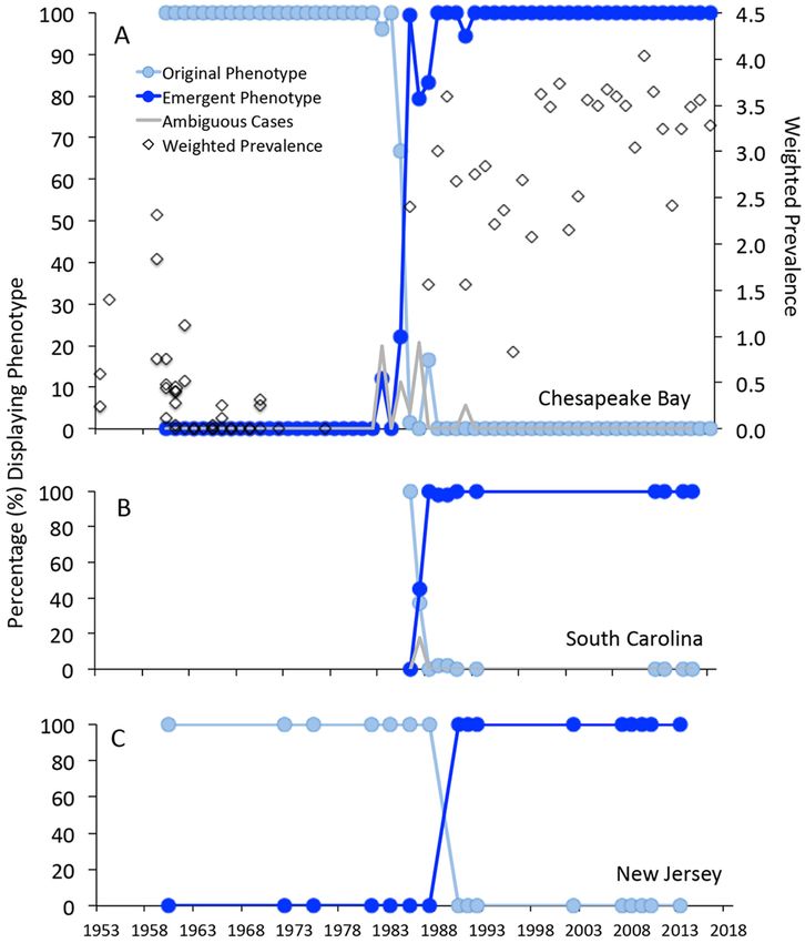
www.nature.com/scientificreports/
1986, dermo became an acute and profoundly destructive disease capable of causing over 70% host mortality
within a period of m onths14. P. marinus also began a progressive range expansion northward to the northern
Atlantic coast of the USA at that time, producing major disease outbreaks along the way: in Delaware Bay in
1990, where it had previously been scarcely detectable and primarily in association with oysters transplanted
from the Chesapeake Bay; and in Long Island Sound and southern New England, USA, between 1991 and 1 99415.
Marine environmental change is recognized as a major driver of marine disease emergence16,17, and the scientific
community has generally accepted the proposition that warming coastal water temperatures in recent decades,
particularly in winter, promoted the range expansion of P. marinus poleward from the Chesapeake Bay r egion15,18.
While warming seawater temperatures may explain the range expansion of P. marinus and dermo disease
to the north since 1990, they fail to fully explain the sudden and persistent intensification of P. marinus activ-
ity in Chesapeake Bay in the mid-1980s14. Nor do they explain an intensification of dermo disease around the
same time as far south as South Carolina in the South Atlantic Bight19, which, in contrast with northern waters
such as Delaware Bay and Long Island Sound, had not warmed significantly20. Elevated salinities associated
with multi-year drought in the Chesapeake Bay region in the 1980s were certainly more favorable to P. marinus
proliferation and t ransmission14. If drought was the primary cause of the intensification of dermo disease, how-
ever, the questions arise as to why more protracted and intense droughts in earlier years (see Supplementary
Table S1) did not produce a similar intensification of disease; and why subsequent wet periods with depressed
salinities distinctly unfavorable to P. marinus, for example from 2003 to 2004 (Supplementary Fig. S1), did not
return the parasite to low levels of infection characteristic of earlier years. In short, environmental factors do not
completely explain a lasting increase in P. marinus infections and associated mortality along the USA Atlantic
Coast from the mid-1980s.
Dermo disease is typically detected in oysters using Ray’s fluid thioglycollate method (RFTM)21, a fluid
culture-based technique that provides sensitive insight into the presence and abundance of P. marinus within
infected hosts. The RFTM method does not, however, generate insights into the specific nature of the host-
parasite interaction in situ or even into the size of the cells infecting the oyster, given that the parasite increases
in size in its transformation from trophozoites into hypnospores in the culture medium. Paraffin histology can
provide more detailed insight into host–pathogen i nteractions22, and is routinely performed for the detection of
co-occurring pathogen H. nelsoni. P. marinus is generally ignored in histological diagnoses, however, in favor of
the more sensitive and semi-quantitative R FTM23. To determine whether histology might nonetheless provide
a window into the intensification of dermo disease in the 1980s, we retrieved histological slides of P. marinus-
infected oysters which were collected from Chesapeake Bay, USA, from 1960 that were archived in a Virginia
Institute of Marine Science histological collection. Then, we evaluated these slides against contemporary histol-
ogy. Unexpectedly, we found a marked difference in the phenotype of P. marinus between slides from 1960 and
the present, which prompted a wider retrospective analysis of histological samples from Chesapeake Bay span-
ning the entire 1960–2018 period. Histological material from New Jersey and South Carolina was added to the
analysis to provide broader geographical perspective. We also drew on RFTM-based P. marinus infection data
for Chesapeake Bay from 1952 to 2018 for additional insight into long-term infection trends. Our analyses show
that while intensification of P. marinus infection in the mid-1980s may have been partly due to climatic factors,
it was more fundamentally associated with the emergence and spread of a hypervirulent parasite phenotype.
Results and discussion
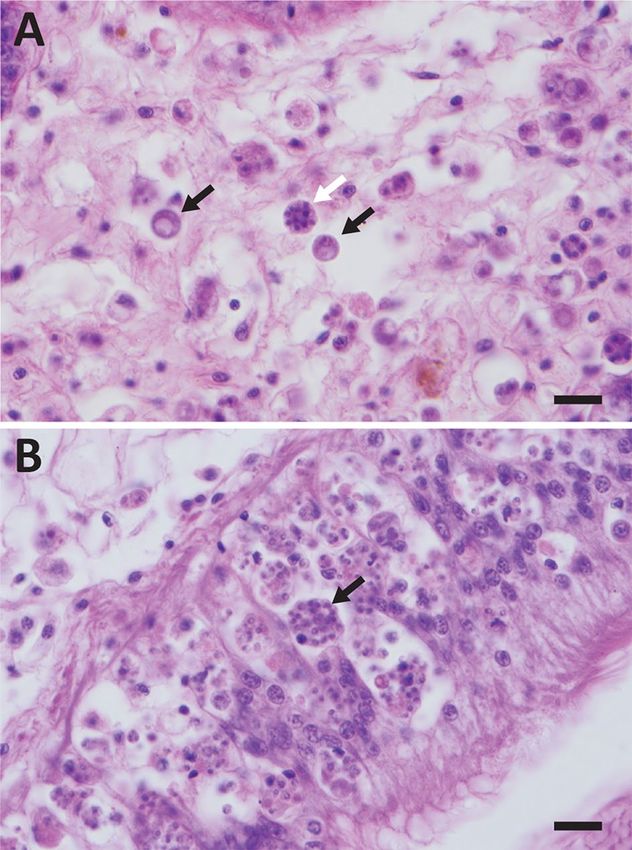
We made two fundamental observations of changes in P. marinus and its interaction with eastern oysters. First,
while P. marinus infections in early years were primarily of hemal spaces in the vesicular connective tissues that
surround oyster digestive and reproductive organs, extending into mantle and gills (Fig. 1A), contemporary P.
marinus infections are typically epithelial in tropism until they become heavy and systemic, with foci of parasite
cells initially and in lighter infections observed in digestive epithelia in particular (Fig. 1B). Even in heavier
pre-1980s infections, digestive epithelia were largely or entirely uncolonized by the parasite; today, P. marinus is
primarily observed in digestive epithelia in most histological examinations.
Second, in addition to a tissue tropism distinct from that of contemporary P. marinus, parasite cells observed
in early-era infections were much larger. Mackin et al.24 described cells of P. marinus, then identified as Der-
mocystidium marinum, to be mostly 5–7 μm in diameter, consistent with our observations of P. marinus in
archival histology from prior to the mid-1980s (Fig. 2). P. marinus schizonts were found to be 6.42 ± 0.14 μm
(mean ± 95% CI) in diameter in Chesapeake Bay samples from 1960 to 1962, and 6.70 ± 0.28 μm in diameter
in samples from 1981–1984. P. marinus cells observed in samples since the mid-1980s, however, are markedly
smaller: 4.42 ± 0.20 μm in diameter in 1992–1993, and 3.90 ± 0.25 μm in 2011–2012 (ANOVA F0.05, 3, 116 = 168.7,
P < 2e−16). Samples collected from Delaware Bay showed a similar reduction in cell size, from 6.58 ± 0.22 μm in
diameter in 1960–1981 to 3.75 ± 0.16 μm in 1992–2010 (Welch’s approximate t-test t = 21.53, df = 52.42, P < 2.2e-
16), as did samples from South Carolina, which decreased in diameter from 5.04 ± 0.71 μm in diameter in 1986
to 3.48 ± 0.22 μm from 1989 to 2016 (Wilcoxon rank sum test W = 65, P = 0.0002334; Fig. 2). Associated with a
reduction in cell size was the loss of distinctly multinucleate schizonts. While vegetative division in P. marinus
has long been known to proceed via schizogony leading to the production of 16 or more daughter cells from a
single trophozoite25, nuclear counts in contemporary P. marinus seldom exceed 2–4, indicating that the parasite
may now be dividing more in a binary mode, and raising the question of whether it is still dividing by schizogony
at all rather than simple binary fission.
Using the histological observations above to define alternative parasite phenotypes—an original phenotype
characterized by larger cells dividing through multinucleate schizonts and primarily infecting oyster connec-
tive tissues, and a contemporary phenotype characterized by smaller cells likely dividing in binary mode or via
more oligonucleate schizonts, and primarily infecting oyster digestive epithelia—we assessed changes in the
Scientific Reports | (2021) 11:12872 | https://doi.org/10.1038/s41598-021-92379-6 2
Vol:.(1234567890)www.nature.com/scientificreports/
Figure 1. Histological presentation of Perkinsus marinus phenotypes. A. Original phenotype, with relatively
small numbers of large P. marinus cells primarily infecting oyster connective tissues. Black arrows indicate two
P. marinus trophozoites; white arrow indicates a multinucleate schizont. B. Contemporary phenotype, with large
numbers of small parasite cells primarily infecting digestive epithelia. Arrow indicates a mass of P. marinus cells
inside a moribund oyster hemocyte. Scale bars for both panels represent 20 microns.
Figure 2. Perkinsus marinus schizont diameters (microns) before (light blue) and after (darker blue) the
emergence of the contemporary P. marinus phenotype, in Chesapeake Bay, South Carolina and New Jersey. Note
that for South Carolina the 1986 data represent trophozoites, as schizonts could not be definitively identified in
the tissue sections. Filled circles represent means with 95% confidence intervals. Reductions in cell diameters
with emergence of the contemporary phenotype within each area were statistically significant, as noted in
the text. For Chesapeake Bay, Tukey multiple comparisons revealed significant cell size reductions between
1981–1984 and 1992–1993 (P = 0.0000000) and between 1992–1993 and 2011–2012 (P = 0.0050495), but not
between 1960–1962 and 1981–1984 (P = 0.2709134).
Scientific Reports | (2021) 11:12872 | https://doi.org/10.1038/s41598-021-92379-6 3
Vol.:(0123456789)www.nature.com/scientificreports/
Figure 3. Timing of the Perkinsus marinus phenotype transition in Chesapeake Bay (n = 7932) (A), South
Carolina (n = 323) (B) and New Jersey (n = 100) (C). Data presented are the percentages of analyzed oysters
from each year and location displaying the original (light blue symbols and lines) or emergent (darker blue)
phenotypes. The gray lines in the Chesapeake Bay and South Carolina panels represent ambiguous cases during
the period of transition. For Chesapeake Bay, weighted prevalence of P. marinus in disease sentinel samples is
also presented (diamonds), illustrating the increase in disease (as expressed in weighted prevalence) concurrent
with the emergence of the contemporary phenotype. We refer the reader to Supplementary Tables S2–S4 for the
annual data and sample sizes.
frequency of these phenotypes in oysters sampled from Chesapeake Bay as well as Delaware Bay and South
Carolina between 1960 and 2018. In Chesapeake Bay, only the original phenotype was observed from 1960 to
1982 (Fig. 3A), and the contemporary phenotype has been observed exclusively since 1988. The years 1983–1988
were a transitional period, with some infections exhibiting an ambiguous phenotype, and potentially represent-
ing mixed infections. There was a major transition from the original to the contemporary phenotype, however,
occurring between 1985 and 1986, between which the contemporary phenotype increased in frequency from
22.2 to 99.3% of observations. The replacement of the original phenotype with the contemporary form coincided
closely with the intensification of the weighted prevalence of dermo disease (a pathogen abundance metric and
traditional measure of dermo disease in a population, the sum of infection intensity scores divided by the size, n,
of a sample) as measured using RFTM analyses—infection producing greater intensities of infection more rapidly
than in earlier years (Fig. 3A), and associated with sharply higher mortality14. In South Carolina, the transition
between the original and contemporary phenotypes occurred between 1986 and 1988 (Fig. 3B), coincident
with increased dermo disease in oyster populations there. In Delaware Bay (Fig. 3C), no histological material
was available for examination from 1988 or 1989, and only the historical phenotype was observed in limited
material available for examination before that, as P. marinus was rarely detected in Delaware Bay before 1990. In
re-analysis of samples collected from the major dermo disease outbreak in Delaware Bay in 1990, however, the
contemporary phenotype was present in 100% of observations, and the original phenotype was never observed
in Delaware Bay samples from subsequent years.
The picture that emerges, therefore, is of the rise of a virulent new phenotype of P. marinus along the Mid-
Atlantic coast of the USA, which dispersed from there and supplanted a form that previously had been widely
distributed in Atlantic estuaries. While changes in pathogen virulence, including virulence increases, have been
documented in other s ystems26, the scope of changes to pathogen life history we have seen, including a change
in tropism within the host, cell size, and potentially mode of division, occurring rapidly over a wide geographical
area, is unusual. Three points should be made with regard to these phenotypic changes. First, however different
the original and contemporary phenotypes appear in histological sections of oyster hosts, the evidence strongly
Scientific Reports | (2021) 11:12872 | https://doi.org/10.1038/s41598-021-92379-6 4
Vol:.(1234567890)www.nature.com/scientificreports/
indicates that these are the same parasite species. In situ hybridization conducted using a DNA probe designed for
P. marinus-specificity based on DNA sequences from the contemporary phenotype27 unambiguously hybridized
to an oyster displaying the original phenotype (Supplementary Fig. S2). While we might expect, and indeed we
would hypothesize, that genetic differences underlie the respective phenotypes, these do not represent species-
level differences. We also note that while genetic analyses over years have uncovered geographic differentiation
of P. marinus populations within and between the Atlantic and Gulf28,29, no study has uncovered cryptic species-
level diversity of Perkinsus parasites in oyster populations, including in the Gulf of Mexico where the original
phenotype can still be observed (Supplementary Fig. S2).
The second point we would make with regard to the phenotypic changes is that they may represent a shorten-
ing of the parasite’s life cycle that would be strongly adaptive with regard to important changes in Chesapeake
Bay, specifically the decrease in both oyster abundance and longevity, in the years following the emergence of
H. nelsoni in 1959. The exotic oyster pathogen H. nelsoni caused mortality exceeding 90% in oysters on private
Virginia aquaculture leases and public reefs in mesohaline waters by 196130, resulting in a decline in oyster
aquaculture production from 3,347,170 bushels in 1959 (a bushel representing approximately 200–250 oysters)
to 361,792 bushels by 1 98331. This production decline of 89% represented a depopulation of the overall aqua-
cultured oyster population in Virginia by an estimated 1.8 billion oysters, a decrease in oyster abundance that
was compounded by simultaneous reductions from wild populations that are impossible to directly quantify. A
large reduction in host abundance would be detrimental to a directly transmissible pathogen entirely dependent
on a single host, such as P. marinus in oysters. In fact, P. marinus did decline sharply in abundance beginning in
1959 as a likely reflection of this32. Recent theoretical modeling has underscored the possibility that an increase
in parasite virulence could be a consequence of such reduction in host resources33.
Similarly detrimental to P. marinus as it existed from the 1950s to early 1980s would have been a reduction in
oyster longevity. Wild oysters cultivated on aquaculture leases or growing on natural reefs would typically take
three or more years to reach “market” size (3 inches or 76.2 mm), and many would live substantially longer, as
evidenced by the larger size of oysters in historical shell middens and other d eposits34. P. marinus is primarily
transmitted through release into the water column with the deaths of infected hosts35, which in early years did not
typically occur until oysters were in their third year of P. marinus exposure13. Today, high disease-caused mortal-
ity limits the age of most oysters in parasite-endemic waters to under three years36. P. marinus today contributes
substantially to this contemporary mortality. The initial cause of the decrease in oyster longevity was H. nelsoni,
however, which caused very high mortality upon its emergence37 and which is capable of killing oysters of a year
or less in age38. As with the reduction in host abundance, the reduction in host longevity must have disadvantaged
a directly transmissible pathogen that required several years for transmission to occur. Evolution of a parasite
phenotype characterized by more rapid infection and within-host proliferation, and capable of rapidly causing
high mortality, would clearly have been beneficial for exploiting the relatively sparse populations of short-lived
oyster hosts that existed after the emergence of H. nelsoni.
The significance of the shift in parasite tropism from deeper connective tissues to more superficial digestive
epithelial tissues is less clear, but it may reflect adaptation on the part of P. marinus to allow for gradual trans-
mission via the feces in advance of host d eath35. This would contrast with the single release to the environment
with host death that would be possible for pathogens infecting the deeper tissues, and it could be particularly
advantageous when hosts are sparse. Understanding whether or not there are fitness advantages for P. marinus
to infect digestive epithelial tissues rather than connective tissues will require further study.
Finally, the phenotypic changes in P. marinus highlight not only the dynamic nature of parasite evolution but
also an unexplored realm of potential response to marine parasite invasion. Effects of parasite invasions are appre-
ciated primarily with regard to impacts on “native host species, communities, and even ecosystems”39. Effects on
native parasite communities have received little attention, yet our results suggest they may be profound. While
eastern North American estuaries such as the Chesapeake Bay have seen other, eutrophication-related changes,
such as increased nutrient loading and exacerbation of seasonal hypoxia with its concomitant effects on sedi-
ment biogeochemistry40, we cannot infer an adaptive response of P. marinus to these changes as we have no clear
basis for relating any of these gradual environmental shifts to the parasite’s theoretical fitness. We can hypoth-
esize, however, that the changes in P. marinus life history represent evolutionary adaptation to the most sudden
and plausibly consequential environmental change in these estuaries, the altered abundance and demographics
(longevity) of the oyster host that were produced by the invasion of H. nelsoni. We can only speculate as to why
the emergence of the phenotype change in P. marinus took so long, a quarter-century after the emergence of H.
nelsoni in Chesapeake Bay, but the delay may simply be due to the low rate of proliferation and thus mutation of
P. marinus when hosts were sparse in the 1960s, and when hosts were still sparse and environmental conditions
(particularly salinities) were unfavorable to the parasite in the 1970s (Supplementary Fig. S1). The phenotype
change in the 1980s occurred when environmental conditions in Chesapeake Bay and throughout the region
had become much more favorable for P. marinus proliferation and transmission14.
In summary, retrospective histological analyses have provided new insights into one of the most historically
significant emergences of marine disease, the lasting increase in dermo disease in eastern oysters, identifying
a rapid change in parasite virulence as associated with the disease intensification. This work underscores the
importance of long-term environmental monitoring and the maintenance of associated natural history collec-
tions, without which this perspective would not have been possible.
Methods
Eastern oysters, C. virginica, were collected during 1960–2018, primarily from natural oyster reefs and planting
grounds, in Chesapeake Bay, South Carolina, and New Jersey. Numbers of samples collected and available for
analyses varied widely. Chesapeake Bay and New Jersey samples were collected from subtidal beds by dredge,
Scientific Reports | (2021) 11:12872 | https://doi.org/10.1038/s41598-021-92379-6 5
Vol.:(0123456789)www.nature.com/scientificreports/
as described previously38,41. South Carolina samples included oysters of both subtidal and intertidal origin42.
South Carolina collections were from locations spanning that state’s coast. New Jersey collections were from
Delaware Bay, with the exception of those from 1983, 1985, and 1987, which were from the Mullica River/Great
Bay system. Once collected, oysters were shucked and transverse sections of soft tissues, including stomach and
intestine, digestive gland, gonad, mantle and gills, were fixed and processed for paraffin histology using standard
methods, including staining with hematoxylin and eosin.
Microscopic analyses of contemporary and archival samples were performed on an Olympus BX51 light
microscope using an Olympus DP71 camera. P. marinus phenotypes were defined after preliminary assessment
of infected oyster samples from Chesapeake Bay spanning the period of the study. Phenotype A (the original
phenotype) was characterized by generally larger P. marinus cells (> 5 μm), an abundance of multinucleate schi-
zonts (dividing forms), and infection primarily of oyster connective tissues. Phenotype B (the contemporary
phenotype) was characterized by generally smaller P. marinus cells (< 5 μm), a scarcity of large schizonts of
nuclear count > 3 or 4, and infection primarily of oyster digestive epithelia, except in advanced, systemic cases.
Across one or more samples for each area (i.e., Chesapeake Bay, South Carolina, and New Jersey) and year, data
were expressed as percentage of oysters presenting either Phenotype A or Phenotype B, with some samples and
years characterized by the presence of ambiguous phenotypes possibly representing mixed infection (Supple-
mentary Tables S2–S4).
In situ hybridization was used to confirm the identity of Phenotype A as P. marinus. We used hybridization
of P. marinus-specific oligonucleotide probe PmarLSU-181DIG27 to an oyster collected from the Gulf of Mexico
coast of Florida in 2013 to determine whether a Perkinsus sp. displaying the original phenotype was P. marinus.
Hybridization of Bonamia exitiosa-specific probe C aBon461DIG43 to a parallel section was used as a negative
control for non-specific probe binding. Hybridization of the P. marinus-specific probe to a clam, Mya arenaria,
infected with Perkinsus chesapeaki was included as an additional control to confirm the P. marinus-specificity of
the reaction. Assays were conducted using an approach based on previously published protocols, with dewax-
ing, rehydration, permeabilization, and hybridization conducted following Carnegie et al.44, with the exception
that pronase (125 µg/mL in phosphate buffered saline at 37 °C for 25 min) rather than proteinase K was used for
permeabilization; and post-hybridization and color reaction steps conducted following Stokes and B urreson45.
All probe concentrations were 7 ng/µL, and the P. marinus-specific assay included two additional unlabeled oli-
gonucleotides at this concentration, PmarLSU-420 and PmarLSU-560 from Reece et al.46, to promote unfolding
of the target rRNA.
For temporal perspective on the intensity of disease caused by P. marinus phenotypes in Chesapeake Bay,
weighted prevalences of P. marinus infection in sentinel oyster deployments were obtained from the data archive
of the long-term Virginia Institute of Marine Science Oyster Disease Monitoring Program32,47. Oysters from one
or more low-salinity populations in Chesapeake Bay characterized by minimal or no P. marinus presence were
deployed in spring of most years to the mesohaline York River, Virginia, where P. marinus infection pressure is
intense. Data presented are the maximum P. marinus weighted prevalences in such sentinel deployments in the
autumn following deployment, with weighted prevalence calculated as follows:
Weighted prevalence = (0.5 ∗ R + 1.0 ∗ L + 3.0 ∗ M + 5.0 ∗ H)/n
where R = the number of rare-intensity infections, L = the number of light-intensity infections, M = the number
of moderate-intensity infections, H = the number of heavy-intensity infections, and n = the sample size analyzed,
including uninfected o ysters48 (Supplementary Table S5).
To provide a more quantitative illustration of the shift in P. marinus phenotypes, P. marinus schizont diam-
eters were measured during selected time spans preceding and following the major transition that was apparent
during the initial phase of analysis. We pooled data across years in this analysis to increase sample sizes. Tem-
poral grouping was as narrow as possible, 2–4 years for Chesapeake Bay, but was necessarily wider for South
Carolina and New Jersey because of more limited data. With the exception of South Carolina samples from 1986,
schizonts were measured, rather than the generally more abundant uninucleate trophozoites which give rise to
the schizonts, because schizont sizes are similar to those of mature trophozoites and do not increase with the
nuclear divisions that produce the multinucleate f orms49. As such, schizont sizes represent both the typical size
of dividing stages irrespective of nuclear count and that of mature trophozoites in a given sample from a given
time. The measurement of these life stages allows for straightforward comparisons among time periods. Measur-
ing uninucleate trophozoites, on the other hand, would necessarily involve the measurement of not only mature
trophozoites but also of many immature trophozoites of sub-maximal size, decreasing estimated mean diameters
by degrees that could differ across samples and complicating comparisons among samples even within a time
period. Trophozoites were measured for South Carolina samples from 1986 because the nature of the fixation
and staining of those samples made it impossible to distinguish P. marinus schizonts from H. nelsoni plasmodia
that were present in abundance. Only South Carolina trophozoites displaying vacuoplasts in their vacuoles were
measured, as this structure can be considered a reasonable indicator of a mature t rophozoite49. The smaller size
and greater size variability of the cells measured from South Carolina in 1986 (see Fig. 2) illustrated the disad-
vantages of measuring this particular cell form. For Chesapeake Bay, pre-transition samples were analyzed in the
1960–1962 and 1981–1984 time periods; post-transition samples were analyzed from 1992–1993 and 2011–2012.
For New Jersey, pre-transition samples were analyzed from 1960 to 1981, and post-transition samples spanned
1992–2010. For South Carolina, pre-transition samples were available only from 1986, while post-transition
samples were evaluated from 1989 to 2016. For each location and time period, the objective was to determine
mean P. marinus schizont diameters for thirty infected oysters, with measurement of fifteen schizonts/oyster
used to calculate the means. Data were expressed as location/time period means of the individual oyster means ±
SD to allow for the calculation of 95% confidence intervals for the means (Supplementary Table S6). Statistical
Scientific Reports | (2021) 11:12872 | https://doi.org/10.1038/s41598-021-92379-6 6
Vol:.(1234567890)www.nature.com/scientificreports/
tests to compare P. marinus diameters between time periods were conducted separately for each site in RStudio
version 1.2.5033, R version 3.6.3 with packages tidyverse, forcats and c ar50–54: analysis of variance with Tukey’s
test for multiple comparisons for Chesapeake Bay data, a Welch’s approximate t-test for New Jersey data, and a
Wilcoxon rank sum test (because of highly uneven sample sizes) for South Carolina, with α = 0.05 for each test55.
To relate the temporal observations of P. marinus phenotypes and disease levels in Chesapeake Bay to a
broader environmental trend, streamflow data were obtained from the US Geological Survey gauge on the
lower James River, Virginia (37°33′47″ N, 77°32′50″ W), a major tributary of the lower Chesapeake Bay. Data
were expressed as percent streamflow anomalies for mean monthly streamflow data relative to the long-term
1935–2018 trend of monthly mean flows (Supplementary Fig. S1). Positive anomalies represented months of
high flow, negative anomalies months of low flow. P. marinus is more pathogenic at salinities above 1 056,57, which
would be more widespread during extended periods of low streamflows—i.e., periods of drought—which would
cause estuarine salinities to increase. Burreson and A ndrews14 cited multi-year drought as a key factor in the
intensification of P. marinus activity in the 1980s (Supplementary Table S1). Periods of elevated streamflows, on
the other hand, tend to depress salinities and suppress P. marinus activity.
Data availability
Data are available as Supplementary Information. Histological materials are available on request via VIMS, the
Haskin Shellfish Research Laboratory (Rutgers), and the South Carolina Department of Natural Resources.
Received: 21 December 2020; Accepted: 7 June 2021
References
1. Lafferty, K. D., Porter, J. W. & Ford, S. E. Are diseases increasing in the ocean?. Annu. Rev. Ecol. Evol. Syst. 35, 31–54 (2004).
2. Lafferty, K. D. et al. Infectious diseases affect marine fisheries and aquaculture economics. Annu. Rev. Mar. Sci. 7, 471–496 (2015).
3. Burreson, E. M., Stokes, N. A. & Friedman, C. S. Increased virulence in an introduced pathogen: Haplosporidium nelsoni (MSX)
in the eastern oyster Crassostrea virginica. J. Aquat. Anim. Health 12, 1–8 (2000).
4. Elston, R. A., Farley, C. A. & Kent, M. L. Occurrence and significance of bonamiasis in European flat oysters Ostrea edulis in North
America. Dis. Aquat. Org. 2, 49–54 (1986).
5. Enzmann, P.-J., Kurath, G., Fichtner, D. & Bergmann, S. M. Infectious hematopoietic necrosis virus: Monophyletic origin of
European isolates from North American genogroup M. Dis. Aquat. Org. 66, 187–195 (2005).
6. Lightner, D. V. The penaeid shrimp viral pandemics due to IHHNV, WSSV, TSV and YHV: History in the Americas and current
status (Proceedings of the 32nd Joint UJNR Aquaculture Panel Symposium, Davis and Santa Barbara, California, USA, 2003).
7. Sutherland, K. P., Shaban, S., Joyner, J. L., Porter, J. W. & Lipp, E. K. Human pathogen shown to cause disease in the threatened
elkhorn coral Acropora palmata. PLoS ONE 6, e23468 (2011).
8. Chang, P. et al. Herpes-like virus infection causing mortality of cultured abalone Haliotis diversicolor supertexta in Taiwan. Dis.
Aquat. Org. 65, 23–27 (2005).
9. Hooper, C., Hardy-Smith, P. & Handlinger, J. Ganglioneuritis causing high mortalities in farmed Australian abalone (Haliotis
laevigata and Haliotis rubra). Aust. Vet. J. 85, 188–193 (2007).
10. Segarra, A. et al. Detection and description of a particular Ostreid herpesvirus 1 genotype associated with massive mortality
outbreaks of Pacific oysters, Crassostrea gigas, in France in 2008. Virus Res. 153, 92–99 (2010).
11. Jenkins, C. et al. Identification and characterisation of an ostreid herpesvirus-1 microvariant (OsHV-1 μ-var) in Crassostrea gigas
(Pacific oysters) in Australia. Dis. Aquat. Org. 105, 109–126 (2013).
12. Mackin, J. G. Oyster disease caused by Dermocystidium marinum and other microorganisms in Louisiana. Pub. Inst. Mar. Sci. Univ.
Texas 7, 132–229 (1962).
13. Andrews, J. D. Epizootiology of the disease caused by the oyster pathogen Perkinsus marinus and its effects on the oyster industry.
Am. Fish. Soc. Spec. Pub. 18, 47–63 (1988).
14. Burreson, E. M. & Andrews, J. D. Unusual intensification of Chesapeake Bay oyster diseases during recent drought conditions. In
Proceeding of the Oceans ’88 Conference, Baltimore, Maryland, USA, 1988) 799–802.
15. Ford, S. E. Range extension by the oyster parasite Perkinsus marinus into the northeastern United States: Response to climate
change?. J. Shellfish Res. 15, 45–56 (1996).
16. Harvell, C. D. et al. Emerging marine diseases: Climate links and anthropogenic factors. Science 285, 1505–1510 (1999).
17. Burge, C. A. et al. Climate change influences on marine infectious diseases: Implications for management and society. Annu. Rev.
Mar. Sci. 6, 249–277 (2014).
18. Cook, T., Folli, M., Klinck, J., Ford, S. & Miller, J. The relationship between increasing sea-surface temperature and the northward
spread of Perkinsus marinus (Dermo) disease epizootics in oysters. Estuar. Coast. Shelf Sci. 46, 587–597 (1998).
19. Crosby, M. P. & Roberts, C. F. Seasonal infection intensity cycle of the parasite Perkinsus marinus (and an absence of Haplosporidium
spp.) in oysters from a South Carolina salt marsh. Dis. Aquat. Org. 9, 149–155 (1990).
20. Shearman, R. K. & Lentz, S. J. Long-term sea surface temperature variability along the U.S. East Coast. J. Phys. Oceanogr. 40,
1004–1017 (2010).
21. Ray, S. M. A culture technique for the diagnosis of infections with Dermocystidium marinus Mackin, Owen, and Collier in oysters.
Science 116, 360–361 (1952).
22. Carnegie, R. B., Arzul, I. & Bushek, D. Managing marine mollusc diseases in the context of regional and international commerce:
Policy issues and emerging concerns. Phil. Trans. R. Soc. B 371, 20150215 (2016).
23. OIE Infection with Perkinsus marinus. In Manual of Diagnostic Tests for Aquatic Animals 7th edn 526–538 (OIE, Paris, 2016).
24. Mackin, J. G., Owen, H. M. & Collier, A. Preliminary note on the occurrence of a new protistan parasite, Dermocystidium marinum
n sp in Crassostrea virginica (Gmelin). Science 111, 328–329 (1950).
25. Perkins, F. O. Ultrastructure of vegetative stages in Labyrinthomyxa marina (Dermocystidium marinum), a commercially significant
oyster pathogen. J. Invertebr. Pathol. 13, 199–222 (1969).
26. Gates, D. E., Valletta, J. J., Bonneaud, C. & Recker, M. Quantitative host resistance drives the evolution of increased virulence in
an emerging pathogen. J. Evol. Biol. 31, 1704–1714 (2018).
27. Moss, J. A., Burreson, E. M. & Reece, K. S. Advanced Perkinsus marinus infections in Crassostrea ariakensis maintained under
laboratory conditions. J. Shellfish Res. 25, 65–72 (2006).
28. Reece, K. S., Bushek, D., Hudson, K. L. & Graves, J. E. Geographic distribution of Perkinsus marinus genetic strains along the
Atlantic and Gulf coasts of the USA. Mar. Biol. 139, 1047–1055 (2001).
Scientific Reports | (2021) 11:12872 | https://doi.org/10.1038/s41598-021-92379-6 7
Vol.:(0123456789)www.nature.com/scientificreports/
29. Thompson, P. C., Rosenthal, B. M. & Hare, M. P. Microsatellite genotypes reveal some long-distance gene flow in Perkinsus marinus,
a major pathogen of the eastern oyster, Crassostrea virginica (Gmelin). J. Shellfish Res. 33, 195–206 (2014).
30. Andrews, J. D. Epizootiology of diseases of oysters (Crassostrea virginica), and parasites of associated organisms in eastern North
America. Helgoländer Meeresuntersuchungen 37, 149–166 (1984).
31. Haven, D. S., Hargis, W. J., Jr. & Kendall, P. C. The oyster industry of Virginia: Its Status, Problems and Promise (VA Institute of
Marine Science Special Papers in Marine Science No. 4, 1978).
32. Andrews, J. D. Perkinsus marinus = Dermocystidium marinum (“Dermo”) in Virginia, 1950–1980 (VA Institute of Marine Science
Data Report No. 16, 1980).
33. Hite, J. L. & Cressler, C. E. Resource-driven changes to host population stability alter the evolution of virulence and transmission.
Phil. Trans. R. Soc. B 373, 20170087 (2018).
34. Rick, T. C. et al. Millennial-scale sustainability of the Chesapeake Bay Native American oyster fishery. Proc. Natl. Acad. Sci. USA
113, 6568–6573 (2016).
35. Bushek, D., Ford, S. E. & Chintala, M. M. Comparison of in vitro-cultured and wild-type Perkinsus marinus. III. Fecal elimination
and its role in transmission. Dis. Aquat. Org. 51, 217–225 (2002).
36. Mann, R., Southworth, M., Harding, J. M. & Wesson, J. A. Population studies of the native eastern oyster, Crassostrea virginica
(Gmelin, 1791) in the James River, Virginia, USA. J. Shellfish Res. 28, 193–220 (2009).
37. Andrews, J. D. Oyster mortality studies in Virginia. IV. MSX in James River public seed beds. Proc. Natl. Shellfish. Assoc. 53, 65–84
(1964).
38. Carnegie, R. B. & Burreson, E. M. Declining impact of an introduced pathogen: Haplosporidium nelsoni in the oyster Crassostrea
virginica in Chesapeake Bay. Mar. Ecol. Prog. Ser. 432, 1–15 (2011).
39. Goedknegt, M. A. et al. Parasites and marine invasions: Ecological and evolutionary perspectives. J. Sea Res. 113, 11–27 (2016).
40. Kemp, W. M. et al. Eutrophication of Chesapeake Bay: Historical trends and ecological interactions. Mar. Ecol. Prog. Ser. 303, 1–29
(2005).
41. Ford, S. E. & Bushek, D. Development of resistance to an introduced marine pathogen by a native host. J. Mar. Res. 70, 205–223
(2012).
42. Bobo, M. Y., Richardson, D. L., Coen, L. D. & Burrell, V. G. A Report on the Protozoan Pathogens Perkinsus marinus (dermo) and
Haplosporidium nelsoni (MSX) in South Carolina shellfish populations (Tech. Rep. No. 86, SC Dept. of Natural Resources, 1997).
43. Hill, K. M. et al. Observation of a Bonamia sp. infecting the oyster Ostrea stentina in Tunisia, and a consideration of its phylogenetic
affinities. J. Invertebr. Pathol. 103, 179–185 (2010).
44. Carnegie, R. B. et al. Molecular detection of the oyster parasite Mikrocytos mackini, and a preliminary phylogenetic analysis. Dis.
Aquat. Org. 54, 219–227 (2003).
45. Stokes, N. A. & Burreson, E. M. A sensitive and specific DNA probe for the oyster pathogen Haplosporidium nelsoni. J. Eukaryot.
Microbiol. 42, 350–357 (1995).
46. Reece, K. S., Dungan, C. F. & Burreson, E. M. Molecular epizootiology of Perkinsus marinus and P. chesapeaki infections among
wild oysters and clams in Chesapeake Bay, USA. Dis. Aquat. Org. 82, 237–248 (2008).
47. Carnegie, R. B. Status of the Major Oyster Diseases in Virginia, 2009–2012: A Summary of the Annual Oyster Disease Monitoring
Program (Virginia Institute of Marine Science, 2013).
48. Andrews, J. D. & Hewatt, W. G. Oyster mortality studies in Virginia II The fungus disease caused by Dermocystidium marinum in
Chesapeake Bay. Ecol. Monogr. 27, 1–26 (1957).
49. Perkins, F. O. The structure of Perkinsus marinus (Mackin, Owen and Collier, 1950) Levine, 1978 with comments on taxonomy
and phylogeny of Perkinsus spp. J. Shellfish Res. 15, 67–87 (1996).
50. RStudio Team. RStudio: Integrated Development for R. (RStudio, Inc., 2019). http://www.rstudio.com/.
51. R Core Team. R: A Language and Environment for Statistical Computing. (R Foundation for Statistical Computing, 2020). https://
www.R-project.org/.
52. Wickham, H. et al. Welcome to the Tidyverse. J. Open Source Softw. 4, 1686 (2019).
53. Wickham, H. forcats: Tools for Working with Categorical Variables (Factors). R Package Version 0.5.1. https://CRAN.R-project.org/
package=forcats (2021).
54. Fox, J. & Weisberg, S. An R Companion to Applied Regression 3rd edn. (Sage Publications, 2019).
55. Zar, J. H. Biostatistical Analysis 3rd edn. (Prentice Hall, 1996).
56. Ragone, L. M. & Burreson, E. M. Effect of salinity on infection progression and pathogenicity of Perkinsus marinus in the eastern
oyster, Crassostrea virginica (Gmelin). J. Shellfish Res. 12, 1–7 (1993).
57. Chu, F.-L.E., Volety, A. K. & Constantin, G. A comparison of Crassostrea gigas and Crassostrea virginica: Effects of temperature
and salinity on susceptibility to the protozoan parasite, Perkinsus marinus. J. Shellfish Res. 15, 375–380 (1996).
Acknowledgements
We are indebted to all the Virginia Institute of Marine Science (VIMS, William & Mary), Haskin Shellfish
Research Laboratory (Rutgers University), and South Carolina Department of Natural Resources staff who
worked over decades to prepare the histological materials used in this study, and to the administrators and other
caretakers of these histological collections who had the foresight to preserve them for future use. We thank
Aswani Volety and Patricia Goodman, Florida Gulf Coast University, for sharing the Florida histological block
on which in situ hybridizations were conducted, and Melanie Kolacy for performing the histology. Support for
our retrospective analyses was provided by the VIMS Foundation A. Marshall Acuff, Sr., Memorial Endowment
for Oyster Disease Research. This publication represents the South Carolina Department of Natural Resources
Marine Resources Research Institute contribution number 846. It is contribution 4022 of the Virginia Institute
of Marine Science, William & Mary.
Author contributions
R.B.C. and E.M.B. conceived the study. R.B.C., R.K.S., S.E.F., and P.K.-S. performed histological analyses. L.M.B.,
L.S.L.S., and L.A.W.-S. performed in situ hybridizations to confirm the identity of the parasites in historical
material. RBC produced the initial draft of the manuscript, with the other co-authors making subsequent con-
tributions, with the exception of SEF who passed away in late 2017.
Competing interests
The authors declare no competing interests.
Scientific Reports | (2021) 11:12872 | https://doi.org/10.1038/s41598-021-92379-6 8
Vol:.(1234567890)www.nature.com/scientificreports/
Additional information
Supplementary Information The online version contains supplementary material available at https://doi.org/
10.1038/s41598-021-92379-6.
Correspondence and requests for materials should be addressed to R.B.C.
Reprints and permissions information is available at www.nature.com/reprints.
Publisher’s note Springer Nature remains neutral with regard to jurisdictional claims in published maps and
institutional affiliations.
Open Access This article is licensed under a Creative Commons Attribution 4.0 International
License, which permits use, sharing, adaptation, distribution and reproduction in any medium or
format, as long as you give appropriate credit to the original author(s) and the source, provide a link to the
Creative Commons licence, and indicate if changes were made. The images or other third party material in this
article are included in the article’s Creative Commons licence, unless indicated otherwise in a credit line to the
material. If material is not included in the article’s Creative Commons licence and your intended use is not
permitted by statutory regulation or exceeds the permitted use, you will need to obtain permission directly from
the copyright holder. To view a copy of this licence, visit http://creativecommons.org/licenses/by/4.0/.
© The Author(s) 2021
Scientific Reports | (2021) 11:12872 | https://doi.org/10.1038/s41598-021-92379-6 9
Vol.:(0123456789)You can also read


