DJ-1 inhibits microglial activation and protects dopaminergic neurons in vitro and in vivo through interacting with microglial p65
←
→
Page content transcription
If your browser does not render page correctly, please read the page content below
www.nature.com/cddis

ARTICLE                  OPEN

DJ-1 inhibits microglial activation and protects dopaminergic
neurons in vitro and in vivo through interacting with microglial
p65
                                                                                                       1✉                             1✉
Zixuan Lin1, Chen Chen1, Dongqin Yang1, Jianqing Ding2, Guanghui Wang                                       and Haigang Ren

© The Author(s) 2021

    Parkinson’s disease (PD), one of the most common neurodegenerative disorders, is characterized by progressive
    neurodegeneration of dopaminergic (DA) neurons in the substantia nigra pars compacta (SNpc). DJ-1 acts essential roles in
    neuronal protection and anti-neuroinflammatory response, and its loss of function is tightly associated with a familial recessive form
    of PD. However, the molecular mechanism of DJ-1 involved in neuroinflammation is largely unclear. Here, we found that wild-type
    DJ-1, rather than the pathogenic L166P mutant DJ-1, directly binds to the subunit p65 of nuclear factor-κB (NF-κB) in the cytoplasm,
    and loss of DJ-1 promotes p65 nuclear translocation by facilitating the dissociation between p65 and NF-κB inhibitor α (IκBα). DJ-1
    knockout (DJ-1−/−) mice exhibit more microglial activation compared with wild-type littermate controls, especially in response to
    lipopolysaccharide (LPS) treatment. In cellular models, knockdown of DJ-1 significantly upregulates the gene expression and
    increases the release of LPS-treated inflammatory cytokines in primary microglia and BV2 cells. Furthermore, DJ-1 deficiency in
    microglia significantly enhances the neuronal toxicity in response to LPS stimulus. In addition, pharmacological blockage of NF-κB
    nuclear translocation by SN-50 prevents microglial activation and alleviates the damage of DA neurons induced by microglial DJ-1
    deficiency in vivo and in vitro. Thus, our data illustrate a novel mechanism by which DJ-1 facilitates the interaction between IκBα
    and p65 by binding to p65 in microglia, and thus repressing microglial activation and exhibiting the protection of DA neurons from
    neuroinflammation-mediated injury in PD.

    Cell Death and Disease (2021)12:715 ; https://doi.org/10.1038/s41419-021-04002-1

INTRODUCTION                                                                               protein, DJ-1 homozygous deletion or point mutations including
Parkinson’s disease (PD) is one of the most common neurode-                                L166P are associated with early-onset autosomal recessive forms
generative diseases, with a prevalence of more than 1% in the                              of PD [11, 12]. Moreover, altered levels of DJ-1 are also found in
population over 65 years old, and up to ~5% over age 85 [1, 2].                            sporadic PD patients [13–16]. DJ-1 protein is abundantly
The typical pathological feature of PD is the progressive loss of                          expressed in both neurons and glial cells in the CNS, and is
dopaminergic (DA) neurons, that is selectively detected in the                             mainly distributed in the cytosol and partially in the nucleus and
substantia nigra pars compacta (SNpc) [3]. Genetic factors, aging,                         mitochondria [11, 17–19]. It has been demonstrated that DJ-1
and neurotoxins contribute to PD pathogenesis [4]. Thus far, more                          protects DA neurons through its multifunctional roles in anti-
than 20 genes including DJ-1/PARK7 have been identified to be                               oxidative ability, transcriptional regulation, mitochondrial function
related to multiple forms of familial PD [5]. A great deal of                              regulation, and signal transduction in neurons [20]. Recent reports
evidence indicates that neuroinflammation-mediated DA neuro-                                also point out that DJ-1 acts a vital role in the neuroinflammatory
toxicity acts a vital role in the pathogenesis of both familial and                        response, and the downregulation of DJ-1 augments neuroin-
sporadic forms of PD [6, 7].                                                               flammation in glial cells [21–24]. However, the role of microglial
   Microglia are macrophages that reside in the central nervous                            DJ-1 in vivo, as well as the potential molecular mechanisms of DJ-1
system (CNS), playing key roles in brain immunity and mediate                              in microglia are largely unclear.
neuroinflammation in response to neuronal injury or dysfunction                                Here, we reveal a novel mechanism by which DJ-1 directly
[8]. Overactivation of microglia leads to excess production of pro-                        binds to p65 in microglial cytoplasm to block neuroinflamma-
inflammatory factors including inducible nitric oxide synthase                              tion. DJ-1 deficiency facilitates the dissociation between p65
(iNOS), cyclooxygenase-2 (COX-2), tumor necrosis factor-α (TNFα),                          and IκBα, leading to p65 nuclear translocation and increases
prostaglandin E2 (PGE2), interleukin-6 (IL-6), and nitric oxide (NO)                       nuclear factor-κB (NF-κB) transcriptional activity. DJ-1−/− mice
[9], which lead to DA neuronal death in PD [10]. A PD-associated                           exhibit more microglial activation than wild-type in response to

1
 Jiangsu Key Laboratory of Translational Research and Therapy for Neuropsychiatric disorders & Department of Pharmacology, College of Pharmaceutical Sciences, Soochow
University, 199 Ren’ai Road, Suzhou, Jiangsu 215123, China. 2Department of Neurology & Institute of Neurology, Ruijin Hospital affiliated to Shanghai Jiao Tong University School
of Medicine, Shanghai 200025, China. ✉email: wanggh@suda.edu.cn; rhg@suda.edu.cn
Edited by P. G. Mastroberardino

Received: 15 February 2021 Revised: 5 July 2021 Accepted: 6 July 2021

Official journal of CDDpress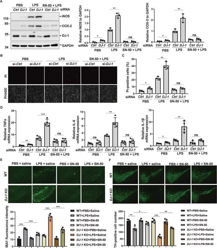
Z. Lin et al.
2
1234567890();,:
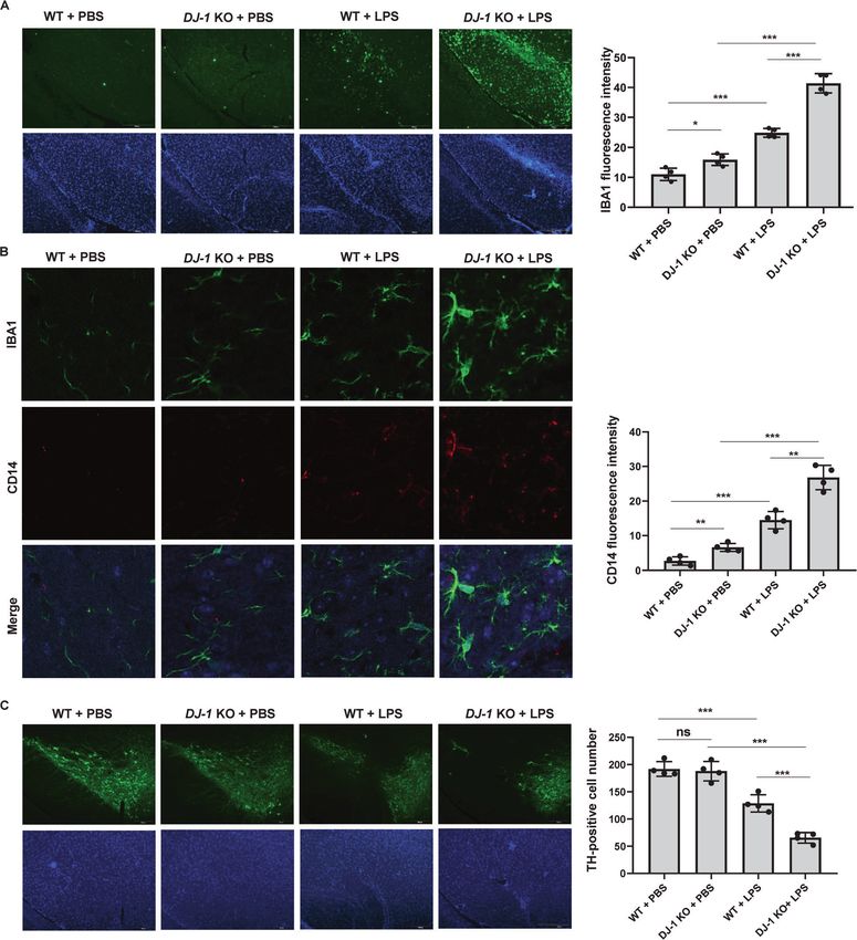
Fig. 1 DJ-1 deficiency leads to microglia activation and DA neuron loss in vivo. A–C DJ-1−/− and the littermate wild-type controls were
microinfused with PBS or LPS into the SN, and 14 days after injection, immunohistochemical staining was conducted. A Slices were stained
with anti-IBA1 antibodies. Scale bar, 200 µm. The fluorescence intensity of IBA1 staining was quantified. n = 4. B Slices were co-stained with
anti-CD14 and anti-IBA1 antibodies. Scale bar, 20 µm. The fluorescence intensity of CD14 staining was quantified. n = 4. C Slices were stained
with anti-TH antibodies. Scale bar, 200 µm. TH-positive neuron numbers in the SN of each slice were quantified. n = 4.
LPS treatment. Moreover, DJ-1 deficiency significantly increases RESULTS
the production of inflammatory factors in microglia and results DJ-1 deficiency leads to microglial activation and DA neuron
in DA neuronal loss in response to LPS stimulus. In addition, NF- loss
κB inhibitors block microglial activation as well as neuronal cell To examine the roles of DJ-1 on microglia in vivo, we first
death induced by DJ-1 deficiency in vivo and in vitro. Thus, our examined the microglia activation in the substantia nigra (SN) in
data illustrate a novel mechanism consisting of DJ-1 inhibiting DJ-1−/− mice and the littermate wild-type controls, with or
neuroinflammation by facilitating the interaction between IκBα without LPS treatment. As shown in Fig. 1A, DJ-1−/− mice
and p65 in microglia. exhibited a greater amount of IBA1 staining than the littermate
Cell Death and Disease (2021)12:715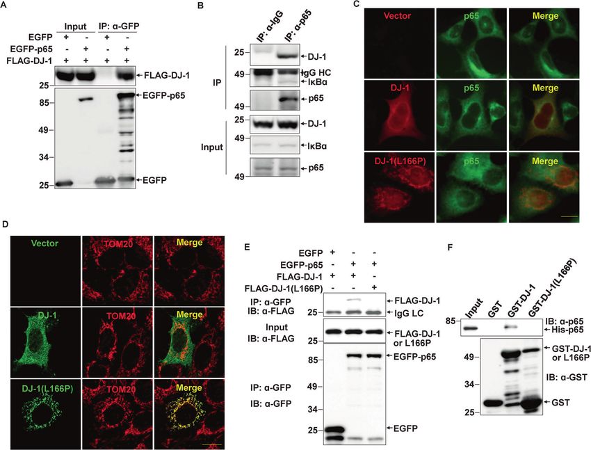
Z. Lin et al.
                                                                                                                                                      3

Fig. 2 DJ-1 deficiency results in microglia activation in vitro. A, B si-Ctrl or si-DJ-1 was transfected into primary microglia (A) or BV2 cells (B)
for 48 h. The cells were then treated with PBS or LPS (100 ng/ml) for 24 h. The cell lysates were analyzed by immunoblotting using the
indicated antibodies. C, D si-Ctrl or si-DJ-1 was transfected into BV2 cells for 72 h. The cells were then treated with PBS or LPS (100 ng/ml) for
6 h and then were subjected to qRT-PCR to measure C iNOS or D COX-2 mRNA levels. n = 3. E–H si-Ctrl or si-DJ-1 was transfected into BV2 cells
for 48 h. The cells were then treated with PBS or LPS (100 ng/ml) for 24 h. The concentration of E NO, F PGE2, G TNFα, or H IL-6 in the cultured
medium was measured. n = 4.

wild-type controls, especially in response to LPS treatment. We             significantly increased mRNA levels of COX-2 and iNOS,
then examined the expression of CD14, a marker of the pro-                  especially in response to LPS treatment (Fig. 2C, D).
inflammatory phenotype of microglia, to access whether the                      Next, we explored the effects of silencing DJ-1 on the release of
increased microglia exhibited pro-inflammatory properties. A                 inflammatory cytokines using BV2 cells. Inflammatory cytokines
greater amount of CD14 expression was detected in DJ-1−/−                   NO and PGE2 are key downstream products of iNOS and COX-2,
mice than that in the littermate wild-type controls. In addition,           respectively [25, 26]. DJ-1 knockdown alone slightly increased NO
most of the IBA1-positive microglia were co-labeled with                    and PGE2 release into the cultured media, whereas DJ-1 deficiency
CD14 staining in DJ-1−/− mice under LPS treatment (Fig. 1B).                dramatically increased the release of NO and PGE2 in response to
Although the number of DA neurons labelled with tyrosine                    LPS stimulation (Fig. 2E, F). In addition, the knockdown of DJ-1 also
hydroxylase (TH) in the SN of DJ-1−/− mice was not significantly             increased the production of TNFα and IL-6 under LPS treatment
different from that of wild-type controls, LPS stimulation                  (Fig. 2G, H). These data suggest that the loss of DJ-1 facilitates
significantly reduced the number of DA neurons in DJ-1−/− mice               microglial activation and intrinsically increases the production of
compared to wild-type controls (Fig. 1C).                                   various pro-inflammatory cytokines, especially in response to LPS
   To explore the mechanism by which DJ-1 deficiency causes                  stimulation.
microglia activation, we performed DJ-1 siRNA experiments in
mouse primary microglia and BV2 microglial cells. We first                   DJ-1 but not its pathogenic L166P mutant represses NF-κB
examined the expression of COX-2 and iNOS, two major                        transcriptional activity
inflammatory mediators. Although knockdown of DJ-1 alone                     Mitogen-activated protein kinases (MAPKs) including extracellular
induced a slight increase in COX-2 and iNOS expression levels               signal-regulated kinase (ERK), Jun N-terminal kinase (JNK), and p38
both in primary microglia and BV2 cells (Fig. 2A, B), DJ-1                  are important in the inflammatory response [27], and DJ-1 has
silencing dramatically increased COX-2 and iNOS levels                      been reported to influence their activity in some types of cells
compared with those in the control in response to LPS                       such as cancer cells, neurons, or astrocytes [28–32]. Thus, we first
stimulation (Fig. 2A, B). In addition, DJ-1 knockdown                       tested whether DJ-1 influenced JNK, p38, or ERK1/2 activity in

Cell Death and Disease (2021)12:715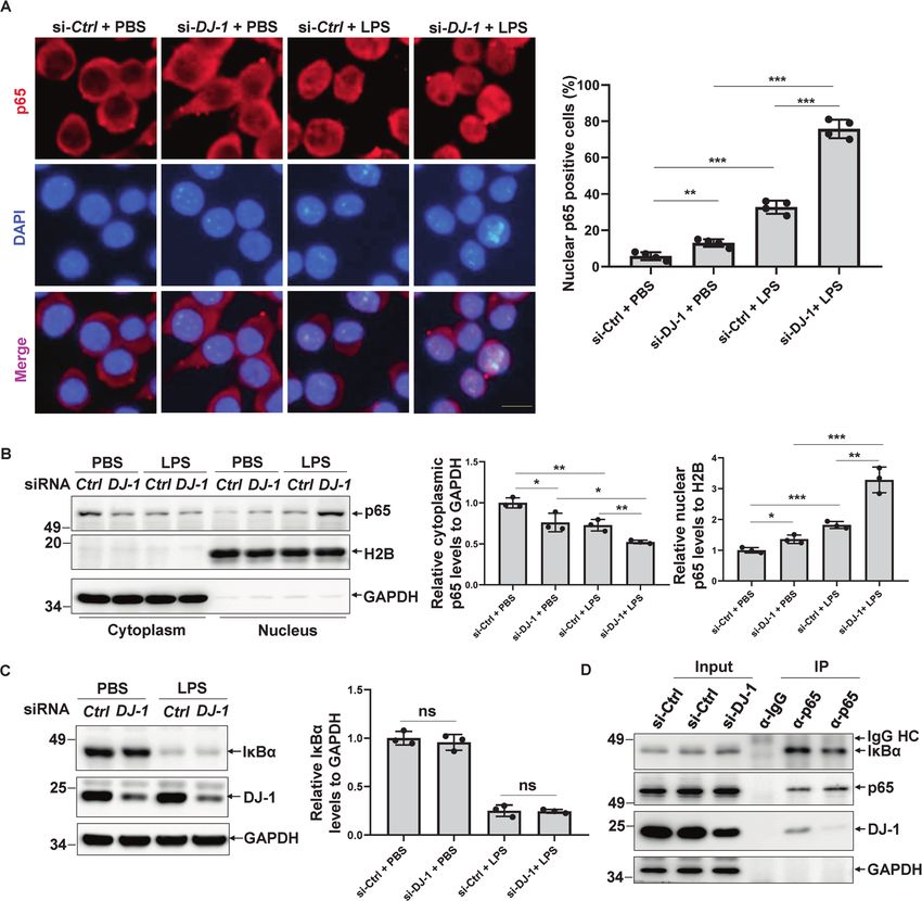
Z. Lin et al.
4

    Fig. 3 DJ-1 rather than L166P mutant represses NF-κB transcriptional activity. A si-Ctrl or si-DJ-1 was transfected into BV2 cells for 72 h.
    Then the cells were treated with PBS or LPS (100 ng/ml) for 15 min. The cell lysates were analyzed by immunoblotting using the indicated
    antibodies. The bar graph shows the relative band intensity of p-JNK1/2/3, p-p38, or p-ERK1/2 to that of GAPDH. n = 3. B BV2 cells harboring
    lentiviral NF-κB-luc were transfected with si-Ctrl or si-DJ-1. After 48 h, the cells were treated with PBS or LPS (100 ng/ml) for 24 h and then
    subjected to a luciferase reporter gene assay. n = 4. C HEK293 cells were transiently transfected with pNF-κB-luc and Renilla along with FLAG or
    FLAG-DJ-1. After 48 h, the cells were subjected to a luciferase reporter gene assay. One-way ANOVA, n = 4. D HEK293 cells were transiently
    transfected with pNF-κB-luc and Renilla along with FLAG, FLAG-DJ-1, or FLAG-DJ-1(L166P). After 48 h, the cells were subjected to a luciferase
    reporter gene assay. One-way ANOVA, n = 4.

    microglial cells. Although LPS treatment immediately activated            co-immunoprecipitated with p65 using anti-p65 antibodies but
    JNK, p38, and ERK1/2 phosphorylation, DJ-1 silencing had no               not control IgG in BV2 cells (Fig. 4B).
    significant effect on JNK, p38, or ERK activation, no matter whether          We next detected the subcellular distribution of p65 and DJ-1
    BV2 cells were treated with LPS or not (Fig. 3A), suggesting that         in cells. DJ-1 and p65 exhibited co-localization in the cytoplasm
    the effect of DJ-1 on microglial activation is not mediated by the        but not in the nucleus (Fig. 4C). In addition, DJ-1(L166P)
    MAPK pathway in microglia.                                                exhibited much more mitochondrial localization and less co-
       The expression levels of inflammatory factors are mainly                localization with p65 in the cytoplasm (Fig. 4C, D). The co-
    regulated by the transcription factor NF-κB [25, 26], and thus,           immunoprecipitation experiment also indicated that wild-type
    we next examined whether DJ-1 regulates NF-κB activity. In BV2            DJ-1 but not L166P mutant interacted with p65 in cells (Fig. 4E).
    cells that stably express Cignal lentiviral NF-κB-luciferase, knock-         To further identify whether there is a direct interaction between
    down of DJ-1 alone significantly increased the NF-κB transcription         DJ-1 and p65, a GST-pull-down assay using purified recombinant
    activity (Fig. 3B), and this activation effect was further augmented      proteins was performed. His-p65 could be directly pulled down by
    under LPS stimulation (Fig. 3B).                                          GST-DJ-1 rather than GST or GST-DJ-1(L166P) (Fig. 4F), indicating
       We next tested the effect of DJ-1 overexpression on NF-κB              that wild-type DJ-1 binds to p65 and represses the transcription
    transcriptional activity. Because the transfection efficiency of           activity of NF-κB, while the pathogenic L166P mutant DJ-1 loses
    overexpression in BV2 cells is very low, we alternatively used            this ability.
    HEK293 cells to perform the reporter gene assay. FLAG-DJ-1
    overexpression but not the control plasmid significantly repressed         DJ-1 represses p65 nuclear translocation in response to LPS
    NF-κB activity (Fig. 3C). However, the pathogenic DJ-1(L166P) lost        treatment
    the ability to repress NF-κB activity (Fig. 3D). These data indicate      We found that DJ-1 interacts with p65 in the cytoplasm and
    that wild-type DJ-1 but not the pathogenic DJ-1(L166P) regulates          inhibits NF-κB transcription activity (Figs. 3B–D and 4A–E), and p65
    microglia-mediated neuroinflammation through the NF-κB signal-             nuclear translocation is necessary for NF-κB transcription activity
    ing pathway.                                                              [26]. Therefore, we next examined whether DJ-1 had an effect on
                                                                              p65 nuclear translocation. Knockdown of DJ-1 induced a slight
    DJ-1 but not the pathogenic L166P mutant binds to the NF-κB               decrease in cytosolic p65 and a slight increase in nuclear p65 (Fig.
    subunit p65                                                               5A, B), whereas DJ-1 silencing resulted in a dramatic nuclear
    Many studies have shown that DJ-1 can influence transcriptional            translocation of p65 in LPS-treated BV2 cells (Fig. 5A, B).
    factors through direct or indirect interactions [33]. We performed           NF-κB inhibitor α (IκBα) is the major partner that interacts with
    co-immunoprecipitation experiments to examine whether DJ-1                and sequesters p65 in the cytoplasm, and thus inhibits NF-κB
    interacts with p65, an NF-κB subunit. In HEK293 cells, Flag-DJ-1          transcription activity, and in response to LPS stimulation, it is
    could be pulled down by EGFP-p65 rather than EGFP using an                phosphorylated by IKK (IκB kinase) and rapidly degraded by the
    anti-GFP antibody (Fig. 4A). Further, endogenous DJ-1 could be            ubiquitin-proteasome system [25, 26]. Therefore, we examined

                                                                                                   Cell Death and Disease (2021)12:715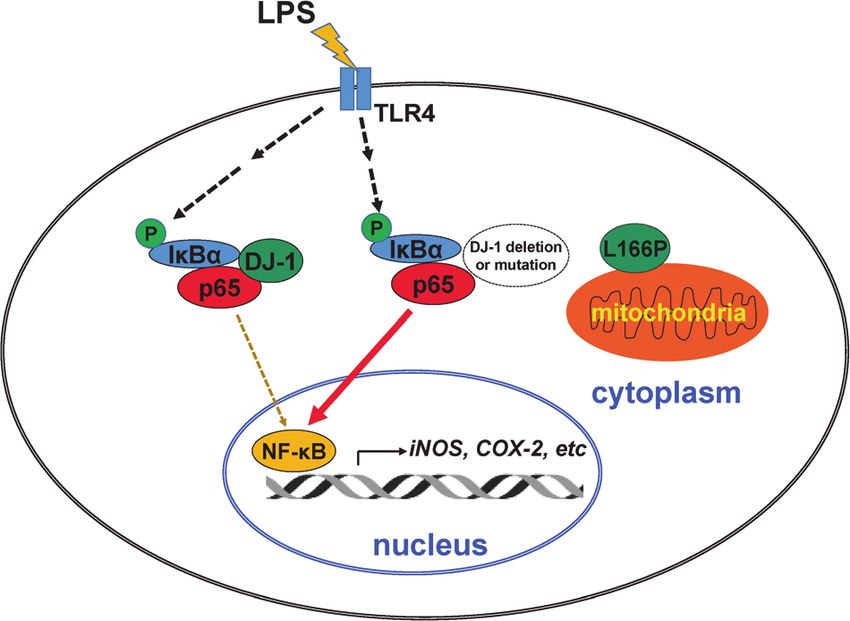
Z. Lin et al.
                                                                                                                                                5

Fig. 4 DJ-1 rather than L166P mutant binds to the p65. A The cell lysate supernatants of HEK293 cells transiently transfected with FLAG-DJ-1
along with EGFP or EGFP-p65 were immunoprecipitated using an anti-GFP antibody. B The supernatants of the BV2 cell lysates were
immunoprecipitated using anti-p65 antibodies or normal rabbit IgG. C HEK293 cells transiently transfected with FLAG, FLAG-DJ-1, or FLAG-DJ-
1(L166P) were subjected to immunocytochemistry using anti-p65 (green) and anti-FLAG (red) antibodies. Scale bar, 10 µm. D HEK293 cells
transiently transfected with FLAG, FLAG-DJ-1, or FLAG-DJ-1(L166P) were subjected to immunocytochemistry using anti-TOM20 (red) and anti-
FLAG (green) antibodies with a confocal microscope. Scale bar, 10 µm. E The cell lysate supernatants of HEK293 cells transiently transfected
with FLAG-DJ-1 or FLAG-DJ-1(L166P) along with EGFP or EGFP-p65 were subjected to immunoprecipitation with anti-GFP antibodies.
F Recombinant GST, GST-DJ-1, or GST-DJ-1(L166P) and His-p65 were subjected to GST-pull-down assay.

whether DJ-1 deletion influences IκBα protein levels or the              Furthermore, conditioned media collected from DJ-1-knockdown
interaction between p65 and IκBα. However, DJ-1 deletion did not        BV2 cells in combination with LPS treatment exhibited a greater
affect IκBα protein levels with or without LPS treatment (Fig. 5C).     amount of cell death as compared to that from control siRNA-
Interestingly, the knockdown of DJ-1 reduced the interaction            knockdown BV2 cells (Fig. 6B, C). Interestingly, SN-50 significantly
between IκBα and p65 in BV2 cells (Fig. 5D). These results indicate     inhibited neurotoxicity caused by loss of microglial DJ-1 combined
that DJ-1 facilitates the interaction between p65 and IκBα by           with LPS stimulation (Fig. 6B, C). We also examined whether SN-50
binding to p65, and loss of DJ-1 promotes p65 nuclear                   inhibits the augmentation of DJ-1 deficiency-induced TNFα, IL-1β,
translocation and thus activates NF-κB transcription activity.          and IL-6 expression in primary microglia in response to LPS
                                                                        stimulation. As shown in Fig. 6D, SN-50 significantly inhibited the
DJ-1 deficiency increases NF-κB-dependent microglial                     mRNA levels of TNFα, IL-1β, and IL-6 induced by the knockdown of
neurotoxicity                                                           DJ-1 under LPS treatment.
To further confirm whether microglia activation by DJ-1 deficiency            We next wondered whether inhibiting the nuclear translocation
is NF-κB dependent, we examined the inflammatory response in             of NF-κB blocks microglial activation and alleviates neurotoxicity
DJ-1-deficient BV2 cells with SN-50, a specific NF-κB inhibitor that      by loss of microglial DJ-1 in vivo. Stereotactic injection of SN-50
directly inhibits NF-κB nuclear transport [34]. SN-50 completely        into the SN dramatically inhibited the activation of microglia in DJ-
blocked iNOS and COX-2 expression induced by DJ-1 knockdown             1−/− mice in response to LPS stimulation (Fig. 6E). SN-50 did not
under LPS stimulation (Fig. 6A). The secretion of inflammatory           affect the number of DA neurons in both wild-type and the
cytokines by activated microglia is considered to be toxic to           littermate DJ-1−/− mice without LPS treatment (Fig. 6F). Whereas,
neuronal cells [35]. To determine whether loss of DJ-1 in microglia     SN-50 treatment dramatically alleviated the loss of DA neurons by
induces neurotoxicity, we performed propidium iodide (PI)               LPS in DJ-1−/− mice (Fig. 6F). Thus, these data revealed that lack of
fluorescence staining to determine the toxic effects of loss of          microglial DJ-1 results in an exaggerated inflammatory response
microglial DJ-1 on N2a cells. Conditioned media from DJ-1-              through the promotion of NF-κB nuclear translocation, which
deficient BV2 cells slightly induced N2a cell death (Fig. 6B, C).        increases DA neurotoxicity, and inhibition of NF-κB nuclear

Cell Death and Disease (2021)12:715
Z. Lin et al.
6
Fig. 5 DJ-1 represses p65 nuclear translocation. A–C BV2 cells were transfected with si-Ctrl or si-DJ-1 for 72 h and then treated with PBS or
LPS (100 ng/ml) for 15 min. A The cells were stained with anti-p65 antibody and DAPI. Scale bar, 10 µm. And the percentage of cells with
nuclear p65 distribution was quantified. n = 4. B The cells were then subjected to a subcellular fractionation assay. The relative band intensity
of cytoplasmic or nuclear p65 to that of GAPDH or H2B was quantified, respectively. n = 3. C Cell lysates were then subjected to
immunoblotting. The relative band intensity of IκBα to that of GAPDH was quantified. n = 3. D The supernatants of the BV2 cells transfected
with si-Ctrl or si-DJ-1 for 72 h were immunoprecipitated using anti-p65 antibodies or normal rabbit IgG.
transport is a potential target for alleviating the damage to DA neuroinflammation is an essential and common pathogenic factor
neurons in PD. in the early stage of PD and is extensively regarded as a
therapeutic target of PD intervention. In addition to the direct
damage to DA neurons, microglia-mediated neuroinflammation
DISCUSSION also induces astrocytes to adopt a neurotoxic function [38].
Microglia play vital roles in CNS hemostasis and are sensitive to Recently, it has been reported that the PD-associated genetic
activation in response to signals derived from dysfunctional factor DJ-1 controls important functions in astrocytes and
neurons or neurotoxins. Activated microglia could produce and microglia [21–24]. However, the role of DJ-1 in microglia in vivo
release pro-inflammatory factors such as TNFα, IL-1α/β, and IL-6, and the potential molecular mechanisms of DJ-1 involves in
leading to damaged DA neurons in the SN [36]. Since it was microglial regulation are largely unclear. In the present study, we
discovered in 1988 that a large number of activated human describe the functions and mechanisms of microglial DJ-1, a PD-
leukocyte antigen (HLA)-positive microglia are present in the SNpc related protein in neuroinflammatory regulation. Wild-type DJ-1
of PD patients [37], there has been a dramatically increased focus rather than its pathogenic mutant L166P blocks activation of
on the microglia in PD in recent years. Microglia-mediated microglia via the NF-κB signaling pathway and protects DA
Cell Death and Disease (2021)12:715Z. Lin et al.
7
Fig. 6 DJ-1 deficiency increases NF-κB-dependent microglial neurotoxicity. A BV2 cells transfected with si-Ctrl or si-DJ-1 for 48 h were
treated with PBS or LPS (100 ng/ml) for 24 h followed by pretreated with SN-50 (20 μM) for 1 h as indicated. After treatment, cell lysates were
subjected to immunoblotting. The relative band intensity of iNOS or COX-2 to that of GAPDH was quantified. n = 3. B N2a cells cultured in
conditioned media collected from BV2 cells with the indicated treatment for 24 h were then stained with PI and visualized with an inverted
microscope. Scale bar, 50 µm. C The percentage of PI-positive cells in (B) was analyzed. n = 3. D Primary microglia transfected with si-Ctrl or si-
DJ-1 for 48 h were treated with PBS or LPS (100 ng/ml) for 6 h followed by pretreated with SN-50 (20 μM) for 1 h as indicated, then the samples
were subjected to qRT-PCR. *P < 0.05, n = 4. E, F DJ-1−/− and the littermate wild-type controls were microinfused with PBS or LPS for 14 days
followed by saline or SN-50 into the SN as indicated, and then the immunohistochemical staining was conducted using (E) anti-IBA1 antibody
or (F) anti-TH antibody. The fluorescence intensity of IBA1 (E) and the number of TH-positive neurons (F) in SN of each slice were quantified.
n = 4.
Cell Death and Disease (2021)12:715Z. Lin et al.
8
neurons from inflammatory damage in vitro and in vivo. In our
observation, wild-type DJ-1 binds to and sequesters p65 in the
cytoplasm, and thus repressing NF-κB hyperactivation, especially
under LPS stimulation. Therefore, lacking DJ-1 in mice, primary
microglia and cultured BV2 cells causes a great increase in the pro-
inflammatory phenotype of microglia compared with the controls,
especially in response to LPS treatment (Figs. 1B and 2). In
addition, it has been reported that DJ-1 has a great antioxidant
function and reactive oxygen species (ROS) scavenging capacity
[20, 33]. Interestingly, ROS are crucial for microglial polarization
regulation and excessive ROS promote the pro-inflammatory
phenotype of microglia [39]. So, we speculated that microglial
activation induced by loss of DJ-1 may also be related to
intracellular oxidative stress.
Two major transcription factors, NF-κB and activator protein 1
(AP-1) are both involved in microglial activation [40, 41]. MAPKs
including ERK, JNK, and p38 are required for AP-1 activation to
induce the expression of inflammatory factors [42, 43]. Although Fig. 7 A schematic diagram shows that DJ-1 functions in
DJ-1 has been reported to affect the activity of these kinases in neuroinflammation in microglia. Wild-type DJ-1 interacts with the
other types of cells [28–32], loss of DJ-1 did not significantly affect subunit p65 of NF-κB and facilitates the interactions between IκBα
their activation in BV2 microglia with or without LPS treatment and p65, thus weakens LPS-induced NF-κB nuclear transport and
(Fig. 3A), suggesting that DJ-1 regulation of the inflammatory microglial activation. Whereas, loss of DJ-1 function by its deletion
response is independent of the AP-1 pathway in microglia. In this or L166P mutation which is mainly distributed in mitochondria and
study, we found that overexpression of DJ-1 inhibits NF-κB fails to bind to p65, augments LPS-induced microglial activation,
activity, and knockdown of DJ-1 activates NF-κB activity (Fig. 3B– and aggravates DA neuron damage.
D). In unstimulated cells, IκB binds to the p65/p50 heterodimer of aggregates microglia-mediated neurotoxicity in an NF-κB-
NF-κB and sequesters NF-κB in the cytosol. Upon stimulation by dependent manner in response to LPS stimulation. In addition,
TNFα, LPS, and ROS, for example, upon LPS recognition, Toll-like inhibition of NF-κB nuclear translocation inhibits microglial
receptor 4 (TLR4) undergoes oligomerization, activates its down- activation and alleviates the loss of DA neurons induced by DJ-1
stream signaling pathway including the IKK complex. Then IκBα is deficiency in vitro and in vivo.
phosphorylated by IKK and subsequently degraded by the
ubiquitin-proteasome system, thus leading to a release in NF-κB
that is transported into the nucleus and subsequently transacti- MATERIALS AND METHODS
vates its target gene expression such as the expression of iNOS, Animals
COX-2, TNFα, IL-1, or IL-6 [25, 26]. DJ-1−/− mice were a kind gift from Dr. Jie Shen at Harvard Medical School
Here, we found that wild-type DJ-1 rather than its pathogenic [45]. The mice were kept in the SPF conditions. All mouse experiments were
mutant L166P directly binds to the p65 subunit and facilitates the approved and carried out in accordance with the Regulations of Experimental
interaction between p65 and IκBα in the cytoplasm, as a DJ-1 Animal Administration, which are issued by the Animal Ethics Committee of
deficiency leads to a reduction in their interaction (Fig. 5D). Loss Soochow University. For drug microfusion in vivo, 6- to 7-week-old mice were
of DJ-1 exacerbates the dissociation of p65 and IκBα, and anesthetized and then fixed on the stereotaxic apparatus (RWD Life Science,
Shenzhen, China). SN-50 (Selleck) was dissolved in ddH2O at a concentration
promotes NF-κB nuclear localization, especially in response to of 0.1 μg/μl and loaded into a 5-μl Hamilton syringe fixed on the stereotaxic
inflammatory stimulation (Fig. 5A, B). Unlike wild-type DJ-1, which apparatus. SN-50 was microinjected into the SN (ML ± 1.2 mm, AP −3.3 mm,
is distributed in the cytoplasm, binds to p65 and inhibits NF-κB DV −4.6 mm) at the rate of 0.2 μl/min (1 μL/mouse). LPS (Sigma) (2 mg/mL)
activity, the L166P mutant DJ-1 translocates to mitochondria and was microinjected into the mouse by the same method (2 μl/mouse).
cannot bind to p65 (Fig. 4C–E). It is also possible that the Fourteen days after LPS treatment, the mice were perfused with 0.9% saline,
translocation of the L166P mutant DJ-1 to the mitochondria may followed by 4% paraformaldehyde. Then, the brains of mice were removed
affect mitochondrial function and promote the activation of and post-fixed overnight, followed by incubation with 30% sucrose solution
microglia by binding to several specific mitochondrial proteins or overnight at 4 °C. Serial 20-µM-thick slices of midbrain were cut with a frozen
affecting mitochondrial metabolism, which needs further inves- microtome for immunohistochemistry. TH+-positive neurons in SN were
counted for each slice under the microscope at a 10x magnification. The
tigation. Thus, these alterations make microglia harboring L166P fluorescence intensity of IBA1 was analyzed using Photoshop7.0 software
mutant DJ-1 more sensitive to inflammatory activators such as (Adobe, CA, USA).
LPS, and augments the inflammatory responses, thus aggravating
the inflammatory damage to DA neurons. Interestingly, the NF-κB
nuclear transport inhibitor SN-50 has a dominant anti- Primary microglia, cell line culture, and transfection
The procedure for extracting primary microglia from the newborn mouse
inflammatory effect and alleviates the neuroinflammation- cortex has been previously described [46]. The mouse microglial BV2 cells
mediated neurotoxicity induced by DJ-1 deficiency combined [47], mouse neuroblastoma Neuro2a (N2a) cells, and HEK293 cells were
with LPS treatment. Many studies have also found that cultured in a DMEM medium (Gibco) with 10% fetal bovine serum,
hyperactivation of the NF-κB pathway is closely associated with penicillin (100 U/ml), and streptomycin (100 µg/ml). All of the cell lines are
PD pathogenesis and is considered a promising intervention authenticated by STR profiling. The small interfering RNAs (siRNAs) and the
target for PD [44]. Increased nuclear translocation and transcrip- expression plasmids were transfected into cells with Lipofectamine
tional activity of NF-κB are observed in PD patients as well as in RNAiMAX reagent (Invitrogen) and Lipofectamine2000 reagent (Invitro-
various PD models [41]. gen), respectively.
In summary, we demonstrated that microglial wild-type
DJ-1 but not DJ-1(L166P) represses neuroinflammation and Immunohistochemistry and immunocytochemistry
neuroinflammation-mediated neurotoxicity by binding to the For immunohistochemistry, the slices of mouse midbrains were stained
p65 subunit of NF-κB, and thus inhibiting NF-κB nuclear overnight with anti-IBA1 antibodies (019-19741, Wako, Japan) for labelling
translocation and activation (Fig. 7). Loss of DJ-1 functions microglia, and anti-TH antibodies (AB152, Millipore) for DA neurons, and
significantly increases microglia activation and dramatically then incubated with the Alexa Fluor 488-conjugated fluorescent secondary
Cell Death and Disease (2021)12:715Z. Lin et al.
9
antibody for 1 h. And anti-CD14 (60253-1-Ig, Proteintech Technology) using PrimeScript RT Master Mix (Takara). Subsequently, quantitative
antibody and the Alexa Fluor 594-conjugated secondary antibody measurement of the target mRNA was subjected to qRT-PCR with SYBR
(Invitrogen) were used for pro-inflammatory microglia staining. Then, the Green Real-Time PCR Master Mix within a 7500 real-time PCR system
slices were labelled with 4′,6-diamidino-2-phenylindole (DAPI, Sigma) for (Applied Biosystems). The following primers were used: mouse COX-2, 5′-
10 min to visualize cellular nuclei. For immunocytochemistry, fixed HEK293 CAGGCTGAACTTCGAAACA-3′ and 5′-GCTCACGAGGCCACTGATACCTA-3′;
cells were incubated with polyclonal anti-p65 (ab32536, Abcam) or anti- mouse iNOS, 5′-TCCCAGCCTGCCCCTTCAAT-3′ and 5′-CGGATCTCTCTCC
TOM20 (42406, Cell Signaling Technology) with monoclonal anti-FLAG TCCTGGG-3′; mouse IL-6, 5′-TAGTCCTTCCTACCCCAATTTCC-3′ and 5′-TT
(F3165, Sigma) antibodies for 4 h, and then incubated with Alexa Fluor 594- GGTCCTTAGCCACTCCTTC-3′; mouse IL-1β, 5′-GCAACTGTTCCTGAACTCAA
and 488-conjugated fluorescent secondary antibody for 2 h. For fixed BV2 CT-3′ and 5′-ATCTTTTGGGGTCCGTCAACT-3′; and mouse TNFα, 5′-CCCTCA-
cells, the primary polyclonal anti-p65 and Alexa Fluor 594-conjugated CACTCAGATCATCTTCT-3′ and 5′-GCTACGACGTGGGCTACAG-3′; mouse
fluorescent secondary antibodies were applied, and then DAPI was used to β-actin, 5′-GACCTGACTGACTACCTC-3′ and 5′-GACAGCGAGGCCAGGATG-
stain the nuclei. Finally, the slices and cells were visualized using an IX71 3′. The relative mRNA levels of these genes to β-actin were calculated by
inverted system microscope (Olympus, Japan), or an LSM800 confocal the 2−ΔΔCT method.
microscope (Zeiss, Germany).
Nuclear and cytoplasmic fractionation assay
Plasmids, siRNAs, and drugs The procedure for extracting nuclear and cytoplasmic fractionation has
The p3xFLAG-myc-cmv-24-DJ-1 and DJ-1(L166P), pGEX-5x-1-DJ-1 and DJ-1 been previously described [50]. Briefly, the BV2 cells were collected and
(L166P) plasmids have been previously described [19, 48]. The EGFP-p65 dissolved in fractionation buffer (2 mM MgAc, 3 mM CaCl2, 320 mM
plasmid was a kind gift from Dr. Yizheng Wang. pET-15b-p65 has been sucrose, 1 mM dithiothreitol (DTT), 0.1 mM EDTA, 0.5% NP-40, and 0.5 mM
previously described [49]. The pNF-κB-luc-containing quadruple NF-κB phenylmethylsulfonyl fluoride (PMSF)) on ice for 20 min. After centrifuga-
response element (GGGAATTTCC) was purchased from Beyotime Biotech- tion at 600 x g at 4 °C for 15 min, the collected supernatant was the
nology Co. Ltd. (Nantong, China). Cignal lentiviral NF-κB-luc was purchased cytoplasmic fraction. The collected pellet was washed once using
from Qiagen. The plasmid fidelities were identified by sequencing. siRNA fractionation buffer without NP-40, and then dissolved in nuclear buffer
sequences targeting mouse DJ-1 mRNA were as follows: sense: 5′ (25% glycerol, 20 mM HEPES (pH 7.9), 1.5 mM MgCl2, 0.2 mM EDTA,
-CGCUUGUUCUCAAAGACUATT-3′, anti-sense: 5′ -UAGUCUUUGAGAACAAG 280 mM KCl, 1 mM DTT, 0.3% NP-40, and 0.5 mM PMSF) as the nuclear
CGGT-3′. fraction.
Immunoprecipitation assay and GST-pull-down assay Luciferase reporter gene assay
The procedures of immunoprecipitation assay using polyclonal anti-GFP BV2 cells stably expressing Cignal lentiviral NF-κB-luc were constructed by
antibodies (Roche), polyclonal anti-p65 antibodies (Cell Signaling Technol- limited dilution method and selected with puromycin (2.5 μg/ml)
ogy), and normal rabbit IgG (Santa Cruz Biotechnology) were according to (Invitrogen) following transfection. BV2 cells stably expressing Cignal
our previous work [19]. Bound proteins and cell lysates (input) were lentiviral NF-κB-luc were transfected with si-Ctrl or si-DJ-1. After 48 h, the
analyzed by immunoblot using the indicated primary antibodies and the cells were treated with 100 ng/ml of LPS or the equal volume PBS for 24 h.
light chain-specific secondary antibodies (Jackson). For the pull-down In HEK293 cells, cells were transfected with FLAG-DJ-1, FLAG-DJ-1(L166P),
assay, GST, GST-DJ-1, and GST-DJ-1(L166P) (20 μg) expressed in Escherichia or empty vector, along with pNF-κB-luc and the Renilla luciferase vector
coli strain JM109 were incubated with glutathione-Sepharose 4B (GE pRL-CMV, which acts as a normalized control. After 48 h, firefly and Renilla
Healthcare) for 30 min in an ice bath, respectively. After washing three luciferase activities were detected with a dual-luciferase reporter system
times, the beads were incubated with His-p65 (50 μg), which is expressed (Promega) using a microplate reader (Infinite M1000 Pro, Tecan).
by Escherichia coli strain BL21 in an ice bath for 4 h. Subsequently, the
beads were washed with cold PBS five times. The input representing 10%
of the proteins used in the immunoprecipitation or pull-down assay and Conditioned medium assays
the interacting proteins were analyzed by immunoblotting. The procedure for conditioned medium assays has been previously
described [51]. Briefly, si-Ctrl or si-DJ-1 were transfected into BV2 cells for
72 h, and then cells were pretreated with or without SN-50 (20 μM) for 1 h,
Immunoblotting and antibodies followed by treatment with or without LPS (100 ng/ml) (Sigma) for 24 h.
Cells were dissolved in cell lysis buffer (0.5% deoxycholate, 1% NP-40, The cells were then washed twice with PBS and cultured for another 24 h
50 mM Tris-HCl pH 7.5, 150 mM NaCl, and protease inhibitor cocktail in fresh media to generate the conditioned media. The conditioned media
(Roche)). The samples were separated by 10 or 15% SDS-PAGE and then of BV2 cells was then collected and filtered by a 0.22 μm filter to culture
transferred onto a polyvinylidene difluoride membrane (PVDF). The PVDF N2a cells for 24 h. The N2a cells were then stained with 1 µM PI (Sigma)
membranes were incubated with the primary antibodies as follows and visualized with an inverted microscope IX71 (Olympus, Japan). The PI-
overnight at 4 °C: polyclonal anti-DJ-1 antibody (AB9212, Chemicon), positive cells were counted and analyzed.
monoclonal anti-GAPDH antibody (MAB374, Millipore), polyclonal anti-
iNOS (ab15323), polyclonal anti-COX2 (ab15191), polyclonal anti-p65
(ab32536) and polyclonal anti-H2B (ab45695) antibodies (Abcam), poly-
Statistical analysis
clonal anti-IκBα antibody (4812, Cell Signaling Technology), monoclonal The densitometric values of three independent immunoblotting experi-
anti-FLAG antibody (F3165, Sigma), and monoclonal anti-DJ-1 (sc-55573) ments were calculated by Photoshop 7.0 software (Adobe). The data
and anti-GFP (sc-9996) antibody (Santa Cruz). The secondary antibodies, presented as the mean ± SD from at least three independent experiments
consisting of horseradish peroxidase-conjugated sheep anti-mouse or anti- were analyzed by GraphPad Prism 8.0 software (San Diego, CA, USA). The
rabbit antibodies (Amersham Pharmacia Biotech), were used for visualiza- statistical significance of the differences between groups was determined
using t-test (two-tailed) following two-way ANOVA unless otherwise
tion with enhanced chemiluminescence (ECL) detection kit (Amersham
Biosciences) and the use of a Chemiluminescence Imaging System specified in the figure legends. The criterion of significance was set at P
(Bioshine ChemiQ 4800). < 0.05. In this study, P values less than 0.001 were shown as ***P < 0.001, P
values less than 0.01 were shown as **P < 0.01, P values less than 0.05 were
shown as *P < 0.05, and no statistical significance was shown as “ns”. The
ELISA assay and NO measurement sample size of experiments is determined on basis of literature in this field.
The levels of PGE2 (Cayman Chemical Company), TNFα, and IL-6 (Boster No sample was excluded from the analysis. Animals were carefully
Biological Technology) in 100 µl of BV2 cultured media were detected assigned based on genotype and age, rather than randomly. There were
using enzyme-linked immunosorbent assay (ELISA) kits. The NO concen- no studies in which investigators were blinded. The replicate numbers are
tration was measured using the Griess method with an NO assay kit described in each figure legend.
(Beyotime Biotechnology) according to the manufacturer’s instructions.
Quantitative real-time PCR (qRT-PCR) REFERENCES
Total RNA was extracted from BV2 cells or primary microglia using TRIzol 1. de Lau LM, Breteler MM. Epidemiology of Parkinson’s disease. Lancet Neurol.
reagent (Invitrogen). Then, the RNA was reverse-transcribed into cDNA 2006;5:525–35.
Cell Death and Disease (2021)12:715Z. Lin et al.
10
2. Li T, Le W. Biomarkers for Parkinson’s disease: how good are they? Neurosci. Bull. regulated kinase pathway by DJ-1 protein through its direct binding to c-Raf
2020;36:183–94. protein. J Biol Chem. 2015;290:17838–47.
3. Poewe W, Seppi K, Tanner CM, Halliday GM, Brundin P, Volkmann J, et al. Par- 32. Xu X, Wang R, Hao Z, Wang G, Mu C, Ding J, et al. DJ-1 regulates tyrosine
kinson disease. Nat Rev Dis Prim. 2017;3:17013. hydroxylase expression through CaMKKbeta/CaMKIV/CREB1 pathway in vitro and
4. Pang SY, Ho PW, Liu HF, Leung CT, Li L, Chang EES, et al. The interplay of aging, in vivo. J Cell Physiol. 2020;235:869–79.
genetics and environmental factors in the pathogenesis of Parkinson’s disease. 33. Ariga H, Takahashi-Niki K, Kato I, Maita H, Niki T, Iguchi-Ariga SM. Neuroprotective
Transl Neurodegener. 2019;8:23. function of DJ-1 in Parkinson’s disease. Oxid Med Cell Longev. 2013;2013:683920.
5. Wang R, Sun H, Wang G, Ren H. Imbalance of lysine acetylation contributes to the 34. Lin YZ, Yao SY, Veach RA, Torgerson TR, Hawiger J. Inhibition of nuclear trans-
pathogenesis of Parkinson’s disease. Int J Mol Sci. 2020;21:7182. location of transcription factor NF-kappa B by a synthetic peptide containing a
6. Wang Q, Liu Y, Zhou J. Neuroinflammation in Parkinson’s disease and its potential cell membrane-permeable motif and nuclear localization sequence. J Biol Chem.
as therapeutic target. Transl Neurodegener. 2015;4:19. 1995;270:14255–8.
7. Kam TI, Hinkle JT, Dawson TM, Dawson VL. Microglia and astrocyte dysfunction in 35. Glass CK, Saijo K, Winner B, Marchetto MC, Gage FH. Mechanisms underlying
parkinson’s disease. Neurobiol Dis. 2020;144:105028. inflammation in neurodegeneration. Cell. 2010;140:918–34.
8. Aloisi F. Immune function of microglia. Glia. 2001;36:165–79. 36. Yang L, Mao K, Yu H, Chen J. Neuroinflammatory responses and Parkinson’ dis-
9. Block ML, Zecca L, Hong JS. Microglia-mediated neurotoxicity: uncovering the ease: pathogenic mechanisms and therapeutic targets. J Neuroimmune Pharm.
molecular mechanisms. Nat Rev Neurosci. 2007;8:57–69. 2020;15:830–7.
10. Hirsch EC, Hunot S. Neuroinflammation in Parkinson’s disease: a target for neu- 37. McGeer PL, Itagaki S, Boyes BE, McGeer EG. Reactive microglia are positive for
roprotection? Lancet Neurol. 2009;8:382–97. HLA-DR in the substantia nigra of Parkinson’s and Alzheimer’s disease brains.
11. Bonifati V, Rizzu P, van Baren MJ, Schaap O, Breedveld GJ, Krieger E, et al. Neurology. 1988;38:1285–91.
Mutations in the DJ-1 gene associated with autosomal recessive early-onset 38. Liddelow SA, Guttenplan KA, Clarke LE, Bennett FC, Bohlen CJ, Schirmer L, et al.
parkinsonism. Science. 2003;299:256–9. Neurotoxic reactive astrocytes are induced by activated microglia. Nature.
12. Abou-Sleiman PM, Healy DG, Quinn N, Lees AJ, Wood NW. The role of pathogenic 2017;541:481–7.
DJ-1 mutations in Parkinson’s disease. Ann Neurol. 2003;54:283–6. 39. Tan HY, Wang N, Li S, Hong M, Wang X, Feng Y. The reactive oxygen species in
13. Waragai M, Wei J, Fujita M, Nakai M, Ho GJ, Masliah E, et al. Increased level of DJ-1 macrophage polarization: reflecting its dual role in progression and treatment of
in the cerebrospinal fluids of sporadic Parkinson’s disease. Biochem Biophys Res human diseases. Oxid Med Cell Longev. 2016;2016:2795090.
Commun. 2006;345:967–72. 40. Shabab T, Khanabdali R, Moghadamtousi SZ, Kadir HA, Mohan G. Neuroin-
14. Hong Z, Shi M, Chung KA, Quinn JF, Peskind ER, Galasko D, et al. DJ-1 and alpha- flammation pathways: a general review. Int J Neurosci. 2017;127:624–33.
synuclein in human cerebrospinal fluid as biomarkers of Parkinson’s disease. 41. Herrero MT, Estrada C, Maatouk L, Vyas S. Inflammation in Parkinson’s disease:
Brain. 2010;133:713–26. role of glucocorticoids. Front Neuroanat. 2015;9:32.
15. Devic I, Hwang H, Edgar JS, Izutsu K, Presland R, Pan C, et al. Salivary alpha- 42. Whitmarsh AJ, Davis RJ. Transcription factor AP-1 regulation by mitogen-activated
synuclein and DJ-1: potential biomarkers for Parkinson’s disease. Brain. 2011;134: protein kinase signal transduction pathways. J Mol Med. 1996;74:589–607.
e178. 43. Wu J, Du J, Gu R, Zhang L, Zhen X, Li Y, et al. Inhibition of neuroinflammation by
16. Shi M, Bradner J, Hancock AM, Chung KA, Quinn JF, Peskind ER, et al. Cere- synthetic androstene derivatives incorporating amino acid methyl esters on
brospinal fluid biomarkers for Parkinson disease diagnosis and progression. Ann. activated BV-2 microglia. ChemMedChem. 2015;10:610–6.
Neurol. 2011;69:570–80. 44. Singh SS, Rai SN, Birla H, Zahra W, Rathore AS, Singh SP. NF-kappaB-mediated
17. Canet-Avilés RM, Wilson MA, Miller DW, Ahmad R, McLendon C, Bandyopadhyay neuroinflammation in Parkinson’s disease and potential therapeutic effect of
S, et al. The Parkinson’s disease protein DJ-1 is neuroprotective due to cysteine- polyphenols. Neurotox Res. 2020;37:491–507.
sulfinic acid-driven mitochondrial localization. Proc Natl Acad Sci USA. 45. Goldberg MS, Pisani A, Haburcak M, Vortherms TA, Kitada T, Costa C, et al.
2004;101:9103–8. Nigrostriatal dopaminergic deficits and hypokinesia caused by inactivation of the
18. Zhang L, Shimoji M, Thomas B, Moore DJ, Yu SW, Marupudi NI, et al. Mito- familial Parkinsonism-linked gene DJ-1. Neuron. 2005;45:489–96.
chondrial localization of the Parkinson’s disease related protein DJ-1: implications 46. Guo DK, Zhu Y, Sun HY, Xu XY, Zhang S, Hao ZB, et al. Pharmacological activation
for pathogenesis. Hum Mol Genet. 2005;14:2063–73. of REV-ERBalpha represses LPS-induced microglial activation through the NF-
19. Ren H, Fu K, Wang D, Mu C, Wang G. Oxidized DJ-1 interacts with the mito- kappaB pathway. Acta Pharm Sin. 2019;40:26–34.
chondrial protein BCL-XL. J Biol Chem. 2011;286:35308–17. 47. Tian LP, Zhang S, Xu L, Li W, Wang Y, Chen SD, et al. Selenite benefits embryonic
20. Repici M, Giorgini F. DJ-1 in Parkinson’s disease: clinical insights and therapeutic stem cells therapy in Parkinson’s disease. Curr Mol Med. 2012;12:1005–14.
perspectives. J Clin Med. 2019;8:1377. 48. Ren H, Fu K, Mu C, Zhen X, Wang G. L166P mutant DJ-1 promotes cell death by
21. Kim JH, Choi DJ, Jeong HK, Kim J, Kim DW, Choi SY, et al. DJ-1 facilitates the dissociating Bax from mitochondrial Bcl-XL. Mol Neurodegener. 2012;7:40.
interaction between STAT1 and its phosphatase, SHP-1, in brain microglia and 49. Fu C, Chen D, Chen R, Hu Q, Wang G. The schizophrenia-related protein
astrocytes: a novel anti-inflammatory function of DJ-1. Neurobiol. Dis. 2013;60:1–10. dysbindin-1A is degraded and facilitates NF-kappa B activity in the nucleus. PLoS
22. Trudler D, Weinreb O, Mandel SA, Youdim MB, Frenkel D. DJ-1 deficiency triggers ONE. 2015;10:e0132639.
microglia sensitivity to dopamine toward a pro-inflammatory phenotype that is 50. Chen D, Li YP, Yu YX, Zhou T, Liu C, Fei EK, et al. Dendritic cell nuclear protein-1
attenuated by rasagiline. J Neurochem. 2014;129:434–47. regulates melatonin biosynthesis by binding to BMAL1 and inhibiting the tran-
23. Ji YJ, Wang HL, Yin BL, Ren XY. Down-regulation of DJ-1 augments neuroin- scription of N-acetyltransferase in C6 cells. Acta Pharm Sin. 2018;39:597–606.
flammation via Nrf2/Trx1/NLRP3 axis in MPTP-induced Parkinson’s disease mouse 51. Yu YX, Li YP, Gao F, Hu QS, Zhang Y, Chen D, et al. Vitamin K2 suppresses
model. Neuroscience. 2020;442:253–63. rotenone-induced microglial activation in vitro. Acta Pharm Sin. 2016;37:1178–89.
24. Peng L, Zhou Y, Jiang N, Wang T, Zhu J, Chen Y, et al. DJ-1 exerts anti-
inflammatory effects and regulates NLRX1-TRAF6 via SHP-1 in stroke. J Neu-
roinflammation. 2020;17:81.
ACKNOWLEDGEMENTS
25. Oeckinghaus A, Ghosh S. The NF-kappaB family of transcription factors and its
We would like to thank Dr. Jie Shen at Harvard Medical School to provide us the DJ-1
regulation. Cold Spring Harb Perspect Biol. 2009;1:a000034.
knockout mice.
26. Napetschnig J, Wu H. Molecular basis of NF-kappaB signaling. Annu Rev Biophys.
2013;42:443–68.
27. Sabio G, Davis RJ. TNF and MAP kinase signalling pathways. Semin Immunol.
2014;26:237–45. AUTHOR CONTRIBUTIONS
28. Mo JS, Kim MY, Ann EJ, Hong JA, Park HS. DJ-1 modulates UV-induced oxidative G.W. and H.R. performed study concept and design. H.R. wrote the paper. G.W.
stress signaling through the suppression of MEKK1 and cell death. Cell Death revised the paper. Z.L. performed the experiments, Z.L., C.C., D.Y., and J.D. analyzed
Differ. 2008;15:1030–41. the data. All authors read and approved the final paper.
29. Waak J, Weber SS, Waldenmaier A, Görner K, Alunni-Fabbroni M, Schell H, et al.
Regulation of astrocyte inflammatory responses by the Parkinson’s disease-
associated gene DJ-1. FASEB J. 2009;23:2478–89.
FUNDING
30. Mo JS, Jung J, Yoon JH, Hong JA, Kim MY, Ann EJ, et al. DJ-1 modulates the p38
This research was funded by the National Natural Science Foundation of China
mitogen-activated protein kinase pathway through physical interaction with
(31970966, 32070970, and 31871023), the National Key Scientific Research and
apoptosis signal-regulating kinase 1. J Cell Biochem. 2010;110:229–37.
Development Program of China (2016YFC1306000), Suzhou Clinical Research Center of
31. Takahashi-Niki K, Kato-Ose I, Murata H, Maita H, Iguchi-Ariga SMM, Ariga H.
Neurological Disease (Szzx201503), and the Priority Academic Program Development of
Epidermal growth factor-dependent activation of the extracellular signal-
Jiangsu Higher Education Institutions.
Cell Death and Disease (2021)12:715Z. Lin et al.
11
ETHICS APPROVAL Publisher’s note Springer Nature remains neutral with regard to jurisdictional claims
The study does not involve human participants, human data, or human tissue. All in published maps and institutional affiliations.
mouse experiments were approved and carried out in accordance with the
Regulations of Experimental Animal Administration, which are issued by the
Animal Ethics Committee of Soochow University.
Open Access This article is licensed under a Creative Commons
Attribution 4.0 International License, which permits use, sharing,
COMPETING INTERESTS adaptation, distribution and reproduction in any medium or format, as long as you give
The authors declare no competing interests. appropriate credit to the original author(s) and the source, provide a link to the Creative
Commons license, and indicate if changes were made. The images or other third party
material in this article are included in the article’s Creative Commons license, unless
indicated otherwise in a credit line to the material. If material is not included in the
ADDITIONAL INFORMATION
article’s Creative Commons license and your intended use is not permitted by statutory
Correspondence and requests for materials should be addressed to G.W. or H.R. regulation or exceeds the permitted use, you will need to obtain permission directly
from the copyright holder. To view a copy of this license, visit http://creativecommons.
Reprints and permission information is available at http://www.nature.com/ org/licenses/by/4.0/.
reprints
© The Author(s) 2021
Cell Death and Disease (2021)12:715You can also read