ZNO NANOPARTICLES: CROSSLINK BETWEEN CYTOTOXICITY ON LIVER CELL LINES AND IN VIVO BIOSAFETY ON DIFFERENT MICE ORGANS - JOURNAL OF APPLIED ...
←
→
Page content transcription
If your browser does not render page correctly, please read the page content below
Journal of Applied Pharmaceutical Science Vol. 9(S1), pp 058-066, March, 2019
Available online at http://www.japsonline.com
DOI: 10.7324/JAPS.2019.S107
ISSN 2231-3354

ZnO nanoparticles: Crosslink between cytotoxicity on liver cell lines
and In Vivo biosafety on different mice organs

Nahla Nabil Kamel1*, Maha Zaki Rizk1, Walaa Gamal Hozayen2, Abdel-Hamid Zaki Abdel-Hamid1
1
 Therapeutic Chemistry Department, National Research Centre, Giza, Egypt.
 Biochemistry Division, Department of Chemistry, Faculty of Science, Beni-Suef University, Beni-Suef, Egypt and Department of Biotechnology and Life
2

 Sciences, Faculty of Postgraduate Studies for Advanced Sciences (PSAS), Beni-Suef University, Beni-Suef, Egypt.

     ARTICLE INFO                        ABSTRACT
     Received on: 03/09/2018             The wide applications of zinc oxide nanoparticles (ZnO NPs) in several biomedical aspects have raised many concerns
     Accepted on: 10/10/2018             about their toxicity effects. This study aimed to explore the cytotoxic effect of different sizes (14 , 30, and 50 nm) of
     Available online: 07/03/2019        ZnO NPs on liver cancer cell lines hepatoma G2 (HepG2), HuH7 cells (HuH7 hepatoma cells) versus normal cells
                                         (THLE2 cells). Cytotoxicity, oxidative stress, and apoptosis were investigated. The results pointed out that the particle
                                         size of 14 nm recorded the highest activity, whereas no cytotoxic effect was observed on THLE2. Also, oxidative stress
     Key words:
                                         elicited a reduction in reduced glutathione with an increase in lipid peroxides and caspase-3. In addition, RT-PCR
     ZnO nanoparticles, HepG2
                                         revealed a significant up-regulation in caspase-3 gene expression. Histopathological investigation confirmed the
     cells, HuH7, liver cancer,
                                         biosafety of the same particle size which revealed the least toxic effect on all mice organs (liver, kidney, lung, and
     cytotoxicity.
                                         brain). In conclusion, this particle size of ZnO NPs could be useful in future therapeutic applications on liver cancer.

INTRODUCTION                                                                         optical, electrical, and thermal performance has been widely used
           Nanotechnology allows the manipulation of materials at                    in the field of catalysis, sunscreen cosmetics, paint materials, and
nanoscale level (1–100 nm), which enables precision engineering                      food packaging materials (Di Pasqua et al., 2008). The chemical and
to control physicochemical properties of these nanoparticles                         physical properties of nanoparticles have a strong influence on the
(NPs), as well as their interactions with biological systems (Wang,                  way they interact with biological components or the environment
2008). Owing to their small size, high number per given mass,                        (Dobrovolskaia and McNeil, 2007) and also on the way they move,
large specific surface area responses (Brown et al., 2002), concern                  accumulate, and clear in the body (Scheringer, 2008). Industrial
regarding their fate in biological systems have raised a research in                 food processing is intended to modify flavor, texture, and storage
this field. Previous studies have indicated that NPs can penetrate                   behavior by mixing with zinc oxide nanoparticles (ZnO NPs).
cell nuclei and directly interfere with DNA structure, causing                       After ingestion of food containing ZnO NPs, mechanical (chewing
increased oxidative stress, induction of apoptosis, genotoxicity, and                and peristalsis) and chemical (interaction with intestinal enzymes)
DNA damage (Ma et al., 2009; Wang et al., 2007). Inorganic NPs,                      processes reduce food into smaller components to maintain
including metal oxides, are promising materials for applications                     physiological processes. Some previous research has shown that
in medicine, such as cell imaging, biosensing, drug/gene delivery,                   ZnO NPs may cause cytotoxicity to several types of cells, such
and cancer therapy (Wu et al., 2011).                                                as osteoblast cancer cells (Nair et al., 2009), human bronchial
           ZnO NPs have at least one dimension in the range of                       epithelial cells (BEAS-2B) (Heng et al., 2011), human alveolar
1–100 nm, a white powder nearly insoluble in water, with a unique                    adenocarcinoma cells (Ahamed et al., 2011a), human hepatocytes,
                                                                                     and embryonic kidney cells (Guan et al., 2012).
                                                                                                ZnO NPs with respect to their specific properties,
*Corresponding Author
                                                                                     such as their relative ease of production, ability to alter their
Nahla Nabil Kamel, Therapeutic Chemistry Department, National
Research Centre, Giza, Egypt. E-mail: nahlanabel_nrc @ yahoo.com                     physiochemical characteristics, their inherent toxicity against
                                                                                     cancerous and bacterial cells, and ability to functionalize them
This article was presented at the 5th Euro-Mediterranean Conference and Expo on
Life Sciences, Pharma and Biomedicine (BioNat-V), Limassol, Cyprus.

© 2019 Nahla Nabil Kamel et al. This is an open access article distributed under the terms of the Creative Commons Attribution 4.0 International License
(https://creativecommons.org/licenses/by/4.0/).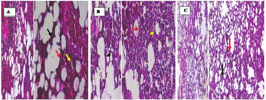
Kamel et al. / Journal of Applied Pharmaceutical Science 9 (S1); 2019: 058-066                            059

with chemotherapeutic drugs and cancer targeting molecules,                 Experimental design
make them an appealing candidate for biomedical applications
(Goharshadi et al., 2011; Moosavi et al., 2010).                            Cell culture
          Exposure to ZnO NPs leads to reactive oxygen species                         Cell Line (HepG2, HuH7, and THLE2 cells) was obtained
(ROS) generation and activation specific signal transduction                from American Type Culture Collection, cells were cultured using
pathways in target cells. Imbalance in the ROS production and               DMEM (Invitrogen/Life Technologies) supplemented with 10%
antioxidant defense system of cells results in several different            FBS (Hyclone,), 10 µg/ml of insulin (Sigma), and 1% penicillin-
outcomes leading to cell death (Sharma et al., 2009). The                   streptomycin. All of the other chemicals and reagents were from
oxidative stress mechanism of ZnO NPs could be attributed to the            Sigma or Invitrogen. Plate cells (cells density 1.2– − 1.8 × 10,000
combination of more than one phenomenon, including generation               cells/well) in a volume of 100 µl complete growth medium + 100
of ROS on the surface of particles (Park et al., 2011), dissolution         µl of the tested compound per well in a 96-well plate for 24 hours
and release of Zn2+ ions in the culture media, and physical                 before the MTT assay.
interaction of ZnO NPs with the membrane wall leading to the
deformation and rupture of membrane (Li et al., 2008).                      Cell viability assay
          The distinct properties of NPs, such as small size, high                    Viability of HepG2, HuH7, and THLE2 cells was
number per given mass, large specific surface area, have aroused            assessed by the MTT assay as described by Mossman with
global concern regarding their fate in biological systems. Previous         some modifications (Ahamed et al., 2011b). Briefly, 1 × 10⁴
studies have indicated that NPs can penetrate cell nuclei of target cells   cells/well were seeded in 96-well plates and exposed to varying
and directly interfere with DNA structure, causing several adverse          concentrations of ZnO NPs for 24 hours. At the end of the
effects represented in increased production of ROS, induction of            exposure, the culture medium was removed from each well to
apoptosis, genotoxicity, and DNA damage (Wang et al., 2007).                avoid interference of ZnO NPs and replaced with new medium
          Nanoparticles can translocate from entry portals into             containing MTT solution (0.5 mg/ml) in an amount equal to 10%
the lymphatic and circulatory systems and ultimately to body                of culture volume and incubated for 3 hours at 37°C until a purple-
tissues and organs due to their small size. Some NPs can produce            colored formazan product developed. The resulting formazan
irreversible damage to cells by oxidative stress or/and organelle           product was dissolved in acidified isopropanol. Further, the 96-
injury due to their composition and size causing severe cytotoxicity        well plate was centrifuged at 2,300 × g for 5 minutes to settle the
(Chernyshev et al., 2018). The distribution of these NPs to other           remaining ZnO NPs. Then, a 100 μl supernatant was transferred to
organs, such as liver, spleen, brain, heart, and kidney may lead to         other fresh wells of a 96-well plate, and absorbance was measured
dysfunction of these organs.                                                at 570 nm by a microplate reader (FLUOstar Omega, Cary, NC).
          A previous study by Sharma et al. (2012) demonstrated that
                                                                            Lipid peroxidation assay
HepG2 cells exposed to 30 nm ZnO nanoparticles for 12 hours showed
a decrease in cell viability and the mode of cell death induced by ZnO                The extent of membrane LPO was estimated by
nanoparticles was apoptosis. Also, Guan et al. (2012) assessed ZnO          measuring the formation of malondialdehyde (MDA) using the
NPs cytotoxicity on human hepatocyte (L02) using 50 nm.                     method of Ohkawa et al. (1979). MDA is one of the end products
          Based on the previous information, this study was                 of membrane LPO. Briefly, a mixture of 0.1 ml cell extract and
designed to demonstrate the impact of different particle sizes of           1.9 ml of 0.1 M sodium phosphate buffer (pH 7.4) was incubated
ZnO NPs on normal and cancer cells to elucidate their cytotoxicity          at 37°C for 1 hour. After the incubation mixture was precipitated
on target cells and their safety on normal cells which may help in          with 5% TCA and centrifuged (2,300 × g for 15 minutes at room
the future application of ZnO NPs as promising therapeutic agents.          temperature) to collect the supernatant. Then, 1.0 ml of 1% TBA
                                                                            was added to the supernatant and placed in the boiling water for
MATERIALS AND METHODS                                                       15 minutes. After cooling to room temperature, the absorbance
                                                                            of the mixture was taken at 532 nm and was converted to MDA
Preparation and characterization of nanoparticles
                                                                            and expressed in nmole MDA/mg protein using molar extinction
           Zinc oxide nanoparticles were synthesized using an               coefficient of 1.56 × 10⁵ M−1 cm−1. A reaction mixture devoid of
aqueous solution of zinc nitrate or zinc acetate and a solution of          cell extract served as control.
potassium hydroxide or sodium hydroxide. The different solutions
were prepared using deionized water while stirring and heating,             Glutathione estimation
respectively. The hydroxide solution was slowly added to the zinc                      GSH level was quantified using Ellman’s (1959) method.
salt solution at room temperature under vigorous stirring, which            Briefly, a mixture of 0.1 ml of cell extract and 0.9 ml of 5% TCA
resulted in the formation of a white precipitate. The precipitate           was centrifuged (2,300 × g for 15 minutes at 4°C). Then, 0.5 ml
was centrifuged at 6,500 rpm while cooling to −10°C for 20                  of supernatant added into 1.5 ml of 0.01% DTNB and the reaction
minutes. The resulted product was washed with distilled water               was monitored at 412 nm.
several times followed by absolute alcohol. The obtained product
was calcined at 500°C in an air atmosphere for 3 hours. The                 Caspase-3 assay enzyme
resulted nanoparticles were tested by TEM in order to evaluate                        Activity of caspase-3 enzyme was measured in exposed
the particle morphology; shape and size. The nanoparticles of               and control cells using standard assay kit (BioVision, Inc.). Crude
zinc oxide were then redisposed in deionized water to give the              cell extract was prepared as described above. This assay is based
requested concentrations (Hingorani et al., 1993).                          on the principle that activated caspases in apoptotic cells cleave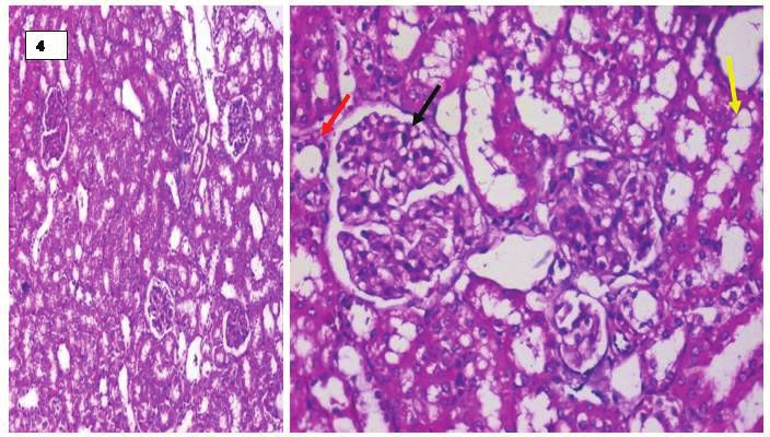
060                             Kamel et al. / Journal of Applied Pharmaceutical Science 9 (S1); 2019: 058-066

the synthetic substrates to release free chromophore p-nitroanilide                Organs were kept in 10% formaldehyde and then
(pNA), which is measured at 405 nm. The pNA was generated after          embedded in paraffin for histopathological examinations (Van
specific action of caspase-3 on tertrapeptide substrates DEVD-           Herck et al., 2001). After deparaffinization and dehydration,
pNA (Ahamed et al., 2010b; Berasain et al., 2005). The reaction          sections of kidney, lung, spleen, heart, and brain of 4 mm thickness
mixture consisted of 50 μl of cell extract protein (50 μg), 50 μl of     were stained with Hematoxylin and Eosin (H&E) and examined
2× reaction buffer (containing 10 mM dithiothreitol) and 5 μl of 4       under the light microscope (Bancroft and Stevens, 1996). All
mM DEVDpNA substrate in a total volume of 105 μl. The reaction           histopathologic processing and assessment of specimens were
mixture was incubated at 37°C for 1 hour and the absorbance of           performed by an experienced observer unaware of the identity of
the product was measured using microplate reader (Synergy-HT,            the sample being examined to avoid any bias.
BioTek) at 405 nm according to the manufacturer’s instruction.
                                                                         Ethical procedure
Total RNA isolation and quantitative real-time PCR analysis                         All procedures related to animal care and treatments
for Caspase-3                                                            strictly adhered to the ethical procedures according to the Guide for
           Human liver cancer HepG2 cells were cultured in six-          Care and Use of Laboratory published by the US National Institute
well plates and exposed to 15 μg/ml ZnO NPs for 24 hours. At the         of Health policies and approved by Animal Care and Use of the
end of the exposure, total RNA was extracted by RNeasy mini Kit          Committee of National Research Centre (approval no:13-088).
(Qiagen, Valencia, CA) according to the manufacturer’s instructions.
Concentration of the extracted RNA was determined using Nanodrop         Statistical analysis
8000 spectrophotometer (Thermo-Scientific, Wilmington, DE) and                    All data were expressed as mean ± SD (n = 5). Statistical
the integrity of RNA was visualized on a 1% agarose gel using a gel      analysis was carried out by one-way analysis of variance
documentation system (Universal Hood II, BioRad, Hercules, CA).          (ANOVA), version 8, coupled with Costat Software Computer
The first strand of cDNA was synthesized from 1 μg of total RNA          program, where unshared letters are significant at p ≤ 0.05.
by reverse transcriptase using M-MLV (Promega, Madison, WI)
and oligo (dT) primers (Promega) according to the manufacturer’s         RESULTS AND DISCUSSION
protocol. Quantitative real-time PCR was performed by QuantiTect         Physical characterization of ZnO NPs
SYBR Green PCR kit (Qiagen) using an ABI PRISM 7900HT
                                                                                    Physical characterization of ZnO NPs with sizes 14, 30,
Sequence Detection System (Applied Biosystems, Foster City, CA).
                                                                         and 50 nm was examined by transmission electron microscopy
Two microliters of template cDNA were added to the final volume of
                                                                         (TEM) to assert their particle size. Results demonstrated the
20 μl of reaction mixture. Real-time PCR cycle parameters included
                                                                         accuracy of preparation of the selected NPs which showed sizes
10 minutes at 95°C followed by 40 cycles involving denaturation at
                                                                         (a) relatively close to 14 nm, (b) close to 30 nm, and (c) close to
95°C for 15 seconds, annealing at 60°C for 20 seconds, and elongation
                                                                         50 nm (Fig. 1).
at 72°C for 20 seconds. The sequences of the specific sets of primer
for caspase-3 (forward: 5′-TGGTTCATCCAGTCGCTTTG-3′                       Cytotoxicity of ZnO nanoparticles
reverse: 5′-CATTCTGTTGCCACCTTTCG-3′), and β-actin
                                                                                   The cytotoxicity of ZnO NPs in HepG2, HuH7, and
(forward:       5′-CCTTCCTGGGCATGGAGTCCT-3′                   reverse:
                                                                         THLE2 cells with different sizes (14, 30, and 50 nm) was
5′-GGAGCAATGATCTTGATCTTC-3′) used in this study are given
                                                                         evaluated by MTT. These cells were exposed to ZnO NPs (0.01–
in our previous publication (Ahamed et al., 2011a). Expressions
                                                                         100 µg/ml) for 24 hours. The results for IC50 using the different
of selected genes were normalized to the β-actin gene, which was
                                                                         particle sizes are demonstrated in Table 1. It could be concluded
used as an internal housekeeping control. All the real-time PCR
                                                                         that the smallest size NPs induced the least toxic effect on
experiments were performed in triplicate, and data were expressed as
                                                                         normal cells and the highest effective toxic concentrations on
the mean of at least three independent experiments.
                                                                         cancer cells followed by the particle size of 50 nm, while the
Histopathological examination                                            highest toxicity on cancer cell lines was demonstrated using a
                                                                         particle size of 30 nm.
          Male albino mice (105), weighing 20–25 g, were obtained
                                                                                   The most important critical features of ZnO NPs are
from the animal house of National Research Centre. Animals were
                                                                         their selective toxicity toward cancerous cells in comparison
housed in cages kept at standardized conditions (22°C ± 5°C, 55%
                                                                         with normal human cells which shows potential benefits of
± 5% humidity, and 12 hour light/dark cycle). They were allowed
                                                                         these NPs as new antitumor drugs. Cytotoxicity results are in
free access to water and pelleted standard chow diet.
                                                                         agreement with published studies which reported that ZnO NPs
          Histopathological examination was performed for
                                                                         significantly induce cytotoxicity to human glioma cells, and no
assessment of cytotoxicity of different size of ZnO NPs in different
                                                                         major effect was observed on normal cells (Ostrovsky et al.,
organs in mice. After careful selection on the basis of health and
                                                                         2009). In addition, ZnO NPs exhibited a preferential ability to
inclusion criteria, animals were left for 1 week for acclimatization
                                                                         kill human myeloblastic leukemia cells (HL60) as compared with
and were randomly divided into two main groups according to the
                                                                         normal peripheral blood mononuclear cells (Premanathan et al.,
following schedule:
                                                                         2011). Hanley et al. (2008) also observed that ZnO NPs exhibit a
          G1: Control group (normal healthy animals) (n = 15).
                                                                         strong preferential ability to kill cancerous T cells compared with
          G2: Animals orally administered different sizes of ZnO-
                                                                         normal cells. Selective killing of cancer cells by ZnO NPs may
NP using a dose (150 mg/kg BW) for 14 consecutive days as
                                                                         be of important clinical interest, as one of the greatest challenges
described by Sharma et al. (2012) (n = 45).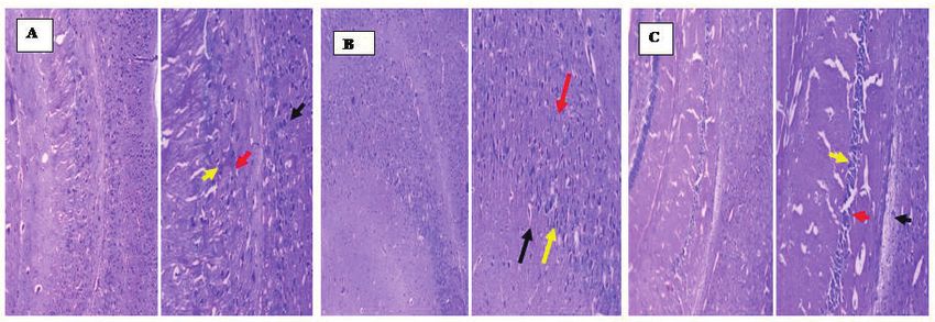
Kamel et al. / Journal of Applied Pharmaceutical Science 9 (S1); 2019: 058-066                                   061

         Figure 1. Characterization of ZnO NPs by TEM.

 Table 1. Cytotoxic effect of ZnO NPs in HepG2, HuH7, and THLE2 cells as           whether by the excessive generation of ROS or by depletion of
                   assessed by a MTT assay for 24 hours.                           cellular antioxidant capacity (Ahamed et al., 2010a; Wise et al.,
                                               IC50 (Ug/ml)                        2010). There has been an increase in biochemical, clinical, and
   ZnO NPs (nm)                                                                    epidemiological evidence that indicate the involvement of ROS
                              HepG2               HuH7            THLE2
          14                   0.048               0.17            202.9
                                                                                   and oxidative stress in various diseases, including cancer (Jomova
                                                                                   and Valko, 2011). Our previous studies highlight that metal and
          30                   0.092               1.17             6.9
                                                                                   metal oxide nanoparticles are able to induce ROS generation and
          50                    0.1                0.22             12.7
                                                                                   oxidative stress in different types of cells (Ahamed et al., 2011a).
The data are presented as the mean (n = 5).                                                  In the present work, ZnO NPs were found to produce
                                                                                   ROS, which consequently causes cell damage, as previously
facing cancer chemotherapy is the inability of anticancer drugs                    shown by Nel et al. (2006) and Park et al. (2010) that cell toxicity
to effectively distinguish between a normal cell and a cancer cell.                is induced by different nanoparticles due to oxidative stress.
Oxidative stress markers                                                           Glutathione reduced
Lipid peroxidation                                                                          Cells exposed to ZnO NPs showed significant depletion
          Lipid peroxidation was examined by measuring                             in GSH level in all tested groups on both cancer cell lines, while no
hydroperoxide concentration. Significant increase in hydroperoxide                 depletion was observed on normal cells as evident from Figure 3.
formation was observed in all compounds as evident from                            These results were reinforced by the study of Sharma et al. (2009),
Figure 2. Oxidative stress has been suggested to play an important                 who showed GSH level was depleted in human epidermal cells
role in the mechanisms of toxicity of a number of nanoparticles                    exposed to ZnO NPs. Furthermore, an increase in the formation
                                                                                   of hydroperoxide compared with control, indicating an increase in

Figure 2. ZnO NPs-induced MDA level in different cell lines. Values are               Figure 3. GSH level in different cell lines. Values are expressed as mean
expressed as mean ± SD, (n = 5). Statistical analysis is carried out using the        ± SD, (n = 5). Statistical analysis is carried out using the SPSS computer
SPSS computer program coupled with Co-Stat computer program (version 8),              program coupled with Co-Stat computer program (version 8), where unshared
where unshared letters between groups are the significance value at p ≤ 0.05.         letters between groups are the significance value at p ≤ 0.05.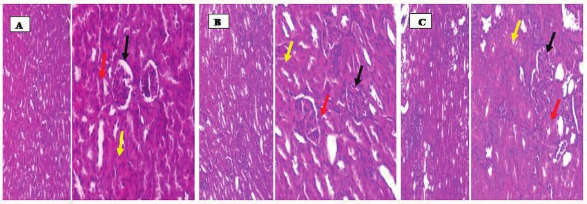
062 Kamel et al. / Journal of Applied Pharmaceutical Science 9 (S1); 2019: 058-066
MDA after exposure to ZnO NPs which coincided with the results brain revealed that size 14 nm induced the most promising non-
obtained by Sharma et al. (2009). toxic effect and no cytotoxic effects were observed in heart and
spleen with any of particle sizes or dose used. A systematic study
Caspase-3 enzyme of the effect of different sizes, doses, and distribution is critical
Cells exposed to ZnO NPs showed a significant increase to the deep understanding of the toxicity mechanism. There are
in caspase 3 level revealing toxicity on both cancer cell lines with impacts factors affecting the properties of nanomaterials to differ
no effect on normal cell lines. ELIZA data are shown in Figure 4 significantly from other materials, including increased relative
and are confirmed by RT-PCR (Table 2). This could be explained surface area and quantum confinement effect. This is in agreement
by the fact that activated caspase-3 is capable of autocatalysis, with the report of Ben-Slama et al. (2015) who pointed out that
as well as cleaving and activating other members of the caspase the histopathological analysis of the kidney showed intratubular
family, which may subsequently cause rapid and irreversible protein deposition but no significant glomerular changes. These
apoptosis (Sánchez-Pérez et al., 2009). Also, in the same aspect, findings were also concurrent with Al Rasheed et al. (2012)
Akhtar et al. (2012) and Sharma et al. (2009) demonstrated up- who declared that renal histopathological examination showed
regulation in mRNA and higher activity of the caspase-3 enzyme alteration of proteinaceous casts in the tubules and renal tubular
in liver cancer cells treated with ZnO NPs. dilatation in the ZnO-NP treated rats. High dose of ZnO-NP showed
Histopathological Findings
Photomicrographs for histopathological study for doses
≤150 mg/kg BW were excluded since no significant effects on
organ pattern were detected. Histopathological investigations on
different organs using a dose of 150 mg/kg BW with the different
sizes are shown in Photomicrographs 1–8. The results showed
that the histopathological disorders in liver, kidney, lung, and
Table 2. Effect of ZnO NPs on mRNA expression level of Caspase-3 in
different cell lines.
Caspase-3
ZnO NPs HepG2 HuH7 THLE2
Conc. IU/ml Conc. IU/ml Conc. IU/ml
14 nm 275.6 ± 2.3a 193.2 ± 2.3b 18.5 ± 0.4h
30 nm 140.6 ± 2.1 d
57.1 ± 0.9 f
21.0 ± 1.4g
50 nm 163.1 ± 1.4 c
82.5 ± 1.7e
7.4 ± 0.3j
Control Cells 7.0 ± 0.4j 12.0 ± 0.4i 4.0 ± 0.2k Figure 4. Caspase-3 enzyme in different cells. Values are expressed as mean ±
Values are expressed as mean ± SD, (n = 5). Statistical analysis is carried out using SPSS SD, (n = 5). Statistical analysis is carried out using the SPSS computer program
computer program coupled with Co-Stat computer program (version 8), where unshared coupled with Co-Stat computer program (version 8), where unshared letters
letters between groups are the significance value at p ≤ 0.05. between groups are the significance value at p ≤ 0.05.
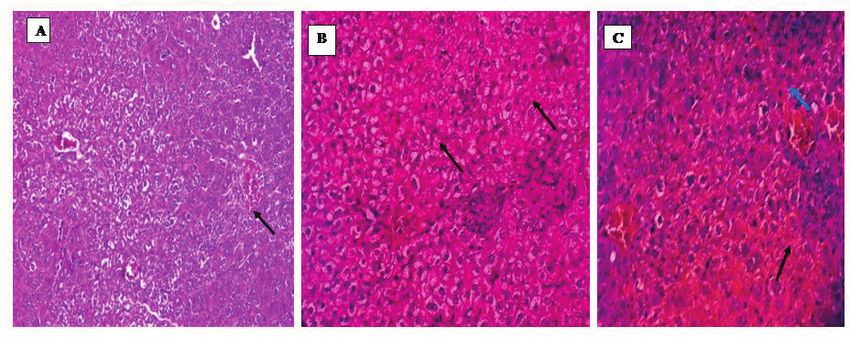
Photomicrograph 1. (A) Liver from the group (14 nm, 150 mg/kg) showed hepatic tissue with preserved (intact) lobular hepatic architecture and mild hydropic
degeneration (black arrow) (H&E, ×400). (B) Liver section from the group (30 nm, 150 mg/kg) showed preserved (intact) lobular hepatic architecture, hepatocyte with
ballooning (red arrow), hydropic degeneration of hepatocytes (black arrow) and binucleated hepatocytes, congested blood vessels with 10% mild necrosis (H&E, ×400).
(C) liver section from the group (50 nm, 150 mg/kg) showed hepatic tissue with binuclear hepatocytes (blue arrow) and sinusoidal dilatations vacuolation of hepatocytes
along with hemorrhageand necrosis (black arrow) (H&E, ×400).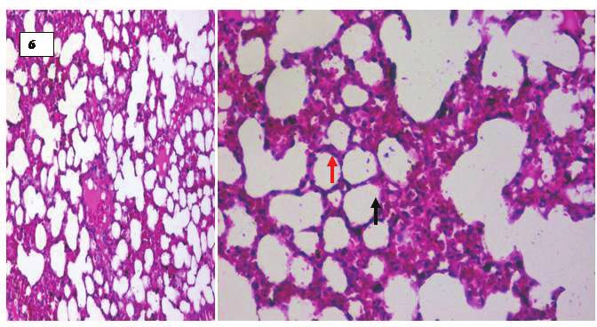
Kamel et al. / Journal of Applied Pharmaceutical Science 9 (S1); 2019: 058-066                                               063

Photomicrograph 2. Liver section showed hepatic tissue with preserved (intact) lobular hepatic architecture and normal morphological appearance (H&E, ×200).

Photomicrograph 3. (A) Kidney section from the group (14 nm, 150 mg/kg) showed most of the corpuscles with high cellularity and obliterated capsular space (black
arrow). Proximal convoluted tubules show destructed epithelial lining (red arrow), destructed epithelial lining of distal convoluted tubules (yellow arrow) (H&E, ×200,
×400). (B) Kidney section from the group (30 nm, 150 mg/kg) showed most of the corpuscles with very high cellularity and obliterated capsular space (black arrow).
Proximal convoluted tubules show destructed epithelial lining (red arrow), destructed epithelial lining of distal convoluted tubules (yellow arrow) (H&E, ×200, ×400).
(C) Kidney section from the group (50 nm, 150 mg/kg) showed most of the corpuscles with very high cellularity and obliterated capsular space (black arrow). Proximal
convoluted tubules show marked destructed epithelial lining (red arrow), destructed epithelial lining of distal convoluted tubules (yellow arrow) (H&E, ×200, ×400).

Photomicrograph 4. Kidney section from control group showed renal cortex of renal corpuscle with normal glomerulus (black arrow), the juxtaglomerular apparatus,
the normal pattern of proximal convoluted (red arrow) and distal convoluted (blue arrow) tubules (H&E, ×200, ×400).
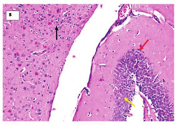
064                                  Kamel et al. / Journal of Applied Pharmaceutical Science 9 (S1); 2019: 058-066

Photomicrograph 5. (A) Lung section from group (14 nm, 150 mg/kg) showing the thick sections, having alveoli with thick intralveolar septum (back arrow), type I
and type II pneumocytes were also clearly seen (red arrow), there is collection of lymphocytes (yellow arrow) (H&E, ×200, ×400). (B) Lung section from group (30
nm, 150 mg/kg) showing the very thick sections, having alveoli with thick intralveolar septum (back arrow), type I and type II pneumocytes were also clearly seen (red
arrow), there is many collection of lymphocytes (yellow arrow) (H&E, ×200, ×400). (C) Lung section from the group (50 nm, 150 mg/kg) showing the thin sections of
control having normal alveoli with thin intralveolar septum (back arrow), type I and type II pneumocytes were also clearly seen (red arrow) with scattered interstitial
lymphocyte (H&E, ×100, ×200).

Photomicrograph 6. Lung section from control group showing the thin sections of control having normal alveoli with thin intralveolar septum (back arrow), type I and
type II pneumocytes were also clearly seen (red arrow) (H&E, ×200, ×400).

Photomicrograph 7. (A) Brain section of group (14 nm, 150 mg/kg) of NCR, showed the cerebellum showed an atrophic molecular layer (black arrow), mildly atrophic
and distorted granular layer (red arrow) and Purkinje layer (yellow arrow) and presence of thin closely packed small cells in the granular layer as well a large Purkinje
cells in the Purkinje cell layer (H&E stain, ×100, x×200). (B) Brain section of group (30 nm, 150 mg/kg) of NCR, showed the cerebellum showed a moderate size
molecular layer (black arrow), mildly atrophic and distorted granular layer (red arrow) and Purkinje layer (yellow arrow) and presence of moderately closely packed
small cells in the granular layer as well a large Purkinje cells in the Purkinje cell layer (H&E stain, ×100, x×200). (C) Brain section of group (50 nm, 150 mg/kg) of
NCR, showed the cerebellum showed well defined molecular (black arrow), granular layer (red arrow) and Purkinje layers (yellow arrow) and presence of numerous
closely packed small cells in the granular layer as well a large Purkinje cells in the Purkinje cell layer (H&E stain, ×100, ×200).
Kamel et al. / Journal of Applied Pharmaceutical Science 9 (S1); 2019: 058-066 065
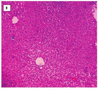
Photomicrograph 8. Brain section of a control group, showed the cerebellum of the group a showed normal histological features, illustrating a well defined molecular
(black arrow), granular (red arrow) and Purkinje layers (yellow arrow) and presence of numerous closely packed small cells in the granular layer, as well a large Purkinje
cells in the Purkinje cell layer. Or showing the normal structure of neuronal cells (H&E stain, ×200).
massive atrophy and fragmentation of numerous glomeruli, the ACKNOWLEDGMENTS
renal tubules showed epithelial exfoliation, degeneration, and The authors would like to thank Prof. Dr. Essam
necrosis. Some of the renal tubules showed casts in their lumina. Rashwan for his support during the in vitro studies.
The brain tissue showed neuronal cell degeneration and vacuoles
were observed in the hippocampus, which is indicative of fatty CONFLICT OF INTEREST
degeneration in the hippocampus. In the lung tissues, inflammatory There is no conflict of interest.
cells, foamy cells, and granulomatous lesions were observed
in TiO2 NPs treated mice (Xu et al., 2013). Park et al. (2011) FUNDING
reported that granulomatous lesions were found in the bronchiole The research is funded by the National Research Centre
and alveoli of the lung after 14 days TiO2 NPs treatment (5, 20, as a grant for Ph.D. Thesis.
and 50 mg/kg) in mice.
The observed cytotoxicity of Zinc oxide nanoparticles on REFERENCES
cancer cells is explained by the fact that these NPs typically have Ahamed M, Akhtar MJ, Raja M, Ahmad I, Siddiqui MK, AlSalhi
neutral hydroxyl groups attached to their surface, which plays a key MS, Alrokayan SA. ZnO nanorod-induced apoptosis in human alveolar
adenocarcinoma cells via p53, survivin and bax/bcl-2 pathways: role of
role in their surface charge behavior. In the aqueous medium and at
oxidative stress. Nanomedicine, 2011a; 7:904–13.
high pH, the chemisorbed protons (H+) move out from the particle Ahamed M, Akhtar MJ, Siddiqui MA, Ahmad J, Musarrat J,
surface leaving a negatively charged surface with partially bonded Al-Khedhairy AA, AlSalhi MS, Alrokayan SA. Oxidative stress mediated
oxygen atoms (ZnO−). At lower pH, protons from the environment apoptosis induced by nickel ferrite nanoparticles in cultured A549 cells.
are likely transferred to the particle surface, leading to a positive Toxicology, 2011b; 283:101–8.
charge from surface ZnOH2+ groups. The isoelectric point indicates Ahamed M, AlSalhi MS, Siddiqui MKJ. Silver nanoparticle
that ZnO nanoparticles will have a strong positive surface charge applications and human health. Clin Chim Acta, 2010a; 411:1841–8.
Ahamed M, Posgai R, Gorey TJ, Nielsen M, Hussain S, Rowe
under physiological conditions. Given that cancer cells frequently
J. Silver nanoparticles induced heat shock protein 70, oxidative stress and
contain a high concentration of anionic phospholipids on their apoptosis in Drosophila melanogaster. Toxicol Appl Pharmacol, 2010b;
outer membrane and large membrane potentials, interactions with 242:263–9.
positively charged ZnO nanoparticles are expected to be driven Akhtar MJ, Ahamed M, Kumar S, Khan MM, Ahmad J,
by electrostatic interactions, thereby promoting cellular uptake, Alrokayan SA. Zinc oxide nanoparticles selectively induce apoptosis in
phagocytosis, and ultimate cytotoxicity. human cancer cells through reactive oxygen species. Int J Nanomedicine,
2012; 7:845–57.
CONCLUSION Al Rasheed N, Abdel Baky NA, Al Rasheed N, Shebly W, Ahmed
AM, Faddah LM. Effect of vitamin E and α-lipoic acid on nano zinc oxide
It could be concluded that the smallest size NPs (14 nm) induced renal cytotoxicity in Rats. Afr J Pharm Pharmacol, 2012; 6:2211–23.
induced the least toxic effect on normal cells and the highest Bancroft JD, Stevens A. Theory and practice of histological
effective toxic concentrations on cancer cells followed by the techniques. 4th edition, Churchill Livingstone, London, UK, p 163, 1996.
particle size of 50 nm, while the highest toxicity on cancer cell lines Ben-Slama I, Mrad I, Rihane N, El Mir L, Sakly M, Amara S.
was demonstrated using the particle size of 30 nm. The marked Sub-acute oral toxicity of zinc oxide nanoparticles in male rats. J Nanomed
difference in cytotoxicity between cancer cells and normal cells Nanotechnol, 2015; 6:1–6.
suggests an exciting potential for ZnO NPs as novel promising Berasain C, Garcia-Trevijano ER, Castillo J, Erroba E,
Santamaria M, Lee DC. Novel role for amphiregulin in protection fromliver
alternatives to cancer therapy. injury. J Biol Chem, 2005; 280:19012–20.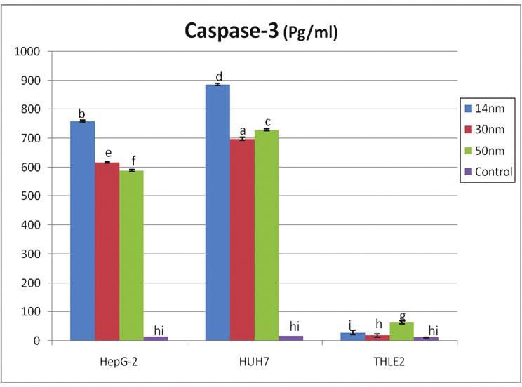
066                                Kamel et al. / Journal of Applied Pharmaceutical Science 9 (S1); 2019: 058-066

            Brown JS, Zeman KL, Bennett WD. Ultrafineparticle deposition                    Park EJ, Kimb H, Kimb Y, Yic J, Choid K, Park K. Inflammatory
and clearance in the healthy and obstructed lung. Am J Respir Crit Care         responses may be induced by a single intratracheal instillation of iron
Med, 2002; 166(9):1240–7.                                                       nanoparticles in mice. Toxicology, 2010; 275:65–71.
            Chernyshev VV, Zakharenko AM, Ugay SM, Hien TT, Hai LH,                         Park SJ, Park YC, Lee SW, Jeong MS, Yu KN, Jung H, Lee JK,
Kholodov AS, Burykina TI, Stratidakis AK, Mezhuev Ya O, Tsatsakis AM,           Kim JS, Cho MH. Comparing the toxic mechanism of synthesized zinc
Golokhvast KS. Morphologic and chemical composition of particulate              oxide nanomaterials by physicochemical characterization and reactive
matter in motorcycle engine exhaust. Toxicol Rep, 2018; 5:224–30.               oxygen species properties. Toxicol Lett, 2011; 207:197–203.
            Di Pasqua AJ, Sharma KK, Shi YL, Toms BB, Ouellette W,                          Premanathan M, Karthikeyan K, Jeyasubramanian K,
Dabrowiak JC, Asefa T. Cytotoxicity of mesoporous silica nanomaterials. J       Manivannan G. Selective toxicity of ZnO nanoparticles toward Gram
Inorg Biochem, 2008; 102:1416–23.                                               positive bacteria and cancer cells by apoptosis through lipid peroxidation.
            Dobrovolskaia MA, McNeil SE. Immunological properties of            Nanomedicine NBM, 2011; 7:184–92.
engineered nanomaterials. Nat Nanotechnol, 2007; 2:469–78.                                  Sánchez-Pérez Y, Chirino YI, Osornio-Vargas AR, Morales-
            Ellman GI. Tissue sulfhydryl groups. Arch Biochem Biophys,          Bárcenas R, Gutiérrez-Ruíz C, Vázquez-López I, García-Cuellar CM. DNA
1959; 82:70–7.                                                                  damage response of A549 cells treated with particulate matter (PM10) of
            Goharshadi EK, Abareshi M, Mehrkhah R, Samiee S, Moosavi            urban air pollutants, Cancer Lett, 2009; 278:192–200.
M, Youssefi A, Nancarrow P. Preparation, structural characterization,                       Scheringer M. Nanoecotoxicology: environmental risks of
semiconductor and photoluminescent properties of zinc oxide                     nanomaterials. Nat Nanotechnol, 2008; 3:322–3.
nanoparticles in a phosphoniumbased ionic liquid. Mat Sci Semicon Proc,                     Sharma V, Shukla RK, Saxena N, Parmar D, Das M, Dhawan A.
2011; 14:69–72.                                                                 DNA damaging potential of zinc oxide nanoparticles in human epidermal
            Guan R, Kang T, Lu F, Zhang Z, Shen H, Liu M. Cytotoxicity,         cells. Toxicol Lett, 2009; 185:211–8.
oxidative stress, and genotoxicity in human hepatocyte and embryonic                        Sharma V, Singh P, Pandey AK, Dhawan A. Induction of
kidney cells exposed to ZnO nanoparticles. Nanoscale Res Lett, 2012;            oxidative stress, DNA damage and apoptosis in mouse liver after sub-acute
7:602.                                                                          oral exposure to zinc oxide nanoparticles. Mutat Res, 2012; 745:84–91.
            Hanley C, Layne J, Punnoose A, Reddy KM, Coombs I, Coombs                       Van Herck H, Baumans V, Brandt CJ, Boere HA, Hesp AP, Van
A, Feris K, Wingett D. Preferential killing of cancer cells and activated       Lith HA. Blood sampling from the retro-orbital plexus, the saphenous vein
human T cells using ZnO nanoparticles. Nanotechnology, 2008; 19:295103.         and the tail vein in rats: comparative effects on selected behavioural and
            Heng BC, Zhao X, Xiong S, Ng KW, Boey FY, Loo JS.                   blood variables. Lab Anim, 2001; 35:131.
Cytotoxicity of zinc oxide (ZnO) nanoparticles is influenced by cell density                Wang JJ, Sanderson JSB, Wang H. Cyto- and genotoxicity of
and culture format. Arch Toxicol, 2011; 85:695–704.                             ultrafine TiO2 particles in cultured human lymphoblastoid cells. Mutat Res,
            Hingorani S, Pillia V, Kumar P, Multani MS, shah DO.                2007; 628:99–106.
Microemulsion mediated synthesis of zinc-oxide nanoparticles for varistor                   Wang ZL. Splendid one-dimensional nanostructures of zinc
studies. Mat Res Bull, 1993; 28:1303–10.                                        oxide: a new nanomaterial family for nanotechnology. ACS Nano, 2008;
            Jomova K, Valko M. Advances in metal-induced oxidative stress       2:1987–92.
and human disease. Toxicology, 2011; 283:65–87.                                             Wise JP, Goodale BC, Wise SS. Silver nanospheres are cytotoxic
            Li N, Xia T, Nel AE. The role of oxidative stress in ambient        and genotoxic to fish cells. Aquat Toxicol, 2010; 97:34–41.
particulate matter-induced lung diseases and its implications in the toxicity               Wu YN, Chen DH, Shi XY, Lian CC, Wang TY, Yeh CS, Ratinac
of engineered nanoparticles. Free Radical Bio Med, 2008; 44:1689–99.            KR, Thordarson P, Braet F, Shieh DB. Cancer-cell-specific cytotoxicity of
            Ma LL, Zhao JF, Wang J, Duan YM, Liu J, Li N, Yan J, Ruan           non-oxidized iron elements in iron core-gold shell NPs. Nanomedicine,
J, Wang H, Hong F. The acute liver injury in micecaused by nano-anatase         2011; 7:420–7.
TiO2. Nanoscale Res Lett, 2009; 4:1275–85.                                                  Xu J, Shi H, Ruth M, Yu H, Lazar L, Zou B, Yang C, Wu A,
            Moosavi M, Goharshadi EK, Youssefi A. Fabrication,                  Zhao J. Acute toxicity of intravenously administered titanium dioxide
characterization, and measurement of some physicochemical properties of         nanoparticles in mice. PLoS One, 2013; 8:1–6.
ZnO nanofluids. Int J Heat Fluid, 2010; 31:599–605.
            Nair S, Sasidharan A, Divya Rani VV, Menon D, Nair S,
Manzoor KS. Role of size scale of ZnO nanoparticles and microparticles
on toxicity toward bacteria and osteoblast cancer cells. J Mater Sci Mater
Med, 2009; 20:235–241.
            Nel A, Xia T, Madler L, Li N. Toxic potential of materials at the     How to cite this article:
nanolevel. Science, 2006; 311:622–27.                                             Kamel NN, Rizk MZ, Hozayen WG, Abdel-Hamid A-HZ.
            Ohkawa H, Ohishi N, Yagi K. Assay for lipid peroxides in animal
tissues by thiobarbituric acid reaction. Anal Biochem, 1979; 95:351–8.
                                                                                  ZnO nanoparticles: Crosslink between cytotoxicity on liver
            Ostrovsky S, Kazimirsky G, Gedanken A, Brodie C. Selective            cell lines and In Vivo biosafety on different mice organs.
cytotoxic effect of ZnO nanoparticles on glioma cells. Nano Res, 2009;            J Appl Pharm Sci, 2019; 9(S1):058–066.
2:882–90.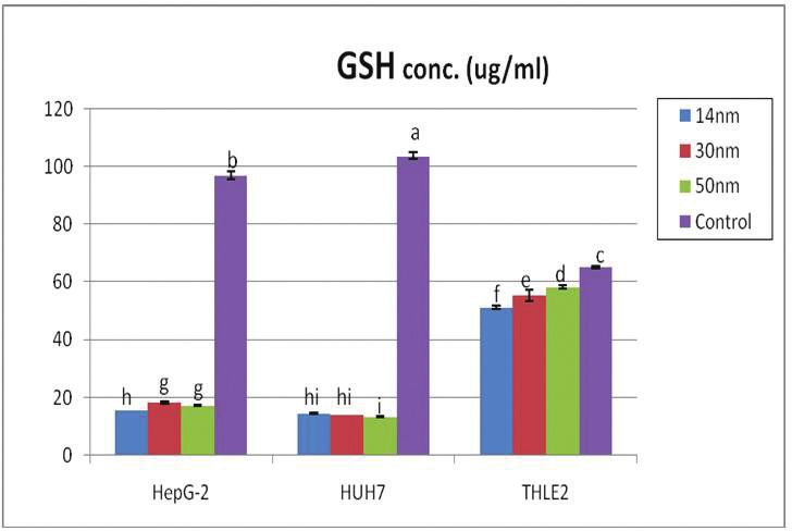
You can also read


