Forsythia Fruit Prevents Fulminant Hepatitis in Mice and Ameliorates Inflammation in Murine Macrophages - MDPI
←
→
Page content transcription
If your browser does not render page correctly, please read the page content below
nutrients
Article
Forsythia Fruit Prevents Fulminant Hepatitis in Mice and
Ameliorates Inflammation in Murine Macrophages
Yun Hee Jeong, Youn-Hwan Hwang                        , Tae In Kim       , You-Chang Oh *         and Jin Yeul Ma *

                                           Korean Medicine (KM)-Application Center, Korea Institute of Oriental Medicine, 70, Cheomdanro, Dong-gu,
                                           Daegu 41062, Korea; runxi0333@kiom.re.kr (Y.H.J.); hyhhwang@kiom.re.kr (Y.-H.H.); tikim@kiom.re.kr (T.I.K.)
                                           * Correspondence: ulivuli@kiom.re.kr (Y.-C.O.); jyma@kiom.re.kr (J.Y.M.); Tel.: +82-53-940-3882 (Y.-C.O.);
                                             +82-53-940-3812 (J.Y.M.)

                                           Abstract: Forsythia Fruit (FF), the fruit of Forsythia suspensa, has been used since ancient times as
                                           an herbal medication in East Asia to treat inflammation, gonorrhea, and pharyngitis. However, the
                                           efficacy of FF against liver damage due to inflammation has not been studied. Here, we explored
                                           the protective effects of FF in a mouse hepatitis model induced by lipopolysaccharide (LPS)/D-
                                           galactosamine (GalN) treatment. We measured inflammatory cytokine and aminotransferase levels
                                           in mouse blood and analyzed the effects of FF on inflammatory gene and protein expression levels
                                           in liver tissue. Our results show that FF treatment effectively lowers inflammatory cytokine and
                                           serum aminotransferase levels in mice and inhibits the expression of hepatic cytokine mRNA and
                                           inflammatory proteins. Furthermore, treatment with FF activated the antioxidant pathway HO-
                                           1/Nrf-2 and suppressed severe histological alteration in the livers of LPS/D-GalN-treated mice.
                                           Further investigation of the effects of FF on inflammatory reactions in LPS-stimulated macrophages
                                 showed that pretreatment with FF inhibits inflammatory mediator secretion and activation of in-

                                           flammatory mechanisms both in a mouse macrophage RAW 264.7 cells and in primary peritoneal
Citation: Jeong, Y.H.; Hwang, Y.-H.;       macrophages. These results show that FF has potential worth as a candidate for the treatment of
Kim, T.I.; Oh, Y.-C.; Ma, J.Y. Forsythia   fulminant inflammatory reactions and subsequent liver injury.
Fruit Prevents Fulminant Hepatitis in
Mice and Ameliorates Inflammation
                                           Keywords: Forsythia Fruit; liver injury; inflammation; antioxidant; lipopolysaccharide; D-galactosamine
in Murine Macrophages. Nutrients
2021, 13, 2901. https://doi.org/
10.3390/nu13082901

                                           1. Introduction
Academic Editor: Pietro Vajro
                                                 Fulminant liver injury is characterized by rapid, widespread liver dysfunction and
Received: 6 August 2021                    can result in encephalopathy, jaundice, and severe coagulopathy [1,2]. It is also a clinical
Accepted: 21 August 2021                   manifestation of sudden and severe hepatic failure, which is difficult to prevent and treat,
Published: 23 August 2021                  resulting in poor prognosis and a high mortality rate [3]. The main causes of acute liver
                                           injury are antigen-induced infections and poisoning by hepatotoxic drugs, but there are also
Publisher’s Note: MDPI stays neutral       many unknown causes [2]. At present, the only effective treatment is liver transplantation,
with regard to jurisdictional claims in    so the development of effective prevention and treatment modalities are necessary [4].
published maps and institutional affil-          Lipopolysaccharide (LPS) is an endotoxin originated from the gram-negative bacteria
iations.                                   E. coli and was initially confirmed as a Toll-like receptor 4 (TLR4) ligand, which causes a
                                           rapid and powerful inflammatory reaction leading to sepsis or multiple organ failure [5].
                                           In addition, LPS plays a pivotal role at the onset of endotoxic damage and increases inflam-
                                           matory cytokine expression, causing liver damage. D-galactosamine (GalN) decreases the
Copyright: © 2021 by the authors.          concentrations of uridine triphosphate, uridine diphosphate, and uridine monophosphate
Licensee MDPI, Basel, Switzerland.         through metabolic disorders of galactose, leading to the inhibition of RNA synthesis, in-
This article is an open access article     filtration of inflammatory cells, necrosis of liver cells, and induction of lesions similar to
distributed under the terms and            hepatitis [6,7]. D-GalN also induces changes in colorectal mucosal permeability, increasing
conditions of the Creative Commons         endotoxin absorption, which interferes with the ability of liver cells to repair membranes
Attribution (CC BY) license (https://      and causes hepatic toxicity [8]. Eventually, D-GalN causes necrosis of the liver during acute
creativecommons.org/licenses/by/           exposure and cirrhosis of the liver and cellular tumors during chronic exposure [9,10].
4.0/).

Nutrients 2021, 13, 2901. https://doi.org/10.3390/nu13082901                                                https://www.mdpi.com/journal/nutrients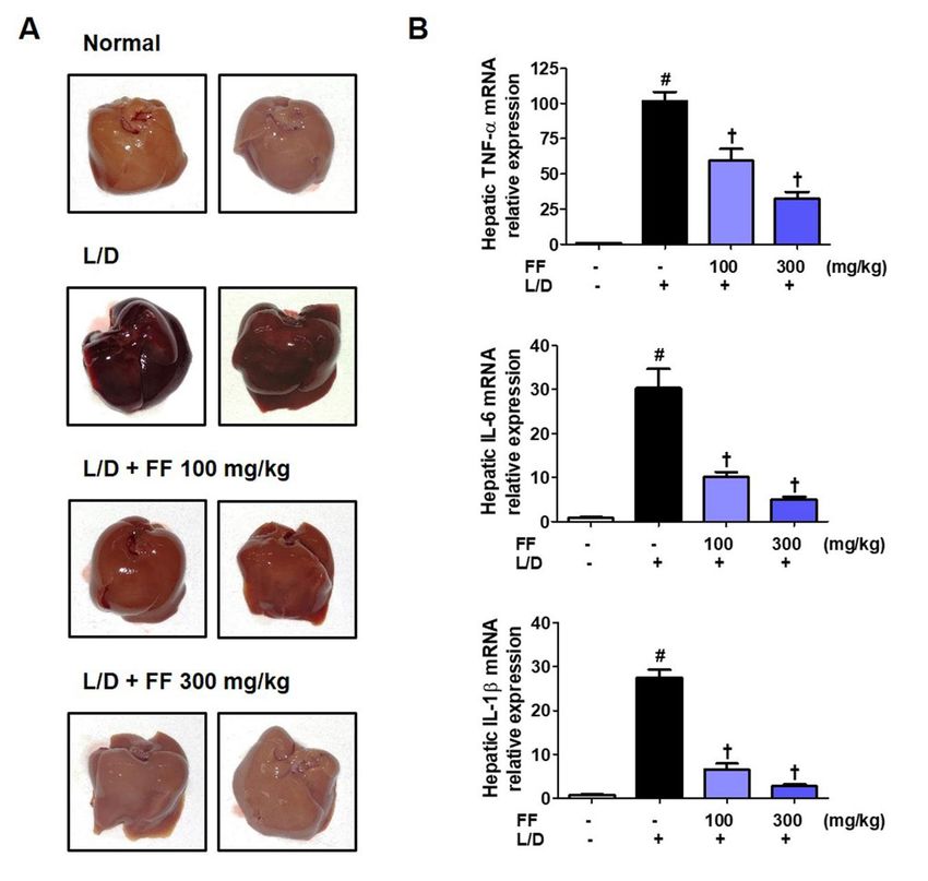
Nutrients 2021, 13, 2901                                                                                           2 of 15

                                 Therefore, D-GalN increases the reactivity of the liver toward endotoxins including
                           LPS, resulting in acute hepatic toxicity within hours, and models of acute hepatic damage
                           caused by LPS/D-GalN show hepatocyte necrosis and apoptosis. Thus, LPS/D-GalN is
                           widely used in studies related to the mechanisms underlying hepatic damage and drug de-
                           velopment [11]. Reactive oxygen species increased by LPS/D-GalN activate macrophages
                           in liver tissue, and the activated macrophages produce inflammatory mediators including
                           tumor necrosis factor (TNF)-α, interleukin (IL)-6, and IL-1β cytokine [12]. These inflam-
                           matory cytokines induce hepatocyte necrosis and reduce antioxidant enzyme activity [13].
                           Consequently, inhibition of inflammatory cytokines and activation of antioxidant enzymes
                           are important factors for the treatment and prevention of acute liver damage caused by
                           LPS/D-GalN.
                                 Inflammation is the central defensive mechanism against external stimuli such as
                           microbial or viral infection, injury, and exposure to endotoxin and is initiated through the
                           activation of microglia and macrophages [14]. Macrophages play a dispensable role in
                           controlling inflammation and produce inflammatory mediators in response to external
                           causes such as LPS [15]. In macrophages with TLR4 activation, inflammatory mechanisms
                           such as nuclear factor (NF)-κB, activator protein (AP)-1, and mitogen-activated protein
                           kinase (MAPK) are also induced, and the expression of inflammatory synthetic enzyme in-
                           ducible nitric oxide synthase (iNOS) and secretion of nitric oxide (NO) are increased [16,17].
                           However, the inflammatory reaction is also effectively inhibited by the activation of the an-
                           tioxidant mechanism nuclear factor erythroid 2-related factor 2 (Nrf-2) and heme oxygenase
                           (HO)-1. HO-1 inhibits the secretion of NO, TNF-α, IL-6, and IL-1β as an important regula-
                           tor of the inflammation and is strongly induced by macrophages [18]. HO-1 expression
                           directly inhibits the production of NO and iNOS and is controlled by the redox-sensitive
                           transcription factor Nrf-2, which regulates various antioxidant enzymes [19]. When the
                           inflammatory response is activated, Nrf-2 translocates to the nucleus and combines to the
                           antioxidant response element to induce HO-1 [19]. Thus, many anti-inflammatory agents
                           act via enhancing HO-1 production via Nrf-2 activation. In addition, mouse peritoneal
                           macrophages are retained within the mouse abdominal cavity by thioglycollate medium
                           and are often used to confirm the efficacy of in vitro inflammation studies [20].
                                 FF is an herbal medicine that has been widely used for a long time in East Asia to treat
                           inflammation, gonorrhea, and pharyngitis [21]. A previous study demonstrated that FF
                           had anti-microbial effects on membrane permeability and apoptosis in Salmonella [22]. In
                           addition, another study reported that FF showed anti-diabetic and anti-hyperlipidemic
                           effects in a streptozotocin-induced diabetes mouse model [23]. Recently, in addition
                           to the pharmacological efficacy of FF, studies on its applicability as a functional food
                           considering nutritional properties have also been reported. FF is rich in vitamin P, and
                           the effect of inhibiting lipid peroxidation in high-cholesterol diet rats through antioxidant
                           action has been reported [24]. In addition, FF was studied for its applicability as a feed
                           additive for effective fattening by reducing the risk of peroxidation in broiler chickens
                           and increasing nutrient digestibility and growth performance in a stress situation due
                           to high temperature [25]. However, the effects of FF on liver damage in mice and on
                           the inflammatory reaction in macrophages and the regulation of FF on its associated
                           mechanisms have not been studied before. Therefore, we investigated the protective
                           efficacy of FF against LPS/D-GalN-induced fulminant hepatic failure and explored how FF
                           impacts related molecular mechanisms. Furthermore, we tested the inhibitory efficacy of
                           FF against the inflammatory reaction in an LPS-stimulated mouse macrophage RAW 264.7
                           and primary macrophages.

                           2. Materials and Methods
                           2.1. Plant Material
                               FF was obtained from Yeongcheonhyundai Herbal Market (Yeongcheon, Korea) and
                           was identified by Prof. KiHwan Bae (Department of Pharmacy, Chungnam National
                           University, Korea). Dried FF (50.0 g) was extracted by heating at 100 ◦ C for 3 h using 1 L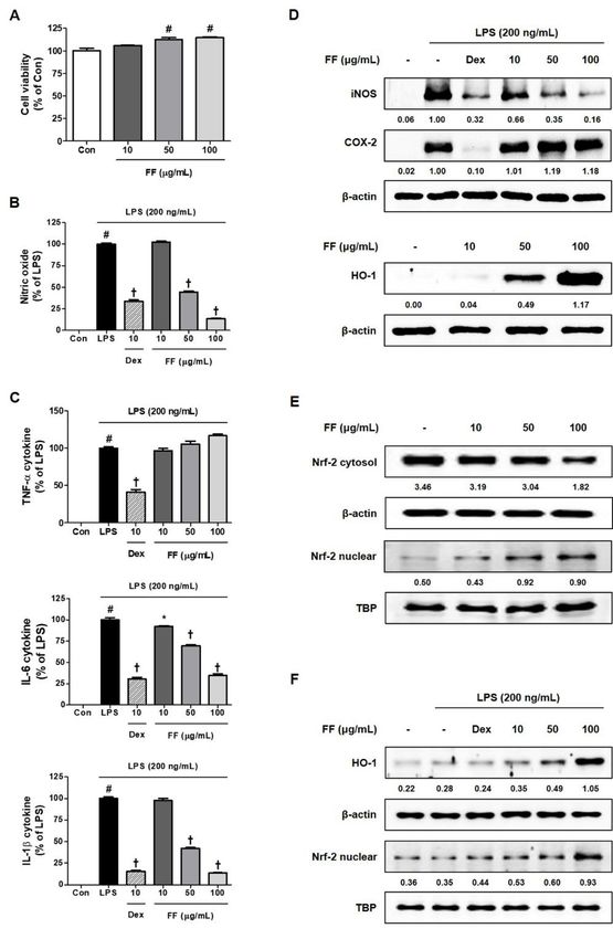
Nutrients 2021, 13, 2901                                                                                        3 of 15

                           distilled water (DW) (Daewoong extractor, Daewoong, Seoul, Korea). Extract solution
                           was filtered using 150 µm sieve, freeze-dried, and stored at −20◦ C until use. The yield
                           was 12.58%.

                           2.2. Materials and Reagents
                                055:B5 LPS from E. coli and D-GalN were acquired from Sigma (St. Louis, MO,
                           USA). Enzyme-linked immunosorbent assay (ELISA) antibody kits were obtained from
                           Thermo (Rockford, IL, USA). Extraction kits for RNA isolation were acquired from iNtRON
                           (Sungnam, Korea). Synthesizing kits for DNA and Master Mix for qPCR were acquired from
                           Bioneer (Daejeon, Korea). Oligonucleotide primers were synthesized by Bioneer. Western
                           blotting antibodies were obtained from Cell Signaling (Boston, MA, USA). Cell culture
                           reagents, including antibiotics, fetal bovine serum (FBS), and Roswell Park Memorial
                           Institute (RPMI) 1640 were obtained from HyClone (Logan, UT, USA). Dexamethasone
                           (Dex) and bovine serum albumin (BSA) were purchased from Sigma. Cell-counting kits
                           (CCK) were acquired from Dojindo (Kumamoto, Japan). Standard compounds, forsythoside
                           A, pinoresinol, and phillygenin were purchased from Chem Faces (Wuhan, china). High-
                           performance liquid chromatography (HPLC)-grade methanol was purchased from Merck
                           (Darmstadt, Germany). ACS reagent-grade acetic acid was obtained Sigma. All water
                           solutions were using a Puris-Evo RO water system (Mirae ST Co., Ltd., Anyang, Korea).
                           HPLC analysis samples were filtered through 0.2 µm membrane filters before use.

                           2.3. Experimental Animals
                                Six weeks old male “imprinting control region” (ICR) mice (30 ± 3 g each) were
                           acquired from Samtako BioKorea (Osan, Korea). All mice were acclimatized for 7 days
                           and were maintained at a room temperature (RT) under a 12 h:12 h light/dark cycle with
                           ad libitum. The mice were subjected to overnight fasting before injection of hepatitis
                           inducers. All experimental procedures in this animal study were carried out depending on
                           the guidelines of the Korea Institute of Oriental Medicine (KIOM)’s Animal Care and Use
                           Committee (Reference number #D-17-020).

                           2.4. Fulminant Hepatitis Mice Model by LPS/D-GalN Injection
                                Briefly, the mice were sorted randomly into four groups (normal controls, LPS/D-
                           GalN, FF 100 mg/kg + LPS/D-GalN, and FF 300 mg/kg + LPS/D-GalN; n = 9 each). Treated
                           mice were orally administered FF once a day for 6 days and intraperitoneally injected
                           with 50 µg/kg LPS and 1 g/kg D-GalN on the last day. Six hours after LPS/D-GalN
                           injection, the animals were anesthetized with isoflurane gas and blood was collected via
                           puncture of the abdominal vena cava. Blood serum was obtained by centrifuging the blood
                           at 2000× g for 15 min. Livers were collected and gently rinsed with phosphate-buffered
                           saline (PBS). Serum cytokine levels were measured with ELISA antibodies. The serum
                           levels of alanine aminotransferase (ALT), aspartate aminotransferase (AST), and alkaline
                           phosphatase (ALP) were determined by an XL-200 automatic clinical chemistry analyzer
                           (Erba, Mannheim, Germany).

                           2.5. RNA Extraction, DNA Synthesis, and Real-Time Reverse Transcription-Polymerase
                           Chain Reaction
                                Isolated total RNA (1 µg) from liver tissue were used for synthesis of cDNA. Sequences
                           of oligonucleotide primer are indicated in Table 1, and real-time reverse transcription-
                           polymerase chain reaction (RT-qPCR) was conducted in accordance with a previously
                           described method [20]. Forty PCR cycles were run using the QuantStudio 6 Flex Real-time
                           PCR System (Thermo), and the samples were compared through the relative CT method.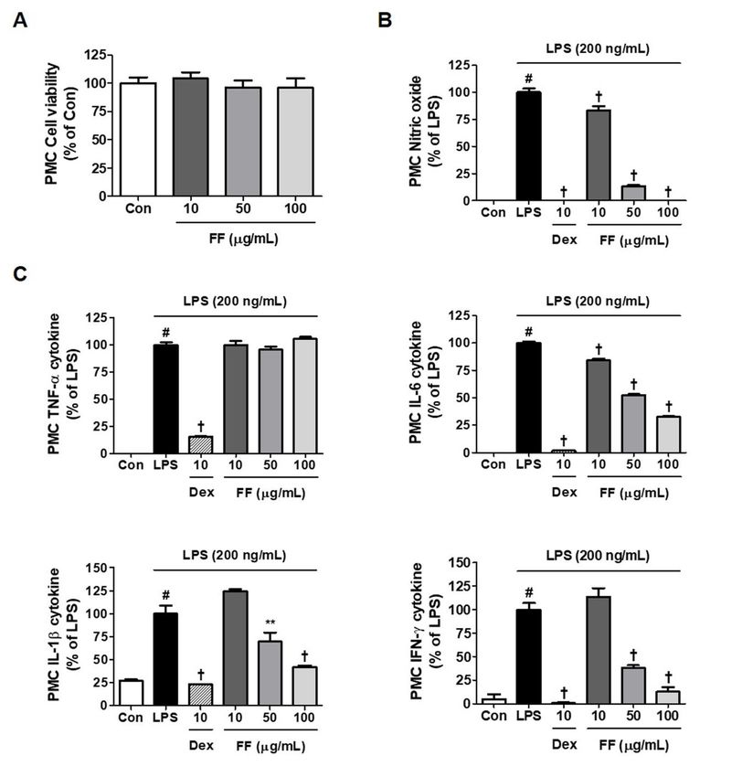
Nutrients 2021, 13, 2901                                                                                           4 of 15

                           Table 1. Primer sequences used for RT-qPCR.

                                      Target Gene                                   Primer Sequence
                                         TNF-α                    F: 50 -TTCTGTCTACTGAACTTCGGGGTGATCGGTCC-30
                                                                 R: 50 -GTATGAGATAGCAAATCGGCTGACGGTGTGGG-30
                                           IL-6                                F: 50 -TCCAGTTGCCTTCTTGGGAC-30
                                                                            R: 50 -GTGTAATTAAGCCTCCGACTTG-30
                                          IL-1β                          F: 50 -ATGGCAACTGTTCCTGAACTCAACT-30
                                                                         R: 50 -CAGGACAGGTATAGATTCTTTCCTTT-30
                                         β-actin                             F: 50 -AGAGGGAAATCGTGCGTGAC-30
                                                                             R: 50 -CAATAGTGATGACCTGGCCGT-30
                           F, forward; R, reverse.

                           2.6. Histopathological Analysis
                                Tissue samples from mouse livers were rinsed with PBS and were fixed in a 10%
                           formaldehyde solution. Liver tissues were then dehydrated in 70–100% ethanol aqueous
                           solution and embedded in paraffin. Paraffin blocks were cut to a thickness of 5 µm by rotary
                           microtome (RM 2165, Leica, Wetzlar, Germany) and were stained using hematoxylin and
                           eosin (H&E). Liver injury in these sections was observed with an Axioskop 40 (Oberkochen,
                           Germany) and was taken at 400× magnification.

                           2.7. Preparation of Protein Extracts and Western Blot Analysis
                                 The liver tissue samples and macrophage cells were lysed in radioimmunoprecip-
                           itation assay buffer (Millipore, Bedford, MA, USA) for total cell protein or in NE-PER
                           extraction reagent (Thermo) for cytosolic and nuclear proteins. Concentrations of total
                           protein were measured by Bradford protein assay reagents (Bio-Rad, Hercules, CA, USA).
                           Equal amount of proteins was separated and then blotted in accordance with a previously
                           described method [20]. Proteins on the membrane were blocked and then incubated with
                           various primary antibodies followed by secondary antibodies (Table 2). Immunoreactive
                           bands of target protein were detected using enhanced chemiluminescence solution (Bio-
                           Rad). Each detected protein band was normalized by internal control proteins and was
                           quantified using ImageJ software (version 1.53k).

                           Table 2. Various antibodies used for Western blot.

                                Antibody             Corporation      Product No.           RRID       Dilution Rate
                                   iNOS              Cell Signaling      #13120          AB_2687529       1:1000
                                 COX-2               Cell Signaling      #4842           AB_2085144       1:1000
                                  HO-1               Cell Signaling      #82206          AB_2799989       1:1000
                                  Nrf-2              Cell Signaling      #12721          AB_2715528       1:1000
                              P-NF-κB p65            Cell Signaling       #3033           AB_331284       1:1000
                                 P-IκBα              Cell Signaling      #2859            AB_561111       1:1000
                                  IκBα               Cell Signaling      #4814            AB_390781       1:1000
                                 P-ERK               Cell Signaling      #4377            AB_331775       1:1000
                                  ERK                Cell Signaling       #9102           AB_330744       1:1000
                                 P-p38               Cell Signaling      #9211            AB_331641       1:1000
                                   p38               Cell Signaling       #9212           AB_330713       1:1000
                                 P-JNK               Cell Signaling      #9251            AB_331659       1:1000
                                  JNK                Cell Signaling       #9252          AB_2250373       1:1000
                                 β-actin             Cell Signaling       #4970          AB_2223172       1:1000
                                  TBP                Cell Signaling      #8515           AB_10949159      1:1000
                             2nd anti-mouse          Cell Signaling       #7076           AB_330924       1:5000
                             2nd anti-rabbit         Cell Signaling      #7074           AB_2099233       1:5000

                           2.8. Culture of Macophage Cell Line
                               RAW 264.7 macrophages were acquired from American Type Culture Collection
                           (Manassas, VA, USA) and were cultured using RPMI 1640 medium containing 10% FBS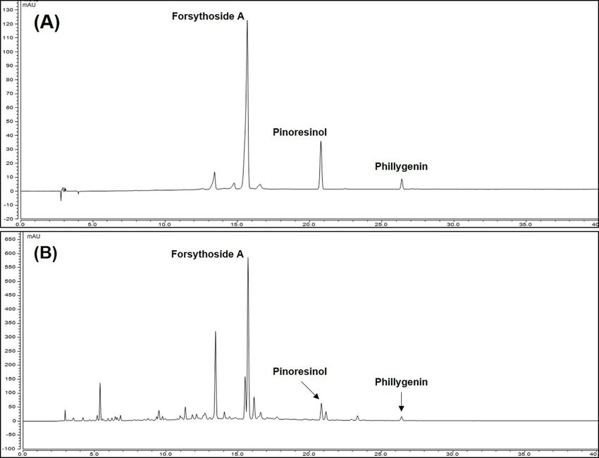
Nutrients 2021, 13, 2901                                                                                         5 of 15

                           and 1% antibiotics in a CO2 incubator. The cells were stimulated by incubating in fresh
                           RPMI 1640 media containing 200 ng/mL LPS in the presence or absence of pretreated FF.

                           2.9. Isolation and Culture of Mouse Peritoneal Macrophages
                                Following intraperitoneal injections of 3% sodium thioglycollate medium (1 mL), five
                           male ICR mice were housed per cage in a 12 h:12 h light/dark cycle. Four days after the
                           injections, the mice were sacrificed and peritoneal macrophage cells (PMC) were collected
                           by flushing with PBS. Red blood cell lysis buffer was then added to the cell suspensions
                           in PBS, after which the samples were incubated for 5 min at RT. After centrifugation at
                           500× g, the supernatants were discarded and PMC were suspended in fresh RPMI 1640
                           medium and incubated with or without FF under the same conditions as those used for
                           RAW 264.7 cells. All experimental procedures for isolation of mouse PMC were carried out
                           depending on the guidelines of the KIOM’s Animal Care and Use Committee (Reference
                           number #D-17-001-1).

                           2.10. Cell Viability Assays
                                Macrophage viability was examined using CCK reagent in accordance with a pre-
                           viously described method [20]. Briefly, macrophages were pre-treated with FF for 24 h,
                           and CCK solution was added, after which the samples were incubated for additional 1 h.
                           The absorbance was then measured at a wavelength of 450 nm using microplate reader
                           (SpectraMax i3, Molecular Devices, San Jose, CA, USA).

                           2.11. Measurement of NO and Inflammatory Cytokine Secretion
                                NO and inflammatory cytokines were measured under the same conditions as in the
                           previous study [20]. Cultured macrophages were pre-treated with FF, stimulated with LPS
                           after 1 h, and incubated for an additional 24 h. NO was detected with Griess reagent and
                           absorbance was measured at 570 nm, and the secretion of inflammatory cytokines in the
                           culture media was quantified by ELISA.

                           2.12. HPLC Instrument
                                HPLC system was set up column oven, an auto sampler, a binary pimp and UV/VIS
                           detector (Dionex Ultimate 3000 system, Dionex Corp., Sunnyvale, CA, USA). All analysis
                           data was processing using Chromeleon 7 software (Thermo, Waltham, MA, USA).

                           2.13. Preperation of Standard and Sample Solutions
                                The FF was dissolved in water at 5 mg/mL concentration using ultrasonicator (JAC Ul-
                           trasonic JAC-3010, Hwaseong, Korea) and after extraction, extract was filtered with a 0.2 µm
                           membrane. 10 µL of extract solution was injected for HPLC analysis. Standard solutions
                           of forsythoside A, pinoresinol, and phillygenin was prepared at 1.0 mg/mL (1000 ppm)
                           using methanol and stored at 4 ◦ C until use. For HPLC analysis, each compound standard
                           solution was diluted with methanol at each standard curve concentration.

                           2.14. HPLC Analysis Method
                                HPLC analysis was conducted to identify of contents of three compounds (forsythoside
                           A, pinoresinol, and phillygenin) in FF. HPLC analysis was performed using X bridge C18
                           column (250 mm × 4.6 mm, 5 µm) connected to a C18 guard cartridge (4.0 mm × 3.0 mm).
                           The mobile phase was eluted at Flow rate 1 mL/min with gradient of 0.3% acetic acid in
                           water (eluent A) and methanol (eluent B). Gradient eluted method was applied: 0–8 min,
                           5–30% B; 8–24 min, 30–57% B; 24–39 min, 57–60% B; 39–50 min, 60–70% B; 50–60 min,
                           70–100% B. The HPLC condition was follows: chromatogram data was detected at 280 nm,
                           the injection volume was 10 µL and temperature of column and auto sampler was keep
                           40◦ C and 20◦ C, respectively (Table 3). Calibration curves, assessed by standard solution
                           and the limits of detection (LOD) and quantification (LOQ) under the chromatographic
                           conditions, were determined by injecting a series of standard solutions. Each samples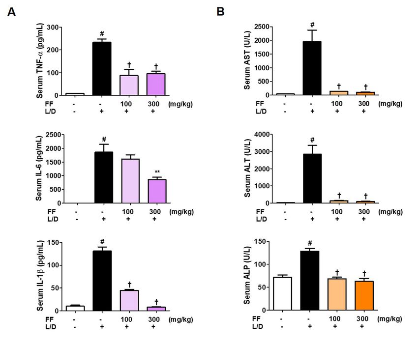
Nutrients 2021, 13, 2901                                                                                                                         6 of 15

                                     were three injected under same condition and data was processed using Chromeleon 7
                                     software (Thermo).

                                     Table 3. HPLC conditions for analysis of compounds and FF.

                                                                                     HPLC Conditions
                                              Detector                                              280 nm
                                               Column                           X bridge C18 Column (250 mm × 4.6 mm, 5 µm)
                                         Column temperature                                          40◦ C
                                          Injection volume                                           10 µL
                                              Flow rate                                          1.0 mL/min
                                            Mobile phase                  Time (min)                   A                   B
                                                                               0.0                          95                           5
                                                                               8.0                          70                          30
                                         A: 0.3% acetic acid in
                                                                              24.0                          43                          57
                                                 water
                                                                              39.0                          40                          60
                                               B: MeOH
                                                                              50.0                          30                          70
                                                                              60.0                          0                          100

                                      2.15. Statistical Analysis
                                            All experimental results are expressed as means ± standard error of the mean. Statis-
                                      tical significance was determined by one-way analysis of variance followed by Dunnett’s
                                      test after comparing each treatment group. Statistical significance was defined as p < 0.05.

                                      3. Results
                                      3.1. Content of Major Compounds of FF
                                          We conduct HPLC analysis to confirm that contents of three compounds forsythoside
                                     A, pinoresinol, and phillygenin in FF that show bioactivity. Each component was selec-
                                     tively detected and identified under HPLC-UV analysis method we established, consistent
                                     with a previous study [26]. The calibration curves the three compounds (forsythoside
                                     A, pinoresinol, and phillygenin) were y = 0.2516x − 3.8826, y = 0.1132x + 0.1922 and
                                     y = 0.1927x + 0.0909 with coefficients of determination of 0.9958, 0.9990, and 0.9994 at
                                     injected concentration ranges (Table 4). These result showed that calibration curve of three
                                     marker compounds has good linearity at the tested concentration range. To confirm the
                                     three compound were showed in FF, we compared the retention time and the UV spectrum
                                     of FF extract and each standard solution (Figure S1). As a result, the three compounds
                                     exhibited the same retention time 15.70, 20.82, and 26.40 min in FF (Figure 1). The area
                                     mean value of FF was calculated for each compounds calibration curve equation. The
                                     content of forsythoside A, pinoresinol, and phillygenin and were 4.54, 1.17, and 0.84%
                                     respectively. Forsythoside A was most abundant constituent in FF and we suggest that it
                                     was marker compound in FF.

                                                   Table 4. Calibration curves of compounds.

  Compound             Range (µg/mL)            Regression Equation                      r2                LOD (µg/mL)             LOQ (µg/mL)
        1                  200.0~500.0           y = 0.2516x − 3.8826                  0.9958                    0.0527                0.1598
        2                   20.0~200.0           y = 0.1132x + 0.1922                  0.9990                    0.0879                0.2664
        3                    2.5~25.0            y = 0.1927x + 0.0909                  0.9994                    0.0517                0.1565
      Forsythoside A (1); Pinoresinol (2); Phillygenin (3). LOD = 3.3 × σ/S. LOQ = 10 × σ/S. σ is the standard deviation of the intercept from the
      regression equation and S is the slope of the calibration curve.

                                     3.2. Regulatory Effects of FF on Serum Cytokine and Aminotransferase Levels in
                                     LPS/D-GalN-Induced Hepatitis in Mice
                                          Inflammatory cytokine levels are important measures of the severeness of inflam-
                                      mation. In addition, ALT, AST, and ALP are markers of hepatic damage. Therefore, we
3.1. Content of Major Compounds of FF
We conduct HPLC analysis to confirm that contents of three compounds forsythoside
A, pinoresinol, and phillygenin in FF that show bioactivity. Each component was selec-
tively detected and identified under HPLC-UV analysis method we established, con-
sistent with a previous study [26]. The calibration curves the three compounds (forsy-
Nutrients 2021, 13, 2901 thoside A, pinoresinol, and phillygenin) were y = 0.2516x − 3.8826, y = 0.1132x + 0.1922 7 of 15
and y = 0.1927x + 0.0909 with coefficients of determination of 0.9958, 0.9990, and 0.9994 at
injected concentration ranges (Table 4). These result showed that calibration curve of three
analyzed
markerthese parameters
compounds to investigate
has good the tested
linearity at the extentconcentration
of fulminantrange.
liver injury and the
To confirm the regu-
latory effects
three of FF.were
compound Serum cytokine,
showed in FF,ALT, AST, andthe
we compared ALP levels were
retention significantly
time and elevated
the UV spec-
6 htrum
after of FF extract and
LPS/D-GalN each standard
treatment. solution
However, (FigureinS1).
as shown As a 2A,B,
Figure result,inthe
thethree com-admin-
groups
pounds
istered with exhibited the same
two doses of FF,retention time 15.70,
inflammatory 20.82, and
cytokine, 26.40
ALT, minand
AST, in FFALP(Figure 1). The
concentrations
area mean value of FF was calculated for each compounds calibration curve
in the mice serum were sharply reduced. IL-6 and IL-1β levels in the serum decreased equation. The in
content of forsythoside A, pinoresinol, and phillygenin and were 4.54,
a dose-dependently, and the other factors were strongly suppressed at both doses. The 1.17, and 0.84%
respectively. Forsythoside A was most abundant constituent in FF and we suggest that it
normal control group did not show any abnormal changes in these measures.
was marker compound in FF.
Figure 1. High-performance liquid chromatography chromatograms of standard solution (A) and FF (B) at 280nm.
Figure 1. High-performance liquid chromatography chromatograms of standard solution (A) and FF (B) at 280 nm.
3.3. FF Protects Mice from Liver Injury and Regulates the Expression of Hepatic Cytokine mRNAs
upon LPS/D-GalN Stimulation
Six hours after LPS/D-GalN was administered, the mice were killed and livers were
collected. To determine the severity of liver injury of each group, liver images were taken.
Livers in the LPS/D-GalN group mice suffered severe damage; in contrast, livers in the
FF-administered group appeared to have a significantly improved pathology in a dose-
dependent manner (Figure 3A). Furthermore, we extracted total RNA from these liver
samples and analyzed the expression of inflammatory cytokines to determine how they
are regulated by FF administration in liver tissue. Results showed that all cytokine mRNA
within the liver tissue were strongly increased by LPS/D-GalN treatment, and they were
dose-dependently significantly inhibited by FF administration (Figure 3B).tory effects of FF. Serum cytokine, ALT, AST, and ALP levels were significantly elevated
6 h after LPS/D-GalN treatment. However, as shown in Figure 2A,B, in the groups admin-
istered with two doses of FF, inflammatory cytokine, ALT, AST, and ALP concentrations
in the mice serum were sharply reduced. IL-6 and IL-1β levels in the serum decreased in
Nutrients 2021, 13, 2901
a dose-dependently, and the other factors were strongly suppressed at both doses. The
8 of 15
normal control group did not show any abnormal changes in these measures.
Figure 2. Effects of Forsythia Fruit (FF) on serum cytokine and aminotransferase levels in a lipopolysaccharide (LPS)/D-
Figure 2. Effects
galactosamine of Forsythia Fruit
(GalN)-induced (FF) on
hepatitis serum
mouse cytokine
model. and
Mice aminotransferase
were levels
pretreated with in a and
FF (100 lipopolysaccharide
300 mg/kg) or (LPS)/D-
vehicle
galactosamine
once per day for(GalN)-induced hepatitis
6 days and 1 h before an mouse model. injection.
LPS/D-GalN Mice wereAfter
pretreated withwas
6 h, blood FF (100 and 300
collected by mg/kg) or vehicle
abdominal once
vena cava
per day for 6 days and 1 h before an LPS/D-GalN injection. After 6 h, blood was collected by abdominal vena cava puncture
puncture and serum was prepared by centrifugation. (A) Serum cytokine levels were determined using enzyme-linked
immunosorbent assay antibodies. (B) Serum aminotransferase was analyzed using an automated clinical chemistry analyzer.
Data are expressed as mean ± standard error of the mean (n = 9). TNF, tumor necrosis factor; IL, interleukin; L/D, LPS/D-
GalN; ALT, alanine aminotransferase; AST, aspartate aminotransferase; ALP, alkaline phosphatase. Statistical significance
was defined as # p < 0.05 (vs. normal controls), ** p < 0.01, and † p < 0.001 (vs. LPS/D-GalN treatment).
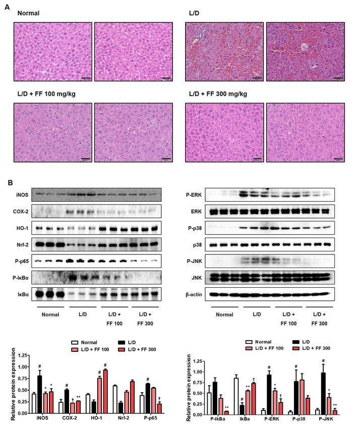
3.4. Hepatoprotective Effects of FF on Histopathological Changes and Regulatory Effects on the
Inflammatioy Proteins Expression
The histopathological findings showed that LPS/D-GalN injection induced atrophy,
hepatocyte necrosis, and infiltration of inflammatory cells. The fulminant changes observed
in the LPS/D-GalN-injected mice significantly improved in those treated with 100 mg/kg
FF, and the 300 mg/kg FF-administered group had no differences from the normal group
(Figure 4A). Next, we analyzed the expression of inflammation-related proteins in the liver
tissue. Cyclooxygenase (COX)-2 and iNOS, which are synthase proteins of prostaglandin
(PG)E2 and NO, respectively, up-regulated in the LPS/D-GalN-administered group and
significantly decreased in the FF-treated group, while the antioxidant mechanism protein
HO-1/Nrf-2 showed opposite patterns (Figure 4B). P-NF-κB p65 and P-inhibitor of NF-κB
alpha proteins were also strongly expressed in the liver tissue of the LPS/D-GalN-treated
mice and were effectively suppressed in the FF-treated group (Figure 4B). Similarly, we
observed that the phosphorylation of extracellular signal-regulated kinase, p38, and c-Jun
NH2 -terminal kinase proteins were effectively inhibited in the FF-treated group (Figure 4B).FF-administered group appeared to have a significantly improved pathology in a dose-
dependent manner (Figure 3A). Furthermore, we extracted total RNA from these liver
samples and analyzed the expression of inflammatory cytokines to determine how they
are regulated by FF administration in liver tissue. Results showed that all cytokine mRNA
Nutrients 2021, 13, 2901 within the liver tissue were strongly increased by LPS/D-GalN treatment, and they9 of were
15
dose-dependently significantly inhibited by FF administration (Figure 3B).
Figure 3. Effects of Forsythia Fruit (FF) on liver injury and expression of hepatic cytokines in lipopolysaccharide (LPS)/D-
Figure 3. Effects
galactosamine of Forsythia Fruit
(GalN)-induced (FF) on
hepatitis. liverwere
Mice injury and expression
pretreated with FFof(100
hepatic
and cytokines
300 mg/kg)in lipopolysaccharide
or vehicle once per (LPS)/D-
day for
galactosamine (GalN)-induced hepatitis. Mice were pretreated with FF (100 and 300 mg/kg) or vehicle once per day for 6
6 days and 1 h before an LPS/D-GalN injection. After 6 h, mice were sacrificed, and livers were collected. (A) Images
days and 1 h before an LPS/D-GalN injection. After 6 h, mice were sacrificed, and livers were collected. (A) Images of
of hepatitis lesions in the mice. (B) mRNA levels of hepatic cytokines were analyzed by real-time reverse transcription-
polymerase chain reaction. Data are expressed as mean ± standard error of the mean. L/D, LPS/D-GalN; TNF, tumor
necrosis factor; IL, interleukin. Statistical significance was defined as # p < 0.05 (vs. normal controls) and † p < 0.001
(vs. LPS/D-GalN treatment).
3.5. Regulatory Effects of FF on the Secretion of Inflammatory Mediators and Activation of
Inflammatory/Antioxidant Pathways in LPS-Stimulated RAW 264.7 Macrophages
Since the pathology of the acute hepatitis mouse model induced by LPS/D-GalN
closely mirrored a fulminant inflammatory response, we investigated the influence of FF on
the LPS-induced mouse macrophage-mediated inflammatory reaction. First, FF had little
effect on RAW 264.7 macrophage viability (Figure 5A), effectively inhibiting the secretion
of inflammatory mediators including LPS-induced NO and cytokines (Figure 5B,C). FF
pretreatment also suppressed the expression of iNOS by LPS in macrophage cells, while
high concentrations of FF treatment (over 50 µg/mL) induced antioxidant protein HO-1
expression (Figure 5D). Treatment with FF induced translocation into the nucleus from the
cytoplasm of Nrf-2, which affected the activation of the antioxidant mechanism (Figure 5E).
In addition, an investigation of the effects of FF on the activation of HO-1/Nrf-2 under LPS
treatment showed that HO-1/Nrf-2 were activated at high concentrations of pretreatment
with FF (Figure 5F).Nutrients 2021, 13, x FOR PEER REVIEW 11 of 16
Nutrients 2021, 13, 2901 10 of 15
Figure 4. Effects of Forsythia Fruit (FF) on histopathological changes in the liver and activation of intracellular signaling
Figure 4. Effects of Forsythia Fruit (FF) on histopathological changes in the liver and activation of intracellular signaling
molecules in lipopolysaccharide (LPS)/D-galactosamine (GalN)-induced hepatitis. Mice were pretreated with FF (100 and
molecules in lipopolysaccharide (LPS)/D-galactosamine (GalN)-induced hepatitis. Mice were pretreated with FF (100 and
300
300mg/kg)
mg/kg) or or vehicle
vehicle once per day
once per day for
for 66 days
daysand
and11hhbefore
beforean anLPS/D-GalN
LPS/D-GalN injection.
injection. After
After 6 h,6 mice
h, mice
werewere sacrificed,
sacrificed, and
and livers
livers were were collected.
collected. (A) Hematoxylin
(A) Hematoxylin and eosin
and eosin staining
staining of mouse
of mouse liver.bars
liver. Scale Scale bars
= 50 μm.=(B)
50 Expression
µm. (B) Expression of
of inflamma-
inflammatory
tory syntheticsynthetic
enzymes,enzymes, inflammatory
inflammatory pathways, pathways, and antioxidant
and antioxidant molecules molecules were determined
were determined by Westernby Western blot
blot analysis.
The histograms
analysis. show protein
The histograms show expression levels relative
protein expression levels to those of
relative to athose
housekeeping protein. Data
of a housekeeping are Data
protein. expressed as mean ±
are expressed
asstandard
mean ± error of the
standard mean.
error L/D,mean.
of the LPS/D-GalN. Statistical significance
L/D, LPS/D-GalN. was definedwas
Statistical significance as #defined
p < 0.05 as
(vs.
# pnormal
< 0.05 control),
(vs. normal*p<
0.05, ** p* p< <
control), 0.01,
0.05,and
** p†Nutrients 2021, 13, x FOR PEER REVIEW 12 of 16
Nutrients 2021, 13, 2901 11 of 15
Figure5.5.Effects
Figure Effectsof
ofForsythia
ForsythiaFruit
Fruit(FF)
(FF)on
onthe
thesecretion
secretionof
ofinflammatory
inflammatorymediators
mediatorsand andexpression
expression
of
of intracellular pathway proteins in RAW 264.7 macrophages. Influence of FF on (A) cell
intracellular pathway proteins in RAW 264.7 macrophages. Influence of FF on (A) cell viability,
viability,
(B)
(B) nitric
nitric oxide secretion,
secretion, and
and(C)
(C)cytokine
cytokineproduction.
production.(D)(D) Expression
Expression of inducible
of inducible nitric
nitric oxideoxide
syn-
thase (iNOS),
synthase cyclooxygenase
(iNOS), cyclooxygenase (COX)-2,
(COX)-2,andandheme
hemeoxygenase
oxygenase(HO)-1,
(HO)-1, (E) nuclear translocation
translocationof of
nuclear factor erythroid 2-related factor 2 (Nrf-2), and (F) HO-1 activation and nuclear translocation
nuclear factor erythroid 2-related factor 2 (Nrf-2), and (F) HO-1 activation and nuclear translocation
ofNrf-2
of Nrf-2under
underlipopolysaccharide
lipopolysaccharide(LPS)
(LPS)stimulation.
stimulation.Control
Controlcells
cellswere
wereincubated
incubatedwith
withthethevehicle
vehicle
alone. Data represent the mean ± standard error of the mean of the results from three
alone. Data represent the mean ± standard error of the mean of the results from three independent independent
experiments. Con, control; Dex, dexamethasone; TNF, tumor necrosis factor; IL, interleukin. # p <
experiments. Con, control; Dex, dexamethasone; TNF, tumor necrosis factor; IL, interleukin. # p < 0.05
0.05 (vs. controls), * p < 0.05, and † p < 0.001 (vs. LPS treatment).
(vs. controls), * p < 0.05, and † p < 0.001 (vs. LPS treatment).
3.6. Inhibitory
3.6. Inhibitory Effects
Effects ofofFF
FFon
onLPS-Induced
LPS-InducedInflammatory
InflammatoryMediator
MediatorLevels
Levelsin
in Primary
Macrophages
Primary Macrophages
Toconfirm
To confirmthe
theinhibitory
inhibitoryactivity
activityofof
FFFF
onon inflammatory
inflammatory response,
response, wewe explored
explored its
its ef-
fects on on
effects LPS-induced
LPS-inducedsecretion of NO
secretion of and
NO inflammatory cytokine
and inflammatory in primary
cytokine macrophages.
in primary macro-
Treatment with FF did
phages. Treatment withnotFFexhibit
did notcytotoxicity up to 100 up
exhibit cytotoxicity µg/mL (Figure
to 100 μg/mL6A), and it
(Figure down-
6A), andNutrients 2021, 13, x FOR PEER REVIEW 13 of 16
Nutrients 2021, 13, 2901 12 of 15
regulated the levels ofthe
it down-regulated NOlevels
and IL-6,
of NOIL-1β,
and and
IL-6,interferon-γ cytokines in cytokines
IL-1β, and interferon-γ a concentration-
in a con-
dependently (Figure 6B,C).(Figure 6B,C).
centration-dependently
Figure 6. Effects of Forsythia Fruit (FF) on the production of inflammatory mediators in mouse peritoneal macrophage cells
Figure
(PMC). 6. Effects
Influence of Forsythia
of FF on (A) cellFruit (FF) (B)
viability, on the production
nitric of inflammatory
oxide secretion, mediators in
and (C) inflammatory mouse production.
cytokine peritoneal macrophage
Primary
cells (PMC). Influence of FF on (A) cell viability, (B) nitric oxide secretion, and (C)
macrophages obtained from five ICR mice were seeded on a 96- or 24-well culture plate and preincubated inflammatory cytokine
for 18production.
h. Then,
Primary macrophages obtained from five ICR mice were seeded on a 96- or 24-well culture plate and preincubated for 18
the cells were pretreated with FF for 1 h and stimulated with lipopolysaccharide (LPS) for another 24 h. Control cells were
h. Then, the cells were pretreated with FF for 1 h and stimulated with lipopolysaccharide (LPS) for another 24 h. Control
incubated with the vehicle alone. Data represent the mean ± standard error of the mean of determinations from three
cells were incubated with the vehicle alone. Data represent the mean ± standard error of the mean of determinations from
independent experiments.
three independent Con, control;
experiments. Con, Dex, dexamethasone;
control; TNF, tumor
Dex, dexamethasone; TNF,necrosis factor; IL,
tumor necrosis interleukin;
factor; IFN, interferon.
IL, interleukin; IFN, inter-
# p feron.
< 0.05 #(vs.
p < controls), ** p < 0.01,
0.05 (vs. controls), ** and † pNutrients 2021, 13, 2901 13 of 15
cytokine levels, and damages liver tissue, so we explored the inhibitory activities of FF on
inflammatory cytokine levels in mouse serum. High concentrations of aminotransferase
are present in the liver and heart, and when liver parenchymal cells are damaged, ALT,
AST, and ALP in the cytoplasm are released into the blood [27], so activity of these enzymes
is an important indicator of liver injury. Thus, the concentrations of ALT, AST, and ALP in
the mouse serum of each group were measured. We found that LPS/D-GalN treatment
strongly induced the inflammatory cytokine and aminotransferase levels in the serum, and
these were then efficiently reduced by FF administration (Figure 2).
Liver damaged by endotoxin or toxic substances has more surface bleeding due to
severe hemorrhage [28], so morphological observation is used to determine the extent of
liver damage. As shown in Figure 3A, we found that the severe hemorrhage caused by
LPS/D-GalN injection significantly improved after FF administration. In addition, we con-
firmed that the expression of hepatic cytokine mRNA in liver tissue was also significantly
and dose-dependently inhibited by FF treatment (Figure 3B). The histopathological changes
examined with H&E staining also showed that FF treatment dramatically improved hep-
atic hemorrhage, hepatocytes necrosis, and inflammatory cell infiltration by LPS/D-GalN
(Figure 4A).
In the model used in this study, the direct cause of liver damage was a fulminant
inflammatory reaction by endotoxin, so we investigated inflammatory protein expres-
sion in liver tissue using immunoblotting. LPS activates MAPK/NF-κB mechanisms via
TLR4 [16,29], which affects the expression of inflammatory mediators such as COX-2 and
iNOS [29,30]. The activation of HO-1/Nrf-2 antioxidant pathways directly impacts the
regulation of an inflammatory reaction, so we tested the effects of FF treatment on the
expression of inflammatory proteins. As the results show in Figure 4B, the expression of
inflammatory proteins activated by LPS/D-GalN injection was strongly repressed by FF
treatment, whereas the antioxidant pathway was effectively activated by FF treatment.
Therefore, 6 days of FF administration was sufficient to suppress severe liver damage in
these mice induced by LPS/D-GalN injection and effectively regulated cytokine production
and aminotransferase secretion.
Next, we investigated how FF affects the inflammatory reaction in endotoxin-stimulated
macrophages. FF pretreatment at a non-toxic concentration strongly inhibited the secre-
tion of NO, IL-6, and IL-1β in RAW 264.7 cells upon LPS stimulation (Figure 5A–C) and
suppressed the expression of the inflammatory enzyme iNOS (Figure 5D). Furthermore,
the production of HO-1 was induced both when the FF was administered alone and in
combination with LPS treatment (Figure 5D,F). In addition, Nrf-2 was activated by FF
treatment and translocated to the nucleus (Figure 5E). In addition, Nrf-2 activation by
FF was also observed under LPS stimulation (Figure 5F). The anti-inflammatory effects
of FF in the macrophage cell line were replicated in primary mouse macrophages, and
pretreatment with FF inhibited the secretion of various inflammatory mediators in PMC in
a pattern similar to those observed in RAW 264.7 macrophages (Figure 6). Taken together,
FF effectively alleviated fulminant liver injury in these mice, and its efficacy is believed to
be associated with a powerful anti-inflammatory activity.
Subsequently, to investigate the relationship between the physiological activities
of FF and its constituents, we performed phytochemical analyses using HPLC. Under
HPLC-DAD analysis conditions, we separated and identified the three main components
including forsythiaside A, pinoresinol, and phillygenin (Figure 1). Previous studies indi-
cated that forsythiaside A exerts protective effect against LPS/D-GalN-induced liver injury
in mice via inhibiting NF-κB activation and up-regulating Nrf-2/HO-1 [31]. Similarly,
forsythiaside A shows hepatoprotective effect against acetaminophen-induced liver injury
in zebrafish through regulation of TNF, matrix metallopeptidase (MMP)9, MMP2, and
phosphatidylinositol 3-kinase [32]. In addition, forsythiaside A exhibits anti-inflammatory
and antioxidant efficacy in BV2 microglia cells through activation of Nrf-2 and HO-1 signal-
ing pathway [33]. Another previous study has shown that pinoresinol has hepatoprotective
effect against carbon tetrachloride (CCl4 )-induced hepatic damage in mice [34]. In addition,Nutrients 2021, 13, 2901 14 of 15
phillygenin inhibits fibrosis by LPS in human hepatic stellate cell LX2 [35] and shows
hepatoprotective effect on CCl4 -induced liver injury in mice by its antioxidant activity
and inhibition on cytochrome P450 2E1 [36]. As can be seen from the results of previous
studies mentioned above, several bioactive components of FF exhibit hepatoprotective,
anti-inflammatory, and antioxidant effects. Based on our HPLC analysis and the results
of previous studies on these constituents, the hepatoprotective, anti-inflammatory, and
antioxidant effects of FF can likely reflect the presence of forsythiaside A, pinoresinol,
and phillygenin.
5. Conclusions
In summary, this work demonstrated that FF mitigates LPS/D-GalN-induced ful-
minant liver injury in mice. FF strongly lowered the levels of inflammatory cytokines
and aminotransferase in mouse serum and inhibited the expression of hepatic cytokine
mRNAs. Furthermore, FF effectively ameliorates a strong inflammatory reaction and
activates antioxidant mechanisms, thereby inhibiting hemorrhage and necrosis in liver
tissue, significantly alleviating liver damage. The anti-inflammatory activities of FF have
also been proved in experimental inflammatory models using a murine macrophage cell
line and primary cells. Based on these results, FF is potentially valuable as a candidate to
prevent or treat intense inflammation and resulting liver damage.
Supplementary Materials: The following are available online at https://www.mdpi.com/article/10
.3390/nu13082901/s1, Figure S1: UV chromatogram of each standard compounds and FF.
Author Contributions: Conceptualization, Y.-C.O., and J.Y.M.; Investigation, Y.H.J., Y.-H.H., T.I.K.,
and Y.-C.O.; Methodology, Y.H.J., Y.-H.H., and Y.-C.O.; Validation, Y.H.J., T.I.K., and Y.-C.O.; Writing–
original draft, Y.H.J.; Writing—review and editing, Y.-C.O., and J.Y.M. All authors have read and
agreed to the published version of the manuscript.
Funding: This research was funded by Korea Institute of Oriental Medicine (KIOM), provided by
the Ministry of Science and ICT, Republic of Korea, grants number K17281 and KSN2021230.
Institutional Review Board Statement: The Animal Care and Use Committee of Korea Institute of
Oriental Medicine approved this animal study (#D-17-020). The study was conducted according to
the guidelines of the Declaration of Helsinki, and approved by the Institutional Review Board (or
Ethics Committee) of Korea Institute of Oriental Medicine (#D-17-020, approved on 3 July 2017).
Informed Consent Statement: Not applicable.
Data Availability Statement: The data are contained within the article.
Conflicts of Interest: The authors declare no conflict of interest.
References
1. Bernuau, J.; Rueff, B.; Benhamou, J.P. Fulminant and subfulminant liver failure: Definitions and causes. Semin. Liver Dis. 1986, 6,
97–106. [CrossRef]
2. Bernal, W.; Auzinger, G.; Dhawan, A.; Wendon, J. Acute liver failure. Lancet 2010, 376, 190–201. [CrossRef]
3. Farazi, P.A.; DePinho, R.A. Hepatocellular carcinoma pathogenesis: From genes to environment. Nat. Rev. Cancer 2006, 6,
674–687. [CrossRef]
4. Ng, K.K.; Lo, C.M. Liver transplantation in Asia: Past, present and future. Ann. Acad. Med. Singapore 2009, 38, 310–322.
5. Soares, J.B.; Pimentel-Nunes, P.; Roncon-Albuquerque, R.; Leite-Moreira, A. The role of lipopolysaccharide/toll-like receptor 4
signaling in chronic liver diseases. Hepatol. Int. 2010, 4, 659–672. [CrossRef]
6. Farber, J.L.; Gill, G.; Konishi, Y. Prevention of galactosamine-induced liver necrosis by uridine. Am. J. Pathol. 1973, 72,
53–62. [PubMed]
7. Yu, K.H.; Lee, S.Y.; Yang, H.M.; Ham, H.A.; Lee, S.U.; Chae, S.W.; Lee, Y.J. Effect of fermented water extracts from Ligularia
fischeri on hepatotoxicity induced by D-galactosamine in rats. J. Korean Soc. Food Sci. Nutr. 2015, 44, 1422–1430. [CrossRef]
8. Chojkier, M.; Fierer, J. D-Galactosamine hepatotoxicity is associated with endotoxin sensitivity and mediated by lymphoreticular
cells in mice. Gastroenterology 1985, 88, 115–121. [CrossRef]
9. Keppler, D.; Lesch, R.; Reutter, W.; Decker, K. Experimental hepatitis induced by D-galactosamine. Exp. Mol. Pathol. 1968, 9,
279–290. [CrossRef]Nutrients 2021, 13, 2901 15 of 15
10. Lesch, R.; Reutter, W.; Keppler, D.; Decker, K. Liver restitution after acute galactosamine hepatitis: Autoradiographic and
biochemical studies in rats. Exp. Mol. Pathol. 1969, 12, 58–69. [CrossRef]
11. Eipel, C.; Kidess, E.; Abshagen, K.; LeMinh, K.; Menger, M.D.; Burkhardt, H.; Vollmar, B. Antileukoproteinase protects against
hepatic inflammation, but not apoptosis in the response of D-galactosamine-sensitized mice to lipopolysaccharide. Br. J. Pharmacol.
2007, 151, 406–413. [CrossRef]
12. Mayer, A.M.; Spitzer, J.A. Modulation of superoxide anion generation by manoalide, arachidonic acid and staurosporine in liver
infiltrated neutrophils in a rat model of endotoxemia. J. Pharmacol. Exp. Ther. 1993, 267, 400–409.
13. Yang, F.; Li, X.; Wang, L.K.; Wang, L.W.; Han, X.Q.; Zhang, H.; Gong, Z.J. Inhibitions of NF-κB and TNF-α result in differential
effects in rats with acute on chronic liver failure induced by d-Gal and LPS. Inflammation 2014, 37, 848–857. [CrossRef] [PubMed]
14. Coussens, L.M.; Werb, Z. Inflammation and cancer. Nature 2002, 420, 860–867. [CrossRef]
15. Wadleigh, D.J.; Reddy, S.; Kopp, T.E.; Ghosh, S.; Herschman, H.R. Transcriptional activation of the cyclooxygenase-2 gene in
endotoxin-treated RAW 264.7 macrophages. J. Biol. Chem. 2000, 275, 6259–6266. [CrossRef]
16. Cheng, B.C.; Ma, X.Q.; Kwan, H.Y.; Tse, K.W.; Cao, H.H.; Su, T.; Shu, X.; Wu, Z.Z.; Yu, Z.L. A herbal formula consisting of Rosae
Multiflorae Fructus and Lonicerae Japonicae Flos inhibits inflammatory mediators in LPS-stimulated RAW 264.7 macrophages. J.
Ethnopharmacol. 2014, 153, 922–927. [CrossRef] [PubMed]
17. Wisdom, R. AP-1: One switch for many signals. Exp. Cell Res. 1999, 253, 180–185. [CrossRef]
18. Ryter, S.W.; Alam, J.; Choi, A.M. Heme oxygenase-1/carbon monoxide: From basic science to therapeutic applications. Physiol.
Rev. 2006, 86, 583–650. [CrossRef] [PubMed]
19. Ryter, S.W.; Otterbein, L.E.; Morse, D.; Choi, A.M. Heme oxygenase/carbon monoxide signaling pathways: Regulation and
functional significance. Mol. Cell Biochem. 2002, 234, 249–263.
20. Jeong, Y.H.; Oh, Y.C.; Cho, W.K.; Yim, N.H.; Ma, J.Y. Hoveniae Semen Seu Fructus Ethanol Extract Exhibits Anti-Inflammatory
Activity via MAPK, AP-1, and STAT Signaling Pathways in LPS-Stimulated RAW 264.7 and Mouse Peritoneal Macrophages.
Mediators Inflamm. 2019, 2019, 9184769. [CrossRef]
21. Piao, X.L.; Jang, M.H.; Cui, J.; Piao, X. Lignans from the fruits of Forsythia suspensa. Bioorg. Med. Chem. Lett. 2008, 18,
1980–1984. [CrossRef]
22. Bae, W.Y.; Kim, H.Y.; Yu, H.S.; Chang, K.H.; Hong, Y.H.; Lee, N.K.; Paik, H.D. Antimicrobial effects of three herbs (Brassica juncea,
Forsythia suspensa, and Inula britannica) on membrane permeability and apoptosis in Salmonella. J. Appl. Microbiol. 2021, 130,
394–404. [CrossRef]
23. Zhang, Y.; Feng, F.; Chen, T.; Li, Z.; Shen, Q.W. Antidiabetic and antihyperlipidemic activities of Forsythia suspensa (Thunb.)
Vahl (fruit) in streptozotocin-induced diabetes mice. J. Ethnopharmacol. 2016, 192, 256–263. [CrossRef]
24. Lee, J.M.; Choi, S.W.; Cho, S.H.; Rhee, S.J. Effect of Forsythia Viridissima Extracts on Antioxidative System and Lipid Peroxidation
of Liver in Rats Fed High-Cholesterol Diet. Korean J. Nutr. 2003, 36, 990–996.
25. Wang, L.; Piao, X.L.; Kim, S.W.; Piao, X.S.; Shen, Y.B.; Lee, H.S. Effects of Forsythia suspensa extract on growth performance,
nutrient digestibility, and antioxidant activities in broiler chickens under high ambient temperature. Poult. Sci. 2008, 87,
1287–1294. [CrossRef]
26. Guo, H.; Liu, A.H.; Li, L.; Guo, D.A. Simultaneous determination of 12 major constituents in Forsythia suspensa by high
performance liquid chromatography–DAD method. J. Pharm. Biomed. Anal. 2007, 43, 1000–1006. [CrossRef] [PubMed]
27. Chung, K.; Cho, S.H.; Sin, E.N.; Choi, K.H.; Choi, Y.S. Effects of alcohol consumption and fat content in diet on chemical
composition and morphology of liver in rat. Korean J. Nutr. 1988, 21, 154–163.
28. Yi, Y.S.; Cho, J.Y.; Kim, D. Cerbera manghas methanol extract exerts anti-inflammatory activity by targeting c-Jun N-terminal
kinase in the AP-1 pathway. J. Ethnopharmacol. 2016, 193, 387–396. [CrossRef] [PubMed]
29. Chun, J.; Choi, R.J.; Khan, S.; Lee, D.S.; Kim, Y.C.; Nam, Y.J.; Lee, D.U.; Kim, Y.S. Alantolactone suppresses inducible nitric oxide
synthase and cyclooxygenase-2 expression by down-regulating NF-kappaB, MAPK and AP-1 via the MyD88 signaling pathway
in LPS-activated RAW 264.7 cells. Int. Immunopharmacol. 2012, 14, 375–383. [CrossRef] [PubMed]
30. Kyriakis, J.M.; Avruch, J. Mammalian MAPK signal transduction pathways activated by stress and inflammation: A 10-year
update. Physiol. Rev. 2012, 92, 689–737. [CrossRef] [PubMed]
31. Pan, C.W.; Zhou, G.Y.; Chen, W.L.; Zhuge, L.; Jin, L.X.; Zheng, Y.; Lin, W.; Pan, Z.Z. Protective effect of forsythiaside A on
lipopolysaccharide/d-galactosamine-induced liver injury. Int. Immunopharmacol. 2015, 26, 80–85. [CrossRef]
32. Gong, L.; Zhou, H.; Wang, C.; He, L.; Guo, C.; Peng, C.; Li, Y. Hepatoprotective effect of forsythiaside a against acetaminophen-
induced liver injury in zebrafish: Coupling network pharmacology with biochemical pharmacology. J. Ethnopharmacol. 2021, 271,
113890. [CrossRef]
33. Wang, Y.; Zhao, H.; Lin, C.; Ren, J.; Zhang, S. Forsythiaside A Exhibits Anti-inflammatory Effects in LPS-Stimulated BV2 Microglia
Cells Through Activation of Nrf2/HO-1 Signaling Pathway. Neurochem. Res. 2016, 41, 659–665. [CrossRef] [PubMed]
34. Kim, H.Y.; Kim, J.K.; Choi, J.H.; Jung, J.Y.; Oh, W.Y.; Kim, D.C.; Lee, H.S.; Kim, Y.S.; Kang, S.S.; Lee, S.H.; et al. Hepatoprotective
effect of pinoresinol on carbon tetrachloride-induced hepatic damage in mice. J. Pharmacol. Sci. 2010, 112, 105–112. [CrossRef]
35. Hu, N.; Wang, C.; Dai, X.; Zhou, M.; Gong, L.; Yu, L.; Peng, C.; Li, Y. Phillygenin inhibits LPS-induced activation and inflammation
of LX2 cells by TLR4/MyD88/NF-kappaB signaling pathway. J. Ethnopharmacol. 2020, 248, 112361. [CrossRef] [PubMed]
36. Song, W.; Wu, J.; Yu, L.; Peng, Z. Evaluation of the Pharmacokinetics and Hepatoprotective Effects of Phillygenin in Mouse.
Biomed. Res. Int. 2018, 2018, 7964318. [CrossRef] [PubMed]You can also read