Salidroside and isorhamnetin attenuate urotensin II induced inflammatory response in vivo and in vitro: Involvement in regulating the RhoA/ROCK II ...
←
→
Page content transcription
If your browser does not render page correctly, please read the page content below
ONCOLOGY LETTERS 21: 292, 2021

        Salidroside and isorhamnetin attenuate urotensin II‑induced
          inflammatory response in vivo and in vitro: Involvement
                 in regulating the RhoA/ROCK II pathway
                           CHENJING WANG1, XIAODONG NAN2, SHUYAN PEI1, YU ZHAO1,
                                 XIAOKUN WANG1, SHIJIE MA1 and GUOYAN MA1

    1
        Department of Pharmacology, School of Basic Medical Sciences, Northwest Minzu University Health Science Center,
               Lanzhou, Gansu 730030; 2Intensive Care Unit, Gansu Provincial Corps Hospital of Chinese People's
                                  Armed Police Force, Lanzhou, Gansu 730050, P.R. China

                                   Received February 16, 2020; Accepted November 11, 2020

                                                   DOI: 10.3892/ol.2021.12553

Abstract. Urotensin II (UII), a vital vasoconstrictor peptide,     isorhamnetin and both in combination attenuated the mRNA
causes an inflammatory response in the pathogenesis of athero‑     and protein expression levels of RhoA and ROCK II in vivo
sclerosis. Previous studies have reported that the Ras homolog     and in vitro, at concentrations corresponding to human thera‑
gene family, member A (RhoA)/Rho kinases (ROCK) pathway            peutic blood plasma concentrations. Thus, these drugs could
modulates the inflammatory response of the atherosclerotic         inhibit the RhoA/ROCK II pathway under UII conditions. The
process. However, to the best of our knowledge, whether the        combination of salidroside and isorhamnetin did not display
RhoA/ROCK pathway mediates the inflammatory effect                 a stronger inhibitory effect on the inflammatory response
of UII has not been previously elucidated. Salidroside and         and the RhoA/ROCK II pathway compared with salidroside
isorhamnetin are two early developed antioxidant Tibetan           and isorhamnetin in isolation. Collectively, the results indi‑
drugs, both displaying cardioprotective effects against            cated that salidroside, isorhamnetin and both in combination
atherosclerosis. Therefore, the aim of the present study was to    inhibited the RhoA/ROCK II pathway, which then attenuated
investigate the protective effects of salidroside, isorhamnetin    the inflammatory response under UII‑induced conditions,
or combination of these two drugs on the UII‑induced inflam‑       resulting in cardioprotection in atherosclerosis.
matory response in vivo (rats) or in vitro [primary vascular
smooth muscle cells (VSMCs)], as well as to examine the role       Introduction
of the RhoA/ROCK pathway in these processes. The levels of
inflammatory markers were measured via ELISA. The mRNA             Atherosclerosis is the leading cause of acute myocardial
and protein expression levels of RhoA and ROCK II were             infarction and stroke, resulting in large global health and
detected using reverse transcription‑quantitative PCR assay        economic burdens (1). It has been reported that atheroscle‑
and western blot analysis. It was demonstrated that salidroside,   rosis is a chronic vascular wall‑related inflammatory disease
isorhamnetin and both in combination decreased the levels of       that occurs within the arterial wall (2). Inflammatory factors
the serum pro‑inflammatory cytokines TNF‑α and IL‑1β, as           are mainly divided into three categories: i) Chemokines,
well as increased the levels of the anti‑inflammatory cytokine     including monocyte chemoattractant protein‑1 (MCP‑1),
IL‑10 and macrophage migration inhibitory factor in rats           fractalkine/CX3CR1 and macrophage colony stimulating
with subacute infusion of UII and in the culture supernatant       factor, whose effects are inhibited by macrophage migra‑
from primary VSMCs‑exposed to UII. Moreover, salidroside,          tion inhibition factor (MIF); ii) pro‑inflammatory factors,
                                                                   including C‑reactive protein, IL‑6, IL‑1 and TNF‑ α; and
                                                                   iii) anti‑inflammatory factors, including IL‑10 and TGF‑β (3).
                                                                   Pharmacological studies have reported that the imbalances
                                                                   between the pro‑atherogenic inflammatory response and
Correspondence to: Dr Chenjing Wang, Department of                 atheroprotective anti‑inflammatory responses serve a key
Pharmacology, School of Basic Medical Sciences, Northwest Minzu
                                                                   role in the initiation and progression of atherosclerosis (4).
University Health Science Center, 1 XibeiXincun, Chengguan,
Lanzhou, Gansu 730030, P.R. China
                                                                   Previous in vivo and in vitro experimental data have revealed
E‑mail: wangcj01002@163.com                                        the key signaling pathways, such as programmed death
                                                                   ligand‑1/programmed cell death protein 1 axis, brain‑derived
Key words: salidroside, isorhamnetin, urotensin II, inflammatory   neurotrophic factor/tyrosine kinase B signaling pathway (5),
response, atherosclerosis, Ras homolog gene family, member A/Rho   Nod‑like receptor protein 3 inflammasome/IL‑1/IL‑18/IL‑6
kinases II pathway                                                 pathway (6) and Toll‑like receptor pathways (7), that mediate
                                                                   the inflammatory response, which may be research hotspots
                                                                   and provide potential preventive targets for atherosclerosis.
2 WANG et al: RhoA/ROCK II IN PROTECTION OF SALIDROSIDE AND ISORHAMNETIN ON UII-INDUCED INFLAMMATION
Therefore, the treatment of atherosclerosis using anti‑inflam‑ Materials and methods
matory drugs may be an attractive strategy (8‑10).
It has been reported that the Ras homolog gene family, Chemicals and reagents. UII (cat. no. U4753), salidroside
member A (RhoA)/Rho kinases (ROCK) pathway is an (cat. no. 05410590) and isorhamnetin (cat. no. 17794) were
important signal transduction system involved in cell prolifer‑ purchased from Sigma‑Aldrich (Merck KGaA). DMEM
ation, endothelial dysfunction, oxidative stress, inflammation, (cat. no. 11965092) and FBS (cat. no. 16140071) were
vascular remodeling and atherosclerosis (11‑13). Previous purchased from Gibco (Thermo Fisher Scientific, Inc.).
studies have suggested that various multiple risk factors and Anti‑RhoA (cat. no. 2117s), anti‑ROCK II (cat. no. 9029s) and
pathological mediators of atherosclerosis can activate the anti‑GAPDH (cat. no. 5174) antibodies were purchased from
RhoA/ROCK pathway to different degrees (14‑16), and the Cell Signaling Technology, Inc.
inhibition of the RhoA/ROCK pathway could be a thera‑
peutic potential target in the treatment of atherosclerosis (16). Rat modeling and animal treatment. All the experimental
Furthermore, the regulatory effect of the RhoA/ROCK procedures were performed in accordance with the National
pathway on the inflammatory response of the atherosclerotic Institutes of Health Guide for the Care and Use of Laboratory
process has been confirmed by previous finding (17,18). The Animals (32), and were approved by the Experimental Animal
study from Shimada and Rajagopalan (19) revealed that ROCK Administration Committee of the School of Basic Medical
mediates lysophosphatidic acid (an inflammatory mediator Sciences, Northwest Minzu University Health Science Center
that is elevated in multiple inflammatory diseases)‑induced (approval no. XBMZ‑YX20130101; March 1, 2013). In total,
IL‑8 and MCP‑1 production in human endothelial cells. The 120 healthy male Wistar rats (weight, 180‑200 g; age, 8 weeks)
ROCK pathway also contributes to hyperglycemia‑activated were provided by Jiangning Qinglongshan Animal Cultivation
macrophages, which results in a pro‑inflammatory phenotype Farm and were housed under laboratory conditions (tempera‑
and eventually contributes to atherosclerosis (17). However, ture 22±2˚C with a relative humidity of 40‑50% and natural
the underlying potential RhoA/ROCK‑regulated signaling light‑dark cycle time of 12/12 h) with free access to food and
pathways in inflammatory response under atherosclerosis water.
remain to be elucidated. For the preparation of the rat model, rats were anaesthetized
Urotensin II (UII), a vasoactive cyclic peptide, and its with pentobarbital sodium [60 mg/kg; intraperitoneal (i.p.)]
high‑affinity G‑protein‑coupled receptor UT are both highly and osmotic mini‑pumps (Alzet Model 2006D; Durect
expressed in the human cardiovascular system, and UII is Corporation) were loaded with either UII or saline alone
involved in the development of cardiovascular homeostasis (vehicle). The rats were randomly divided into eight groups:
disease (20,21). Clinical and experimental studies have identi‑ i) Normal control (saline alone, equal amount as the UII
fied a positive correlation between increased UII levels and the group, n=15); ii) UII group [rats were subcutaneously injected
development of atherosclerosis (22‑24). UII can enhance the with UII (10 ng/kg/min) for 7 consecutive days, n=15];
development of aortic atherosclerotic lesions and destabilizes ii) Salidroside (12 mg/kg) + UII group, (n=15); iv) Salidroside
atherosclerotic plaques (25). UII also exerts a pro‑inflamma‑ (24 mg/kg) + UII group, (n=15); v) Isorhamnetin (12 mg/kg) +
tory effect on vascular wall cells in atherosclerosis (24,26). UII group, (n=15); vi) Isorhamnetin (24 mg/kg) + UII group,
Previous studies have shown that the RhoA/ROCK pathway (n=15); vii) Salidroside + isorhamnetin (Both, 12 mg/kg) +
mediates UII‑induced migration of endothelial progenitor UII group, (n=15); and viii) Salidroside + isorhamnetin (Both,
cells and the formation of macrophage derived foam cells, 24 mg/kg) + UII group, (n=15). The doses of salidroside and
suggesting that the RhoA/ROCK pathway may contribute to isorhamnetin in vivo and in vitro were chosen in accordance
the UII‑induced inflammatory response (27). However, to the with the previous literatures (33‑36).
best of our knowledge, there are no previous reports on the
roles of the RhoA/ROCK pathway in UII‑derived inflamma‑ Isolation and identification of primary vascular smooth
tory effects. muscle cells (VSMCs). A total of 8 adult male Wistar rats
Salidroside and isorhamnetin are two early used anti‑ (age, 10 weeks; weight, 250‑350 g) housed under the same
oxidant Tibetan drugs, possessing a variety of biological aforementioned laboratory conditions were heparinized
activities such as anti‑apoptosis, anti‑oxidative stress and (4 IU/g; i.p.) and then euthanatized via pentobarbital sodium
anti‑inflammation effects (28,29). It has also been reported that (100 mg/kg; i.p.) administration. The procedure for VSMCs
salidroside and isorhamnetin exert a cardioprotective effect on isolation was performed in accordance with a previously
the development of atherosclerosis, which is partly dependent described protocol (31). The aorta was immediately collected,
on their anti‑inflammatory ability (26,30,31). However, the placed into 75% (v/v) alcohol, dissected into sections (length,
anti‑inflammatory or protective properties of salidroside and 3 cm) and subsequently placed in PBS. After removing fibro‑
isorhamnetin against UII‑derived inflammatory effects in blasts that were present in the tunica externa (the external third
atherosclerosis, as well as the underlying molecular mecha‑ of the vessel wall thickness) using forceps, the residual vessels
nisms, are yet not fully understood. were longitudinally cut and the tunica interna was scraped off,
Therefore, the present study aimed to investigate the leaving the tunica media. Then, the tunica media was washed
role of the RhoA/ROCK II pathway in the UII‑induced with DMEM, cut into sections (1 mm 3) and maintained in
inflammatory response, as well as to identify the effects of DMEM containing 10% (v/v) FBS at 37˚C in a Heraeus 5% CO2
salidroside and isorhamnetin treatment on the UII‑induced incubator (Thermo Fisher Scientific, Inc.). The morphology of
inflammatory response and their potential mechanism cultured VSMCs was observed by phase contrast microscopy at
in vivo and in vitro. passages 3‑8 at room temperature. The identification of VSMCsONCOLOGY LETTERS 21: 292, 2021 3
was performed using a Histostain‑streptavidin‑peroxidase kit Scientific, Inc.; cat. no. 11744100) on an ABI Prism 7500
(rabbit; cat. no. SP‑0023; Beijing Biosynthesis Biotechnology system (MP Biomedicals) according to the manufacturer's
Co., Ltd.) according to the manufacturer's protocol. Briefly, instructions. The amplification conditions were as follows:
the third generation of VSMCs was inoculated into a 100‑mm Initial denaturation at 95˚C for 1 min, followed by 40 cycles of
culture. When the cells grew to near fusion state, the cover glass 95˚C for 5 sec, 60˚C for 15 sec and 72˚C for 30 sec. GAPDH
was removed and the cells were fixed with 4% freshly prepared was used as the endogenous control. The mRNA expression
cooled neutral paraformaldehyde buffer at room temperature levels of RhoA and ROCK II were normalized to GAPDH
for 15 min and washed with PBS. Subsequently, after blocking using the 2‑ΔΔCq equation (37). The primer sequences used
non‑specific binding sites with 5% BSA Blocking Reagent were as follows: RhoA forward, 5'‑TCGGAATGATGAGCA
included in the aforementioned immunohistochemical kit CACAA‑3' and reverse, 5'‑GCTTCACAAGATGAGGCAC‑3';
at 37˚C for 30 min, rabbit anti‑a smooth muscle actin poly‑ ROCK II forward, 5'‑CAGCAACTTTGACGACATTGAG‑3'
clonal antibodies (cat. no. bs‑0189R; Beijing Biosynthesis and reverse, 5'‑AGAT TTG CAC TTC TGT TCCAGC‑3'; and
Biotechnology Co., Ltd.) were incubated with the cells over‑ GAPDH forward, 5'‑ACGG CAAGTTCAACGG CACAG‑3'
night at 4˚C. Next, the slides were incubated with secondary and reverse, 5'‑GACGCCAGTAGACTCCACGACA‑3'.
antibodies for 1 h at room temperature and subsequently
stained with a‑smooth muscle actin antibody included in the Western blot analysis. After the indicated treatments, VSMCs
immunohistochemical kit for 10 min at room temperature. The were harvested, lysed in RIPA buffer (Beyotime Institute of
cells were visualized using a laser confocal microscope (Leica Biotechnology; cat. no. P0013K) containing 1% (V/V) PMSF
Microsystems GmbH; magnification, x400). (Beyotime Institute of Biotechnology; cat. no. ST506) on ice
for 30 min, and then centrifuged at 13,000 x g for 10 min
Cell culture and treatment. VSMCs were cultured in DMEM at 4˚C. The protein concentration was confirmed using a
supplemented with 10% FBS in a humidified atmosphere with BCA Protein Assay kit (Beyotime Institute of Biotechnology;
5% CO2 at 37˚C. To investigate the effects of UII on VSMCs, cat. no. P0012). Equal amounts of protein (30 µg/lane) were
cells were treated with different concentration of UII (10 ‑9, separated on 12% SDS‑PAGE, transferred onto PVDF
10‑8, 10‑7 and 10‑6 mol/l) for 24 hat 37˚C. To demonstrate the membranes (EMD Millipore; cat. no. IPVH00010) and
impacts of salidroside and isorhamnetin on VSMCs exposed blocked with blocking buffer [0.1% Tween‑20 in TBS (TBS‑T)
to UII, cells were pretreated with salidroside (1, 3 or 10 µM), supplemented with 5% fat‑free milk] for 2 h at room tempera‑
isorhamnetin (1, 3 or 10 µM) or both salidroside (3 or 10 µM) ture. After washing with TBS‑T buffer, the membranes were
and isorhamnetin (3 or 10 µM) for 1 h, followed by treatment incubated with anti‑RhoA, anti‑ROCK II and anti‑GAPDH
with UII (10‑6 mol/l) for 24 hat 37˚C. antibodies (1:2,000) overnight at 4˚C. GAPDH was used as
a loading control. Then, the membranes were incubated with
Measurement of inflammatory markers in the culture HRP‑conjugated secondary antibody (1:5,000; cat. no. 7077;
supernatant using ELISA. VSMCs were seeded into a 6‑well Cell Signaling Technology, Inc.) for 2 h at room temperature.
plate at a density of 1x10⁶ cells/ml. After incubation for 24 h as The immunoreactive bands were visualized using a chemilu‑
aforementioned, the culture supernatants were collected, centri‑ minescence imaging analysis system (cat. no. 32106; Pierce;
fuged at 1,000 x g for 5 min at room temperature, and used to Thermo Fisher Scientific, Inc.). The densities of protein
assess the levels of TNF‑α (Nanjing Jiancheng Bioengineering expression were semi‑quantified using Bio‑Rad ChemiDoc
Research Institute; cat. no. H052), IL‑1β (Nanjing Jiancheng XRS (version 4.3.0;Bio‑Rad Laboratories, Inc.).
Bioengineering Research Institute; cat. no. H002), IL‑10
(Nanjing Jiancheng Bioengineering Research Institute; Statistical analysis. Data are presented as the mean ± SD from
cat. no. H009) and MIF (BioLegend ® Legend Max TM ≥3 different experiments. Data were analyzed using SPSS.18
Human Active MIF; cat. no. 438408; BioLegend, Inc.) with software (SPSS, Inc.). Comparison among multiple relevant
ELISA kits, according to manufacturer's instructions. The groups was performed using a one‑way ANOVA followed by
supernatants (500 µM) were seeded into enzyme labelling Bonferroni's multiple comparison test. P4 WANG et al: RhoA/ROCK II IN PROTECTION OF SALIDROSIDE AND ISORHAMNETIN ON UII-INDUCED INFLAMMATION Figure 1. Effects of SAL and IRN on inflammatory cytokines in the serum of rats following UII treatment. Levels of (A) TNF‑α, (B) IL‑1β, (C) IL‑10 and (D) MIF in the serum of rats were analyzed via ELISA. Data are from ≥3 independent experiments and presented as the mean ± SD. *P
ONCOLOGY LETTERS 21: 292, 2021                                                                5

Figure 2. Effects of SAL and IRN on the RhoA/ROCK II pathway in the thoracic aorta of rats following subacute infusion of UII. mRNA expression levels of
(A) RhoA and (B) ROCK II were measured via reverse transcription‑quantitative PCR. Protein expression levels of (C) RhoA and (D) ROCK II were detected
using western blot analysis. Data are from ≥3 independent experiments and presented as the mean ± SD. *P
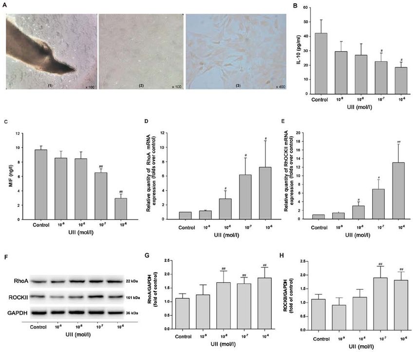
6           WANG et al: RhoA/ROCK II IN PROTECTION OF SALIDROSIDE AND ISORHAMNETIN ON UII-INDUCED INFLAMMATION

Figure 3. Effects of UII on the inflammatory response and the RhoA/ROCK II pathway in primary vascular smooth muscle cells. (A) Observation of cell
morphology. (A‑1) After 4‑5 days of culture, cell morphology was observed under the phase contrast microscope (magnification, x100). (A‑2) After 10‑12 days
of culture, cell morphology was observed under an invert microscope (magnification, x100). (A‑3) α‑actin immunocytochemical staining (magnification, x400).
Inflammatory markers (B) IL‑10 and (C) MIF in the culture supernatant were measured via ELISA. mRNA expression levels of (D) RhoA and (E) ROCK II
were detected using reverse transcription‑quantitative PCR. Protein expression levels of (G) RhoA and (H) ROCK II were detected using (F) western blot anal‑
ysis. Data are from ≥3 independent experiments and presented as the mean ± SD. #P
ONCOLOGY LETTERS 21: 292, 2021                                                                  7

Figure 4. Effects of SAL and IRN on inflammatory response and the RhoA/ROCKII pathway in VSMCs stimulated with UII. (A) VSMCs were pretreated with
SAL or IRN (1, 3 and 10 µM) alone or SAL and IRN (3 and 10 µM) in combination for 1 h followed by treatment with UII (10‑6 mol/l) for 24 h. Quantitative
analysis of (A) TNF‑α, (B) IL‑1β and (C) IL‑10 levels via ELISA. mRNA expression levels of (D) RhoA, (E) ROCK I and (F) ROCK II were detected via reverse
transcription‑quantitative PCR. (G) Western blot analysis of the protein expression levels of (H) ROCK I and (I) ROCK II were measured using. Data are from
≥3 independent experiments and presented as the mean ± SD. *P
8          WANG et al: RhoA/ROCK II IN PROTECTION OF SALIDROSIDE AND ISORHAMNETIN ON UII-INDUCED INFLAMMATION

knowledge, the present results were the first to demonstrate          of salidroside and isorhamnetin on inflammatory response
that UII stimulation results in an inflammatory response, as          were inconsistent. The combination of low and high concen‑
shown by the increases in the levels of pro‑inflammatory              trations of salidroside and isorhamnetin both eliminated
cytokines (TNF‑α and IL‑1β) and the decreases in the levels           the UII‑induced inflammatory response in vivo and in vitro.
of anti‑inflammatory cytokines (IL‑10 and MIF) in the serum           Furthermore, the anti‑inflammatory effect of the combination
of rats and the culture supernatant of VSMCs. Therefore, it           of salidroside and isorhamnetin was not significantly different
was indicated that inflammatory factors induced by UII led to         compared with the single drug alone during UII conditions in
an inflammatory response that is involved in the pathological         atherosclerosis.
process of atherosclerosis.                                               Subsequently, based on the role of the RhoA/ROCK
     RhoA and its downstream effector ROCK, which exists              pathway in UII‑induced inflammatory response, the present
in two isoforms, ROCK1 and ROCK2, serve significant roles             study further investigated the effects of salidroside, isorham‑
in multiple cellular processes, such as proliferation, apoptosis      netin and both in combination on the RhoA/ROCK pathway. It
and migration (47). Abnormal activation of the RhoA/ROCK              was found that salidroside, isorhamnetin and both in combina‑
pathway has been reported to be involved in various types of          tion inhibited the RhoA/ROCK pathway in the thoracic aorta of
diseases including diabetes, osteoarthritis, cardiovascular and       rats following subacute infusion of UII and in UII‑stimulated
cerebrovascular diseases (11‑13). In recent years, the regula‑        VSMCs. Consistent with the in vivo results, the inhibitory
tory effect of the RhoA/ROCK pathway on the inflammatory              effects of low concentrations of salidroside and isorhamnetin
process of atherosclerosis has been revealed. For instance,           on the RhoA/ROCK pathway were inconsistent, while the
upregulation of the RhoA/ROCK signaling cascade has been              high concentrations of salidroside and isorhamnetin both
identified in atherosclerosis (11). Moreover, RhoA‑mediated           reversed UII‑induced the enhancement of the RhoA/ROCK
NF‑ κ B signaling pathways lead to vascular endothelial               pathway in vivo and in vitro. The combination of low and high
dysfunction in diabetes (48). ROCK pathways also contribute           concentrations of salidroside and isorhamnetin both attenu‑
to hyperglycemia‑activated macrophages, which result in a             ated the RhoA/ROCK pathway under UII in vivo and in vitro.
more pro‑inflammatory phenotype and eventually lead to                There was no significant difference between salidroside and
atherosclerosis (17). Previous studies have shown that ROCK I         isorhamnetin in isolation and in combination. Thus, the results
is predominantly increased in the process of macrophage               suggested that, to a certain extent, the RhoA/ROCK pathway
adherence, and ROCK1‑deficiency decreases atherosclerosis             contributed to the anti‑inflammatory effects of salidroside,
in bone marrow‑derived cells (49), indicating that ROCK I             isorhamnetin and both in combination under UII simulation
serves an important role in the development of atherosclerosis.       in atherosclerosis.
However, whether ROCK II inhibition could also be beneficial              However, there are a few limitations to the present study.
in attenuating atherosclerosis remain to be investigated. In line     First, gain‑of‑function and loss‑of function experiments were
with these previous results, the present findings suggested that      not used to investigate the role of the RhoA/ROCK pathway in
UII stimulation promoted the RhoA/ROCK pathway in rats                UII‑induced inflammatory or the anti‑inflammatory effects of
and VSMCs, implying the involvement of the RhoA/ROCK II               salidroside, isorhamnetin and both in combination. This will
pathway in UII‑induced inflammatory response.                         require further examination in future studies. In addition, the
     It has been revealed that salidroside, an early antioxidant      potential mechanism of salidroside, isorhamnetin and both in
Tibetan medicine, has anti‑inflammatory effects in atheroscle‑        combination in the inhibition of the RhoA/ROCK pathway
rosis (50,51). Li et al (50) reported that salidroside can decrease   under UII has not yet been elucidated. The present study
the generation of inflammatory cytokines, such as IL‑6, IL‑1β         also did not examine the comparison of the two drugs with
and MCP‑1, in TNF‑α‑induced cardiac microvascular endothe‑            currently, widely‑used anti‑atherosclerosis drugs and other
lial cells, alleviating vascular inflammation and atherosclerosis.    reported anti‑inflammatory drugs in atherosclerosis, which
Another previous study revealed that salidroside attenuated           may be interesting to evaluate in the subsequent experiment
endothelial cellular senescence via reducing the expression           plan.
of inflammatory cytokines, thus mitigating the pathogenesis               In conclusion, the present findings indicated that UII stim‑
of atherosclerosis (51). In addition, isorhamnetin, a flavonoid       ulation resulted in an inflammatory response, accompanied
monomer extracted from seabuckthorn fruit, has been shown             by the enhancement of the RhoA/ROCK pathway in vivo and
to possess anti‑cancer, anti‑oxidant, anti‑inflammatory and           in vitro. In addition, to the best of our knowledge, the present
anti‑atherosclerotic activities (34,52,53). However, there are        results provided the first evidence that salidroside, isorham‑
few studies on the anti‑inflammatory and anti‑atherosclerotic         netin and both in combination attenuated the UII‑induced
effects of salidroside, isorhamnetin and both in combination.         inflammatory response, which was partly dependent on
     To the best of our knowledge, the present study was the          inhibition of the RhoA/ROCK pathway. Moreover, there is no
first to demonstrate that salidroside, isorhamnetin and both in       significant difference between salidroside and isorhamnetin,
combination decreased TNF‑α and IL‑1β levels, and increased           both in isolation or in combination. The present study may
IL‑10 level in the serum of UII‑treated rats and in the culture       provide a novel theoretical basis for the separate used of
supernatant of UII‑stimulated VSMCs, at concentrations                the two drugs or their combination in the treatment against
corresponding to human therapeutic blood plasma concentra‑            atherosclerosis. Furthermore, the present results may provide
tions, thus eliminating the UII‑induced inflammatory response.        additional theoretical guidance for the clinical combined use
High concentrations of salidroside and isorhamnetin both              of the Tibetan medicines salidroside and isorhamnetin in
reversed the UII‑induced inflammatory response in vivo and            the prevention against atherosclerosis‑related cardiovascular
in vitro. However, the inhibitory effects of low concentration        diseases.
ONCOLOGY LETTERS 21: 292, 2021 9
Acknowledgements 7. Koushki K, Shahbaz SK, Mashayekhi K, Sadeghi M, Zayeri ZD,
Taba MY, Banach M, Al‑Rasadi K, Johnston TP and Sahebkar A:
Anti‑inflammatory action of statins in cardiovascular disease:
Not applicable. The role of inflammasome and toll‑like receptor pathways.
Clin Rev Allergy Immunol: May 6, 2020 (Epub ahead of print).
doi: 10.1007/s12016-020-08791-9.
Funding 8. Luque‑Martin R, Van den Bossche J, Furze RC, Neele AE,
van der Velden S, Gijbels MJJ, van Roomen CPPA, Bernard SG,
The present study was supported by grants from National de Jonge WJ, Rioja I, et al: Targeting histone deacetylases in
myeloid cells inhibits their maturation and inflammatory func‑
Natural Science Foundation of China (grant no. 81360490), tion with limited effects on atherosclerosis. Front Pharmacol 10:
research funds for Institutions of Higher Learning of Gansu 1242, 2019.
Province (grant no. 2017B‑81), the Fundamental Research 9. Mog B, Asase C, Chaplin A, Gao H, Rajagopalan S and
Maiseyeu A: Nano‑antagonist alleviates inflammation and allows
Funds for the Central Universities (grant nos. 31920180023 for MRI of atherosclerosis. Nanotheranostics 3: 342‑355, 2019.
and 31920190106) and the Natural Science Foundation of 10. Yang G, Zhuo J, Lin Y, Zhang M, Liu L, Chen X and Gao R:
Gansu Province, China (grant no. 20JR5RA503). Ginsenoside Rb1 prevents dysfunction of endothelial cells
by suppressing inflammatory response and apoptosis in the
high‑fat diet plus balloon catheter‑injured rabbit model via the G
Availability of data and materials protein‑coupled estrogen receptor‑mediated phosphatidylinositol
3‑kinases (PI3K)/akt pathway. Med Sci Monit 25: 7407‑7417, 2019.
11. Cai A, Li L and Zhou Y: Pathophysiological effects of RhoA and
The datasets used and/or analyzed during the current study Rho‑associated kinase on cardiovascular system. J Hypertens 34:
are available from the corresponding author on reasonable 3‑10, 2016.
request. 12. Deng Z, Jia Y, Liu H, He M, Yang Y, Xiao W and Li Y:
RhoA/ROCK pathway: Implication in osteoarthritis and thera‑
peutic targets. Am J Transl Res 11: 5324‑5331, 2019.
Authors' contributions 13. Strzelecka‑Kiliszek A, Mebarek S, Roszkowska M, Buchet R,
Magne D and Pikula S: Functions of Rho family of small
GTPases and Rho‑associated coiled‑coil kinases in bone cells
CW and XN participated in the design of the study and during differentiation and mineralization. Biochim Biophys Acta
conducted the experiment. CW, SP and YZ wrote the manu‑ Gen Subj 1861: 1009‑1023, 2017.
script and analyzed data. XW, SM and GM collaborated in 14. Li N, Chen J, Zhao J and Wang T: MicroRNA‑3188 targets
ETS‑domain protein 4 and participates in RhoA/ROCK pathway
the analysis and interpretation of data. All authors read and to regulate the development of atherosclerosis. Pharmazie 72:
approved the final manuscript. 687‑693, 2017.
15. Surma M, Wei L and Shi J: Rho kinase as a therapeutic target in
cardiovascular disease. Future Cardiol 7: 657‑671, 2011.
Ethics approval and consent to participate 16. Z hou Q, G ensch C a nd Liao J K: R ho ‑a sso ciat e d
coiled‑coil‑forming kinases (ROCKs): Potential targets for
All animal experimental procedures were performed in accor‑ the treatment of atherosclerosis and vascular disease. Trends
Pharmacol Sci 32: 167‑173, 2011.
dance with the National Institutes of Health Guide for the 17. Cheng CI, Chen PH, Lin YC and Kao YH: High glucose acti‑
Care and Use of Laboratory Animals, and were approved by vates Raw264.7 macrophages through RhoA kinase‑mediated
the Experimental Animal Administration Committee of the signaling pathway. Cell Signal 27: 283‑292, 2015.
18. Loirand G, Guerin P and Pacaud P: Rho kinases in cardiovascular
School of Basic Medical Sciences, Northwest Minzu University physiology and pathophysiology. Circ Res 98: 322‑334, 2006.
Health Science Center (approval no. XBMZ‑YX20130101; 19. Shimada H and Rajagopalan LE: Rho‑kinase mediates lyso‑
March 1, 2013). phosphatidic acid‑induced IL‑8 and MCP‑1 production via p38
and JNK pathways in human endothelial cells. FEBS Lett 584:
2827‑2832, 2010.
Patient consent for publication 20. Desche P, Couderc LJ and Epardeau B: Sjogren's syndrome and
pulmonary lymphangiomyomatosis. Chest 94: 898, 1988.
21. Douglas SA and Ohlstein EH: Human urotensin‑II, the most
Not applicable. potent mammalian vasoconstrictor identified to date, as a
therapeutic target for the management of cardiovascular disease.
Competing interests Trends Cardiovasc Med 10: 229‑237, 2000.
22. Albanese I, Daskalopoulou SS, Yu B, You Z, Genest J,
Alsheikh‑Ali A and Schwertani AG: The urotensin II system
The authors declare that they have no competing interests. and carotid atherosclerosis: A role in vascular calcification. Front
Pharmacol 7: 149, 2016.
23. Demirpence M, Guler A, Yilmaz H, Sayin A, Pekcevik Y,
References Turkon H, Colak A, Ari EM, Aslanipour B, Kocabas GU and
Calan M: Is elevated urotensin II level a predictor for increased
1. Libby P, Bornfeldt KE and Tall AR: Atherosclerosis: Successes, cardiovascular risk in subjects with acromegaly? J Endocrinol
surprises, and future challenges. Circ Res 118: 531‑534, 2016. Invest 42: 207‑215, 2019.
2. Chistiakov DA, Kashirskikh DA, Khotina VA, Grechko AV and 24. Şatıroğlu Ö, Durakoğlugil ME, Çetin M, Çiçek Y, Erdoğan T
Orekhov AN: Immune‑inflammatory responses in atheroscle‑ and Duman H: The role of urotensin II and atherosclerotic risk
rosis: The role of myeloid cells. J Clin Med 8: 1798, 2019. factors in patients with slow coronary flow. Interv Med Appl
3. Hanna A and Frangogiannis NG: Inflammatory cytokines and Sci 8: 158‑163, 2016.
chemokines as therapeutic targets in heart failure. Cardiovasc 25. Li Y, Zhao S, Wang Y, Chen Y, Lin Y, Zhu N, Zheng H, Wu M,
Drugs Ther 34: 849‑863, 2020. Cheng D, Li Y, et al: Urotensin II promotes atherosclerosis in
4. Taleb S: Inflammation in atherosclerosis. Arch Cardiovasc cholesterol‑fed rabbits. PLoS One 9: e95089, 2014.
Dis 109: 708‑715, 2016. 26. Lu D, Peng F, Li J, Zhao J, Ye X, Li B and Ding W: Urotensin
5. Veluswamy P, Wacker M, Scherner M and Wippermann J: II promotes secretion of LTB4 through 5‑lipoxygenase via the
Delicate role of PD‑L1/PD‑1 axis in blood vessel inflamma‑ UT‑ROS‑Akt pathway in RAW264.7 macrophages. Arch Med
tory diseases: Current insight and future significance. Int J Mol Sci 15: 1065‑1072, 2019.
Sci 21: 8159, 2020. 27. Xu S, Jiang H, Wu B, Yang J and Chen S: Urotensin II induces
6. Sethwala AM, Goh I and Amerena JV: Combatting inflammation migration of endothelial progenitor cells via activation of the
in cardiovascular disease. Heart Lung Circ 30: 197‑206, 2021. RhoA/Rho kinase pathway. Tohoku J Exp Med 219: 283‑288, 2009.10 WANG et al: RhoA/ROCK II IN PROTECTION OF SALIDROSIDE AND ISORHAMNETIN ON UII-INDUCED INFLAMMATION
28. Saito K, Yonekura‑Sakakibara K, Nakabayashi R, Higashi Y, 42. Diebold I, Petry A, Burger M, Hess J and Görlach A: NOX4
Yamazaki M, Tohge T and Fernie AR: The flavonoid biosyn‑ mediates activation of FoxO3a and matrix metalloproteinase‑2
thetic pathway in arabidopsis: Structural and genetic diversity. expression by urotensin‑II. Mol Biol Cell 22: 4424‑4434, 2011.
Plant Physiol Biochem 72: 21‑34, 2013. 43. Yang Y, Zhang J, Chen X, Wu T, Xu X, Cao G, Li H and Li Y:
29. Zhong Z, Han J, Zhang J, Xiao Q, Hu J and Chen L: UII/GPR14 is involved in NF‑κ B‑mediated colonic inflammation
Pharmacological activities, mechanisms of action, and safety in vivo and in vitro. Oncol Rep 36: 2800‑2806, 2016.
of salidroside in the central nervous system. Drug Des Devel 44. Fiordelisi A, Iaccarino G, Morisco C, Coscioni E and Sorriento D:
Ther 12: 1479‑1489, 2018. NFkappaB is a key player in the crosstalk between inflammation
30. Boesch‑Saadatmandi C, Loboda A, Wagner AE, Stachurska A, and cardiovascular diseases. Int J Mol Sci 20: 1599, 2019.
Jozkowicz A, Dulak J, Döring F, Wolffram S and Rimbach G: 45. Li Q, Zhao W, Zeng X and Hao Z: Ursolic acid attenuates
Effect of quercetin and its metabolites isorhamnetin and quer‑ atherosclerosis in apoE(‑/‑) mice: Role of LOX‑1 mediated by
cetin‑3‑glucuronide on inflammatory gene expression: Role of ROS/NF‑κ B pathway. Molecules 23: 1101‑1108, 2018.
miR‑155. J Nutr Biochem 22: 293‑299, 2011. 46. Zhang J, Wang X, Vikash V, Ye Q, Wu D, Liu Y and Dong W:
31. Lodi F, Jimenez R, Moreno L, Kroon PA, Needs PW, Hughes DA, ROS and ROS‑mediated cellular signaling. Oxid Med Cell
Santos‑Buelga C, Gonzalez‑Paramas A, Cogolludo A, Longev 2016: 4350965, 2016.
Lopez‑Sepulveda R, et al: Glucuronidated and sulfated metabo‑ 47. Bustelo XR, Sauzeau V and Berenjeno IM: GTP‑binding
lites of the flavonoid quercetin prevent endothelial dysfunction but proteins of the Rho/Racfamily: Regulation, effectors and func‑
lack direct vasorelaxant effects in rat aorta. Atherosclerosis 204: tions in vivo. Bioessays 29: 356‑370, 2007.
34‑39, 2009. 48. Tang ST, Zhang Q, Tang HQ, Wang CJ, Su H, Zhou Q, Wei W,
32. National Research Council (US): Committee for the Update of Zhu HQ and Wang Y: Effects of glucagon‑like peptide‑1 on
the Guide for the Care and Use of Laboratory Animals: Guide for advanced glycation endproduct‑induced aortic endothelial
the Care and Use of Laboratory Animals. 8th edition. National dysfunction in streptozotocin‑induced diabetic rats: Possible
Academies Press, Washington, DC,2011. roles of Rho kinase‑ and AMP kinase‑mediated nuclear factor κ B
33. Chen L, Liu P, Feng X and Ma C: Salidroside suppressing signaling pathways. Endocrine 53: 107‑116, 2016.
LPS‑induced myocardial injury by inhibiting ROS‑mediated 49. Wang HW, Liu PY, Oyama N, Rikitake Y, Kitamoto S,
PI3K/Akt/mTOR pathway in vitro and in vivo. J Cell Mol Gitlin J, Liao JK and Boisvert WA: Deficiency of ROCK1 in
Med 21: 3178‑3189, 2017. bone marrow‑derived cells protects against atherosclerosis in
34. Jamali‑Raeufy N, Baluchnejadmojarad T, Roghani M, Keimasi S LDLR‑/‑mice. FASEB J 22: 3561‑3570, 2008.
and Goudarzi M: Isorhamnetin exerts neuroprotective effects 50. Li R, Dong Z, Zhuang X, Liu R, Yan F, Chen Y, Gao X and Shi H:
in STZ‑induced diabetic rats via attenuation of oxidative stress, Salidroside prevents tumor necrosis factor‑ α‑induced vascular
inflammation and apoptosis. J Chem Neuroanat 102: 101709, 2019. inflammation by blocking mitogen‑activated protein kinase and
35. Zhang N, Pei F, Wei H, Zhang T, Yang C, Ma G and Yang C: NF‑κ B signaling activation. Exp Ther Med 18: 4137‑4143, 2019.
Isorhamnetin protects rat ventricular myocytes from ischemia 51. Xing SS, Li J, Chen L, Yang YF, He PL, Li J and Yang J: Salidroside
and reperfusion injury. Exp Toxicol Pathol 63: 33‑38, 2011. attenuates endothelial cellular senescence via decreasing the
36. Hwang SM, Lee YJ, Lee YP, Yoon JJ, Lee SM, Cha JD, Choi KM, expression of inflammatory cytokines and increasing the expres‑
Kang DG and Lee HS: Anti‑proliferative effect of an aqueous sion of SIRT3. Mech Ageing Dev 175: 1‑6, 2018.
extract of prunella vulgaris in vascular smooth muscle cells. Evid 52. Luo Y, Sun G, Dong X, Wang M, Qin M, Yu Y and Sun X:
Based Complement Alternat Med 2013: 936463, 2013. Isorhamnetin attenuates atherosclerosis by inhibiting macro‑
37. Livak KJ and Schmittgen TD: Analysis of relative gene expres‑ phage apoptosis via PI3K/AKT activation and HO‑1 induction.
sion data using real‑time quantitative PCR and the 2(‑Delta Delta PLoS One 10: e120259, 2015.
C(T)) method. Methods 25: 402‑408, 2001. 53. Park C, Cha HJ, Choi EO, Lee H, Hwang‑Bo H, Ji SY, Kim MY,
38. Zhou J, Yin G, Yu T, Zhang Y, Tian X, Xia D and Shi L: Kim SY, Hong SH, Cheong JH, et al: Isorhamnetin induces cell
Rosuvastatin reduces expression of tissue factor through inhibiting cycle arrest and apoptosis via reactive oxygen species‑mediated
RhoA/ROCK pathway to ameliorate atherosclerosis. Panminerva AMP‑activated protein kinase signaling pathway activation in
Med: Sep 24, 2019 (Epub ahead of print). doi: 10.23736/S0031- human bladder cancer cells. Cancers (Basel) 11: 1494, 2019.
0808.19.03761-3.
39. Pereira‑Castro J, Bras‑Silva C and Fontes‑Sousa AP: Novel This work is licensed under a Creative Commons
insights into the role of urotensin II in cardiovascular disease. Attribution-NonCommercial-NoDerivatives 4.0
Drug Discov Today 24: 2170‑2180, 2019. International (CC BY-NC-ND 4.0) License.
40. Yu QQ, Cheng DX, Xu LR, Li YK, Zheng XY, Liu Y, Li YF, Liu HL,
Bai L, Wang R, et al: Urotensin II and urantide exert opposite effects
on the cellular components of atherosclerotic plaque in hypercholes‑
terolemic rabbits. Acta Pharmacol Sin 41: 546‑553, 2019.
41. Tsai CS, Loh SH, Liu JC, Lin JW, Chen YL, Chen CH and
Cheng TH: Urotensin II‑induced endothelin‑1 expression and
cell proliferation via epidermal growth factor receptor transac‑
tivation in rat aortic smooth muscle cells. Atherosclerosis 206:
86‑94, 2009.You can also read