ORIGINAL ARTICLE PROLONGED WARM ISCHEMIA AGGRAVATES HEPATIC MITOCHONDRIA DAMAGE AND APOPTOSIS IN DCD LIVER BY REGULATING CA2+/CAM/CAMKII SIGNALING ...
←
→
Page content transcription
If your browser does not render page correctly, please read the page content below
Int J Clin Exp Pathol 2019;12(1):217-228
www.ijcep.com /ISSN:1936-2625/IJCEP0088927

Original Article
Prolonged warm ischemia aggravates hepatic
mitochondria damage and apoptosis in DCD liver
by regulating Ca2+/CaM/CaMKII signaling pathway
Li Zhang1,2, Sheng-Ning Zhang1, Li Li1, Xi-Bing Zhang1, Rui-Chao Wu1, Jun-Han Liu1, Zhao-Yu Huang1, Wang
Li1, Jiang-Hua Ran1
1
 Department of Hepatopancreatobiliary Surgery, The Affiliated Calmette Hospital of Kunming Medical University,
The First People’s Hospital of Kunming, Calmette Hospital, Kunming, Yunnan Province, China; 2Department
of Hepatopancreatobiliary, The First People’s Hospital of Yunnan Province, The Affiliated Hospital of Kunming
University of Science and Technology, Kunming, Yunnan Province, China
Received November 26, 2018; Accepted December 14, 2018; Epub January 1, 2019; Published January 15, 2019

Abstract: This study was conducted to investigate the effect of warm ischemia duration on hepatocyte mitochondrial
damage after liver transplantation, and confirm the role of CaMKIIγ in this process. Rat donation after cardiac death
(DCD) liver transplantation model was established by exposing donor liver to 0 (W0 group), 15 (W15 group), and 30
(W30 group) min warm ischemia. Some rats in W15 group were transfected with CaMKIIγ and CaMKIIγ-shRNA lentivi-
rus. On day 1, 3, and 7 post-transplantation, a series of experiments, including HE staining, TEM observation, ALT
and AST measurement, flow cytometry analysis, qRT-PCR, and Western blotting were performed to evaluate the ex-
tent of hepatic and mitochondria damage. Within 7 days post-transplantation, prolonged ischemia led to an obvious
deterioration of hepatic and mitochondria damage, presenting with a marked increase of apoptotic hepatocytes,
ALT and AST levels, cells with low MMP, and AIF and Cyt C expression. CaMKIIγ overexpression caused the significant
ultrastructural damage of hepatic cells, increase of cells with low MMP, enhancement of AIF and Cyt C expression,
and augmented Ca2+/CaM/CaMKIIγ, while blocking CaMKIIγ showed an opposite result. In conclusion, ischemia
duration is proportional to the extent of hepatic mitochondria damage, and CaMKIIγ plays a negative regulatory role
in this process by regulating the Ca2+/CaM/CaMKII signaling pathway.

Keywords: Warm ischemia, mitochondria damage, DCD liver, Ca2+/CaM/CaMKII

Introduction                                                   an important indicator of liver function recovery
                                                               after hepatectomy and transplantation [1].
Liver transplantation is an effective form of                  Severe hepatic I/R injury could contribute to
therapy for patients with end-stage liver diseas-              liver failure that is negatively associated with
es. However, the shortage of organ donors has                  the prognosis of disease, success rate of oper-
become a bottleneck impeding liver transplan-                  ation, and survival rate of patients [2]. In early
tation development. Thus, liver donation after                 post transplantation, about 10% graft failure is
cardiac death (DCD) and donation after brain                   caused by I/R injury [3]; meanwhile, the inci-
death (DBD) have become the important ways                     dence of acute and chronic graft rejection after
to acquire human organs. However, some prob-                   transplantation may also increase markedly [4].
lems resulting from the widespread use of DCD                  Therefore, how to protect ischemic liver and
donor present gradually, that is, DCD donors                   reduce reperfusion-induced injury is an urgent
inevitably undergo warm ischemia, cold preser-                 problem to be solved in the medical field.
vation, and reperfusion injury, causing detri-
mental effect of prolonged warm-ischemia on                    As the main productive organelle of eukaryotic
the graft function. Ischemia-reperfusion (I/R)                 cells, mitochondria are one of the target organ-
injury, the usual clinical practice for organ and              elles in I/R injury. Upon the action of various
tissue injury in liver transplantation surgery, is             injury factors, mitochondria undergo osmotic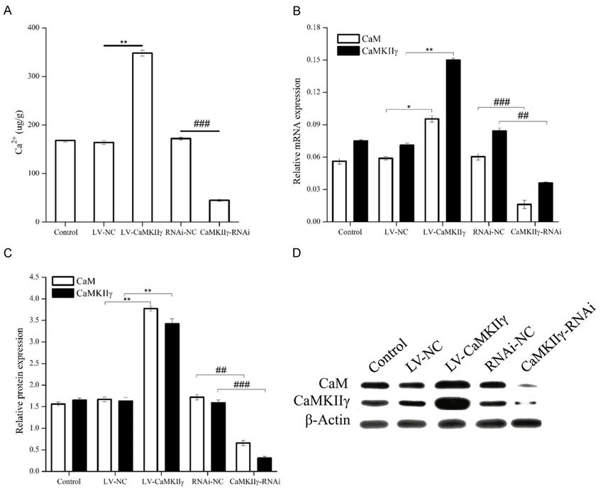
Warm ischemia duration affects hepatocyte mitochondrial damage after liver transplant

displacement, then leads the related proteins        I/R injury, we successfully established a rat
between the mitochondrial bilayer membranes          model of DCD liver transplantation, and ass-
to go into the cytoplasm, and eventually initi-      essed the effect of the ischemia duration on
ates apoptosis [5, 6]. Previous studies observed     liver function mitochondrial damage, and the
severe mitochondrial damage in the liver model       related protein expression. Further, we also
of I/R injury, manifesting as swelling mitochon-     evaluated the potential role of CaMKIIγ in isch-
dria and mitochondrial cavity, a disorganized        emia caused-hepatocyte mitochondria apopto-
cristal body, and the loose matrix accompanied       sis after DCD orthotopic liver transplantation.
by varying degrees of vacuolation and mito-          Our study may provide a valuable theoretical
chondrial membrane rupture, which has a high         basis for clinical application of DCD and a novel
positive correlation with the increase of serum      research point for improving the quality of liver
ALT and AST levels [7, 8]. Thus, the mitochon-       donation.
drial apoptosis pathway plays a key role in liver
warm ischemia-reperfusion injury because             Materials and methods
mitochondrial dysfunction will significantly re-
                                                     Animals
duce the activity of transplanted organs [9].

Ca2+ is one of the first discovered factors in-      One hundred thirty-eight healthy Sprague-
volved in the progress of ischemic injury. The       Dawley (SD) rats, aged 10-12 weeks, weight
corresponding Ca2+ channels affect the severity      250-280 g were obtained from Experimental
                                                     Animal Center of Fudan University (Shanghai,
of I/R injury by regulating the intracellular Ca2+
                                                     China). Before formal experimentation, all ani-
level [10]. Calmodulin (CaM), a major Ca2+ car-
                                                     mals were housed under standard laboratory
rier protein in non-muscle cells, and its down-
                                                     conditions for 7 days. Animals were randomly
stream signaling pathways were revealed to be
                                                     divided into three groups: 0 min-ischemic
implicated in the regeneration and proliferation
                                                     group, namely sham-operated group (n = 36),
of liver cells [11]. CaMKII (CaM kinase II) is an
                                                     15 min-ischemic group (n = 66), and 30 min-
important member of the calmodulin-depen-
                                                     ischemic group (n = 36). In 0 min-ischemic and
dent kinases cascade family that is a class of
                                                     30 min-ischemic groups, half of the group was
proteins activated by the presence of Ca2+ and
                                                     used as the donor and the other half as the
calmodulin. Kang et al. [12] reported that
                                                     recipient for liver transplantation. In 15 min-
CaMKII plays a negative role in cardiomyocyte
                                                     ischemic group, thirty rats were selected to as
apoptosis in heart failure. In vitro in human
                                                     the recipient of lentiviral transfection. All exper-
macrophages, over expression of CaMKII ser-          imental procedures were approved by the ani-
ves as an independent factor to trigger macro-       mal ethics committee of Kunming Medical
phage death [13]. Similarly, CaMKII can also         University (Approval No.: KMMU2018015) and
induce the apoptosis of mitochondria and car-        in accordance with the institutional guidelines
diomyocytes by direct action on mitochondria         for the Care and Use of Laboratory Animals
in myocardial cells undergoing irreversible I/R      (National Institutes of Health 2003).
injury, while the blocking of CaMKII causes the
opposite result [14]. Considering this evidence,     Animal model of warm ischemia
we speculate that the Ca2+/CaM/CaMKII signal-
ing pathway may play an important role in mito-      After ether aspiration anesthesia, a midline
chondrial apoptosis.                                 laparotomy was performed in the supine posi-
                                                     tion to expose the heart. All animals were intra-
At present, most of the studies involving hepa-      venously injected with 1 ml of heparin sodium
tocyte damage post-liver transplantation con-        solution (25 U heparin sodium) by the dorsum
centrate more on oxygen radicals [15], anti-         of penis vein to heparinize the donor liver. Next,
body-dependent cell-mediated cytotoxicity (AD-       the basilar part of the heart was clamped to
CC), and T cell-mediated apoptosis [16]. Wh-         block the thoracic aorta for 15 and 30 min.
ereas, the relationship between warm ischemia        Thus, a donor liver warm ischemia model was
time (WIT) of donor liver and hepatocytes mito-      established. Sham-operated rats underwent
chondria damage, as well as Ca2+/CaM/CaMKII          the same protocol without clamping of the
signaling pathway has not been reported. In          heart vessels. According to the duration of
this study, to explore the mechanism of hepatic      warm ischemia, the rats were randomly divided

218                                                        Int J Clin Exp Pathol 2019;12(1):217-228
Warm ischemia duration affects hepatocyte mitochondrial damage after liver transplant
into three groups: W0 (WIT: 0 min), W15 (WIT: the hepatic artery, the portal vein, and the
15 min), and W30 (WIT: 30 min). infra-hepatic inferior vena cava were all resect-
ed. The collected donor liver specimens were
Construction of lentiviral vector targeting stored in 4°C Ringer’s solution. Rat DCD liver
CaMKIIγ transplantation surgery was performed accord-
ing to the method reported by Kamada and
The recombinant lentivirus CaMKIIγ protein Calne [17] with minor modification. In this study,
(LV-CaMKIIγ), lentivirus-encoding short hairpin the cold ischemia time was 50 min, and no ani-
RNA targeting CaMKIIγ (LV-CaMKIIγ-RNAi), and mals died during the study. On days 1, 3, and 7
their corresponding empty vectors including LV- after liver transplantation, the rats in three
NC and RNAi-NC were constructed by Shanghai groups were randomly executed and the he-
GenePharma Co. Ltd (Shanghai, China). The fol- patic tissues were collected for subsequent
lowing sequences were used: 5’-CCAACTT- analysis.
TGTGCCAACCGGTCGCCACCATGGCCACCACCG
CCACCTGCAC-3’ and 3’-AATGCCAACTCTGAGC- Determination of liver function
TTCTGCAGCGGTGCAGCAGGGGCTC-5’ for LV-Ca-
MKIIγ; 5’-GGAGAGCGTCAACCACATC-3’ and 3’- Serum alanine aminotransferase (ALT) and
GATGTGGTTGACGCTCT CC-5’ for LV-CaMKIIγ- aspartate aminotransferase (AST) were ass-
RNAi. ayed by a 7170-A automatic analyzer (Hitachi,
Tokyo, Japan) using commercially available kits
Lentiviral transfection (Boehringer Mannheim, Lewes, East Sussex,
UK). Both values were showed as units/liter
Prior to orthotopic liver transplantation, half of
(U/L).
the rats in W15 group were randomly divided
into five groups (6 rats in each group): 1) Con- Liver morphological examination
trol group: the rats were injected with 500 μl
normal saline; 2) LV-NC group: the rats were Liver tissues were fixed in 10% neutral buffered
injected with 500 μl saline containing 108 TU/ formalin for 24 h, embedded in paraffin, cut
ml LV-CaMKIIγ empty vector; 3) LV-CaMKIIγ into 3 μm thick sections, and then stained with
group: the rats were injected with 500 μl saline hematoxylin-eosin (HE). For transmission elec-
containing 108 TU/ml LV-CaMKIIγ protein; 4) tron microscopy, liver samples were fixed in
RNAi-NC group: the rats were injected with 500 2.5% glutaraldehyde, embedded, sectioned,
μl saline containing 108 TU/ml RNAi empty vec- and then observed using an electron micro-
tor; 5) CaMKIIγ-RNAi group: the rats were inject- scope (JEOL JEM-1400, Japan).
ed with 500 μl saline containing 108 TU/ml
LV-CaMKIIγ-RNAi. All rats were injected with Measurement of mitochondrial membrane
saline by the penile dorsal vein. After 6 h of potential
administration, we detected the transfection
efficiency by fluorescence analysis. Real-time Mitochondrial membrane potential was deter-
PCR and western blotting were used to iden- mined using JC-10 probe (Invitrogen, Eugene,
tify overexpression or knockdown efficiency of OR, USA) that emits red-orange fluorescent
CaMKIIγ. from the cells with high mitochondrial potential
(HMP) or green fluorescent light from the cells
Animal model of liver transplantation with low (LMM) and medium (MMM) mitochon-
drial potential. Samples were analyzed by flow
After warm ischemia duration, 20 mL lactic cytometry excited at 488 nm, and measured at
acid Ringer’s solution (0-4°C, containing 50 u/ 590 nm. Data analysis was conducted using
mL heparin sodium) was perfused into the FlowJo software.
abdominal aorta by an infusion pump. Then, we
dissected the liver ligaments and ligated the Measurement of Ca2+
pyloric vein adjacent to the portal vein when the
portal vein and hepatic proper artery were The total Ca2+ of hepatic tissues was deter-
freed. After isolating the infra-hepatic inferior mined using an atomic absorption spectropho-
vena cava, the right suprarenal vein and right tometer (FAAS; TAS-986, Persee; China) accord-
renal vein, the supra-hepatic inferior vena cava, ing to the manufacturer’s instructions.
219 Int J Clin Exp Pathol 2019;12(1):217-228Warm ischemia duration affects hepatocyte mitochondrial damage after liver transplant
Cell apoptosis analysis unpaired Student’s t-test or one-way analysis of
variance (ANOVA) is used to evaluate the signifi-
Briefly, the hepatic cells were collected and cance between groups. P < 0.05 was consid-
then transferred into a flow tube. Annexin ered significant.
V-FITC and Propidium Iodide (PI) were added to
each sample and incubated in the dark for 15 Results
min. Samples were analyzed using a flow
cytometer (FACStar, BD Biosciences). The duration of ischemia is proportional to the
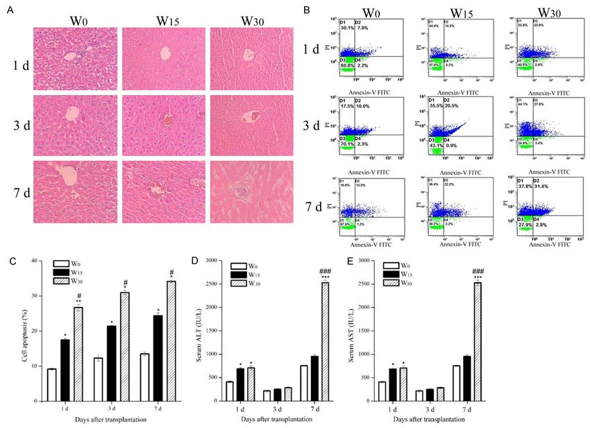
extent of hepatic damage
RT-qPCR
To evaluate the link between the duration of
RNA was extracted from liver tissues using liver ischemia and hepatocellular injury in rats,
Trizol reagent (Life Technologies, Carlsbad, CA, H&E staining was first performed to observe
USA). Quantitative real time-PCR was carried the manifestation of hepatocellular damage
out as previously described [18]. The primer and the results are shown in Figure 1A. At 1 d
sequences were as follows: CaM, forward post transplantation, the non-ischemic group
5’-GACGGTAATGGCACAATC-3’, reverse 5’-ACG- showed approximately normal morphology of
GAACGCTTCTCTAAT-3’; CaMKII, forward 5’-AC- liver tissues with intact liver tissues structure.
ACAGGAATATGCTGCAAAAAT-3’, reverse 5’-CAG- In group W15, hepatic damage was represented
TAACAAGGTCAAACACGAGG-3’; AIF, forward TA- by slight hepatocyte swelling, necrosis, vacu-
GAGAACAGCGAGTCCCGA, reverse GACCCGC- oles, and neutrophil infiltration. These patho-
TTGTCTACGACTT; Cyt C (Cytochrome C), for- logic changes were dramatically aggravated in
ward TGGACCCAATCTGCATGGTC, reverse TCC- W30 group, with severe vacuolar degeneration
AGGTACTCGAACAGGGT; β-actin, forward 5’-TG- necrosis in liver cells and infiltration of massive
GGTATGGAATCCTGTGGCA-3’, reverse 5’-TGTTG- neutrophils. With prolongation of postoperative
GCATAGAGGTCTTTACGG-3’. Gene expression time, hepatic damage in all groups gradually
levels were calculated as 2-ΔΔCt values normal- deteriorated, represented by the hepatic sinu-
ized to β-actin levels. soid narrowing and edema, and derangement
of extensive hepatocytes with turgescence. At
Western blot analysis
7 d post transplantation, livers in the 30-min
Protein extraction and western blotting were ischemia group were entirely pale and had dis-
performed as previously described with minor organized liver morphology.
modification [19]. Briefly, equal amounts of pro-
In the results of the apoptosis assay, we ob-
tein were separated on 10% sodium dodecyl
served that both W15 and W30 groups showed a
sulfate polyacrylamide gel electrophoresis
significant increase of the proportion of apop-
(SDS-PAGE, Bio-Rad, Hercules, USA) and then
totic liver cells compared with W0 group within 7
transferred to a PVDF membrane (Millipore,
days post-transplantation (P < 0.05, P < 0.01),
Bedford, USA). After incubation with 5% non-fat
and the same trend was also found in the com-
milk in Tris-buffered saline (TBS), the mem-
parison of W15 and W30 groups (P < 0.05) (Figure
branes were probed with primary antibody (all
1B, 1C). Also, warm ischemia also caused hep-
antibodies were purchased from Abcam, Cam-
atotoxicity in liver tissues, as indicated by the
bridge, UK) overnight at 4°C, and then with the
elevation of the serum ALT and AST levels in the
corresponding secondary antibody conjugated
rats tolerating 15 and 30 min of ischemia at 1
with HRP (1:1000, Cell Signaling Technology,
d after surgery (P < 0.05) (Figure 1D, 1E). The
Danvers, MA, USA) at room temperature for 1 h.
Finally, the membranes were detected using a serum ALT and AST levels almost returned to
multispectral imaging system (UVP, California, normal levels in three groups at 3 d after trans-
USA). plantation and no significant difference was
found, but they significantly increased again in
Statistical analysis livers in the 30-min ischemia group at 7 d post
transplantation (P < 0.001) (Figure 1D, 1E).
All statistical analyses were performed using This may be associated with the hepatic dam-
SPSS 19.0 software. Values are shown as age induced by reperfusion. The above results
mean ± standard deviation (SD). Two-tailed strongly confirmed that the extent of hepatocel-
220 Int J Clin Exp Pathol 2019;12(1):217-228Warm ischemia duration affects hepatocyte mitochondrial damage after liver transplant 221 Int J Clin Exp Pathol 2019;12(1):217-228
Warm ischemia duration affects hepatocyte mitochondrial damage after liver transplant

Figure 1. The duration of ischemia is proportional to the extent of hepatic damage. The rats were randomly divided
into W0, W15, and W30 groups, and then respectively exposed to 0, 15, and 30 min-warm ischemia. At 1, 3, and 7
d post transplantation, the rats were sacrificed and the liver tissues were collected for analysis. A. Representative
hematoxylin and eosin-stained liver sections (400 × magnification). B. Representative results of the cell apoptotic
rate detected by flow cytometry. C. Statistical data of the apoptosis rate. D. Serum aspartate aminotransferase (AST)
level. E. Serum alanine aminotransferase (ALT) level. *P < 0.05, **P < 0.01, ***P < 0.001 versus W0 group; #P <
0.05, ##P < 0.01, ###P < 0.001 versus W15 group.

Figure 2. The duration of ischemia is proportional to the extent of mitochondrial apoptosis. The rats were randomly
divided into W0, W15, and W30 groups, and then respectively exposed to 0, 15, and 30 min-warm ischemia. At 1, 3,
and 7 d post transplantation, the rats were sacrificed and the liver tissues were collected for analysis. (A) The sta-
tistic data of the cells with low MMP in liver tissues detected by JC-10 staining. (B, C) Statistical data of the relative
protein expression of AIF (B) and Cyt C (C). (D) The relative protein expression of AIF and Cyt C measured by western
blotting. (E) The relative protein expression of CaMKIIγ measured by western blotting and the corresponding statisti-
cal data. *P < 0.05, **P < 0.01, ***P < 0.001 versus W0 group; #P < 0.05, ##P < 0.01, ###P < 0.001 versus W15
group.

lular damage is proportional to the duration of                   group at 3 or 7 d post transplantation (P <
ischemia.                                                         0.05); meanwhile, the relative protein expres-
                                                                  sion of AIF and Cyt C was also increased with
The duration of ischemia is proportional to the                   the prolongation of ischemia implemented
extent of mitochondrial apoptosis                                 within 7 days after DCD orthotopic liver trans-
                                                                  plantation (P < 0.05, P < 0.01) (Figure 2B, 2C).
Next, we evaluated the effect of WIT on hepatic                   The above manifestations were attributed to
mitochondrial damage and apoptosis. As shown                      mitochondrial apoptosis under the stimulation
in Figure 2A, ischemia treatment contributed to                   of ischemia. Previous evidence reported that
a significantly increased number of the cells                     CaMKIIγ was abnormally expressed in several
with lower MMP in the livers in W15 and W30                       injured tissues or cells induced by various risk
groups compared with the control non-ischemic                     factors [20, 21]. Thus, we assumed that warm

222                                                                      Int J Clin Exp Pathol 2019;12(1):217-228
Warm ischemia duration affects hepatocyte mitochondrial damage after liver transplant
Figure 3. CaMKIIγ aggravates I/R-induced hepatic mitochondria apoptosis. Rats in W15 group were randomly di-
vided into five groups and then respectively injected with saline, LV-CaMKIIγ empty vector, LV-CaMKIIγ protein, LV-
CaMKIIγ-RNAi, and RNAi empty vector. On day 7 after liver transplantation, the rats in five groups were randomly
executed and the hepatic tissues were collected for subsequent analysis. A. Ultrastructural changes of liver tissues
determined by transmission electron microscopy (15000 × magnification). B. Statistical data of the cells with low
MMP in liver tissues detected by JC-10 staining. C. Statistical data of the relative mRNA expression of AIF and Cyt C
measured by RT-qPCR. D. Statistical data of the relative protein expression of AIF and Cyt C. E. Results of the relative
protein expression of AIF and Cyt C tested by western blotting. D. E. *P < 0.05, **P < 0.01, ***P < 0.001 versus
LV-NC group; #P < 0.05, ##P < 0.01, ###P < 0.001 versus RNAi-NC group.
ischemia may result in an aberrant expression sion lentivirus into the rats exposed to 15 min-
of CaMKIIγ in the liver tissues. As expected, the ischemia, and then evaluated mitochondrial
protein expression of CaMKIIγ in rats exposed damage at 7 d post transplantation. As shown
to 15 or 30 min ischemia was much higher than in Figure 3A, ischemia only caused insignificant
that in the W0 group at 3 and 7 d after trans- ultrastructural damage of liver tissues in con-
plantation (P < 0.05, P < 0.01) (Figure 2E), con- trol group, including slight hepatocyte edema
firming the positive association between WIT and mitochondrial condensation, but the mito-
and CaMKIIγ level. Thus, it can be concluded chondria exhibited regular cristae and intact
that the duration of ischemia is proportional to membrane without swelling. Similar changes
the degree of mitochondrial apoptosis and the were also observed in the LV-NC and RNAi-NC
expression of CaMKIIγ. groups. However, the ultrastructural damage
was much worse in the rats after transferred
CaMKIIγ aggravates I/R-induced hepatic mito-
chondria apoptosis with CaMKIIγ, as shown by obvious mitochon-
drial swelling and hepatocellular necrosis, me-
In consideration of the significant increase in mbrane rupture and atrophy, and the loss of
CaMKIIγ expression in the livers subjected to mitochondrial cristae. When CaMKIIγ expres-
with warm ischemia, we assumed that CaMKII sion was blocked, damaged morphology was
may be implicated in the process of I/R-induced improved markedly, in which the hepatic cells
mitochondrial injury. To identify this, we trans- were normal and no swollen mitochondrial or
ferred CaMKIIγ-RNAi and CaMKIIγ overexpres- hepatocytes were found.
223 Int J Clin Exp Pathol 2019;12(1):217-228Warm ischemia duration affects hepatocyte mitochondrial damage after liver transplant
Figure 4. CaMKIIγ upregulates Ca2+/CaM/CaMKIIγ level in rat model of I/R injury. The rats in W15 group were ran-
domly divided into five groups and then respectively injected with saline, LV-CaMKIIγ empty vector, LV-CaMKIIγ
protein, LV-CaMKIIγ-RNAi, and RNAi empty vector. On day 7 after liver transplantation, the rats in five groups were
randomly executed and the hepatic tissues were collected for subsequent analysis. A. The results of Ca2+ level de-
tected by an atomic absorption spectrophotometer. B. Statistical data of the relative mRNA expression of CaM and
CaMKIIγ measured by RT-qPCR. C. Statistical data of the relative protein expression of CaM and CaMKIIγ. D. Relative
protein expression of CaM and CaMKIIγ measured by western blotting. *P < 0.05, **P < 0.01, ***P < 0.001 versus
LV-NC group; #P < 0.05, ##P < 0.01, ###P < 0.001 versus RNAi-NC group.
Next, JC-10 staining was performed in vitro, for 0.05, P < 0.01, P < 0.001), as confirmed by
assessing mitochondrial damage. Control gro- both RT-qPCR (Figure 3C) and western blotting
up exhibited relatively few cells with low MMP (Figure 3D, 3E) assays. Collectively, these find-
at 7 d after surgery, and there were no differ- ings revealed a destructive effect of CaMKIIγ
ences between control and empty vector gro- on I/R-induced hepatic mitochondrial apopto-
ups. CaMKIIγ overexpression resulted in a sig- sis post-orthotopic liver transplantation.
nificant increase of the percentage of the cells
CaMKIIγ upregulates Ca2+/CaM/CaMKIIγ levels
with low MMP relative to the empty vector
in rat model of I/R injury
group (P < 0.05), suggesting apoptosis in the
mitochondria; whereas, CaMKIIγ knockdown To further explore the potential mechanism of
caused an opposite result (P < 0.01) (Figure CaMKIIγ involving I/R-induced mitochondrial
3B). Similarly, AIF and Cyt C release was signi- injury, we then detected the changes of Ca2+/
ficantly enhanced by CaMKIIγ overexpression CaM/CaMKIIγ expression in different treatment
or reduced by CaMKIIγ-RNAi when compared groups. Clearly, there was an obvious increase
with the corresponding negative control (P < of Ca2+ level in rats administered with CaMKIIγ
224 Int J Clin Exp Pathol 2019;12(1):217-228Warm ischemia duration affects hepatocyte mitochondrial damage after liver transplant
overexpression compared with empty vector groups gradually deteriorated. It is well known
group (P < 0.01); whereas, the Ca2+ level in that apoptosis is the primary pathogenesis of
CaMKIIγ-RNAi group was much lower than that I/R injury [24], thus the Annexin V-FITC/PI dou-
in RNAi-NC group because of the downregula- ble staining was subsequently performed to
tion of CaMKIIγ (P < 0.001). We also found that explore the apoptotic rate of hepatocytes. As
the relative expression of CaM and CaMKIIγ expected, the percentage of apoptotic liver
was upregulated markedly in the CaMKIIγ group cells was also proportional to the duration of
and downregulated significantly in the CaMKIIγ- ischemia within 7 days after transplantation
RNAi group when compared with their negative surgery, which further confirmed the observa-
controls at both the mRNA (P < 0.05, P < 0.01, tion of H&E staining. ALT and AST are the most
P < 0.001) (Figure 4B) and protein levels (P < useful indicators of liver function. Enhanced
0.01, P < 0.001) (Figure 4C, 4D). Combined levels of ALT and AST indirectly reflect the
with above and prior results, we can conclude degree of hepatocyte damage [25]. In this
that the overexpression of CaMKIIγ that aggra- study, the serum ALT and AST improved rapidly
vates I/R-induced mitochondrial injury in liver at 1 d after operation and then almost returned
tissues may be achieved by upregulating of to the normal level at 3 d, which was consistent
Ca2+/CaM/CaMKIIγ. with the previous finding [26] that showing the
activities of ALT and AST increased markedly
Discussion within a few hours after transplantation, then
reached a peak within 24 h, and finally dropped
Orthotopic liver transplantation has been wide- rapidly. However, we also found that the levels
ly recognized as the only effective method for of ALT and AST significantly increased again on
treating end-stage liver disease [22]. Currently, 7 days, which may be caused by reperfusion
donor liver is one of the main sources of liver induced-hepatic injury post-operation of liver
transplantation, and its global demand has transplantation.
been growing steadily over the past few years
[23]. However, ischemia-reperfusion caused- Mitochondrial dysfunction, a key pathological
liver injury has been identified as an important process of I/R injury, can directly affect cell
risk factor that determined the success rate of energy metabolism and induce necrosis or
liver transplantation by affecting the prognosis apoptosis [27, 28]. The decline in mitochondrial
of disease, success rate of operation, and sur- membrane potential is the most significant
vival rate of patients [2]. Therefore, to maintain manifestation of damaged mitochondria. In our
the functional activity of donor liver and improve research, the proportion of cells with low MMP
the success rate of liver transplantation, it is rose gradually with the increase of ischemic
necessary to explore the effects of warm isch- time within 7 days after orthotopic liver trans-
emia duration on transplanted liver, as well as plantation, and the difference was statistically
identify the potential mechanism and related significant, indicating a positive relationship
target molecules. between the extent of mitochondria apoptosis
and the duration of warm ischemia. As for the
To evaluate the effect of warm ischemic time mechanism of I/R induced mitochondria injury,
on hepatocellular damage, we first established a previous study revealed that ischemic treat-
a rat model of liver I/R injury by exposing the ment generally caused mitochondrial osmotic
donor liver to 0, 15, and 30 min-ischemia translocation through the action of risk factors,
respectively, and then assessed the extent of then caused the related proteins (including
liver damage in rats on 1, 3, and 7 d post trans- cytochrome C and apoptosis inducing factor,
plantation. The representative images of H & E AIF) between the mitochondrial bilayer mem-
staining revealed that the livers that tolerated brane to move into the cytoplasm [5], and final-
30-min ischemia showed the worst histomor- ly initiated apoptosis [6]. Not surprisingly, the
phology, exhibited by a large amount of neutro- protein expression levels of AIF and Cyt C were
phil infiltration and severe vacuolar degenera- found to be proportional to the duration of
tion necrosis. The morphology of liver tissues in warm ischemia in this study. Similarly, we also
W15 group was much better than the W30 group observed a significant upregulation of CaMKIIγ
but worse than the W0 group. With prolongation expression when warm ischemia time was
of postoperative time, hepatic damage in all increased, which revealed that CaMKIIγ may be
225 Int J Clin Exp Pathol 2019;12(1):217-228Warm ischemia duration affects hepatocyte mitochondrial damage after liver transplant
one potential target molecule involved in I/R by many factors. For example, activation of the
induced-mitochondria damage. Actually, CaM- Ca2+/CaM/CAMKII pathway could positively reg-
KIIγ was found to be overexpressed in several ulate high glucose-induced apoptosis in human
injured cells caused by various wounding fac- umbilical vein endothelial cells [34], and also
tors, such as in the brain tissues in rats with was confirmed to be implicated in myocardial
acute severe carbon monoxide poisoning [20] ischemia induced-heart injury [35]. In this
and in PC12 cells exposed to fluoride [21]; study, CaMKIIγ overexpression caused a signifi-
these studies partly agreed with our results. cant increase of Ca2+ level, as well as the
expression of CaM and CaMKIIγ at both the
Considering the abnormal upregulation of mRNA and protein levels, and an opposite
CaMKIIγ, we tested a hypothesis that CaMKII result was found when CaMKIIγ was downregu-
plays an important role in the process of mito- lated. The above observation indicated that the
chondrial apoptosis induced by I/R. To verify induction of mitochondrial damage by I/R may
this, we overexpressed or down CaMKIIγ level be achieved by activating Ca2+/CaM/CaMKII
in the rats exposed to 15 min-ischemia by signaling pathway, which is supported by the
transfecting CaMKIIγ overexpression and Ca- prior article that confirming the involvement of
MKIIγ-RNAi lentiviral vectors. On 7 d post trans- the Ca2+/CaM/CaMKII pathway in oxidative
plantation, CaMKIIγ overexpression caused a stress-induced mitochondrial permeability
significant deterioration of ultrastructural mor- transition and apoptosis in isolated rat hepato-
phology in liver tissues compared with the neg- cytes [36].
ative control, while blocking CaMKIIγ exhibited
opposite effect. These findings preliminarily Based on our results, the potential mechanism
revealed the negative regulatory role of CaMKIIγ of hepatocyte apoptosis induced by warm isch-
in I/R induced mitochondria injury, which is emia can be tentatively summarized as follows:
supported by several previous studies that during DCD orthotopic liver transplantation,
revealing CaMKII regulates ischemia mediated- hepatocellular I/R injury contributes to the
cardiomyocyte apoptosis and necrosis by abnormal expression of CaMKIIγ. Upregulation
directly inducing mitochondrial apoptosis [29, of CaMKIIγ induces a decline and permeability
30]. Membrane potential is involved in main- shift of mitochondrial membrane potential,
taining mitochondrial function and plays a cru- then leads to mitochondrial swelling, rupture,
cial part in inhibiting hepatocyte apoptosis [31]. and apoptosis. Next, AIF and Cyt C proteins
Previous research observed that the apoptotic enter into the cytoplasm and initiate apoptosis,
rate of renal tubular epithelial cells induced by and eventually promote hepatocyte apoptosis.
clindamycin in CaMKIIγ knockout rats was sig- Considering the auxo-action of CaMKIIγ overex-
nificantly lower than the control group. Similar pression in ischemia induced-mitochondrial
results were also found in the loss of mitochon- apoptosis, it can be inferred that specific block-
drial membrane potential and mitochondrial age of the CaMKII signaling pathway post DCD
apoptosis [32, 33]. In our study, an obvious liver transplantation may effectively relieve
increased percentage of cells with low MMP hepatocyte damage and improve prognosis.
was observed under the administration of
Acknowledgements
CaMKIIγ, which directly suggested that CaMKIIγ
can induce the permeability transition and the
This study was supported by Joint project funds
disruption of the mitochondrial membrane of Yunnan Provincial Science and Technology
potential. In addition, the protein level of Cyt C Department-Kunming Medical University, P. R.
and AIF was also up-regulated when CaMKIIγ China (Grant No.: 2015FA010).
was up-regulated, which reflected that CaMKIIγ
upregulates the level of apoptosis-related pro- Disclosure of conflict of interest
teins, such as Cyt C and AIF, by acting on the
mitochondrial membrane, and then initiate None.
apoptosis.
Address correspondence to: Jiang-Hua Ran, De-
According to much evidence, we learned that partment of Hepatopancreatobiliary Surgery, The
Ca2+/CaM/CaMKII signaling pathway was asso- Affiliated Calmette Hospital of Kunming Medical
ciated with cell damage or apoptosis triggered University, The First People’s Hospital of Kunming,
226 Int J Clin Exp Pathol 2019;12(1):217-228Warm ischemia duration affects hepatocyte mitochondrial damage after liver transplant
Calmette Hospital, 1228 Beijing Road, Panlong and hypertrophy in the cardiac signaling net-
District, Kunming 650224, Yunnan Province, China. work. Sci Rep 2017; 7: 34.
Tel: +86-13700631170; Fax: +86-871-67390506; [13] Timmins JM, Ozcan L, Seimon TA, Li G, Malage-
E-mail: ranjianghuakm@163.com lada C, Backs J, Backs T, Bassel-Duby R, Olson
EN, Anderson ME, Tabas I. Calcium/calmodu-
References lin-dependent protein kinase II links ER stress
with Fas and mitochondrial apoptosis path-
[1] Tashiro H, Kuroda S, Mikuriya Y and Ohdan H. ways. J Clin Invest 2009; 119: 2925-2941.
Ischemia-reperfusion injury in patients with [14] Salas MA, Valverde CA, Sánchez G, Said M, Ro-
fatty liver and the clinical impact of steatotic driguez JS, Portiansky EL, Kaetzel MA, Ded-
liver on hepatic surgery. Surg Today 2014; 44: man JR, Donoso P and Kranias EG. The signal-
1611-1625. ling pathway of camkii-mediated apoptosis
[2] Eliasmiró M, Jiménezcastro MB, Rodés J and and necrosis in the ischemia/reperfusion inju-
Peralta C. Current knowledge on oxidative ry. J Mol Cell Cardiol 2010; 48: 1298-1306.
stress in hepatic ischemia/reperfusion. Arch [15] Kostopanagiotou G, Tierris J, Arkadopoulos N,
Med Sci 2013; 47: 555-568. Theodoraki K, Deliconstantinos G, Matsota P,
[3] De RO, Dutkowski P and Clavien PA. Biological Smyrniotis V and Pandazi A. Liver transplanta-
modulation of liver ischemia-reperfusion inju- tion in pigs: NO, oxygen free radicals, pulmo-
ry. Curr Opin Organ Transplant 2010; 15: 183- nary hemodynamics. J Surg Res 2008; 149:
9. 231-235.
[4] Toledopereyra LH, Toledo AH, Walsh J and [16] Jiang Z, Chen Y, Feng X, Jiang J, Chen T, Xie H,
Lopezneblina F. Molecular signaling pathways Zhou L and Zheng S. Hepatic stellate cells pro-
in ischemia/reperfusion. Exp Clin Transplant mote immunotolerance following orthotopic
2004; 2: 174-7. liver transplantation in rats via induction of T
[5] Lin NC, Liu CS, Chang CJ, Loong CC, Hsia CY cell apoptosis and regulation of Th2/Th3-like
and Tsai HL. Changes in mitochondrial respira- cell cytokine production. Exp Ther Med 2013;
tory enzyme activity after ischemia-reperfu- 5: 165-169.
sion injury inliving-donor liver transplantation. [17] Kamada N and Calne RY. Orthotopic liver
Transplant Proc 2010; 42: 721-724. transplantation in the rat. Technique using cuff
[6] Norberg E, Orrenius S and Zhivotovsky B. Mito- for portal vein anastomosis and biliary drain-
chondrial regulation of cell death: processing age. Transplantation 1979; 28: 47-50.
of apoptosis-inducing factor (AIF). Biochem [18] Mühlbauer M, Bosserhoff AK, Hartmann A,
Biophys Res Commun 2010; 396: 95-100. Thasler WE, Weiss TS, Herfarth H, Lock G,
[7] Ran J, Guo Y, Li L, Zhang S, Liu J, Li Z and Li L. Schölmerich J and Hellerbrand C. A novel MCP-
Protective and repaire of recombinant human 1 gene polymorphism is associated with he-
growth hormone (rhGH) in ischemic reperfu- patic MCP-1 expression and severity of HCV-
sion injury of rat liver. Journal of Hepatobiliary related liver disease. Gastroenterology 2003;
Surgery 2008; 16: 300-303. 125: 1085-1093.
[8] Ran J, Guo Y, Li L and Zhang S. Protective role [19] Dorn C, Kraus B, Motyl M, Weiss TS, Gehrig M,
of recombinant human growth hormone in Schölmerich J, Heilmann J, Hellerbrand C.
ischemic reperfusion injury of rat liver. Chin J Xanthohumol, a chalcon derived from hops,
Base General Surg 2006; 13: 555-559. inhibits hepatic inflammation and fibrosis. Mol
[9] Lin J, He F, Wu L, Huang H and Zeng Z. Isch- Nutr Food Res 2010; 54: S205-S213.
emic postconditioning alleviating liver isch- [20] Li Q, Ding X, Bi W, Wang J and Zou Y. [Effects of
emia-reperfusion injury in rats via mitochon- N-butylphthalide on the expressions of calpain
drial pathway. Chin J Organ Transplant 2012; 1 and CaMK II in hippocampus in rats with
33. acute severe carbon monoxide poisoning].
[10] Chang WJ, Chehab M, Kink S and Toledoperey- Zhonghua Wei Zhong Bing Ji Jiu Yi Xue 2017;
ra LH. Intracellular calcium signaling pathways 29: 1127-1132.
during liver ischemia and reperfusion. J Invest [21] Liao Q, Zhang R, Wang X, Nian W, Ke L, Ouyang
Surg 2010; 23: 228-238. W and Zhang Z. Effect of fluoride exposure on
[11] Orellana D, Liu X, Wang GL, Jin J, Iakova P and mRNA expression of cav1.2 and calcium signal
Timchenko NA. Calmodulin controls liver prolif- pathway apoptosis regulators in PC12 cells.
eration via interactions with C/EBPbeta-LAP Environ Toxicol Pharmacol 2017; 54: 74-79.
and C/EBPbeta-LIP. J Biol Chem 2010; 285: [22] Pompili M, Francica G, Ponziani FR, Iezzi R and
23444-56. Avolio AW. Bridging and downstaging treat-
[12] Kang JH, Lee HS, Park D, Kang YW, Kim SM, ments for hepatocellular carcinoma in patients
Gong JR and Cho KH. Context-independent es- on the waiting list for liver transplantation.
sential regulatory interactions for apoptosis World J Gastroenterol 2013; 19: 7515-7530.
227 Int J Clin Exp Pathol 2019;12(1):217-228Warm ischemia duration affects hepatocyte mitochondrial damage after liver transplant
[23] Fujita S, Mizuno S, Fujikawa T, Reed AI, Kim [31] Xie Y, Xiao F, Luo L and Zhong C. Activation of
RD, Howard RJ and Hemming AW. Liver trans- autophagy protects against ROS-mediated mi-
plantation from donation after cardiac death: a tochondria-dependent apoptosis in L-02 hepa-
single center experience. Transplantation tocytes induced by Cr(VI). Cell Physiol Biochem
2007; 84: 46-9. 2014; 33: 705-716.
[24] Yuan J, Zeng L, Sun Y, Wang N, Sun Q, Cheng Z [32] Moser MJ, Geiser JR and Davis TN. Ca2+-
and Wang Y. SH2B1 protects against OGD/R- calmodulin promotes survival of pheromone-
induced apoptosis in PC12 cells via activation induced growth arrest by activation of calci-
of the JAK2/STAT3 signaling pathway. Mol Med neurin and Ca2+-calmodulin-dependent pro-
Rep 2018; 18: 2613-2620. tein kinase. Mol Cell Biol 1996; 16: 4824-
[25] Dhibi M, Brahmi F, Mnari A, Houas Z, Chargui I, 4831.
Bchir L, Gazzah N, Alsaif MA and Hammami M. [33] Schlegel A, Kron P, Graf R, Dutkowski P and
The intake of high fat diet with different trans Clavien PA. Warm vs. cold perfusion tech-
fatty acid levels differentially induces oxidative niques to rescue rodent liver grafts. J Hepatol
stress and non alcoholic fatty liver disease 2014; 61: 1267-1275.
(NAFLD) in rats. Nutr Metab 2011; 8: 65. [34] Chen Z and Wu X. [Salidroside attenuates high
[26] Biasi F, Bosco M, Chiappino I, Chiarpotto E, glucose-induced apoptosis in human umbilical
Lanfranco G, Ottobrelli A, Massano G, Donadio vein endothelial cells via activating the Ca(2)+/
PP, Vaj M and Andorno E. Oxidative damage in CaM/CAMKIIδ/eNOS pathway]. Zhonghua Xin
human liver transplantation. Free Radic Biol Xue Guan Bing Za Zhi 2014; 42: 327.
Med 1995; 19: 311-317. [35] Zhao Y, Hu HY, Sun DR, Feng R, Sun XF, Guo F
[27] Li Y, Jiang Z, Xue D, Deng G, Li M, Liu X and and Hao LY. Dynamic alterations in the CaV1.2/
Wang Y. Mycoplasma ovipneumoniae induces CaM/CaMKII signaling pathway in the left ven-
sheep airway epithelial cell apoptosis through tricular myocardium of ischemic rat hearts.
an ERK signalling-mediated mitochondria DNA Cell Biol 2014; 33: 282-290.
pathway. BMC Microbiol 2016; 16: 222. [36] Toledo FD, Pérez LM, Basiglio CL, Ochoa JE,
[28] Pan Q, Xue M, Xiao SS, Wan YJ and Xu DB. A Sanchez Pozzi EJ and Roma MG. The Ca²⁺-
combination therapy with baicalein and taxol calmodulin-Ca²⁺/calmodulin-dependent pro-
promotes mitochondria-mediated cell apopto- tein kinase II signaling pathway is involved in
sis: involving in akt/β-catenin signaling path- oxidative stress-induced mitochondrial perme-
way. DNA Cell Biol 2016; 35: 646. ability transition and apoptosis in isolated rat
[29] Vilapetroff M, Salas MA, Said M, Valverde CA, hepatocytes. Arch Toxicol 2014; 88: 1695-
Sapia L, Portiansky E, Hajjar RJ, Kranias EG, 709.
Mundiñaweilenmann C and Mattiazzi A. CaM-
KII inhibition protects against necrosis and
apoptosis in irreversible ischemia-reperfusion
injury. Cardiovasc Res 2007; 73: 689-98.
[30] Chang H, Sheng JJ, Zhang L, Yue ZJ, Jiao B, Li
JS and Yu ZB. ROS-induced nuclear transloca-
tion of calpain-2 facilitates cardiomyocyte
apoptosis in tail-suspended rats. J Cell Bio-
chem 2015; 116: 2258-2269.
228 Int J Clin Exp Pathol 2019;12(1):217-228You can also read