Proliferative Pathways of Vascular Smooth Muscle Cells in Response to Intermittent Hypoxia - MDPI
←
→
Page content transcription
If your browser does not render page correctly, please read the page content below
International Journal of
Molecular Sciences
Review
Proliferative Pathways of Vascular Smooth Muscle
Cells in Response to Intermittent Hypoxia
Yoji Kyotani 1, *, Shin Takasawa 2 and Masanori Yoshizumi 1
1 Department of Pharmacology, Nara Medical University School of Medicine, Kashihara 634-8521, Japan;
yoshizu@naramed-u.ac.jp
2 Department of Biochemistry, Nara Medical University School of Medicine, Kashihara 634-8521, Japan;
shintksw@naramed-u.ac.jp
* Correspondence: cd147@naramed-u.ac.jp; Tel.: +81-744-29-8831
Received: 28 April 2019; Accepted: 30 May 2019; Published: 1 June 2019
Abstract: Obstructive sleep apnea (OSA) is characterized by intermittent hypoxia (IH) and is a risk
factor for cardiovascular diseases (e.g., atherosclerosis) and chronic inflammatory diseases (CID). The
excessive proliferation of vascular smooth muscle cells (VSMCs) plays a pivotal role in the progression
of atherosclerosis. Hypoxia-inducible factor-1 and nuclear factor-κB are thought to be the main factors
involved in responses to IH and in regulating adaptations or inflammation pathways, however,
further evidence is needed to demonstrate the underlying mechanisms of this process in VSMCs.
Furthermore, few studies of IH have examined smooth muscle cell responses. Our previous studies
demonstrated that increased interleukin (IL)-6, epidermal growth factor family ligands, and erbB2
receptor, some of which amplify inflammation and, consequently, induce CID, were induced by IH
and were involved in the proliferation of VSMCs. Since IH increased IL-6 and epiregulin expression
in VSMCs, the same phenomenon may also occur in other smooth muscle cells, and, consequently,
may be related to the incidence or progression of several diseases. In the present review, we describe
how IH can induce the excessive proliferation of VSMCs and we develop the suggestion that other
CID may be related to the effects of IH on other smooth muscle cells.
Keywords: intermittent hypoxia; vascular smooth muscle cells; epiregulin; interleukin
1. Introduction
Obstructive sleep apnea (OSA) is characterized by repeated episodes of intermittent hypoxia
(IH), i.e., transient oxygen (O2 ) desaturation, and resaturation. In clinical practice, OSA is commonly
diagnosed by polysomnography and its severity is classified by the apnea hypopnea index (AHI) as
follows: mild, AHI ≥5; moderate, AHI ≥15; severe, AHI ≥30 [1,2]. It is a highly prevalent disorder [3,4];
Peppard et al. estimated that the prevalence of moderate to severe sleep-disordered breathing is
10% and 3% among 30- to 49-year-old men and women, respectively, and 17% and 9% among 50- to
70-year-old men and women, respectively [3]. Furthermore, OSA is well known as a risk factor for
diabetes, systematic hypertension, and cardiovascular diseases [5–16], and also increases mortality
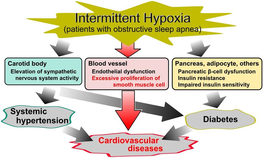
from cardiovascular diseases (Figure 1) [17,18].
Continuous positive airway pressure (CPAP) is a clinically effective strategy for treating several
diseases that derive from OSA. A number of studies have shown that CPAP decreases hemoglobin
A1c levels, blood pressure, and inflammatory markers, as well as the frequency of cardiovascular
events [19–22]. However, some studies have reported no significant effects of CPAP on glycemic control,
serum lipids, hypertension, or cardiovascular events [23–26]. Additionally, patient compliance with
CPAP treatment is often unsatisfactory [27–29]. Therefore, a clarification of the mechanisms underlying
atherosclerosis in response to IH is important for establishing prophylaxis against OSA-related diseases.
Int. J. Mol. Sci. 2019, 20, 2706; doi:10.3390/ijms20112706 www.mdpi.com/journal/ijms
Int. J. Mol. Sci. 2019, 20, 2706 2 of 14
Int. J. Mol. Sci. 2018, 19, x FOR PEER REVIEW 2 of 14
FigureFigure 1. Cause
1. Cause and and effect
effect diagram
diagram of ofobstructive
obstructive sleep
sleep apnea
apnea(OSA)-related
(OSA)-related diseases. Although
diseases. Although
intermittent hypoxia (IH) in OSA is a known risk factor for diabetes, systematic hypertension,
intermittent hypoxia (IH) in OSA is a known risk factor for diabetes, systematic hypertension, and
cardiovascular diseases, the cellular mechanisms underlying the relationship between IH and
and cardiovascular diseases, the cellular mechanisms underlying the relationship between IH and
cardiovascular diseases remain elusive. Despite a large number of studies of IH, the molecular
cardiovascular diseases remain elusive. Despite a large number of studies of IH, the molecular
mechanism of IH on vascular smooth muscle cells is less established.
mechanism of IH on vascular smooth muscle cells is less established.
Continuous positive airway pressure (CPAP) is a clinically effective strategy for treating several
Atherosclerosis is well
diseases that derive fromknown
OSA. Aas a major
number risk factor
of studies have for cardiovascular
shown diseases
that CPAP decreases that can result
hemoglobin
A1c
in heart levels, blood
diseases pressure,Itand
and stroke. inflammatory markers,
is characterized as well as the
by the formation frequency
of lesions, of cardiovascular
foam cells, and fibrous
events
plaques. The[19–22]. However,in
major features some studies have reported
the progression no significant
of atherosclerosis areeffects of CPAP on
inflammation, theglycemic
dysfunction
of the control,
endothelialserum lipids,
barrier, hypertension,
oxidative stress,or andcardiovascular
the excessiveevents [23–26].ofAdditionally,
proliferation vascular smooth patientmuscle
compliance with CPAP treatment is often unsatisfactory [27–29]. Therefore,
cells (VSMCs) [30,31]. However, the pathophysiology of these cardiovascular diseases in OSA remains a clarification of the
mechanisms underlying atherosclerosis in response to IH is important for establishing prophylaxis
incompletely understood. OSA-related cardiovascular diseases are generally thought to be caused
against OSA-related diseases.
by variousAtherosclerosis
pathophysiological triggers, such as sympathetic nervous system overactivity, systemic
is well known as a major risk factor for cardiovascular diseases that can result
inflammation, and oxidative stress, which in turn
in heart diseases and stroke. It is characterized lead
by the to metabolic
formation dysregulation,
of lesions, foam cells, and hypertension,
fibrous
plaques. The major features in the progression of atherosclerosis are inflammation, the dysfunction
and endothelial dysfunction [32,33]. In vitro and in vivo models of IH have allowed researchers to
of thethe
investigate endothelial
influencesbarrier,
of IHoxidative
on severalstress, and the
tissues and excessive
cells, and proliferation
although of vascular
articles onsmooth muscleeffects
the vascular
cellscardiovascular
in IH and (VSMCs) [30,31]. However,
diseases the pathophysiology
in OSA syndrome have of been these cardiovascular
previously diseases
published, theineffects
OSA of IH
remains
on VSMCs, incompletely
including understood.
its molecular OSA-related have
mechanisms, cardiovascular
not beendiseases
describedare [14,15].
generallyFurthermore,
thought to be there
caused by various pathophysiological triggers, such as sympathetic nervous system overactivity,
are few in vitro or in vivo studies of IH in other smooth muscle cells.
systemic inflammation, and oxidative stress, which in turn lead to metabolic dysregulation,
Recently, our laboratory demonstrated that IH directly increased the number of VSMCs by
hypertension, and endothelial dysfunction [32,33]. In vitro and in vivo models of IH have allowed
increasing the epidermal
researchers growth
to investigate factor (EGF)
the influences family
of IH ligands
on several andand
tissues the cells,
EGF andreceptor erbB2,
although which
articles on were
the mediated
partially vascular effects
by the in IH-induced
IH and cardiovascular
increase of diseases in OSA(IL)-6
interleukin syndrome haveInbeen
[34,35]. previously
the present review,
published, the
we summarize theeffects
effects of of
IH IHon VSMCs,
on VSMCs, including its molecular
focusing on the mechanisms,
intracellularhavemechanisms
not been described
related to
[14,15]. Furthermore,
atherosclerosis, and develop therea are few in vitro
discussion or in vivo
of other studies
chronic of IH in otherdiseases
inflammatory smooth muscle
(CID).cells.
Recently, our laboratory demonstrated that IH directly increased the number of VSMCs by
increasing
2. Vascular Smooththe epidermal growth
Muscle Cells factor (EGF)
(VSMCs) family ligands and the EGF receptor erbB2, which
in Atherosclerosis
were partially mediated by the IH-induced increase of interleukin (IL)-6 [34,35]. In the present
Typically,
review, weVSMCs havethe
summarize been regarded
effects asVSMCs,
of IH on key players in the
focusing progression
on the ofmechanisms
intracellular atherosclerosis because
related
to atherosclerosis,
their excessive and develop
proliferation a discussion
promotes plaqueof formation,
other chronicand
inflammatory
then theirdiseases (CID).
presence in the advanced
plaques prevent the rupture of the plaques’ fibrous caps. VSMCs in normal arterial media have a
spindle shape, termed the contractile phenotype, however, in damaged vessels, VSMCs develop a
proinflammatory phenotype that produces proinflammatory mediators responsible for proliferation
and chemotaxis. Thus, in both beneficial and detrimental ways, inflammatory responses and an
excessive proliferation of VSMCs are responsible for the progression of atherosclerosis [36,37].Int. J. Mol. Sci. 2019, 20, 2706 3 of 14
3. Reactive Oxygen Species (ROS) and Transcriptional Factors in Intermittent Hypoxia (IH)
A large number of previous in vivo and in vitro studies have shown that IH-induced intracellular
mechanisms are mainly classified into two different transcription pathways, where the hypoxia-inducible
factor (HIF)-1 and the nuclear factor (NF)-κB play central roles [16,33,38]. In carotid bodies, IH-induced
ROS generation is associated with HIF-1 activity and results in a sensory long-term facilitation of
carotid bodies [39–41]. Several in vivo and in vitro studies have also observed that IH induces the
activation of NF-κB in cardiovascular tissues and endothelial cells [42–45]. Our previous research,
using reporter gene assays, also confirmed that IH induces the activation of NF-κB in cultured rat
aortic smooth muscle cells (RASMCs) [34]. Taken together, all of these studies suggest that IH activates
alternative transcriptional pathways depending on the tissue and cell types. Similarly, Kaczmarek et al.
showed that IH decreased HIF-1α expression in human dermal microvascular endothelial cells but
increased HIF-1α expression in human coronary artery endothelial cells, indicating that endothelial
cells in cultures originating from distinct vascular beds respond differently to IH stress [46].
3.1. Reactive Oxygen Species (ROS)
ROS, such as superoxide anion, hydrogen peroxide, and hydroxyl radical, are well known as
products of a partial reduction of oxygen. They are generated either in the processes of mitochondrial
oxidative phosphorylation or during cellular responses to exogenous sources. Excessive ROS cause
oxidative stress, which in turn results in macromolecular damage and is implicated in various diseases,
including atherosclerosis [47]. In fact, it has been suggested that ROS are generated in patients with
OSA [38,48–51] and are associated with the pathogenesis of cardiovascular diseases [52]. Furthermore,
it has also been found that the mRNA molecules of heme oxygenase 1, superoxide dismutase (SOD) 1
and 2, and catalase, which are all involved in the modulation of ROS, are also changed in patients
with OSA [53].
Makarenko et al. conducted an in vitro study using human lung microvascular endothelial cells
and found that IH increased ROS levels and led to the reorganization of cytoskeleton and junction
proteins via the ROS-dependent activation of p38 mitogen-activated protein kinases (MAPK), which
resulted in endothelial barrier dysfunction [54]. Similarly, Recoquillon et al. used human aortic
endothelial cells and also reported that IH increased ROS and nitric oxide production, p65-NF-κB
activation, and IL-6 secretion [44]. In contrast, Hoffmann et al. found that, in human coronary artery
endothelial cells, IH increased manganese SOD activity via an increased dual-specificity phosphatase 1
(DUSP1) expression, and that overnight IH induced the expression of DUSP1 in mononuclear cells and
granulocytes from patients with OSA [55]. Therefore, there is some evidence that ROS play a pivotal
role in mediating the cardiovascular pathology associated with IH.
However, Hayakawa et al. suggested that ROS were unlikely to mediate the activation of
NF-κB [56], while Ryan et al. did not detect any influence on NF-κB activation from the presence of ROS
scavenger N-acetyl-L-cysteine [43]. With respect to VSMCs, few studies have examined the involvement
of ROS in IH. In our previous study, TEMPOL (1-oxyl-2,2,6,6-tetramethyl-4-hydroxypiperidine), a
SOD mimic, did not exhibit any inhibitory effects on the IH-induced proliferation of RASMCs [34].
Therefore, further investigation is needed to elucidate the roles of ROS in VSMC responses in IH.
3.2. Nuclear Factor (NF)-κB
The eukaryotic transcription factor, NF-κB, is a key mediator involved in the control of a large
number of cellular processes, especially in immune and inflammatory responses [57,58]. In the NF-κB
activation pathway, degradation of the inhibitor of NF-κB (I-κB) results in the translocation of NF-κB
to the nucleus. This in turn causes an increase of inflammatory cytokines, such as IL-6 and IL-8 [59,60].Int. J. Mol. Sci. 2019, 20, 2706 4 of 14
A number of in vivo and in vitro studies have found that IH activates NF-κB in cardiovascular
tissues and endothelial cells [42,44,45]. Ryan et al. demonstrated that IH activates NF-κB, rather than
HIF-1α, via p38 MAPK, in both HeLa cells and bovine aortic endothelial cells [43,61]. The well-known
stress-activated protein kinase, p38 MAPK, is frequently activated by a wide range of environmental
stresses and cytokines and induces inflammation [62]. Therefore, it seems likely that p38 MAPK is
a key player in the IH-induced activation of NF-κB in cardiovascular tissues and endothelial cells.
However, although the IH-induced activation of NF-κB has been observed in RASMCs [34], we did
not find the phosphorylation of p38 MAPK in IH for 24 h [63]. We did, however, confirm the transient
phosphorylation of extracellular signal-regulated kinase (ERK) 1/2 and protein kinase B (Akt) induced
by IH in RASMCs [63]. Imano et al. also found that IH increased the expression of ERK1/2 and
NF-κB in human cardiac microvascular endothelial cells [45]. Taking into account the relationships
among ERK1/2, Akt, HIF-1, and NF-κB [64–66], these factors, in addition to p38 MAPK, may also play
important roles in the IH-induced activation of NF-κB.
3.3. Hypoxia-Inducible Factor (HIF)-1
HIFs are well-characterized transcriptional factors that are one of primary regulators of oxygen
homeostasis in every cell of the body. IH exposure creates an imbalance between the activities of
HIF-1 and HIF-2 via ROS generation, which leads in turn to oxidative stress, resulting in pathological
states like hypertension and breathing abnormalities [16,38]. It has been well established that IH
induces the activation of HIF-1 in carotid bodies. However, few studies have shown any IH-induced
activation of HIF-1 either in other tissues and cells or in vascular smooth muscles. Polotsky et al.
suggested that, in human aortic endothelial cells, IH (16% and 0% O2 ) and sustained hypoxia (4% O2 )
induced the mRNA expression of antioxidant genes, including heme oxygenase-1 and nuclear factor
(erythroid-derived 2)-like 2 (NRF2), excluding HIF-1-related genes, such as endothelin and glucose
transporter (GLUT)1 [67]. Furthermore, Kaczmarek et al. demonstrated that IH-induced changes of
HIF-1α expression were quite different between endothelial cells in cultures originating from distinct
vascular beds [46]. On the other hand, our study of RASMCs found that sustained hypoxia (1% O2 )
induced a large increase of GLUT1 mRNA and IH which led to a very slight increase of GLUT1
mRNA [63]. Furthermore, IH has been shown to significantly increase IL-6 expression, which is mainly
mediated by NF-κB rather than HIF-1 [35]. In conjunction with the studies showing the involvement of
NF-κB in IH, these results suggest that, in VSMCs, HIF-1 has more difficulty functioning in response to
IH, as compared with NF-κB . Thus, NF-κB, but not HIF-1, probably plays an important role in VSMC
responses in IH.
3.4. Interaction Between Nuclear Factor (NF)-κB and Hypoxia-Inducible Factor (HIF)-1
A significant relationship between NF-κB and HIF-1 has been established, along with their
independent roles in hypoxia. The NF-κB binding site is located in the promoter region of the
HIF-1 gene, and NF-κB regulates basal HIF-1α expression [68,69]. I-κB kinase-β (IKKβ), whose
catalytic activity is repressed by O2 -sensitive prolyl hydroxylases (PHDs), is a key factor in the mRNA
expression of HIF-1α [64,70]. It has also been demonstrated that HIF-1 regulates NF-κB activity in
human peripheral blood neutrophils, and that the overexpression of HIF-1 results in an increased
NF-κB activity and an enhanced inflammatory response in HIF-1 transgenic mice [71,72]. Therefore,
NF-κB and HIF-1 are thought to have interdependent roles and pathways that are important for
modulating inflammatory responses to intermittent hypoxia (Figure 2) [73,74]. Furthermore, the
susceptibility between NF-κB and HIF-1, depending on cells, may cause the different response to IH.Int. J. Mol. Sci. 2018, 19, x FOR PEER REVIEW 5 of 14
NF-κB and HIF-1 are thought to have interdependent roles and pathways that are important for
modulating inflammatory responses to intermittent hypoxia (Figure 2) [73,74]. Furthermore,5 of
Int. J. Mol. Sci. 2019, 20, 2706
the14
susceptibility between NF-κB and HIF-1, depending on cells, may cause the different response to IH.
Intermittent hypoxia
PHD • Dysfunction of endothelial barrier
• Recruitment of leukocytes
• Accumulation of macrophage
• Increase of collagenases by foam cell
IKKβ • Proliferation and migration of VSMC
NF-κB Inflammatation
• Hypertension via carotid body chemosensory reflex
• Reprogramming of metabolic process
HIF-1α mRNA • Suppression of ROS generation
• Angiogenesis
HIF-1α Adaptation
Figure
Figure 2.2. Model
Modelofofthe the hypoxia-inducible
hypoxia-inducible factor
factor (HIF)-1
(HIF)-1 and
and nuclear
nuclear factor
factor (NF)-κB
(NF)-κB activation
activation
mechanisms
mechanismsin inresponse
response toto intermittent
intermittenthypoxia
hypoxia(IH).
(IH).IH-induced
IH-inducedhypoxic
hypoxiccondition
conditiondecreases
decreases PHD
PHD
activity. As a result, NF-κB is induced to activate via the activation of I-κB kinase-β (IKKβ), which
activity. As a result, NF-κB is induced to activate via the activation of I-κB kinase-β (IKKβ), which
activates
activatesboth
boththetheNF-κB
NF-κBmediated
mediatedinflammation
inflammationpathway
pathway and
andthethe
NF-κB
NF-κB mediated
mediated up-regulation of
up-regulation
HIF-1. Activation of NF-κB and HIF-1 induces inflammation and adaptation to IH, resulting
of HIF-1. Activation of NF-κB and HIF-1 induces inflammation and adaptation to IH, resulting in
in
angiogenesis
angiogenesis viavia the
the proliferation
proliferation and
and migration
migration ofof VSMC.
VSMC. PHD:
PHD: prolyl
prolyl hydroxylases.
hydroxylases.
4. Interleukin (IL)-6
4. Interleukin (IL)-6
Several clinical studies have demonstrated a significant correlation of OSA with inflammatory
Several clinical studies have demonstrated a significant correlation of OSA with inflammatory
markers, such as C-reactive proteins, interleukins, intracellular adhesion molecules, and tumor necrosis
markers, such as C-reactive proteins, interleukins, intracellular adhesion molecules, and tumor
factor-α [75–78]. These studies indicated that IH in patients with OSA induces systemic inflammation,
necrosis factor-α [75–78]. These studies indicated that IH in patients with OSA induces systemic
which is involved in the progression of atherosclerosis. In fact, in a meta-analysis of 29 population-based
inflammation, which is involved in the progression of atherosclerosis. In fact, in a meta-analysis of
prospective studies, IL-6 and IL-18 were associated with increases in the adjusted relative risks for
29 population-based prospective studies, IL-6 and IL-18 were associated with increases in the
nonfatal myocardial infarctions and coronary heart disease deaths [79].
adjusted relative risks for nonfatal myocardial infarctions and coronary heart disease deaths [79].
Our in vitro model confirmed that IH increased IL-6 in human coronary artery smooth muscle
Our in vitro model confirmed that IH increased IL-6 in human coronary artery smooth muscle
cells (hCASMCs) [35]. Similarly, increases of IL-6 and IL-8 with IH have been observed in both human
cells (hCASMCs) [35]. Similarly, increases of IL-6 and IL-8 with IH have been observed in both
endothelial cells and cardiac myocytes [44,67,80]. These results suggest that IH causes inflammation
human endothelial cells and cardiac myocytes [44,67,80]. These results suggest that IH causes
in vessel walls or proximal tissues, since IL-6 and IL-8 function as autocrine/paracrine inflammatory
inflammation in vessel walls or proximal tissues, since IL-6 and IL-8 function as autocrine/paracrine
cytokines. The IL-6 amplifier may also be present, due to the simultaneous activations of the nuclear
inflammatory cytokines. The IL-6 amplifier may also be present, due to the simultaneous activations
factor NF-κB and the signal transducer and activator of transcription 3 (STAT3), which increases the
of the nuclear factor NF-κB and the signal transducer and activator of transcription 3 (STAT3), which
expression of chemokines in non-immune cells and induces inflammation via a NF-κB loop, resulting in
increases the expression of chemokines in non-immune cells and induces inflammation via a NF-κB
the accumulation of various immune cells and the dysregulation of homeostasis. The IL-6 amplifier is
loop, resulting in the accumulation of various immune cells and the dysregulation of homeostasis.
also thought to be associated with a number of diseases and disorders [81–83] as well as cardiovascular
The IL-6 amplifier is also thought to be associated with a number of diseases and disorders [81–83]
diseases, including atherosclerosis, in patients with OSA, since IH activates both NF-κB (via hypoxic
as well as cardiovascular diseases, including atherosclerosis, in patients with OSA, since IH activates
conditions) and STAT3 (via increased IL-6) [34,35,43,61,83].
both NF-κB (via hypoxic conditions) and STAT3 (via increased IL-6) [34,35,43,61,83].
In addition to inflammation, the creation of macrophage foam cells is also an important feature of
In addition to inflammation, the creation of macrophage foam cells is also an important feature
the progression of atherosclerosis. Likely, IL-6 partially contributes to increases in major scavenger
of the progression of atherosclerosis. Likely, IL-6 partially contributes to increases in major
receptors such as scavenger receptor A and CD36, in macrophages, and likely induces macrophage
scavenger receptors such as scavenger receptor A and CD36, in macrophages, and likely induces
foam cell formation [84]. Given the increases of IL-6 from VSMCs and other vascular tissues, IH
macrophage foam cell formation [84]. Given the increases of IL-6 from VSMCs and other vascular
may facilitate macrophage foam cell formations in lesions, and thereby contribute to the progression
tissues, IH may facilitate macrophage foam cell formations in lesions, and thereby contribute to the
of atherosclerosis.
progression of atherosclerosis.Int. J. Mol. Sci. 2019, 20, 2706 6 of 14
5. Epiregulin
Epiregulin belongs to the EGF family and is expressed as type 1 transmembrane precursors, which
are cleaved by disintegrin and metalloproteinase enzymes to release mature forms. The mature growth
factors bind to members of the erbB family of receptor tyrosine kinases to regulate the proliferation,
differentiation,Int.and
J. Mol. variation ofPEER
Sci. 2018, 19, x FOR mature
REVIEW cell functions. Epiregulin also plays an6 important of 14 role in
angiogenesis and vascular
5. Epiregulin
remodeling, particularly during inflammation [85–87].
Takahashi et al. showed that epiregulin is released from ERK1/2- and p38 MAPK-activated VSMCs
Epiregulin belongs to the EGF family and is expressed as type 1 transmembrane precursors,
and it acts as awhich
major areautocrine/paracrine
cleaved by disintegrin andfactor metalloproteinase
for VSMC enzymes to release mature and
dedifferentiation, forms.thus
The proliferation,
suggesting thatmature growth factors bind to members of the erbB family of receptor tyrosine kinases to regulate
epiregulin regulates vascular remodeling such as atherosclerosis [88]. Similarly, our
the proliferation, differentiation, and variation of mature cell functions. Epiregulin also plays an
previous studyimportant
found that role inIH inducedand
angiogenesis thevascular
proliferation
remodeling, ofparticularly
VSMCs during via aninflammation
increase of epiregulin [34]. We
[85–87].
confirmed that theTakahashi
phosphorylationet al. showedlevelthat epiregulin
of ERK1/2 is released from ERK1/2- and
was significantly p38 MAPK-activated
increased by IH and then became
VSMCs and it acts as a major autocrine/paracrine factor for VSMC dedifferentiation, and thus
a decreased level as compared with that of normoxia in RASMCs, and that IH increased DUSP1 mRNA
proliferation, suggesting that epiregulin regulates vascular remodeling such as atherosclerosis [88].
Similarly,
in RASMCs [63,89]. our previous
Hoffman study
et al. alsofound that IH induced
reported the proliferation
an IH-induced of VSMCs
increase via an increase
of DUSP1 of
expression in human
coronary arteryepiregulin
endothelial [34]. We confirmed that the phosphorylation level of ERK1/2 was significantly increased
cells [55]. Since DUSP1 is one of the mitogen-activated, stress-inducible
by IH and then became a decreased level as compared with that of normoxia in RASMCs, and that
and dual-specificity
IH increased MAPK DUSP1 phosphatases
mRNA in RASMCs [90], the
[63,89]. imbalance
Hoffman et al. alsobetween
reported anERK1/2
IH-inducedand DUSP1 activities
increase
of DUSP1
likely contributes to theexpression
IH-induced in human coronary of
increase artery endothelialexpression
epiregulin cells [55]. SinceinDUSP1
VSMCs.is one of the
mitogen-activated, stress-inducible and dual-specificity MAPK phosphatases [90], the imbalance
With respect
betweento the IL-6and
ERK1/2 amplifier, Murakami
DUSP1 activities et al. demonstrated
likely contributes to the IH-induced the involvement
increase of epiregulinof epiregulin in
the development expression in VSMCs.
of inflammatory diseases [91]. However, we found that IH up-regulated IL-6 which
With respect to the IL-6 amplifier, Murakami et al. demonstrated the involvement of epiregulin
in turn increased epiregulin expression in hCASMCs [35]. These results suggest that IH-induced IL-6
in the development of inflammatory diseases [91]. However, we found that IH up-regulated IL-6
and epiregulin which
cooperatively
in turn increased induce inflammation,
epiregulin expression resulting
in hCASMCs in [35].
the dysregulation of homeostasis
These results suggest that in the
vessel tissues ofIH-induced
patientsIL-6 withand epiregulin
OSA (Figure cooperatively
3). induce inflammation, resulting in the dysregulation of
homeostasis in the vessel tissues of patients with OSA (Figure 3).
Figure 3. Model of vascular smooth muscle cell (VSMC) cellular responses to intermittent hypoxia
Figure 3. Model of vascular smooth muscle cell (VSMC) cellular responses to intermittent hypoxia
(IH). IH induces the up-regulation of interleukin (IL)-6 via activation of the nuclear factor (NF)-κB
(IH). IH induces
andthe up-regulation
the signal transducer andof interleukin
activator (IL)-6
of transcription via activation
3 (STAT3), of the
which involves theIL-6
nuclear
amplifierfactor (NF)-κB
and the signal (red arrow). Thisand
transducer results in an increased
activator expression of epiregulin
of transcription and otherwhich
3 (STAT3), cytokines that proliferate
involves the IL-6 amplifier
VSMCs, leading to atherosclerosis.
(red arrow). This results in an increased expression of epiregulin and other cytokines that proliferate
VSMCs, leading Interestingly, in our promoter assay using VSMCs, where an epiregulin promoter-luciferase
to atherosclerosis.
reporter were transiently expressed, IH exhibited no significant effect on epiregulin promoter
activity despite its up-regulation of epiregulin mRNA [35]. This suggests that the IH-induced
Interestingly, in our promoter assay using VSMCs, where an epiregulin promoter-luciferase
increase of epiregulin mRNA does not depend on transcriptional activation, including any
reporter were transiently expressed,
activation of NF-κB and HIF-1.IH exhibited
Therefore, no significant
the underlying mechanisms effect
in theon epiregulin
IH-induced promoter
increase of activity
epiregulin remain
despite its up-regulation ofunclear and are very
epiregulin mRNA attractive areasThis
[35]. for future researchthat
suggests to establish prophylaxis
the IH-induced increase of
epiregulin mRNA does not depend on transcriptional activation, including any activation of NF-κB
and HIF-1. Therefore, the underlying mechanisms in the IH-induced increase of epiregulin remain
unclear and are very attractive areas for future research to establish prophylaxis for cardiovascular
diseases in patients with OSA. Recently, there has been increased evidence that IH induces changes in
microRNA expression in several types of cells in patients with sleep disorders [92–95], and therefore
microRNAs may play pivotal roles in cellular responses to IH, including the increase of epiregulin
expression in VSMCs.Int. J. Mol. Sci. 2019, 20, 2706 7 of 14
6. Chronic Inflammatory Diseases (CID)
IL-6 and epiregulin are associated with a several CID, such as cancer, asthma and other pulmonary
diseases, and Crohn’s disease [85,96–99]. Furthermore, inflammation amplifiers, especially IL-6 and
epiregulin, cause inflammation and consequently are associated with CID. IH increases the expression
of IL-6 and epiregulin in VSMCs, and therefore may increase the incidence of CID in other tissues, as
well as in vessel walls.
Martínez-García et al. attempted to analyze the relationship between OSA and cancer but found that
the evidence was limited by a number of factors, including inadequately assessed IH, nonspecific cancer
sites, and the inclusion of studies designed to serve other purposes. They concluded that the evidence
was not strong enough to infer a relationship between OSA and cancer incidence or progression [100]. In
contrast, Gozal et al. reported that the presence of OSA may increase the risk of cancer incidence and
worsen cancer prognoses [101]. However, the underlying molecular mechanisms of IH in cancer are not
fully understood. IL-6 exhibits immune-suppressive effects on T cell-mediated anti-tumor immunity and
is well known as a pivotal player in immunosuppressive states in tumor microenvironments, and in
the development and metastasis of various cancers such as prostate and ovarian cancers [96,97,102–104].
Epiregulin, meanwhile, appears to contribute to the progression of several different human cancers,
including bladder, stomach, colon, breast, and other cancers [85]. The IH-induced increases of IL-6 and
epiregulin in VSMCs, therefore, let us speculate that IH also causes an increase of IL-6 and epiregulin,
and consequently, chronic inflammation in other smooth muscle tissues, resulting in the progression of
several cancers. This is consistent with the previously proposed paradigm that activation of inflammation
amplifiers is associated with the development of various tumors [105].
IL-6 has been shown to play a pivotal role in the pathogenesis of lung diseases and to act as a key
modulator of overall immune response, as well as non-immune cell responses [99]. The up-regulation of
epiregulin has also been shown to increase IL-8 production, which contributes to the inflammation and
tissue remodeling associated with asthma, bacterial pneumonia, and chronic obstructive pulmonary
disease [85]. Given that IH increases IL-6 and epiregulin expression in VSMCs, the same phenomenon
may occur in other smooth muscle cells, and thus could contribute to the pathogenesis or progression
of diseases in the lungs and airways.
Crohn’s disease is a CID that in 30% to 50% of patients is complicated by intestinal fibrosis
and stricture formation as a result of dysregulated wound healing over time. Inflammation, cellular
hyperplasia, and increased extracellular matrix production from smooth muscle cells are important
factors in the development of fibrosis in patients with Crohn’s disease [106]. Indeed, intestinal smooth
muscle cells contribute to fibrosis via production of large amounts of extracellular matrix proteins,
cytokines, and growth factors, including IL-6.
Taken together, these facts suggest the possibility that smooth muscle cells in each tissue play
pivotal roles in several diseases, including cancer, asthma and other pulmonary diseases, and Crohn’s
disease. Therefore, in patients with OSA, IH could contribute to the progression of these diseases at a
molecular level.
7. Summary and Perspective
OSA, which is characterized by IH, is a highly prevalent respiratory disorder associated with
morbidity and mortality from cardiovascular diseases. Atherosclerosis is a major chronic inflammatory
cardiovascular disease that is characterized by an excessive proliferation and migration of VSMCs in
lesions and plaques. Therefore, research into the intracellular mechanisms underlying IH in VSMCs
can provide new insights that may help to establish effective preventive methods for cardiovascular
diseases in patients with OSA.
Over the past decade, in vitro and in vivo models of IH have demonstrated intracellular responses
to IH in several tissues and cells. In almost all in vitro studies, the effects of IH are compared with that
in normoxia or SH. However, misgivings about the definition of hypoxia have recently arisen, because
21% O2 as normoxia in the in vitro model of hypoxia represents hyperoxia. It is thought that fewInt. J. Mol. Sci. 2019, 20, 2706 8 of 14
studies of IH, including our previous studies, describe how to define normoxic and hypoxic conditions.
For better investigation of IH, it is important to confirm the normoxic and hypoxic conditions in cell
culture media based on PO2 or SpO2 of blood in healthy human, patients with OSA and in vivo study
as well as describing the setting of those conditions.
ROS and the transcriptional factors HIF-1 and NF-κB are commonly thought to regulate the
inflammation and adaptation of tissues and cells in IH. However, the precise involvement of ROS
and the predominant transcriptional factors in VSMCs due to IH have not yet been uncovered. We
previously confirmed that IH induces the activation of NF-κB in RASMCs [34], but our promoter assay
revealed that the transcriptional factors, such as NF-κB or HIF-1, may not be involved in the IH-induced
increase of epiregulin mRNA. The mechanisms underlying IH-induced epiregulin increases thus
require further investigation. However, did demonstrate that IH causes the proliferation of VSMCs,
and that this is mediated by epiregulin, which in turn is up-regulated via IL-6 [34,35]. Given this, IL-6
amplifier, IL-6, and epiregulin may be key modulators of inflammation in vessels and other tissues in
patients with OSA. Therefore, additional research is needed to establish the mechanisms that underly
the responses of VSMCs and other smooth muscle cells to IH, and to yield novel therapeutic and
prophylactic targets for CID, including cardiovascular diseases in patients with OSA.
Author Contributions: Conceptualization, Y.K. and S.T.; writing—original draft preparation, Y.K.; writing—review
and editing, Y.K.; visualization, Y.K.; supervision, S.T.; project administration, M.Y.
Funding: This research received no external funding.
Acknowledgments: Our previous findings reported and discussed here were obtained thanks to members of
Departments of Pharmacology and Biochemistry, Nara Medical University School of Medicine.
Conflicts of Interest: The authors declare no conflict of interest.
Abbreviations
Akt Protein kinase B
CID Chronic inflammatory diseases
CPAP Continuous positive airway pressure
DUSP1 Dual specificity protein phosphatase 1
EGF Epidermal growth factor
erbB EGF receptor
ERK Extracellular signal-regulated kinase
GLUT Glucose transporter
hCASMC Human coronary artery smooth muscle cell
HIF Hypoxia-inducible factor
IH Intermittent hypoxia
I-κB Inhibitor of NF-κB
IKKβ I-κB kinase-β
IL Interleukin
MAPK Mitogen-activated protein kinase
NF-κB Nucleus factor-κB
NRF2 Nuclear factor (erythroid-derived 2)-like 2
OSA Obstructive sleep apnea
PHD Prolyl hydroxylases
RASMC Rat aortic smooth muscle cell
ROS Reactive oxygen species
SOD Superoxide dismutase
STAT3 Signal transducer and activator of transcription 3
VSMC Vascular smooth muscle cellInt. J. Mol. Sci. 2019, 20, 2706 9 of 14
References
1. Kapur, V.K.; Auckley, D.H.; Chowdhuri, S.; Kuhlmann, D.C.; Mehra, R.; Ramar, K.; Harrod, C.G. Clinical
Practice Guideline for Diagnostic Testing for Adult Obstructive Sleep Apnea: An American Academy of
Sleep Medicine Clinical Practice Guideline. J. Clin. Sleep Med. 2017, 13, 479–504. [CrossRef] [PubMed]
2. Laratta, C.R.; Ayas, N.T.; Povitz, M.; Pendharkar, S.R. Diagnosis and treatment of obstructive sleep apnea in
adults. CMAJ. 2017, 189, E1481–E1488. [CrossRef]
3. Peppard, P.E.; Young, T.; Barnet, J.H.; Palta, M.; Hagen, E.W.; Hla, K.M. Increased prevalence of
sleep-disordered breathing in adults. Am. J. Epidemiol. 2013, 177, 1006–1014. [CrossRef]
4. Young, T.; Palta, M.; Dempsey, J.; Skatrud, J.; Weber, S.; Badr, S. The occurrence of sleep-disordered breathing
among middle-aged adults. N. Engl. J. Med. 1993, 328, 1230–1235. [CrossRef] [PubMed]
5. Kent, B.D.; Grote, L.; Ryan, S.; Pépin, J.-L.; Bonsignore, M.R.; Tkacova, R.; Saaresranta, T.; Verbraecken, J.;
Lévy, P.; Hedner, J.; et al. Diabetes mellitus prevalence and control in sleep-disordered breathing. Chest 2014,
146, 982–990. [CrossRef] [PubMed]
6. Wang, X.; Bi, Y.; Zhang, Q.; Pan, F. Obstructive sleep apnoea and the risk of Type 2 diabetes: A meta-analysis
of prospective cohort studies: OSA and Type 2 diabetes. Respirology 2013, 18, 140–146. [CrossRef]
7. Sahlin, C.; Sandberg, O.; Gustafson, Y.; Bucht, G.; Carlberg, B.; Stenlund, H.; Franklin, K.A. Obstructive sleep
apnea is a risk factor for death in patients with stroke: A 10-year follow-up. Arch. Intern. Med. 2008, 168, 297.
[CrossRef]
8. Sorajja, D.; Gami, A.S.; Somers, V.K.; Behrenbeck, T.R.; Garcia-Touchard, A.; Lopez-Jimenez, F. Independent
association between obstructive sleep apnea and subclinical coronary artery disease. Chest 2008, 133, 927–933.
[CrossRef] [PubMed]
9. Durán, J.; Esnaola, S.; Rubio, R.; Iztueta, Á. Obstructive sleep apnea–hypopnea and related clinical features
in a population-based sample of subjects aged 30 to 70 yr. Am. J. Respir. Crit. Care Med. 2001, 163, 685–689.
[CrossRef] [PubMed]
10. Nieto, F.J. Association of sleep-disordered breathing, sleep apnea, and hypertension in a large
community-based study. JAMA 2000, 283, 1829. [CrossRef] [PubMed]
11. Peppard, P.E. Longitudinal study of moderate weight change and sleep-disordered breathing. JAMA 2000,
284, 3015. [CrossRef] [PubMed]
12. Ryan, S. Adipose tissue inflammation by intermittent hypoxia: Mechanistic link between obstructive sleep
apnoea and metabolic dysfunction. J. Physiol. 2017, 595, 2423–2430. [CrossRef]
13. Conde, S.V.; Sacramento, J.F.; Guarino, M.P.; Gonzalez, C.; Obeso, A.; Diogo, L.N.; Monteiro, E.C.; Ribeiro, M.J.
Carotid body, insulin, and metabolic diseases: Unraveling the links. Front. Physiol. 2014, 5, 418. [CrossRef]
[PubMed]
14. Garvey, J.F.; Taylor, C.T.; McNicholas, W.T. Cardiovascular disease in obstructive sleep apnoea syndrome:
The role of intermittent hypoxia and inflammation. Eur. Respir. J. 2009, 33, 1195–1205. [CrossRef] [PubMed]
15. Kanagy, N.L. Vascular effects of intermittent hypoxia. ILAR J. 2009, 50, 282–288. [CrossRef]
16. Prabhakar, N.R.; Semenza, G.L. Adaptive and maladaptive cardiorespiratory responses to continuous and
intermittent hypoxia mediated by hypoxia-inducible factors 1 and 2. Physiol. Rev. 2012, 92, 967–1003.
[CrossRef]
17. Azarbarzin, A.; Sands, S.A.; Stone, K.L.; Taranto-Montemurro, L.; Messineo, L.; Terrill, P.I.; Ancoli-Israel, S.;
Ensrud, K.; Purcell, S.; White, D.P.; et al. The hypoxic burden of sleep apnoea predicts cardiovascular
disease-related mortality: The osteoporotic fractures in men study and the sleep heart health study. Eur.
Heart J. 2019, 40, 1149–1157. [CrossRef]
18. Young, T.; Finn, L.; Peppard, P.E.; Szklo-Coxe, M.; Austin, D.; Nieto, F.J.; Stubbs, R.; Hla, K.M. Sleep
disordered breathing and mortality: Eighteen-year follow-up of the Wisconsin sleep cohort. Sleep 2008, 31,
1071–1078.
19. Martínez-Cerón, E.; Barquiel, B.; Bezos, A.-M.; Casitas, R.; Galera, R.; García-Benito, C.; Hernanz, A.;
Alonso-Fernández, A.; Garcia-Rio, F. Effect of continuous positive airway pressure on glycemic control in
patients with obstructive sleep apnea and Type 2 diabetes. A randomized clinical trial. Am. J. Respir. Crit.
Care Med. 2016, 194, 476–485. [CrossRef]Int. J. Mol. Sci. 2019, 20, 2706 10 of 14
20. Barbé, F.; Durán-Cantolla, J.; Capote, F.; de la Peña, M.; Chiner, E.; Masa, J.F.; Gonzalez, M.; Marín, J.M.;
Garcia-Rio, F.; de Atauri, J.D.; et al. Long-term effect of continuous positive airway pressure in hypertensive
patients with sleep apnea. Am. J. Respir. Crit. Care Med. 2010, 181, 718–726. [CrossRef]
21. Gottlieb, D.J.; Punjabi, N.M.; Mehra, R.; Patel, S.R.; Quan, S.F.; Babineau, D.C.; Tracy, R.P.; Rueschman, M.;
Blumenthal, R.S.; Lewis, E.F.; et al. CPAP versus oxygen in obstructive sleep apnea. N. Engl. J. Med. 2014,
370, 2276–2285. [CrossRef] [PubMed]
22. Marin, J.M.; Carrizo, S.J.; Vicente, E.; Agusti, A.G. Long-term cardiovascular outcomes in men with
obstructive sleep apnoea-hypopnoea with or without treatment with continuous positive airway pressure:
An observational study. Lancet 2005, 365, 1046–1053. [CrossRef]
23. Yu, J.; Zhou, Z.; McEvoy, R.D.; Anderson, C.S.; Rodgers, A.; Perkovic, V.; Neal, B. Association of positive
airway pressure with cardiovascular events and death in adults with sleep apnea: A systematic review and
meta-analysis. JAMA 2017, 318, 156. [CrossRef] [PubMed]
24. Shaw, J.E.; Punjabi, N.M.; Naughton, M.T.; Willes, L.; Bergenstal, R.M.; Cistulli, P.A.; Fulcher, G.R.;
Richards, G.N.; Zimmet, P.Z. The effect of treatment of obstructive sleep apnea on glycemic control in Type 2
diabetes. Am. J. Respir. Crit. Care Med. 2016, 194, 486–492. [CrossRef] [PubMed]
25. Myhill, P.C.; Davis, W.A.; Peters, K.E.; Chubb, S.A.P.; Hillman, D.; Davis, T.M.E. Effect of continuous positive
airway pressure therapy on cardiovascular risk factors in patients with Type 2 diabetes and obstructive sleep
apnea. J. Clin. Endocrinol. Metab. 2012, 97, 4212–4218. [CrossRef]
26. Barbé, F.; Durán-Cantolla, J.; Sánchez-de-la-Torre, M.; Martínez-Alonso, M.; Carmona, C.; Barceló, A.;
Chiner, E.; Masa, J.F.; Gonzalez, M.; Marín, J.M.; et al. Effect of continuous positive airway pressure on the
incidence of hypertension and cardiovascular events in nonsleepy patients with obstructive sleep apnea: A
randomized controlled trial. JAMA 2012, 307, 2161–2168. [CrossRef]
27. Campos-Rodriguez, F.; Martinez-Alonso, M.; Sanchez-de-la-Torre, M.; Barbe, F. Long-term adherence to
continuous positive airway pressure therapy in non-sleepy sleep apnea patients. Sleep Med. 2016, 17, 1–6.
[CrossRef] [PubMed]
28. Hussain, S.F.; Irfan, M.; Waheed, Z.; Alam, N.; Mansoor, S.; Islam, M. Compliance with continuous positive
airway pressure (CPAP) therapy for obstructive sleep apnea among privately paying patients- a cross
sectional study. BMC Pulm. Med. 2014, 14, 188. [CrossRef]
29. Sawyer, A.M.; Gooneratne, N.S.; Marcus, C.L.; Ofer, D.; Richards, K.C.; Weaver, T.E. A systematic review
of CPAP adherence across age groups: Clinical and empiric insights for developing CPAP adherence
interventions. Sleep Med. Rev. 2011, 15, 343–356. [CrossRef]
30. Russell, R. Atherosclerosis—An inflammatory disease. N. Engl. J. Med. 1999, 340, 115–126.
31. Lusis, A.J. Atherosclerosis. Nature 2000, 407, 233–241. [CrossRef] [PubMed]
32. Levy, P.; Ryan, S.; Oldenburg, O.; Parati, G. Sleep apnoea and the heart. Eur. Respir. Rev. 2013, 22, 333–352.
[CrossRef]
33. Ryan, S.; Taylor, C.T.; McNicholas, W.T. Systemic inflammation: A key factor in the pathogenesis of
cardiovascular complications in obstructive sleep apnoea syndrome? Postgrad. Med. J. 2009, 85, 693–698.
[CrossRef] [PubMed]
34. Kyotani, Y.; Ota, H.; Itaya-Hironaka, A.; Yamauchi, A.; Sakuramoto-Tsuchida, S.; Zhao, J.; Ozawa, K.;
Nagayama, K.; Ito, S.; Takasawa, S.; et al. Intermittent hypoxia induces the proliferation of rat vascular
smooth muscle cell with the increases in epidermal growth factor family and ErbB2 receptor. Exp. Cell Res.
2013, 319, 3042–3050. [CrossRef] [PubMed]
35. Kyotani, Y.; Itaya-Hironaka, A.; Yamauchi, A.; Sakuramoto-Tsuchida, S.; Makino, M.; Takasawa, S.;
Yoshizumi, M. Intermittent hypoxia-induced epiregulin expression by IL-6 production in human coronary
artery smooth muscle cells. FEBS Open Bio. 2018, 8, 868–876. [CrossRef]
36. Chistiakov, D.A.; Orekhov, A.N.; Bobryshev, Y.V. Vascular smooth muscle cell in atherosclerosis. Acta Physiol.
2015, 214, 33–50. [CrossRef] [PubMed]
37. Bennett, M.R.; Sinha, S.; Owens, G.K. Vascular smooth muscle cells in atherosclerosis. Circ. Res. 2016, 118,
692–702. [CrossRef]
38. Semenza, G.L.; Prabhakar, N.R. The role of hypoxia-inducible factors in carotid body (patho) physiology:
HIF and the carotid body. J. Physiol. 2018, 596, 2977–2983. [CrossRef]Int. J. Mol. Sci. 2019, 20, 2706 11 of 14
39. Peng, Y.-J.; Nanduri, J.; Yuan, G.; Wang, N.; Deneris, E.; Pendyala, S.; Natarajan, V.; Kumar, G.K.;
Prabhakar, N.R. NADPH oxidase is required for the sensory plasticity of the carotid body by chronic
intermittent hypoxia. J. Neurosci. 2009, 29, 4903–4910. [CrossRef]
40. Peng, Y.-J.; Overholt, J.L.; Kline, D.; Kumar, G.K.; Prabhakar, N.R. Induction of sensory long-term facilitation
in the carotid body by intermittent hypoxia: Implications for recurrent apneas. Proc. Natl. Acad. Sci. 2003,
100, 10073–10078. [CrossRef]
41. Peng, Y.-J.; Yuan, G.; Ramakrishnan, D.; Sharma, S.D.; Bosch-Marce, M.; Kumar, G.K.; Semenza, G.L.;
Prabhakar, N.R. Heterozygous HIF-1α deficiency impairs carotid body-mediated systemic responses and
reactive oxygen species generation in mice exposed to intermittent hypoxia: HIF-1 and intermittent hypoxia.
J. Physiol. 2006, 577, 705–716. [CrossRef] [PubMed]
42. Greenberg, H.; Ye, X.; Wilson, D.; Htoo, A.K.; Hendersen, T.; Liu, S.F. Chronic intermittent hypoxia activates
nuclear factor-κB in cardiovascular tissues in vivo. Biochem. Biophys. Res. Commun. 2006, 343, 591–596.
[CrossRef] [PubMed]
43. Ryan, S.; McNicholas, W.T.; Taylor, C.T. A critical role for p38 map kinase in NF-κB signaling during
intermittent hypoxia/reoxygenation. Biochem. Biophys. Res. Commun. 2007, 355, 728–733. [CrossRef]
[PubMed]
44. Recoquillon, S.; Gómez-Guzmán, M.; Rodier, M.; Koffi, C.; Nitiéma, M.; Gagnadoux, F.; Martínez, M.C.;
Andriantsitohaina, R. Non-muscular myosin light chain kinase triggers intermittent hypoxia-induced
interleukin-6 release, endothelial dysfunction and permeability. Sci. Rep. 2017, 7, 13664. [CrossRef]
45. Imano, H.; Kato, R.; Tanikawa, S.; Yoshimura, F.; Nomura, A.; Ijiri, Y.; Yamaguchi, T.; Izumi, Y.; Yoshiyama, M.;
Hayashi, T. Factor Xa inhibition by rivaroxaban attenuates cardiac remodeling due to intermittent hypoxia.
J. Pharmacol. Sci. 2018, 137, 274–282. [CrossRef] [PubMed]
46. Kaczmarek, E.; Bakker, J.P.; Clarke, D.N.; Csizmadia, E.; Kocher, O.; Veves, A.; Tecilazich, F.; O’Donnell, C.P.;
Ferran, C.; Malhotra, A. Molecular biomarkers of vascular dysfunction in obstructive sleep apnea. PLoS ONE
2013, 8, e70559. [CrossRef] [PubMed]
47. Ray, P.D.; Huang, B.-W.; Tsuji, Y. Reactive oxygen species (ROS) homeostasis and redox regulation in cellular
signaling. Cell. Signal. 2012, 24, 981–990. [CrossRef]
48. Prabhakar, N.R.; Peng, Y.-J.; Yuan, G.; Nanduri, J. Reactive oxygen radicals and gaseous transmitters in
carotid body activation by intermittent hypoxia. Cell Tissue Res. 2018, 372, 427–431. [CrossRef]
49. Semenza, G.L.; Prabhakar, N.R. Neural regulation of hypoxia-inducible factors and redox state drives the
pathogenesis of hypertension in a rodent model of sleep apnea. J. Appl. Physiol. 2015, 119, 1152–1156.
[CrossRef]
50. Dyugovskaya, L.; Lavie, P.; Lavie, L. Increased adhesion molecules expression and production of reactive
oxygen species in leukocytes of sleep apnea patients. Am. J. Respir. Crit. Care Med. 2002, 165, 934–939.
[CrossRef]
51. Schulz, R.; Mahmoudi, S.; Hattar, K.; Sibelius, U.; Olschewski, H.; Mayer, K.; Seeger, W.; Grimminger, F.
Enhanced release of superoxide from polymorphonuclear neutrophils in obstructive sleep apnea: Impact of
continuous positive airway pressure therapy. Am. J. Respir. Crit. Care Med. 2000, 162, 566–570. [CrossRef]
[PubMed]
52. Kattoor, A.J.; Pothineni, N.V.K.; Palagiri, D.; Mehta, J.L. Oxidative stress in atherosclerosis. Curr. Atheroscler.
Rep. 2017, 19, 42. [CrossRef] [PubMed]
53. Hoffmann, M.S.; Singh, P.; Wolk, R.; Romero–Corral, A.; Raghavakaimal, S.; Somers, V.K. Microarray studies
of genomic oxidative stress and cell cycle responses in obstructive sleep apnea. Antioxid. Redox Signal. 2007,
9, 661–669. [CrossRef] [PubMed]
54. Makarenko, V.V.; Usatyuk, P.V.; Yuan, G.; Lee, M.M.; Nanduri, J.; Natarajan, V.; Kumar, G.K.; Prabhakar, N.R.
Intermittent hypoxia-induced endothelial barrier dysfunction requires ROS-dependent MAP kinase activation.
Am. J. Physiol.-Cell Physiol. 2014, 306, C745–C752. [CrossRef] [PubMed]
55. Hoffmann, M.S.; Singh, P.; Wolk, R.; Narkiewicz, K.; Somers, V.K. Obstructive sleep apnea and intermittent
hypoxia increase expression of dual specificity phosphatase 1. Atherosclerosis 2013, 231, 378–383. [CrossRef]
[PubMed]
56. Hayakawa, M. Evidence that reactive oxygen species do not mediate NF-κB activation. EMBO J. 2003, 22,
3356–3366. [CrossRef]
57. Hayden, M.S.; Ghosh, S. Shared principles in NF-κB signaling. Cell 2008, 132, 344–362. [CrossRef]Int. J. Mol. Sci. 2019, 20, 2706 12 of 14
58. Li, Q.; Verma, I.M. NF-κB regulation in the immune system. Nat. Rev. Immunol. 2002, 2, 725–734. [CrossRef]
59. Son, Y.-H.; Jeong, Y.-T.; Lee, K.-A.; Choi, K.-H.; Kim, S.-M.; Rhim, B.-Y.; Kim, K. Roles of MAPK and NF-κB
in interleukin-6 induction by lipopolysaccharide in vascular smooth muscle cells. J. Cardiovasc. Pharmacol.
2008, 51, 71–77. [CrossRef]
60. Kunsch, C.; Rosen, C.A. NF-kappa B subunit-specific regulation of the interleukin-8 promoter. Mol. Cell.
Biol. 1993, 13, 6137–6146. [CrossRef]
61. Ryan, S.; Taylor, C.T.; McNicholas, W.T. Selective activation of inflammatory pathways by intermittent
hypoxia in obstructive sleep apnea syndrome. Circulation 2005, 112, 2660–2667. [CrossRef] [PubMed]
62. Coulthard, L.R.; White, D.E.; Jones, D.L.; McDermott, M.F.; Burchill, S.A. p38MAPK : Stress responses from
molecular mechanisms to therapeutics. Trends Mol. Med. 2009, 15, 369–379. [CrossRef] [PubMed]
63. Kyotani, Y.; Ota, H.; Itaya-Hironaka, A.; Yamauchi, A.; Sakuramoto-Tsuchida, S.; Zhao, J.; Nagayama, K.;
Ozawa, K.; Takasawa, S.; Kimura, H.; et al. Sleep apnea syndrome as an emerging risk factor for type 2
diabetes and atherosclerosis: Evidence and underlying mechanism. In Proceedings of the 9th Metabolic
Syndrome, Type 2 Diabetes and Atherosclerosis Congress (MSDA 2014), Kyoto, Japan, 12–14 September 2014.
64. Rius, J.; Guma, M.; Schachtrup, C.; Akassoglou, K.; Zinkernagel, A.S.; Nizet, V.; Johnson, R.S.; Haddad, G.G.;
Karin, M. NF-κB links innate immunity to the hypoxic response through transcriptional regulation of HIF-1α.
Nature 2008, 453, 807–811. [CrossRef] [PubMed]
65. Madrid, L.V.; Mayo, M.W.; Reuther, J.Y.; Baldwin, A.S. Akt stimulates the transactivation potential of the
RelA/p65 subunit of NF-κB through utilization of the IκB kinase and activation of the mitogen-activated
protein kinase p38. J. Biol. Chem. 2001, 276, 18934–18940. [CrossRef]
66. Minet, E.; Arnould, T.; Michel, G.; Roland, I.; Mottet, D.; Raes, M.; Remacle, J.; Michiels, C. ERK activation
upon hypoxia: Involvement in HIF-1 activation. FEBS Lett. 2000, 468, 53–58. [CrossRef]
67. Polotsky, V.Y.; Savransky, V.; Bevans-Fonti, S.; Reinke, C.; Li, J.; Grigoryev, D.N.; Shimoda, L.A. Intermittent
and sustained hypoxia induce a similar gene expression profile in human aortic endothelial cells. Physiol.
Genomics 2010, 41, 306–314. [CrossRef] [PubMed]
68. BelAiba, R.S.; Bonello, S.; Zähringer, C.; Schmidt, S.; Hess, J.; Kietzmann, T.; Görlach, A. Hypoxia up-regulates
hypoxia-inducible factor-1α transcription by involving phosphatidylinositol 3-kinase and nuclear factor κB
in pulmonary artery smooth muscle cells. Mol. Biol. Cell 2007, 18, 4691–4697. [CrossRef] [PubMed]
69. Van Uden, P.; Kenneth, N.S.; Rocha, S. Regulation of hypoxia-inducible factor-1α by NF-κB. Biochem. J. 2008,
412, 477–484. [CrossRef]
70. Cummins, E.P.; Berra, E.; Comerford, K.M.; Ginouves, A.; Fitzgerald, K.T.; Seeballuck, F.; Godson, C.;
Nielsen, J.E.; Moynagh, P.; Pouyssegur, J.; et al. Prolyl hydroxylase-1 negatively regulates IκB kinase-β,
giving insight into hypoxia-induced NFκB activity. Proc. Natl. Acad. Sci. USA 2006, 103, 18154–18159.
[CrossRef] [PubMed]
71. Walmsley, S.R.; Print, C.; Farahi, N.; Peyssonnaux, C.; Johnson, R.S.; Cramer, T.; Sobolewski, A.;
Condliffe, A.M.; Cowburn, A.S.; Johnson, N.; et al. Hypoxia-induced neutrophil survival is mediated
by HIF-1α–dependent NF-κB activity. J. Exp. Med. 2005, 201, 105–115. [CrossRef]
72. Scortegagna, M.; Cataisson, C.; Martin, R.J.; Hicklin, D.J.; Schreiber, R.D.; Yuspa, S.H.; Arbeit, J.M. HIF-1
regulates epithelial inflammation by cell autonomous NFκB activation and paracrine stromal remodeling.
Blood 2008, 111, 3343–3354. [CrossRef] [PubMed]
73. Taylor, C.T. Interdependent roles for hypoxia inducible factor and nuclear factor-κB in hypoxic inflammation.
J. Physiol. 2008, 586, 4055–4059. [CrossRef] [PubMed]
74. Eltzschig, H.K.; Carmeliet, P. Hypoxia and inflammation. N. Engl. J. Med. 2011, 364, 656–665. [CrossRef]
[PubMed]
75. Nadeem, R.; Molnar, J.; Madbouly, E.M.; Nida, M.; Aggarwal, S.; Sajid, H.; Naseem, J.; Loomba, R. Serum
inflammatory markers in obstructive sleep apnea: A meta-analysis. J. Clin. Sleep Med. 2013, 9, 1003–1012.
[CrossRef]
76. Wang, J.; Yu, W.; Gao, M.; Zhang, F.; Gu, C.; Yu, Y.; Wei, Y. Impact of obstructive sleep apnea syndrome on
endothelial function, arterial stiffening, and serum inflammatory markers: An updated meta-analysis and
metaregression of 18 studies. J. Am. Heart Assoc. 2015, 4, e002454. [CrossRef] [PubMed]
77. Ciccone, M.; Scicchitano, P.; Zito, A.; Cortese, F.; Boninfante, B.; Falcone, V.; Quaranta, V.; Ventura, V.;
Zucano, A.; Di Serio, F.; et al. Correlation between inflammatory markers of atherosclerosis and carotid
intima-media thickness in obstructive sleep apnea. Molecules 2014, 19, 1651–1662. [CrossRef] [PubMed]You can also read