Single and Double Strand Sperm DNA Damage: Different Reproductive Effects on Male Fertility - MDPI
←
→
Page content transcription
If your browser does not render page correctly, please read the page content below
G C A T
T A C G
G C A T
           genes
Review
Single and Double Strand Sperm DNA Damage:
Different Reproductive Effects on Male Fertility
Jordi Ribas-Maynou *          and Jordi Benet *
 Unitat de Biologia Cel·lular i Genètica Mèdica, Departament de Biologia Cel·lular, Fisiologia i Immunologia,
 Facultat de Medicina, Universitat Autònoma de Barcelona, 08193 Bellaterra, Spain
 * Correspondence: jordi_ri@hotmail.com (J.R.-M.); jordi.benet@uab.cat (J.B.); Tel.: +34-93-581-1773 (J.B.)

 Received: 30 December 2018; Accepted: 29 January 2019; Published: 31 January 2019

 Abstract: Reproductive diseases have become a growing worldwide problem and male factor plays
 an important role in the reproductive diagnosis, prognosis and design of assisted reproductive
 treatments. Sperm cell holds the mission of carrying the paternal genetic complement to the oocyte in
 order to contribute to an euploid zygote with proper DNA integrity. Sperm DNA fragmentation had
 been used for decades as a male fertility test, however, its usefulness have arisen multiple debates,
 especially around Intracytoplasmic Sperm Injection (ICSI) treatments. In the recent years, it has been
 described that different types of sperm DNA breaks (single and double strand DNA breaks) cause
 different clinical reproductive effects. On one hand, single-strand DNA breaks are present extensively
 as a multiple break points in all regions of the genome, are related to oxidative stress and cause a
 lack of clinical pregnancy or an increase of the conception time. On the other hand, double-strand
 DNA breaks are mainly localized and attached to the sperm nuclear matrix as a very few break
 points, are possibly related to a lack of DNA repair in meiosis and cause a higher risk of miscarriage,
 low embryo quality and higher risk of implantation failure in ICSI cycles. The present work also
 reviews different studies that may contribute in the understanding of sperm chromatin as well as
 treatments to prevent sperm DNA damage.

 Keywords: sperm DNA damage; DNA fragmentation; infertility; assisted reproduction;
 miscarriage; implantation

1. Introduction
     Different fertility societies around the globe and the World Health Organization estimate that
infertility is present in between 7% and 15% of couples in reproductive age [1,2]. In a high number of
cases female factors and especially female age [3], are the most important causes of infertility, however,
different male factors are present in at least 50% of the couples presenting this disorder [4]. Due to
the high percentage of incidence in the pathology, recent research suggests that sperm cell and sperm
DNA may have a major influence not only in natural conception but also in fertility treatments [5,6].
     In front of a fertility disorder or a fertility treatment, microscopic semen analysis measuring
sperm concentration, motility and morphology has been the traditional and important first approach
to male infertility and, although a high decrease of these parameters had been associated to a lack of
achievement of natural pregnancy [7] and nowadays home-based technologies in order to advance
the first diagnosis are emerging [8]. However, in most cases these parameters are not indicative of
the positive performance of assisted reproduction techniques (ART) [5,9]. In fact, although they are
improving, ICSI treatments reached limited implantation rates [10]. Because of that, a deeper study is
necessary in most cases to elucidate the alteration in order to design the best treatment in each case.

Genes 2019, 10, 105; doi:10.3390/genes10020105                                    www.mdpi.com/journal/genes
Genes 2019, 10, 105 2 of 13
2. Sperm DNA and Sperm DNA Damage
Spermatogenesis is a very complex cellular process that implies both meiosis
Genes 2019, 10, x FOR PEER REVIEW
and cell
2 of 12
differentiation. The main stage of meiosis is in prophase I where, spermatocytes deliberately produce
44 2. Sperm DNA
double-strand DNA and Sperm (DSB)
breaks DNA Damage
through Spo11 protein [11,12]. These DSB are necessary for
homologous
45 chromosomes to allow
Spermatogenesis is a very complex DNA recombination.
cellular process Then,thatafter strand
implies bothinvasion,
meiosisDSB andactivate
cell the
46 differentiation.
DNA repair machinery The through
main stagetheofprotein
meiosiskinase ataxia-telangiectasia
is in prophase mutated (ATM)
I where, spermatocytes in order to
deliberately
47 produce
repair the freedouble-strand
ends and thereforeDNA breaks (DSB) the
generate through Spo11by
chiasma protein [11,12]. These
homologous DSB are necessary
recombination and ATM is
48 for homologous chromosomes to allow DNA recombination. Then,
also responsible of inhibiting the formation of new DSB by Spo11 [12,13]. After meiosis, after strand invasion, DSB round
haploid
49 activate the DNA repair machinery through the protein kinase ataxia-telangiectasia mutated (ATM)
spermatids suffer a cell differentiation, loosing most part of their cytoplasm and acquiring midpiece
50 in order to repair the free ends and therefore generate the chiasma by homologous recombination
and flagellum in order to possess motility after ejaculation [14]. However, in terms of chromatin,
51 and ATM is also responsible of inhibiting the formation of new DSB by Spo11 [12,13]. After meiosis,
the
52 most important
haploid change happening
round spermatids suffer a cellin spermatids loosing
differentiation, is the exchange
most part of of their
histones by protamines,
cytoplasm and
53 acquiring
which extraordinarily
midpiece compact about
and flagellum in 85%
order of
to the human
possess sperm
motility after DNA in toroidal
ejaculation structures
[14]. However, in tied
54 terms
between them and bond
of chromatin, the to theimportant
most nuclearchange
matrixhappening
by the matrix attachment
in spermatids regionsof(MAR
is the exchange histones regions)
55 by1).
(Figure protamines,
These MAR which extraordinarily
regions remain compact
compacted about by85% of the human
histones sperm DNA
and represent in toroidal
a very small part of
56 structures tied between them and bond to the nuclear matrix by the
the genome estimated to be around 15% of the human sperm chromatin [15,16]. This high-grade of matrix attachment regions
57 (MAR regions) (Figure 1). These MAR regions remain compacted by histones and represent a very
DNA compaction with protamines, coupled to a motile architecture of the cell, give the sperm the
58 small part of the genome estimated to be around 15% of the human sperm chromatin [15,16]. This
perfect features to
59 high-grade carry male genetic material to oocyte to form the zygote. It is obvious that if this
of DNA compaction with protamines, coupled to a motile architecture of the cell, give
male
60 genetic
the sperm material contains
the perfect alterations,
features to carry malethese maymaterial
genetic affect the zygotetosomehow
to oocyte [17]. In
form the zygote. It fact,
is it is
61 obvious
undeniable that DNA
that if thisbreaks inducematerial
male genetic a cellular response
contains in somatic
alterations, thesecells
may leading
affect thetozygote
an activation
somehowof DNA
62 [17].
repair In fact, itapoptosis
machinery, is undeniable thattransformation,
or cell DNA breaks induce a cellular
being response
the basis in somatic
of cancer cells leading
and other diseases to [18,19].
63 an activation of DNA repair machinery, apoptosis or cell transformation,
Different works in embryos analysing the effect of induced DNA breaks in animal sperm cells throughbeing the basis of cancer
64 and other
radiation diseases
observed [18,19]. chromosomal
multiple Different works alterations
in embryos analysing
such as the effect of induced
chromosome DNA
breaks, breaks
translocations,
65 in animal sperm cells through radiation observed multiple chromosomal alterations such as
fusions and acentric fragments in the zygote [17,20].
66 chromosome breaks, translocations, fusions and acentric fragments in the zygote [17,20].
67
68 Figure 1.
Figure 1: Schematic
Schematic structure
structureofofthe
thesperm DNA
sperm DNAcompacted in protamines
compacted that form
in protamines that toroid structures
form toroid (red)
structures
69 linked by MAR regions (matrix attachment regions) compacted in histones (blue) and attached
(red) linked by MAR regions (matrix attachment regions) compacted in histones (blue) and attached to the nuclear
70 matrix (green). (A) represents an intact chromatin. (B) represents chromatin with single-strand breaks (red
to the nuclear matrix (green). (A) represents an intact chromatin. (B) represents chromatin with
71 lines). (C) represents chromatin with extensive double-strand breaks (red cross). (D) represents chromatin with
72 single-strand breaks (red lines). (C) represents chromatin with extensive double-strand breaks (red
localized double-strand breaks attached to the nuclear matrix (yellow circle).
cross). (D) represents chromatin with localized double-strand breaks attached to the nuclear matrix
(yellow circle).
Genes 2019, 10, 105                                                                              3 of 13

      In the last decade, the previous evidences suggested the incorporation of the sperm DNA
fragmentation tests as a promising analysis in male reproduction and multiple studies were performed
in the field since then [21]. Regarding natural conception, multiple works show a relation of sperm
DNA fragmentation (SDF) to a lack of clinical pregnancy and an increase of time of conception [22–24].
However, after ICSI procedures, opposite results were found by different research groups regarding
embryo quality, implantation and pregnancy outcomes, being some studies that show a positive
relation of SDF [25–28] and others that show a negative relation of SDF to clinical outcomes [29–33].
This controversy, coupled that only a few studies were conducted in a prospective and double blind
manner, led the American Society for Reproductive Medicine to refuse its routine use in 2013 [34].
However, some promising results arisen in the last years might be the explanation why the traditionally
measured sperm DNA damage present a lack of predictive power in ICSI.
      The debate in sperm DNA fragmentation started regarding which of all DNA analysis techniques,
that rely on different mechanisms for DNA breaks detection, was the best for the male infertility
diagnosis. Understanding the basis of each technique and the correlations between them is critical to
understand their implications in the male fertility diagnosis and to compare between them. Techniques
are explained in the following part of the review and are summarized in Table 1.
      On one hand, the most used techniques for the analysis of sperm DNA fragmentation have
traditionally been the Terminal deoxynucleotidyl transferase dUTP nick end labelling (TUNEL),
Sperm Chromatin Structure Assay (SCSA) and Sperm Chromatin Dispersion (SCD) test. These techniques
offer a unique value of sperm with DNA fragmentation, independently of the type (single and
double-strand DNA breaks) and the region (toroids compacted in protamines or MAR regions compacted
in histones).
      TUNEL assay [35] relies on a terminal TdT transferase for the labelling of 30 free ends of DNA,
resulting in a higher labelling on fragmented sperm cells. Different modifications have been introduced
in the protocol in order to increase its sensitivity in sperm cells, such as the use of a previous DNA
decompaction using dithiothreitol (DTT) or the use of flow cytometer [36–38].
      SCSA is based on an acid denaturation of the chromatin and staining with acridine orange.
When DNA breaks are present, chromatin is more susceptible to denaturation and acridine orange
accumulates in the DNA emitting in red fluorescence. When DNA breaks are not present, acridine
orange intercalates in the double helix and emits in green fluorescence. Fluorescence is captured using
a cytometer in order to determine DNA fragmentation [39].
      SCD test uses a sperm lysis solution based on DTT, sodium dodecyl sulphate (SDS) and NaCl to
remove the sperm membrane and protamines, that causes the formation of DNA haloes, which allow
the differentiation of fragmented and non-fragmented sperm cells [40].
Genes 2019, 10, 105 4 of 13
Table 1. Techniques for the detection of different types of DNA damage.
Technique Basic Principle Advantages Disadvantages Type of DNA Damage Detected Clinical Effect
· Need of flow cytometer for higher number
of analysed cells.
Labelling of 30 free ends with a TdT · Single-strand breaks.
TUNEL · Highly standardized protocol. · Sensitivity for the detection of DNA breaks · Pregnancy achievement.
transferase. Breaks are directly labelled. · Extensive DSB.
in sperm cells.
· No detection of MAR-region attached DSB.
Acid denaturation followed by staining · Standardized and fast protocol.
· Need of flow cytometer. · Single-strand breaks.
SCSA with Acridine Orange. DNA with breaks · Differentiation of immature sperm · Pregnancy achievement.
· No detection of MAR-region attached DSB. · Extensive DSB.
is more susceptible to denaturating. cells (HDS%).
Acid denaturation, lysis of sperm
membranes and extraction of protamines
using detergent and salt. · Non-standardized analysis.
· Single-strand breaks.
SCD Non-fragmented sperm cells form a halo · Highly standardized protocol. · Number of analysed sperm cells · Pregnancy achievement.
· Extensive DSB.
and fragmented sperm cells do not form · No detection of MAR-region attached DSB.
halo (form a huge halo that cannot be
seen at the optic microscope)
· Differentiation of mostly single
strand DNA breaks at 4 minutes of
Lysis of sperm membranes and · Technique and analysis are not · Pregnancy achievement (4 min.
electrophoresis.
extraction of protamines, alkaline standardized between laboratories. · Mostly single-strand breaks (4 electrophoresis time).
Alkaline · Modulation: longer electrophoresis
denaturation and electrophoresis at · No detection of MAR-region DSB. min. electrophoresis). · Some studies related alkaline
Comet time may allow elucidating total
alkaline pH. DNA breaks migrate · Studies comparing different electrophoresis · Probably extensive DSB. Comet to ICSI success using
DNA damage.
towards cathode forming a DNA tail. times are needed. longer times of electrophoresis.
· Allow quantification of DNA breaks
with specific software.
Lysis of sperm membranes and · First trimester miscarriage risk.
extraction of protamines and · MAR-region specific double · Risk of implantation failure in
Neutral · Differentiation of MAR-region · Technique and analysis are not
electrophoresis at neutral pH. DNA strand breaks. ICSI cycles.
Comet specific DSB. standardized between laboratories.
breaks migrate towards cathode forming · Extensvie DSB. · May be associated to slower
a DNA tail. embryo kinetics.
Lysis of sperm membranes and · Single-strand breaks.
extraction of protamines. First, neutral · Extensive DSB.
· Detection of single and double · Technique not standardized · Pregnancy achievement.
Two-tailed electrophoresis and, after alkaline · Not known if MAR-region
strand DNA breaks in the same · Difficult interpretation. Requires · Need of human clinical studies
Comet denaturation and rotation of slide 90º, specific double strand breaks
sperm cell. experienced observer. regarding ICSI.
alkaline electrophoresis. Sperm present (lack of studies comparing to
two DNA tails. neutral Comet alone).
HDS: High DNA Stainable sperm; TUNEL: Terminal deoxynucleotidyl transferase dUTP nick end labelling; SCSA: Sperm Chromatin Structure Assay; SCD: Sperm Chromatin Dispersion;
ICSI: Intracytoplasmic sperm injection.Genes 2019, 10, 105 5 of 13
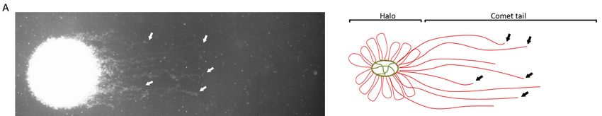
On the other hand, Comet assay [41] relies on a DNA decompaction and protein depletion coupled
to a single-cell electrophoresis in an agarose micro gel. DNA molecules that contain breaks move
towards the cathode and the length of the “comet tail” can be measured to determine the grade of DNA
fragmentation at a single cell level. This technique has been applied in multiple different protocols,
which usually vary in agarose concentrations and in electrophoresis times [42,43]. As the Comet assay
can be performed in alkaline or neutral pH, different types of DNA breaks can be detected (Table 1)
(Figure 1): (i) alkaline Comet assay performed in a small electrophoresis time (about four minutes)
detect mostly single-strand DNA breaks affecting both toroidal regions and MAR regions in a high
number of break points [44,45] and (ii) neutral Comet assay can detect two types of double-strand
DNA breaks (Figure 2): (a) extensive DSB, which represent a very small part of total DSB and can be
observed as very long comet tails separated from the sperm core; and (b) localized DSB localized and
attached to the MAR region, as demonstrated in pulsed-field gel electrophoresis [43–46], being the
most common DSB. Although extensive DSB result in longer Comet tails, they cannot be distinguished
from localized DSB in a single Comet. However, when a semen sample present high number of sperm
cells with extensive DSB (long tails), single-strand DNA damage is also present in a high amount
Genes 2019, 10, x FOR
(Ribas-Maynou PEER REVIEW
personal observation). Previous studies had shown that localized DSB represent4very
of 12
few break points in the genome, as long chromatin fibres with a break point in the end can be seen in a
125 long chromatin fibres with a break point in the end can be seen in a detailed neutral Comet image
detailed neutral Comet image (Figure 2A), which is supported by Kaneko et al., using pulsed field
126 (Figure 2A), which is supported by Kaneko et al., using pulsed field gel electrophoresis [47]. We
gel electrophoresis [47]. We demonstrated that localized DSB remain attached to the sperm nuclear
127 demonstrated that localized DSB remain attached to the sperm nuclear matrix [45], maybe through a
matrix [45], maybe through a TOP2B or similar protein [45,46], a very important feature taking into
128 TOP2B or similar protein [45,46], a very important feature taking into account that the nuclear
account that the nuclear matrix is inherited to the male pronucleus in the zygote [46,48–50], giving a
129 matrix is inherited to the male pronucleus in the zygote [46,48–50], giving a chance to the embryo to
chance to the embryo to repair the DSB.
130 repair the DSB.
131
132 Figure 2: (A)2.Picture
Figure and scheme
(A) Picture of neutral
and scheme CometComet
of neutral with localized DSB (double-strand
with localized DNA breaks)
DSB (double-strand attached to
DNA breaks)
133 attached
the nuclear to the(green).
matrix nuclear Comet
matrix (green). Cometinhalo
halo consists consists in non-fragmented
non-fragmented chromatin and chromatin
comet tail andis comet
formed by
134 chromatin fibres attached to the nuclear matrix with low number of DNA breaks at the end (arrows). (B) at
tail is formed by chromatin fibres attached to the nuclear matrix with low number of DNA breaks Picture
135 the endof
and scheme (arrows). (B) Picture
neutral Comet withand schemeDSB.
extensive of neutral
CometComet
tail is with extensive
formed by DNADSB. Comet tail
fragments thatisare
formed by
not attached
136 to theDNA
nuclearfragments that are
matrix. This notalso
comet attached
showstopart
the of
nuclear matrix.
localized DNAThis comet
breaks also shows
attached to thepart
MAR ofregion
localized
(arrow).
DNA breaks attached to the MAR region (arrow).
137
138 Studies using
Studies using all
all the
the techniques
techniques showed
showed that
that oxidative
oxidative damage
damage detected
detected byby alkaline
alkaline Comet
Comet
139 assay presented
assay presented aa good
good correlation
correlation to
to TUNEL,
TUNEL, SCSA
SCSA and
and SCD
SCD techniques
techniques [23,51,52].
[23,51,52]. Although
Although these
these
140 techniques may potentially detect double-strand breaks, a study conducted by our group
techniques may potentially detect double-strand breaks, a study conducted by our group analysing the analysing
141 the same
same semen
semen samples
samples with with five methodologies
five methodologies showedshowed
that nothat no correlation
correlation was present
was present with thewith the
neutral
142 neutral assay
Comet Comet[23].
assay [23].the
Then, Then, thewould
latter latter would be the
be the only only technique
technique that is
that is able to able to differentially
differentially detect
143 MAR-region double-strand breaks [23,44], whereas TUNEL, SCSA and SCD may detect extensivedetect
detect MAR-region double-strand breaks [23,44], whereas TUNEL, SCSA and SCD may DSB.
144 extensive
A DSB. variant
Comet assay A Comet assay variant
(two-tailed (two-tailed
Comet Comet assay)
assay) applying applying
both alkaline andboth alkaline
neutral andassay
Comet neutral
in
145 Comet assay in the same slide by turning it 90º between electrophoresis allows to distinguish single
146 and double-strand DNA breaks on the same sperm cell [53]. However, no studies have been
147 performed comparing these techniques and alkaline or neutral Comet assay separately in order to
148 elucidate if double-strand breaks detected in two-tailed Comet assay correspond to MAR region
149 localized DSB.Genes 2019, 10, 105 6 of 13
the same slide by turning it 90º between electrophoresis allows to distinguish single and double-strand
DNA breaks on the same sperm cell [53]. However, no studies have been performed comparing these
techniques and alkaline or neutral Comet assay separately in order to elucidate if double-strand breaks
detected in two-tailed Comet assay correspond to MAR region localized DSB.
3. Oxidative DNA Damage, Alkaline Comet Assay and Pregnancy Achievement
Using alkaline Comet assay in different cohorts, an study published in 2012 [43] showed that the
extensive single-strand DNA breaks were reversely associated to the achievement of natural pregnancy
independently of the neutral Comet results (Figure 1 and Table 1). This was confirmed and compared
with TUNEL, SCSA and SCD tests in 2013, demonstrating also that alkaline Comet is the most sensitive
technique for the prediction of natural pregnancy achievement [23,43]. Which is also in accordance to
the numerous studies from other research groups that find similar association in natural pregnancy
using TUNEL, SCSA, SCD and Comet assay tests [5,51,54–58].
Single-strand breaks are produced mainly due to reactive oxygen species (ROS) [42,53,59],
which may come from exogenous sources such as environmental toxicants, smoking, alcohol, diet,
radiation and so forth or from endogenous sources such as an increase of leukocytes, presence of
varicocele or even the ROS generated by mitochondria for the movement of sperm cell [60–62].
Free radicals may cause lipid peroxidation, mitochondrial and nuclear DNA base modifications such
as 8-OH-guanine and 8-OH-20 -deoxyguanosine (8-OHdG), an oxidized base adduct that destabilize
DNA structure and cause a DNA break [63–65]. This affectation does not find a restriction by DNA
condensation and therefore may affect both toroids compacted in protamines and MAR regions
compacted in histones [44]. Then, if such an extensive damage happens to the sperm DNA due
to oxidative stress, the sperm membranes would also be affected and usually sperm motility is
lost. Because of that, a strong negative relation between progressive motility and oxidative damage
(single-strand DNA damage) analyzed using TUNEL, SCSA, SCD and alkaline Comet [55,61,66].
As mentioned before in this review, controversial results are found in different studies regarding
ICSI outcomes: some of them which found predictive value of oxidative damage [25–28] and other
with opposite results [29–33]. If single-strand DNA breaks present a correlation to progressive motility
and sperm morphology and ICSI procedures use the most motile sperm cells with better morphology,
paternal genome should be free of oxidative damage. In this regard, a work by Gosalvez et al. [67]
demonstrated that motile sperm organelle morphology examination (MSOME) selected sperm cells
were free of DNA damage analysed by SCD test. Moreover, a work using Comet assay suggested that
grade I and II sperm cells present lower incidence of oxidative DNA damage than grade III and IV [68].
These results need to be further confirmed in conventional ICSI sperm selection. However, our data
suggest that no relation is present between alkaline Comet and embryo quality, embryo kinetics or
implantation [69].
4. Double-Strand DNA Damage, Recurrent Miscarriage and Preimplantation Failure in
ICSI Cycles
Analysing the data of the patients and donors with high DSB, a specific profile was observed
with low oxidative damage and high neutral comet values in patients with first trimester recurrent
miscarriage where all related female factors were discarded and in one subgroup of fertile donors [44].
In a recent study, our group has found that patients with this profile who undergo ICSI treatments
produce embryos with a delayed embryo development to blastocyst, which also cause lower
implantation rates [69]. Other works also show that double-strand breaks may contribute to a higher
implantation failure risk [6,25]. Since implantation failures in ICSI cycles and miscarriages present
similar profiles with high DSB, one may think that they might have similar origin. In fact, small number
of DNA breaks localized in concrete regions of the genome might induce a cell failure where the affected
regions are necessary for the development. In our last study, embryos that achieved implantationGenes 2019, 10, 105 7 of 13
presented faster embryo kinetics than those that did not achieve implantation [69]. In fact, faster
embryo kinetics had been associated to embryo euploidy [70–72].
DSB are the most lethal alteration that may happen in a zygote, since paternal and maternal
pronucleus remain separated in early mammalian embryos and, therefore, no complementary chain
would be available for DNA repair [73–75] and a few number of DSB are sufficient to delay cell
cycle [76]. It is important to note that paternal double-strand breaks remain attached to the nuclear
matrix and probably to other proteins such as TOP2B [20,46,77] and the nuclear matrix is inherited at
male pronucleus until first mitotic division [49,78]. This may be crucial at the zygote, because it may
give a chance to correctly repair both free ends of the double-strand break. There is a consensus point
that oocyte quality may play a role in this DNA repair, since different studies proved that early embryos
are able to repair DNA damage [79–84]. In this sense, in patients with DSB, the most significant delay
observed in the embryo kinetics was just after fertilization, indicating that DNA repair machinery
may be active in this stage [69]. Recent studies in sperm cells demonstrated that MAR regions are
required as a scaffold for DNA replication after fertilization [48] and, in somatic cells, nuclear matrix
also is involved in transcription, cell regulation and replication [85,86]. In mammals, inducing DSB in
sperm cells and used these sperm cells to fertilize eggs observed chromosomal alterations in paternal
genome of the embryo and showing also a delay in the first embryo cleavage [17,20,87]. Moreover,
studies inducing double-strand DNA breaks in mice sperm through radiation observed a p53 and p21
related response and less number of foetuses [88,89] or less survival of offspring in a dose dependent
manner [90].
5. Prevention of DNA Damage
The data presented in the studies referenced before supports that oxidative damage may affect
the pregnancy achievement capacity due to misbalanced levels of oxidants/antioxidants [61,91].
The use of antioxidants has been widely applied in subfertile males [92]. Several works
demonstrated that they are a positive contribution on sperm count, motility, morphology and also
proved that they help reducing oxidative DNA fragmentation [93–96]. Although there are very few
studies with randomized and placebo controls, Cochrane review suggests that the use of antioxidants
causes from 1.8 to 4.6 fold increase in the chances of achieving a natural pregnancy. However, up to
a 6.5 fold increase in miscarriages might be observed [97]. In ICSI treatments, it is still not clear
if antioxidants could help on improving pregnancy and birth rates [98–100]. High quality studies
including different groups of patients are necessary in order to elucidate the need of antioxidants in
ICSI procedures.
Treatments for the reduction of double-strand sperm DNA damage should also reduce the
miscarriage risk and the implantation failure risk in ICSI cycles, showing also less delay on embryo
kinetics. Until our knowledge, no validated treatment reduce the incidence of MAR-region localized
DSB. However, a study conducted in humans in 2006 by Schmid and colleagues demonstrated that
men with daily caffeine consumption presented increased values of DSB measured with neutral
Comet independently of male age in healthy non-smokers [101]. Caffeine is a known inhibitor of
DNA repair, as it has been described that inhibits ATM kinase [102,103] and DNA resection in
homologous recombination through Rad51 [104,105]. Also, it has been reported to affect cell cycle
at both G1/S and G2/M checkpoints and inducing programmed cell death through p53-dependent
pathway [106]. Studies in animals reported that caffeine administration to rats caused an impairment
of pregnancy [107]. Other studies inducing DNA strand breaks in sperm cells through radiation and
cultivating the oocytes and the produced embryos in caffeine demonstrated that chromosome and
chromatid aberrations persist in the zygote, indicating oocyte DNA repair is inhibited by caffeine [17].
Since spermatocytes must produce double-strand breaks through Spo11 in prophase I in order
to perform DNA recombination and later, they need to repair these DSB. According to previous
results, the consumption of caffeine would impair ATM kinase and/or resection of double-strand
breaks [104,105] and may induce that a few double-strand breaks would not be repaired, causing thatGenes 2019, 10, 105 8 of 13
mature sperm cells present DSB [101]. Further basic studies are needed to explain how a spermatocyte
with double-strand breaks can escape the pachytene checkpoint [108,109]. Reducing the incidence
of DSB in sperm cell would improve clinical outcomes in terms of miscarriage and implantation in
ICSI cycles.
Author Contributions: J.R.-M. and J.B. contributed in manuscript writing and revision.
Funding: This research was funded by the European Regional Development Fund and Instituto de Salud Carlos
III (Economy, Industry and Competitiveness Ministry, Madrid, Spain; Project PI14/Q1 00119) and Generalitat de
Catalunya (Project 2017SGR1796).
Conflicts of Interest: The authors declare no conflict of interest.
References
1. Thoma, M.E.; McLain, A.C.; Louis, J.F.; King, R.B.; Trumble, A.C.; Sundaram, R.; Buck Louis, G.M. Prevalence
of infertility in the United States as estimated by the current duration approach and a traditional constructed
approach. Fertil. Steril. 2013, 99, 1324–1331. [CrossRef] [PubMed]
2. Louis, J.F.; Thoma, M.E.; Sørensen, D.N.; McLain, A.C.; King, R.B.; Sundaram, R.; Keiding, N.; Buck Louis, G.M.
The prevalence of couple infertility in the United States from a male perspective: Evidence from a nationally
representative sample. Andrology 2013, 1, 741–748. [CrossRef] [PubMed]
3. Baird, D.T.; Collins, J.; Egozcue, J.; Evers, L.H.; Gianaroli, L.; Leridon, H.; Sunde, A.; Templeton, A.;
Van Steirteghem, A.; Cohen, J.; et al. Fertility and ageing. Hum. Reprod. Update 2005, 11, 261–276. [PubMed]
4. De Kretser, D.M. Male infertility. Lancet 1997, 349, 787–790. [CrossRef]
5. Lewis, S.E.M.; Simon, L. Clinical implications of sperm DNA damage. Hum. Fertil. (Camb.) 2010, 13, 201–207.
[CrossRef] [PubMed]
6. Simon, L.; Murphy, K.; Shamsi, M.B.; Liu, L.; Emery, B.; Aston, K.I.; Hotaling, J.; Carrell, D.T. Paternal
influence of sperm DNA integrity on early embryonic development. Hum. Reprod. 2014, 29, 2402–2412.
[CrossRef] [PubMed]
7. Cooper, T.G.; Noonan, E.; von Eckardstein, S.; Auger, J.; Baker, H.W.G.; Behre, H.M.; Haugen, T.B.; Kruger, T.;
Wang, C.; Mbizvo, M.T.; et al. World Health Organization reference values for human semen characteristics.
Hum. Reprod. Update 2010, 16, 231–245. [CrossRef] [PubMed]
8. Yu, S.; Rubin, M.; Geevarughese, S.; Pino, J.S.; Rodriguez, H.F.; Asghar, W. Emerging technologies for
home-based semen analysis. Andrology 2018, 6, 10–19. [CrossRef] [PubMed]
9. Wang, C.; Swerdloff, R.S. Limitations of semen analysis as a test of male fertility and anticipated needs from
newer tests. Fertil. Steril. 2014, 102, 1502–1507. [CrossRef] [PubMed]
10. Kupka, M.S.; D’Hooghe, T.; Ferraretti, A.P.; De Mouzon, J.; Erb, K.; Castilla, J.A.; Calhaz-Jorge, C.;
De Geyter, C.; Goossens, V. Assisted reproductive technology in Europe, 2011: Results generated from
European registers by ESHRE. Hum. Reprod. 2015, 31, 233–248.
11. Love, C.C.; Kenney, R.M. Scrotal heat stress induces altered sperm chromatin structure associated with a
decrease in protamine disulfide bonding in the stallion. Biol. Reprod. 1999, 60, 615–620. [CrossRef] [PubMed]
12. Keeney, S.; Lange, J.; Mohibullah, N. Self-organization of meiotic recombination initiation: General principles
and molecular pathways. Annu. Rev. Genet. 2014, 48, 187–214. [CrossRef] [PubMed]
13. Cooper, T.J.; Wardell, K.; Garcia, V.; Neale, M.J. Homeostatic regulation of meiotic DSB formation by
ATM/ATR. Exp. Cell Res. 2014, 329, 124–131. [CrossRef] [PubMed]
14. Alberts, B.; Johnson, A.; Lewis, J.; Raff, M.; Roberts, K.; Walter, P. Molecular Biology of the Cell, 4th ed.; Garland
Sc.: New York, NY, USA, 2002.
15. Ward, W.S.; Coffey, D.S. Specific organization of genes in relation to the sperm nuclear matrix. Biochem. Biophys.
Res. Commun. 1990, 173, 20–25. [CrossRef]
16. Ward, W.S. Function of sperm chromatin structural elements in fertilization and development.
Mol. Hum. Reprod. 2010, 16, 30–36. [CrossRef] [PubMed]
17. Genescà, A.; Caballín, M.R.; Miró, R.; Benet, J.; Germà, J.R.; Egozcue, J. Repair of human sperm chromosome
aberrations in the hamster egg. Hum. Genet. 1992, 89, 181–186. [CrossRef]
18. Oh, J.; Symington, L. Role of the Mre11 complex in preserving genome integrity. Genes (Basel) 2018, 9, 589.
[CrossRef]Genes 2019, 10, 105 9 of 13
19. Barnes, J.L.; Zubair, M.; John, K.; Poirier, M.C.; Martin, F.L. Carcinogens and DNA damage. Biochem. Soc. Trans.
2018, 46, 1213–1224. [CrossRef]
20. Gawecka, J.E.; Marh, J.; Ortega, M.; Yamauchi, Y.; Ward, M.A.; Ward, W.S. Mouse zygotes respond to severe
sperm DNA damage by delaying paternal DNA replication and embryonic development. PLoS ONE 2013,
8, e56385. [CrossRef]
21. Lewis, S.E.M.; John Aitken, R.; Conner, S.J.; De Iuliis, G.; Evenson, D.P.; Henkel, R.; Giwercman, A.;
Gharagozloo, P. The impact of sperm DNA damage in assisted conception and beyond: Recent advances in
diagnosis and treatment. Reprod. Biomed. Online 2013, 27, 325–337. [CrossRef]
22. Evenson, D.P. Sperm chromatin structure assay (SCSA®). Methods Mol. Biol. 2013, 927, 147–164. [PubMed]
23. Ribas-Maynou, J.; García-Peiró, A.; Fernández-Encinas, A.; Abad, C.; Amengual, M.J.; Prada, E.; Navarro, J.;
Benet, J. Comprehensive analysis of sperm DNA fragmentation by five different assays: TUNEL assay, SCSA,
SCD test and alkaline and neutral Comet assay. Andrology 2013, 1, 715–722. [CrossRef]
24. Peluso, G.; Palmieri, A.; Cozza, P.P.; Morrone, G.; Verze, P.; Longo, N.; Mirone, V. The study of spermatic
DNA fragmentation and sperm motility in infertile subjects. Arch. Ital. di Urol. e Androl. 2013, 85, 8.
[CrossRef] [PubMed]
25. Garolla, A.; Cosci, I.; Bertoldo, A.; Sartini, B.; Boudjema, E.; Foresta, C. DNA double strand breaks in human
spermatozoa can be predictive for assisted reproductive outcome. Reprod. Biomed. Online 2015, 31, 100–107.
[CrossRef] [PubMed]
26. Simon, L.; Brunborg, G.; Stevenson, M.; Lutton, D.; McManus, J.; Lewis, S.E.M. Clinical significance of sperm
DNA damage in assisted reproduction outcome. Hum. Reprod. 2010, 25, 1594–1608. [CrossRef] [PubMed]
27. Simon, L.; Proutski, I.; Stevenson, M.; Jennings, D.; McManus, J.; Lutton, D.; Lewis, S.E.M. Sperm DNA
damage has a negative association with live-birth rates after IVF. Reprod. Biomed. Online 2013, 26, 68–78.
[CrossRef] [PubMed]
28. Meseguer, M.; Santiso, R.; Garrido, N.; García-Herrero, S.; Remohí, J.; Fernandez, J.L. Effect of sperm DNA
fragmentation on pregnancy outcome depends on oocyte quality. Fertil. Steril. 2011, 95, 124–128. [CrossRef]
29. Esbert, M.; Pacheco, A.; Vidal, F.; Florensa, M.; Riqueros, M.; Ballesteros, A.; Garrido, N.; Calderón, G. Impact
of sperm DNA fragmentation on the outcome of IVF with own or donated oocytes. Reprod. Biomed. Online
2011, 23, 704–710. [CrossRef]
30. Thomson, L.K.; Zieschang, J.-A.; Clark, A.M. Oxidative deoxyribonucleic acid damage in sperm has a
negative impact on clinical pregnancy rate in intrauterine insemination but not intracytoplasmic sperm
injection cycles. Fertil. Steril. 2011, 96, 843–847. [CrossRef]
31. Pregl Breznik, B.; Kovačič, B.; Vlaisavljević, V. Are sperm DNA fragmentation, hyperactivation and
hyaluronan-binding ability predictive for fertilization and embryo development in in vitro fertilization
and intracytoplasmic sperm injection? Fertil. Steril. 2013, 99, 1233–1241. [CrossRef]
32. Anifandis, G.; Bounartzi, T.; Messini, C.I.; Dafopoulos, K.; Markandona, R.; Sotiriou, S.; Tzavella, A.;
Messinis, I.E. Sperm DNA fragmentation measured by Halosperm does not impact on embryo quality and
ongoing pregnancy rates in IVF/ICSI treatments. Andrologia 2015, 47, 295–302. [CrossRef] [PubMed]
33. Haghpanah, T.; Salehi, M.; Ghaffari Novin, M.; Masteri Farahani, R.; Fadaei-Fathabadi, F.;
Dehghani-Mohammadabadi, M.; Azimi, H. Does sperm DNA fragmentation affect the developmental
potential and the incidence of apoptosis following blastomere biopsy? Syst. Biol. Reprod. Med. 2016, 62, 1–10.
[CrossRef] [PubMed]
34. ASRM. The clinical utility of sperm DNA integrity testing: A guideline. Fertil. Steril. 2013, 99, 673–677.
[CrossRef] [PubMed]
35. Gorczyca, W.; Gong, J.; Darzynkiewicz, Z. Detection of DNA strand breaks in individual apoptotic cells by
the in situ terminal deoxynucleotidyl transferase and nick translation assays. Cancer Res. 1993, 53, 1945–1951.
[PubMed]
36. Sharma, R.; Masaki, J.; Agarwal, A. Sperm DNA fragmentation analysis using the TUNEL assay.
Methods Mol. Biol. 2013, 927, 121–136. [PubMed]
37. Mitchell, L.A.; De Iuliis, G.N.; Aitken, R.J. The TUNEL assay consistently underestimates DNA damage in
human spermatozoa and is influenced by DNA compaction and cell vitality: Development of an improved
methodology. Int. J. Androl. 2011, 34, 2–13. [CrossRef]Genes 2019, 10, 105 10 of 13
38. Domínguez-Fandos, D.; Camejo, M.I.; Ballescà, J.L.; Oliva, R. Human sperm DNA fragmentation: correlation
of TUNEL results as assessed by flow cytometry and optical microscopy. Cytometry. A 2007, 71, 1011–1018.
[CrossRef]
39. Evenson, D.; Jost, L. Sperm chromatin structure assay is useful for fertility assessment. Methods Cell Sci. 2000,
22, 169–189. [CrossRef]
40. Fernández, J.L.; Muriel, L.; Goyanes, V.; Segrelles, E.; Gosálvez, J.; Enciso, M.; LaFromboise, M.; De Jonge, C.
Simple determination of human sperm DNA fragmentation with an improved sperm chromatin dispersion
test. Fertil. Steril. 2005, 84, 833–842. [CrossRef]
41. Singh, N.P.; McCoy, M.T.; Tice, R.R.; Schneider, E.L. A simple technique for quantitation of low levels of
DNA damage in individual cells. Exp. Cell Res. 1988, 175, 184–191. [CrossRef]
42. Simon, L.; Carrell, D.T. Sperm DNA damage measured by comet assay. Methods Mol. Biol. 2013, 927, 137–146.
[CrossRef] [PubMed]
43. Ribas-Maynou, J.; Garca-Peiro, A.; Abad, C.; Amengual, M.J.; Navarro, J.; Benet, J. Alkaline and neutral
Comet assay profiles of sperm DNA damage in clinical groups. Hum. Reprod. 2012, 27, 652–658. [CrossRef]
44. Ribas-Maynou, J.; García-Peiró, A.; Fernandez-Encinas, A.; Amengual, M.J.; Prada, E.; Cortés, P.; Navarro, J.;
Benet, J. Double stranded sperm DNA breaks, measured by comet assay, are associated with unexplained
recurrent miscarriage in couples without a female factor. PLoS ONE 2012, 7. [CrossRef]
45. Ribas-Maynou, J.; Gawecka, J.E.; Benet, J.; Ward, W.S. Double-stranded DNA breaks hidden in the neutral
Comet assay suggest a role of the sperm nuclear matrix in DNA integrity maintenance. Mol. Hum. Reprod.
2014, 20, 330–340. [CrossRef] [PubMed]
46. Gawecka, J.E.; Ribas-Maynou, J.; Benet, J.; Ward, W.S. A model for the control of DNA integrity by the sperm
nuclear matrix. Asian J. Androl. 2015, 17.
47. Kaneko, S.; Yoshida, J.; Ishikawa, H.; Takamatsu, K. Single-cell pulsed-field gel electrophoresis to detect the
early stage of DNA fragmentation in human sperm nuclei. PLoS ONE 2012, 7, e42257. [CrossRef] [PubMed]
48. Shaman, J.A.; Yamauchi, Y.; Ward, W.S. Function of the sperm nuclear matrix. Arch. Androl. 2007, 53, 135–140.
[CrossRef]
49. Mohar, I.; Szczygiel, M.A.; Yanagimachi, R.; Ward, W.S. Sperm nuclear halos can transform into normal
chromosomes after injection into oocytes. Mol. Reprod. Dev. 2002, 62, 416–420. [CrossRef] [PubMed]
50. Shaman, J.A.; Yamauchi, Y.; Ward, W.S. The sperm nuclear matrix is required for paternal DNA replication.
J. Cell. Biochem. 2007, 102, 680–688. [CrossRef]
51. Simon, L.; Liu, L.; Murphy, K.; Ge, S.; Hotaling, J.; Aston, K.I.; Emery, B.; Carrell, D.T. Comparative analysis
of three sperm DNA damage assays and sperm nuclear protein content in couples undergoing assisted
reproduction treatment. Hum. Reprod. 2014, 29, 904–917. [CrossRef]
52. Chohan, K.R.; Griffin, J.T.; Lafromboise, M.; De Jonge, C.J.; Carrell, D.T. Comparison of chromatin assays for
DNA fragmentation evaluation in human sperm. J. Androl. 2006, 27, 53–59. [CrossRef] [PubMed]
53. Enciso, M.; Sarasa, J.; Agarwal, A.; Fernández, J.L.; Gosálvez, J. A two-tailed Comet assay for assessing DNA
damage in spermatozoa. Reprod. Biomed. Online 2009, 18, 609–616. [CrossRef]
54. Saleh, R.A.; Agarwal, A.; Nelson, D.R.; Nada, E.A.; El-Tonsy, M.H.; Alvarez, J.G.; Thomas, A.J.; Sharma, R.K.
Increased sperm nuclear DNA damage in normozoospermic infertile men: A prospective study. Fertil. Steril.
2002, 78, 313–318. [CrossRef]
55. Belloc, S.; Benkhalifa, M.; Cohen-Bacrie, M.; Dalleac, A.; Amar, E.; Zini, A. Sperm deoxyribonucleic acid
damage in normozoospermic men is related to age and sperm progressive motility. Fertil. Steril. 2014, 101,
1588–1593. [CrossRef] [PubMed]
56. Cho, C.-L.; Agarwal, A. Role of sperm DNA fragmentation in male factor infertility: A systematic review.
Arab J. Urol. 2018, 16, 21–34. [CrossRef]
57. Santi, D.; Spaggiari, G.; Simoni, M. Sperm DNA fragmentation index as a promising predictive tool for male
infertility diagnosis and treatment management – meta-analyses. Reprod. Biomed. Online 2018, 37, 315–326.
[CrossRef] [PubMed]
58. Simon, L.; Lutton, D.; McManus, J.; Lewis, S.E.M. Sperm DNA damage measured by the alkaline Comet
assay as an independent predictor of male infertility and in vitro fertilization success. Fertil. Steril. 2011, 95,
652–657. [CrossRef]
59. Agarwal, A.; Prabakaran, S.A. Mechanism, measurement and prevention of oxidative stress in male
reproductive physiology. Indian J. Exp. Biol. 2005, 43, 963–974.Genes 2019, 10, 105 11 of 13
60. Agarwal, A.; Virk, G.; Ong, C.; du Plessis, S.S. Effect of oxidative stress on male reproduction. World J.
Mens. Health 2014, 32, 1. [CrossRef]
61. Aitken, R.J.; De Iuliis, G.N. On the possible origins of DNA damage in human spermatozoa. Mol. Hum. Reprod.
2010, 16, 3–13. [CrossRef]
62. Sakkas, D.; Alvarez, J.G. Sperm DNA fragmentation: mechanisms of origin, impact on reproductive outcome
and analysis. Fertil. Steril. 2010, 93, 1027–1036. [CrossRef] [PubMed]
63. Lord, T.; Aitken, R.J. Fertilization stimulates 8-hydroxy-20 -deoxyguanosine repair and antioxidant activity to
prevent mutagenesis in the embryo. Dev. Biol. 2015, 406, 1–13. [CrossRef] [PubMed]
64. De Iuliis, G.N.; Thomson, L.K.; Mitchell, L.A.; Finnie, J.M.; Koppers, A.J.; Hedges, A.; Nixon, B.; Aitken, R.J.
DNA damage in human spermatozoa is highly correlated with the efficiency of chromatin remodeling and
the formation of 8-hydroxy-20 -deoxyguanosine, a marker of oxidative stress. Biol. Reprod. 2009, 81, 517–524.
[CrossRef] [PubMed]
65. Santiso, R.; Tamayo, M.; Gosálvez, J.; Meseguer, M.; Garrido, N.; Fernández, J.L. Simultaneous determination
in situ of DNA fragmentation and 8-oxoguanine in human sperm. Fertil. Steril. 2010, 93, 314–318. [CrossRef]
[PubMed]
66. Evgeni, E.; Lymberopoulos, G.; Touloupidis, S.; Asimakopoulos, B. Sperm nuclear DNA fragmentation and
its association with semen quality in Greek men. Andrologia 2015, 47, 1166–1174. [CrossRef] [PubMed]
67. Gosálvez, J.; Migueles, B.; López-fernández, C.; Sanchéz-martín, F.; Sáchez-martín, P. Single sperm selection
and DNA fragmentation analysis: The case of MSOME/IMSI. Nat. Sci. Res. 2013, 5, 7–14. [CrossRef]
68. Pastuszek, E.; Kiewisz, J.; Skowronska, P.; Liss, J.; Lukaszuk, M.; Bruszczynska, A.; Jakiel, G.; Lukaszuk, K.
An investigation of the potential effect of sperm nuclear vacuoles in human spermatozoa on DNA
fragmentation using a neutral and alkaline Comet assay. Andrology 2017, 5, 392–398. [CrossRef]
69. Casanovas, A.; Ribas-Maynou, J.; Lara-Cerrillo, S.; Jimenez-Macedo, A.R.; Hortal, O.; Benet, J.; Carrera, J.;
Garcia-Peiró, A. Double-stranded sperm DNA damage is a cause of delay in embryo development and can
impair implantation rates. Fertil. Steril. 2018, in press.
70. Campbell, A.; Fishel, S.; Laegdsmand, M. Aneuploidy is a key causal factor of delays in blastulation:
author response to “A cautionary note against aneuploidy risk assessment using time-lapse imaging”.
Reprod. Biomed. Online 2014, 28, 279–283. [CrossRef]
71. Basile, N.; del Carmen Nogales, M.; Bronet, F.; Florensa, M.; Riqueiros, M.; Rodrigo, L.; García-Velasco, J.;
Meseguer, M. Increasing the probability of selecting chromosomally normal embryos by time-lapse
morphokinetics analysis. Fertil. Steril. 2014, 101, 699–704. [CrossRef]
72. Campbell, A.; Fishel, S.; Bowman, N.; Duffy, S.; Sedler, M.; Thornton, S. Retrospective analysis of outcomes after
IVF using an aneuploidy risk model derived from time-lapse imaging without PGS. Reprod. Biomed. Online 2013,
27, 140–146. [CrossRef] [PubMed]
73. Bernstein, K.A.; Rothstein, R. At loose ends: Resecting a double-strand break. Cell 2009, 137, 807–810.
[CrossRef] [PubMed]
74. Price, B.D.; D’Andrea, A.D. Chromatin remodeling at DNA double-strand breaks. Cell 2013, 152, 1344–1354.
[CrossRef] [PubMed]
75. Reichmann, J.; Nijmeijer, B.; Hossain, M.J.; Eguren, M.; Schneider, I.; Politi, A.Z.; Roberti, M.J.; Hufnagel, L.;
Hiiragi, T.; Ellenberg, J. Dual-spindle formation in zygotes keeps parental genomes apart in early mammalian
embryos. Science 2018, 361, 189–193. [CrossRef] [PubMed]
76. Van den Berg, J.; G Manjón, A.; Kielbassa, K.; Feringa, F.M.; Freire, R.; Medema, R.H. A limited number
of double-strand DNA breaks is sufficient to delay cell cycle progression. Nucleic Acids Res. 2018, 46,
10132–10144. [CrossRef] [PubMed]
77. Yamauchi, Y.; Shaman, J.A.; Ward, W.S. Topoisomerase II-mediated breaks in spermatozoa cause the specific
degradation of paternal DNA in fertilized oocytes. Biol. Reprod. 2007, 76, 666–672. [CrossRef]
78. Ward, W.S.; Kimura, Y.; Yanagimachi, R. An intact sperm nuclear matrix may be necessary for the mouse
paternal genome to participate in embryonic development. Biol. Reprod. 1999, 60, 702–706. [CrossRef]
79. Zheng, P.; Schramm, R.D.; Latham, K.E. Developmental regulation and in vitro culture effects on expression
of DNA repair and cell cycle checkpoint control genes in rhesus monkey oocytes and embryos. Biol. Reprod.
2005, 72, 1359–1369. [CrossRef] [PubMed]Genes 2019, 10, 105 12 of 13
80. Marchetti, F.; Bishop, J.; Gingerich, J.; Wyrobek, A.J. Meiotic interstrand DNA damage escapes paternal
repair and causes chromosomal aberrations in the zygote by maternal misrepair. Sci. Rep. 2015, 5, 7689.
[CrossRef] [PubMed]
81. Menezo, Y.; Russo, G.; Tosti, E.; El Mouatassim, S.; Benkhalifa, M. Expression profile of genes coding for
DNA repair in human oocytes using pangenomic microarrays, with a special focus on ROS linked decays.
J. Assist. Reprod. Genet. 2007, 24, 513–520. [CrossRef]
82. Jaroudi, S.; Kakourou, G.; Cawood, S.; Doshi, A.; Ranieri, D.M.; Serhal, P.; Harper, J.C.; SenGupta, S.B.
Expression profiling of DNA repair genes in human oocytes and blastocysts using microarrays. Hum. Reprod.
2009, 24, 2649–2655. [CrossRef] [PubMed]
83. Martin, J.H.; Bromfield, E.G.; Aitken, R.J.; Lord, T.; Nixon, B. Double strand break DNA repair occurs via
non-homologous end-joining in mouse MII oocytes. Sci. Rep. 2018, 8, 9685. [CrossRef] [PubMed]
84. García-Rodríguez, A.; Gosálvez, J.; Agarwal, A.; Roy, R.; Johnston, S. DNA Damage and repair in human
reproductive cells. Int. J. Mol. Sci. 2018, 20, 31. [CrossRef] [PubMed]
85. Pardoll, D.M.; Vogelstein, B.; Coffey, D.S. A fixed site of DNA replication in eucaryotic cells. Cell 1980, 19,
527–536. [CrossRef]
86. Wilson, R.H.C.; Coverley, D. Relationship between DNA replication and the nuclear matrix. Genes Cells 2013,
18, 17–31. [CrossRef] [PubMed]
87. Tusell, L.; Latre, L.; Ponsa, I.; Miró, R.; Egozcue, J.; Genescà, A. Capping of radiation-induced DNA breaks in
mouse early embryos. J. Radiat. Res. 2004, 45, 415–422. [CrossRef] [PubMed]
88. Adiga, S.K.; Toyoshima, M.; Shiraishi, K.; Shimura, T.; Takeda, J.; Taga, M.; Nagai, H.; Kumar, P.; Niwa, O.
p21 provides stage specific DNA damage control to preimplantation embryos. Oncogene 2007, 26, 6141–6149.
[CrossRef]
89. Toyoshima, M. Analysis of p53 dependent damage response in sperm-irradiated mouse embryos. J. Radiat. Res.
2009, 50, 11–17. [CrossRef]
90. Kumar, D.; Upadhya, D.; Salian, S.R.; Rao, S.B.S.; Kalthur, G.; Kumar, P.; Adiga, S.K. The extent of paternal
sperm DNA damage influences early post-natal survival of first generation mouse offspring. Eur. J. Obstet.
Gynecol. Reprod. Biol. 2013, 166, 164–167. [CrossRef]
91. Majzoub, A.; Agarwal, A. Antioxidant therapy in idiopathic oligoasthenoteratozoospermia. Indian J. Urol.
2017, 33, 207.
92. Gharagozloo, P.; Aitken, R.J. The role of sperm oxidative stress in male infertility and the significance of oral
antioxidant therapy. Hum. Reprod. 2011, 26, 1628–1640. [CrossRef] [PubMed]
93. Menezo, Y.; Evenson, D.; Cohen, M.; Dale, B. Effect of Antioxidants on Sperm Genetic Damage. In Genetic
Damage in Human Spermatozoa; Advances in Experimental Medicine and Biology; Springer: New York, NY,
USA, 2014; Volume 791, pp. 173–189.
94. Zini, A.; San Gabriel, M.; Baazeem, A. Antioxidants and sperm DNA damage: A clinical perspective. J. Assist.
Reprod. Genet. 2009, 26, 427–432. [CrossRef] [PubMed]
95. Rahman, S.; Huang, Y.; Zhu, L.; Feng, S.; Khan, I.; Wu, J.; Li, Y.; Wang, X. therapeutic role of green tea
polyphenols in improving fertility: A review. Nutrients 2018, 10, 834. [CrossRef] [PubMed]
96. Imamovic Kumalic, S.; Pinter, B. Review of clinical trials on effects of oral antioxidants on basic semen and
other parameters in idiopathic oligoasthenoteratozoospermia. Biomed Res. Int. 2014, 2014, 1–11. [CrossRef]
[PubMed]
97. Showell, M.G.; Mackenzie-Proctor, R.; Brown, J.; Yazdani, A.; Stankiewicz, M.T.; Hart, R.J. Antioxidants for
male subfertility. Cochrane Database Syst. Rev. 2014, CD007411. [CrossRef] [PubMed]
98. Agarwal, A.; Durairajanayagam, D.; du Plessis, S.S. Utility of antioxidants during assisted reproductive
techniques: An evidence based review. Reprod. Biol. Endocrinol. 2014, 12, 112. [CrossRef]
99. Greco, E.; Romano, S.; Iacobelli, M.; Ferrero, S.; Baroni, E.; Minasi, M.G.; Ubaldi, F.; Rienzi, L.; Tesarik, J.
ICSI in cases of sperm DNA damage: Beneficial effect of oral antioxidant treatment. Hum. Reprod. 2005, 20,
2590–2594. [CrossRef] [PubMed]
100. Tremellen, K.; Miari, G.; Froiland, D.; Thompson, J. A randomised control trial examining the effect of an
antioxidant (Menevit) on pregnancy outcome during IVF-ICSI treatment. Aust. N. Z. J. Obstet. Gynaecol.
2007, 47, 216–221. [CrossRef] [PubMed]Genes 2019, 10, 105 13 of 13
101. Schmid, T.E.; Eskenazi, B.; Baumgartner, A.; Marchetti, F.; Young, S.; Weldon, R.; Anderson, D.; Wyrobek, A.J.
The effects of male age on sperm DNA damage in healthy non-smokers. Hum. Reprod. 2006, 22, 180–187,
Epub 19 October 2006. [CrossRef]
102. Meng, A.; Jiang, L. Induction of G2/M arrest by pseudolaric acid B is mediated by activation of the ATM
signaling pathway. Acta Pharmacol. Sin. 2009, 30, 442–450. [CrossRef]
103. Sabisz, M.; Skladanowski, A. Modulation of cellular response to anticancer treatment by caffeine: inhibition
of cell cycle checkpoints, DNA repair and more. Curr. Pharm. Biotechnol. 2008, 9, 325–336. [CrossRef]
[PubMed]
104. Tsabar, M.; Eapen, V.V.; Mason, J.M.; Memisoglu, G.; Waterman, D.P.; Long, M.J.; Bishop, D.K.; Haber, J.E.
Caffeine impairs resection during DNA break repair by reducing the levels of nucleases Sae2 and Dna2.
Nucleic Acids Res. 2015, 43, 6889–6901. [CrossRef] [PubMed]
105. Tsabar, M.; Mason, J.M.; Chan, Y.-L.; Bishop, D.K.; Haber, J.E. Caffeine inhibits gene conversion by displacing
Rad51 from ssDNA. Nucleic Acids Res. 2015, 43, 6902–6918. [CrossRef] [PubMed]
106. Bode, A.M.; Dong, Z. The enigmatic effects of caffeine in cell cycle and cancer. Cancer Lett. 2007, 247, 26–39.
[CrossRef] [PubMed]
107. Moskovtsev, S.I.; Willis, J.; White, J.; Mullen, J.B.M. Disruption of telomere-telomere interactions associated
with DNA damage in human spermatozoa. Syst. Biol. Reprod. Med. 2010, 56, 407–412. [CrossRef] [PubMed]
108. Roeder, G.S.; Bailis, J.M. The pachytene checkpoint. Trends Genet. 2000, 16, 395–403. [CrossRef]
109. Tsubouchi, H.; Argunhan, B.; Tsubouchi, T. Exiting prophase I: No clear boundary. Curr. Genet. 2018, 64,
423–427. [CrossRef]
© 2019 by the authors. Licensee MDPI, Basel, Switzerland. This article is an open access
article distributed under the terms and conditions of the Creative Commons Attribution
(CC BY) license (http://creativecommons.org/licenses/by/4.0/).You can also read