Recent Insights into NCL Protein Function Using the Model Organism Dictyostelium discoideum
←
→
Page content transcription
If your browser does not render page correctly, please read the page content below
cells
Review
Recent Insights into NCL Protein Function Using the
Model Organism Dictyostelium discoideum
Meagan D. McLaren, Sabateeshan Mathavarajah and Robert J. Huber *
 Department of Biology, Trent University, 1600 West Bank Drive, Peterborough, ON K9L 0G2, Canada;
 meaganmclaren@trentu.ca (M.D.M.); smathavarajah@trentu.ca (S.M.)
 * Correspondence: roberthuber@trentu.ca; Tel.: +1-705-748-1011 (ext. 7316)

 Received: 23 December 2018; Accepted: 30 January 2019; Published: 2 February 2019

 Abstract: The neuronal ceroid lipofuscinoses (NCLs) are a group of devastating neurological disorders
 that have a global distribution and affect people of all ages. Commonly known as Batten disease,
 this form of neurodegeneration is linked to mutations in 13 genetically distinct genes. The precise
 mechanisms underlying the disease are unknown, in large part due to our poor understanding
 of the functions of NCL proteins. The social amoeba Dictyostelium discoideum has proven to be an
 exceptional model organism for studying a wide range of neurological disorders, including the NCLs.
 The Dictyostelium genome contains homologs of 11 of the 13 NCL genes. Its life cycle, comprised of
 both single-cell and multicellular phases, provides an excellent system for studying the effects of
 NCL gene deficiency on conserved cellular and developmental processes. In this review, we highlight
 recent advances in NCL research using Dictyostelium as a biomedical model.

 Keywords: Batten disease; neuronal ceroid lipofuscinosis; Dictyostelium discoideum; TPP1/CLN2;
 CLN3; CLN5; development

1. Neuronal Ceroid Lipofuscinosis
     The neuronal ceroid lipofuscinoses (NCLs), collectively known as Batten disease, are forms of
neurodegeneration that affect people of all ages and ethnic backgrounds [1]. The pathological hallmark of
the disease is the accumulation of autofluorescent storage bodies in almost every cell type and organ [2].
Storage body accumulation is caused by lysosomal dysfunction, which gradually leads to vision loss,
epileptic seizures, impaired cognitive and motor function, and premature death [2,3]. Mutations in any
one of 13 genetically distinct genes can cause Batten disease (CLN1-8, CLN10-14) [1]. These genes encode
lysosomal enzymes (PPT1/CLN1, TPP1/CLN2, CLN5, CTSD/CLN10, CTSF/CLN13), proteins that
peripherally associate with membranes (DNAJC5/CLN4, KCTD7/CLN14), proteins that are present in
the secretory pathway (CLN5, PGRN/CLN11), and several transmembrane domain-containing proteins
(CLN3, CLN6, MFSD8/CLN7, CLN8, ATP13A2/CLN12) [4]. The mechanisms underlying Batten disease
are not well understood as the physiological functions of these proteins have not been fully established.

2. Studying the Functions of NCL Proteins Using the Model Organism Dictyostelium Discoideum
      Various genetic models have been used to study the functions of NCL proteins [5]. One such
organism is the eukaryotic microbe Dictyostelium discoideum, which is firmly established as a model
system for biomedical and human disease research [6,7]. Its 34 Mb haploid genome is fully sequenced,
annotated, and encodes approximately 12,500 proteins [8]. The 24-h life cycle of Dictyostelium is
comprised of distinct single-cell and multicellular phases, which allows for the study of conserved
cellular and developmental processes [9]. Moreover, the ability to knockout genes using homologous
recombination or CRISPR/Cas9-mediated targeting has made Dictyostelium a powerful model system
for studying the functions of proteins linked to human disease [10,11].

Cells 2019, 8, 115; doi:10.3390/cells8020115                                    www.mdpi.com/journal/cells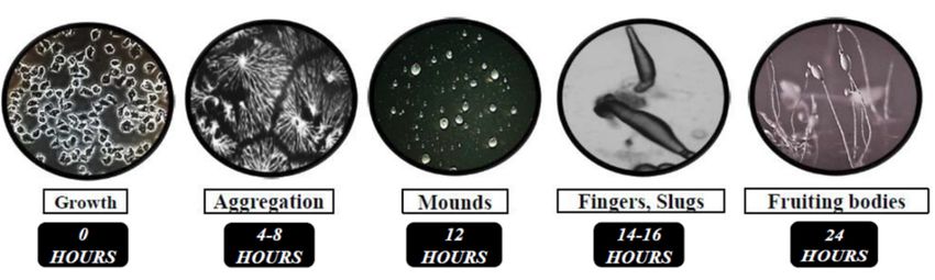
Cells 2019, 8, x 2 of 16
cellular and developmental processes [9]. Moreover, the ability to knockout genes using homologous
Cells 2019, 8, 115 2 of 16
recombination or CRISPR/Cas9-mediated targeting has made Dictyostelium a powerful model system
for studying the functions of proteins linked to human disease [10,11].
In Innutrient-rich
nutrient-rich conditions,
conditions, Dictyostelium
Dictyostelium grows
grows as as
single cells,
single multiplying
cells, multiplying bybymitosis
mitosis andand
obtaining
obtaining nutrients
nutrientsthrough
through endocytosis
endocytosis (Figure 1) [9].
(Figure Removal
1) [9]. of nutrients
Removal prompts
of nutrients a 24-hour
prompts a 24-h
developmental
developmental program
program consisting
consistingof of
a sequence
a sequence of of
well-defined
well-defined events
events (Figure 1).1).
(Figure Cells first
Cells undergo
first undergo
chemotactic aggregation towards 0 3’,5’-cyclic
0 adenosine monophosphate
chemotactic aggregation towards 3 ,5 -cyclic adenosine monophosphate (cAMP) to form multicellular (cAMP) to form
multicellular
mounds (Figure mounds (Figure then
1). Mounds 1). Mounds
undergothen undergo
a series a series of morphological
of morphological changes to formchanges to form
fingers that fall on
fingers that fall on the surface to generate motile pseudoplasmodia, also known
the surface to generate motile pseudoplasmodia, also known as slugs (Figure 1). Finally, the majority as slugs (Figure 1).
Finally,
of cellsthe majority
within of cells
the slug within differentiate
terminally the slug terminally differentiate
into either stalk cells into either forming
or spores, stalk cells or spores,
a fruiting body
forming a fruiting body that completes the life cycle (Figure 1). Spore are
that completes the life cycle (Figure 1). Spore are then dispersed and germinate in the presence of then dispersed and
germinate
nutrients, inrestarting
the presencethe of
lifenutrients,
cycle. restarting the life cycle.
Figure The 24-h
Figure1.1.The life cycle
24-hour life of Dictyostelium
cycle discoideum.discoideum.
of Dictyostelium In nutrient-rich conditions, Dictyostelium
In nutrient-rich conditions,grow
as single cells and feed on readily available nutrients and bacteria. Removal
Dictyostelium grow as single cells and feed on readily available nutrients and bacteria. of the food source initiates
multicellular
Removal of thedevelopment.
food source Duringinitiatesthe initial stages development.
multicellular of development,During
cells chemotactically aggregate
the initial stages of
0 0
towards 3 ,5 -cyclic adenosine monophosphate aggregate
(cAMP) to formtowards
multicellular mounds. Cellsadenosine
then undergo
development, cells chemotactically 3’,5’-cyclic
a series of structural
monophosphate changes
(cAMP) totoform
form multicellular
a finger followed by a motile
mounds. pseudoplasmodium,
Cells then undergo or slug. Finally,
a series of
the majority of cells within the slug terminally differentiate to
structural changes to form a finger followed by a motile pseudoplasmodium, orform either stalk cells or spores in aslug.
fruiting
body. Spores are dispersed and then germinate when nutrients become available, restarting the life cycle.
Finally, the majority of cells within the slug terminally differentiate to form either stalk cells
or Like
spores in a fruiting
metazoan body. Spores growth
cells, Dictyostelium are dispersed and then germinate
and development when nutrients
relies on fundamental processes
become available, restarting the life cycle.
including cell movement, cell sorting, cell differentiation, intracellular trafficking, autophagy, and signal
transduction [9]. As a result, uncharacterized genes or undefined biological pathways can be thoroughly
Like metazoan cells, Dictyostelium growth and development relies on fundamental processes
studied in Dictyostelium, and the results of these studies can then be translated to mammalian
including cell movement, cell sorting, cell differentiation, intracellular trafficking, autophagy, and
systems [12–14]. Work in Dictyostelium has made valuable contributions to our understanding of
signal transduction [9]. As a result, uncharacterized genes or undefined biological pathways can be
the functions of proteins linked to human neurological disorders, including epilepsy, prion diseases,
thoroughly studied in Dictyostelium, and the results of these studies can then be translated to
lissencephaly, Alzheimer’s disease, Parkinson’ disease, and Huntington’s disease [15–20]. In addition,
mammalian systems [12–14]. Work in Dictyostelium has made valuable contributions to our
Dictyostelium has proven to be an exceptional organism for studying the cellular and molecular
understanding of the functions of proteins linked to human neurological disorders, including
mechanisms underlying Batten disease [7]. The Dictyostelium genome encodes homologs of 11 of the 13
epilepsy, prion diseases, lissencephaly, Alzheimer’s disease, Parkinson’ disease, and Huntington’s
NCL genes, which is more than other model organisms including yeast, C. elegans, and D. melanogaster [7].
disease [15–20]. In addition, Dictyostelium has proven to be an exceptional organism for studying the
Recent work on Dictyostelium has provided fresh new insight into the functions of TPP1/CLN2, CLN3,
cellular and molecular mechanisms underlying Batten disease [7]. The Dictyostelium genome encodes
and CLN5. In this review, we highlight these discoveries and discuss how these new findings have
homologs of 11 of the 13 NCL genes, which is more than other model organisms including yeast, C.
enhanced our knowledge of NCL protein function in humans.
elegans, and D. melanogaster [7]. Recent work on Dictyostelium has provided fresh new insight into the
functions
3. UsingofDictyostelium
TPP1/CLN2, CLN3, andCLN2
to Study CLN5.Disease
In this review, we highlight these discoveries and discuss
how these new findings have enhanced our knowledge of NCL protein function in humans.
3.1. Human TPP1
3. Using Dictyostelium to Study CLN2 Disease
Mutations in tripeptidyl peptidase 1 (TPP1) cause a late infantile form of NCL referred to as CLN2
disease [1]. Mutations in TPP1/CLN2 are also linked to autosomal recessive spinocerebellar ataxia
3.1. Human TPP1
7 (SCAR7) [21]. However, unlike in CLN2 disease where the activity of TPP1/CLN2 is completely
Mutations
abolished, theinactivity
tripeptidyl
of thepeptidase
enzyme in1 (TPP1)
SCAR7 cause a late
patients infantile
is merely form of
reduced NCL
[21]. As referred to as
a result, SCAR7
CLN2 disease [1]. Mutations in TPP1/CLN2 are also linked to autosomal recessive spinocerebellar
patients do not exhibit vision loss or epilepsy [21]. TPP1/CLN2 is an acid-activated serine protease
ataxia 7 (SCAR7)
that localizes [21].lysosomal
to the However, unlike
matrix in As
[22]. CLN2 disease
a serine whereTPP1/CLN2
protease, the activityisof TPP1/CLN2
involved is
in several
processes such as macroautophagy and endocytosis [23]. The study of TPP1/CLN2 in model organisms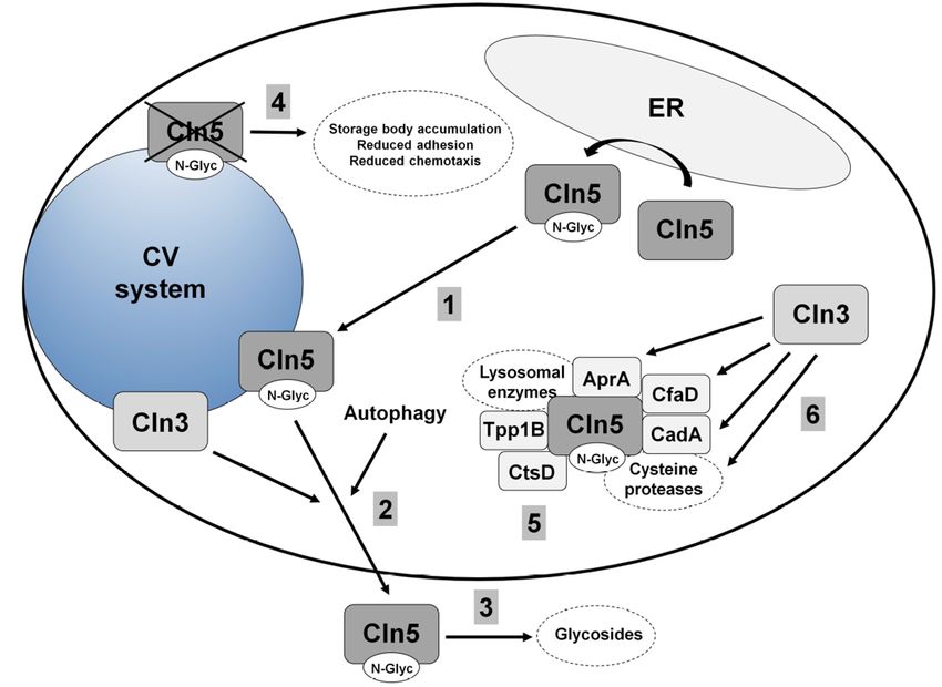
late stages of development, loss of tpp1A causes cells to develop precociously and form abnormal
spores [24] (Figure 2). In addition, the development of tpp1A- cells is severely compromised in the
presence of the lysosomotropic agent chloroquine, which is consistent with a role for Tpp1A at the
lysosome [24]. By exploiting the genetic tractability of Dictyostelium, researchers used restriction
enzyme-mediated
Cells 2019, 8, 115       integration (REMI) mutagenesis to identify stpA (suppressor of Tpp1 A) as              3 ofa16
second site suppressor of tpp1A-deficiency [24] (Figure 2). StpA shares some similarity to oxysterol-
binding proteins, which function in lipid transport and metabolism [24,26]. Intriguingly, altered lipid
has   been limited
homeostasis        has due
                       beentolinked
                               the absence  of homologs
                                     to the NCLs  [27,28]. in
                                                           Foryeast, C. elegans,
                                                                example,         and D. melanogaster
                                                                           lipid accumulation          [7].observed
                                                                                                has been    However,
Dictyostelium       has six genes   that  encode  proteins   that share  a  significant amount   of
in neural stem cells derived from induced pluripotent stem cells generated from CLN2 disease patientsimilarity   with
human
fibroblastsTPP1/CLN2          work intpp1A
                [27]. Thus,(genes:           tpp1B, tpp1C,
                                       Dictyostelium         tpp1D, tpp1E,
                                                      has provided    valuable    tpp1F;
                                                                              andnew      proteins:
                                                                                      insight       Tpp1A,
                                                                                              into the        Tpp1B,
                                                                                                        potential of
Tpp1C,     Tpp1D,     Tpp1E,   and  Tpp1F)   [24,25] (Figure  2).
targeting other genes that may reduce the effects of loss of function mutations in human TPP1.

      Figure2.2.Current
     Figure      Currentmodel
                            model  of of Tpp1
                                      Tpp1      function
                                            function       in Dictyostelium.
                                                       in Dictyostelium.  (1) The(1)Dictyostelium
                                                                                     The Dictyostelium
                                                                                                  genome  genome
                                                                                                             encodes
     six proteins that show similarity to human TPP1/CLN2. These proteins likely all contribute toall
      encodes   six proteins   that   show   similarity   to human    TPP1/CLN2.        These  proteins  likely   the
      contribute
     total         to theintotal
           TPP1 activity          TPP1 activity
                             Dictyostelium.        in Dictyostelium.
                                             (2) Tpp1A                 (2) Tpp1A
                                                         and Tpp1F localize     to theand  Tpp1F
                                                                                       endocytic   localizeincluding
                                                                                                 pathway     to the
      endocytic
     acidic       pathway(e.g.,
            compartments      including     acidic
                                    lysosomes).  (3) compartments      (e.g.,storage
                                                     Loss of tpp1A causes       lysosomes).   (3) Loss ofimpaired
                                                                                      body accumulation,     tpp1A
      causes  storage   body     accumulation,      impaired    autophagy,      precocious    development,
     autophagy, precocious development, and impaired spore formation. (4) StpA functions as a second-site      and
      impaired of
     suppressor   spore   formation. in
                     tpp1A-deficiency    (4)Dictyostelium.
                                              StpA functions       as a and
                                                             (5) Tpp1B   second-site
                                                                               Tpp1F bind suppressor   of regulator
                                                                                            the Golgi pH    tpp1A-
     (GPHR). (5,6) Tpp1F also localizes to the endoplasmic reticulum (ER) and extracellular space.

3.2. Loss of the Lysosomal Enzyme Tpp1A Impairs Autophagy and Multicellular Development in Dictyostelium
      Homologous recombination was used to knockout the tpp1A gene in Dictyostelium [24].
tpp1A-deficiency in Dictyostelium reduces overall Tpp1 activity and results in an accumulation of
autofluorescent storage material in starved cells [24] (Figure 2). Like human TPP1/CLN2, Tpp1A
localizes to the lysosome [22,24] (Figure 2). The growth and viability of tpp1A− cells is impaired
in autophagy-stimulating media, which is consistent with previous work that reported reduced
autophagosome formation in CLN2 disease patient fibroblasts [23,24] (Figure 2). During the mid-to-late
stages of development, loss of tpp1A causes cells to develop precociously and form abnormal spores [24]
(Figure 2). In addition, the development of tpp1A− cells is severely compromised in the presence of
the lysosomotropic agent chloroquine, which is consistent with a role for Tpp1A at the lysosome [24].
By exploiting the genetic tractability of Dictyostelium, researchers used restriction enzyme-mediated
integration (REMI) mutagenesis to identify stpA (suppressor of Tpp1 A) as a second site suppressor
of tpp1A-deficiency [24] (Figure 2). StpA shares some similarity to oxysterol-binding proteins,
which function in lipid transport and metabolism [24,26]. Intriguingly, altered lipid homeostasis
has been linked to the NCLs [27,28]. For example, lipid accumulation has been observed in neural stem
cells derived from induced pluripotent stem cells generated from CLN2 disease patient fibroblasts [27].
Thus, work in Dictyostelium has provided valuable new insight into the potential of targeting other
genes that may reduce the effects of loss of function mutations in human TPP1.
Cells 2019, 8, 115 4 of 16
3.3. Tpp1B and Tpp1F Interact with the Golgi pH Regulator in Dictyostelium
As mentioned above, the Dictyostelium genome contains six genes that encode proteins similar
to human TPP1/CLN2, with all six proteins likely contributing to the total TPP1 activity in the
cell [24,25] (Figure 2). In addition to Tpp1A, recent work has also studied the function of Tpp1B and
Tpp1F [25]. In Dictyostelium, both proteins bind the Golgi pH regulator (GPHR) [25] (Figure 2). GPHR is
a transmembrane anion channel that acidifies compartments of the Golgi complex and influences its
morphology as well as the morphology of the ER [29,30]. In Dictyostelium, the GPHR plays a role in
regulating growth and the later stages of multicellular development [31]. In addition to the Golgi
complex, Tpp1F localizes to the ER, V-ATPase-positive vesicles, and the extracellular space [25,32]
(Figure 2). Like Tpp1A, Tpp1F also has serine protease activity [25]. However, tpp1F-deficiency in
Dictyostelium has no obvious effects on growth or development, likely from the compensatory activities
provided by the other Tpp1 proteins in Dictyostelium (expression of tpp1B is the highest during growth
and development followed by tpp1F and tpp1A) [25,33]. In total, this work revealed a novel interaction
of Tpp1 proteins with the GPHR in Dictyostelium, which should fuel research in mammalian models of
CLN2 disease to determine if TPP1 interacts with the GPHR in human cells and how this interaction
may contribute to the pathology underlying NCL.
4. Using Dictyostelium to Study CLN3 Disease
4.1. Human CLN3
Mutations in CLN3 (ceroid lipofuscinosis neuronal 3) cause a juvenile form of NCL, which is the
most common subtype of the disease [1]. CLN3 encodes a 438-amino acid transmembrane protein that
localizes to the late endosomal and lysosomal membranes [34,35]. Research in a diversity of genetic
models has speculated that the function of CLN3 is linked to adhesion, apoptosis, autophagy, cell cycle
control, cell proliferation, endocytosis, neurogenesis, osmoregulation, pH and ion homeostasis,
and protein trafficking and secretion [32,36–48]. However, the precise function of the protein has not
been defined.
4.2. Loss of Cln3 Causes Pleiotropic Effects in Dictyostelium that are Consistent with its Localization to the
Contractile Vacuole System
The Dictyostelium homolog of human CLN3 (gene: cln3, protein: Cln3) encodes a 421-amino acid
transmembrane protein. In both growth and starved conditions, Cln3 localizes predominantly to the
contractile vacuole (CV) system, and to a lesser extent, compartments of the endocytic pathway and
Golgi complex [32,40,45] (Figure 3). During growth, cln3− cells display increased cell proliferation,
aberrant cytokinesis, and defects in osmoregulation [40,48] (Figure 3). During multicellular development,
cln3− cells display reduced cell-cell and cell-substrate adhesion, delayed aggregation, aberrant protein
secretion, and precocious multicellular development [32,40,45] (Figure 3). Importantly, evidence from
yeast and mammalian cell models also supports a role for CLN3 in these processes, highlighting that the
molecular function of CLN3 is likely conserved from Dictyostelium to human [36–39,41,42,46,47].
The localization of Cln3 to the CV system has provided clues into the mechanism underlying
cln3-deficiency phenotypes in Dictyostelium. The contractile vacuole (CV) system is a dynamic
organelle that is linked to osmoregulation, protein secretion, and ion homeostasis [49–51]. The effect of
cln3-deficiency on osmoregulation and protein secretion in Dictyostelium has been studied in detail and
will be described below [32,48]. Previous work has also shown that cln3-deficiency phenotypes during
development can be suppressed by treating cells with the calcium chelator egtazic acid (EGTA) [40,45].
These results are consistent with work showing aberrant calcium homeostasis in mouse models of
CLN3 disease [43,46,52]. However, further work is needed to clarify the exact role of Cln3 in regulating
ion balance in Dictyostelium.Cells 2019, 8, 115 5 of 16
Cells 2019, 8, x 5 of 16
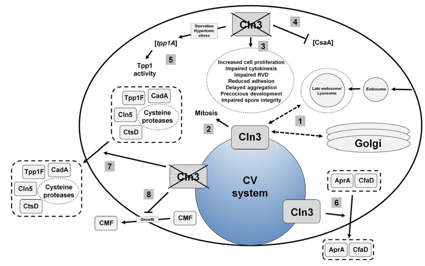
3. Current
Figure 3.
Figure Currentmodel
modelof of
Cln3 function
Cln3 in Dictyostelium.
function (1) Cln3
in Dictyostelium. (1)localizes primarilyprimarily
Cln3 localizes to the contractile
to the
vacuole (CV)
contractile system, (CV)
vacuole and tosystem,
a smallerandextent,
to acompartments
smaller extent,of the endocytic pathway
compartments of theand the Golgi
endocytic
complex. (2)
pathway andCln3
thefunction is linked to(2)
Golgi complex. mitosis.
Cln3 (3) Loss of cln3
function increases
is linked the rate of(3)
to mitosis. cellLoss
proliferation,
of cln3
alters cytokinesis, decreases the efficiency of regulatory volume decrease
increases the rate of cell proliferation, alters cytokinesis, decreases the efficiency(RVD), reduces adhesion,
of
delays aggregation,
regulatory volumecauses cells(RVD),
decrease to develop precociously,
reduces adhesion,and delays
impairsaggregation,
spore integrity.causes
(4) Loss of cln3
cells to
reduces the intracellular level of CsaA protein. (5) Loss of cln3 increases the expression of tpp1A
develop precociously, and impairs spore integrity. (4) Loss of cln3 reduces the intracellular
during osmotic stress and increases TPP1 enzymatic activity. (6) Cln3 modulates the secretion of AprA
level of CsaA protein. (5) Loss of cln3 increases the expression of tpp1A during osmotic
and CfaD. (7) Loss of cln3 increases the secretion of Tpp1F, Cln5, CtsD, CadA, and selected cysteine
stress and increases TPP1 enzymatic activity. (6) Cln3 modulates the secretion of AprA and
proteases. (8) Loss of cln3 reduces the secretion of CMF during growth.
CfaD. (7) Loss of cln3 increases the secretion of Tpp1F, Cln5, CtsD, CadA, and selected
cysteine proteases.
4.3. Cln3 Regulates (8) Loss of in
Osmoregulation cln3 reduces the secretion of CMF during growth.
Dictyostelium
During osmoregulation, the CV system regulates intracellular water balance by collecting excess water
The localization of Cln3 to the CV system has provided clues into the mechanism underlying
from the cytosol and then expelling the water out of the cell [49]. CLN3 has been shown to play a role in
cln3-deficiency phenotypes in Dictyostelium. The contractile vacuole (CV) system is a dynamic
osmoregulation
organelle that isinlinked
mammalian models of CLN3
to osmoregulation, disease
protein [37–39]. and
secretion, In a baby hamster kidney
ion homeostasis cell line,
[49–51]. Theosmotic
effect
stress affects the expression and localization of CLN3 [38]. In mice, CLN3 regulates
of cln3-deficiency on osmoregulation and protein secretion in Dictyostelium has been studied in detail renal control of water
and potassium balance [37]. Finally, osmotic stress induces an abnormal blood–brain
and will be described below [32,48]. Previous work has also shown that cln3-deficiency phenotypes barrier response in
brain endothelial cells obtained from Cln3-deficient mice [39]. During
during development can be suppressed by treating cells with the calcium chelator egtazic acidhypotonic stress, the expulsion of
water from
(EGTA) the cytosol
[40,45]. Theseisresults
knownare as regulatory
consistentvolume
with workdecrease (RVD),aberrant
showing which is calcium
a conserved process in in
homeostasis all
eukaryotic cells [53]. In Dictyostelium, previous work showed that cln3 − cells display defects in RVD and
mouse models of CLN3 disease [43,46,52]. However, further work is needed to clarify the exact role
exhibit
of Cln3 aindelay in theirion
regulating ability to recover
balance from hypotonic stress [48] (Figure 3). This delay is exacerbated
in Dictyostelium.
−
when cln3 cells are treated with ammonium chloride, a lysosomotropic compound that elevates the pH of
intracellular
4.3. compartments
Cln3 Regulates [48,54].in
Osmoregulation More specifically, following hypotonic stress, the ability of cln3− cells
Dictyostelium
and V-ATPase-positive compartments in cln3− cells to reduce in size is delayed compared to wild-type
cellsDuring
[48]. The osmoregulation,
sensitivity of cln3 the−CV system
cells regulates
to hypotonic intracellular
stress perpetuates water balance
into by collecting
multicellular excess
development
water from −the cytosol and then expelling the water out of the cell [49]. CLN3
where cln3 cells display delayed development under hypotonic stress, and arrested development at has been shown to play
the
aslug
rolestage
in osmoregulation in mammalian models of CLN3 disease [37–39]. In a
when developed in hypotonic conditions with ammonium chloride [48]. These data suggest thatbaby hamster kidney
cell line, osmoticagents
lysosomotropic stressaffect
affects the
the expression
ability of cln3−and localization
cells to cope with of osmotic
CLN3 [38]. In mice,
stress. CLN3 cln3
In addition, regulates
− cells
renal control of water and potassium balance [37]. Finally, osmotic stress induces an
display reduced viability under hypotonic stress, which also compromises the integrity of cln3− spores [48]. abnormal blood–
brain
Finally,barrier
loss of response in brain
cln3 also impairs theendothelial
viability andcells obtainedoffrom
development Cln3-deficient
cells in mice [39].
response to hypertonic During
stress [48].
hypotonic stress, the expulsion of water from the cytosol is known as regulatory volume decrease
(RVD), which is a conserved process in all eukaryotic cells [53]. In Dictyostelium, previous work
showed that cln3− cells display defects in RVD and exhibit a delay in their ability to recover from
hypotonic stress [48] (Figure 3). This delay is exacerbated when cln3− cells are treated with ammoniumCells 2019, 8, 115 6 of 16
RNA sequencing was used to examine the pathways regulating the response of cln3− cells
to osmotic stress. This analysis revealed 320 genes that were differentially expressed in cln3− cells
compared to wild-type cells during hypotonic stress, and 162 genes that were differentially expressed
during hypertonic stress [48]. The resulting datasets were then examined using GO term enrichment
and STRING protein–protein interaction network analyses [55,56]. These analyses revealed that the
differentially expressed genes are linked to developmental processes, which is consistent with the
aberrant development of cln3− cells during osmotic stress [48]. Additionally, cln3− cells subjected to
hypotonic stress displayed differential expression of genes linked to metabolic processes [48]. In both
osmotic stress conditions, there was an enrichment of differentially expressed genes involved in transport
and catalysis [48]. These results are consistent with the role of Cln3 in protein secretion, specifically
the aberrant secretion of proteases by cln3− cells and the enhanced activity of Tpp1 in cln3− cells
during hypertonic stress [32,48]. Finally, the proteins encoded by genes differentially expressed during
hypotonic stress localize to the cell periphery and extracellular region, while proteins encoded by genes
differentially expressed during hypertonic stress localize to membranes (e.g., intrinsic component of
membrane) [48].
In Dictyostelium, GFP-Cln3 localizes to the CV system during mitosis and cytokinesis [48] (Figure 3).
During cytokinesis, water efflux from CV system bladders facilitates the formation of the cleavage furrow,
which is a transient structure that divides the two daughter cells [57–59]. Not surprisingly, Dictyostelium
osmoregulatory mutants display defects in cytokinesis [60–62]. Aligning with the osmoregulatory defects
observed in cln3− cells, loss of cln3 increases the number of multi-nucleated cells in growth culture [48]
(Figure 3). Importantly, these results are consistent with cytokinesis defects observed in a yeast model of
CLN3 disease [36]. In total, this work links the function of Cln3 to osmoregulation in Dictyostelium and
provides valuable new insight into the mechanisms underlying this function.
4.4. Cln3 Regulates Protein Secretion in Dictyostelium
In addition to osmoregulation, the CV system has also been linked to protein secretion in
Dictyostelium [50]. Work has shown that the enhanced proliferation of cln3− cells may be due to
the aberrant secretion of proteins linked to growth, specifically autocrine proliferation repressor A
(AprA) and counting factor-associated protein A (CfaD) [40] (Figure 3). AprA and CfaD function
together to repress cell proliferation and facilitate chemorepulsion [63–65]. A preliminary analysis into
the mechanism underlying the aberrant adhesion and aggregation of cln3− cells revealed that loss of
cln3 decreased the intracellular amount of the cell–cell adhesion protein contact site A (CsaA) and
increased the amount of soluble extracellular calcium-dependent cell adhesion molecule A (CadA) [45]
(Figure 3). The delayed aggregation of cln3− cells has also been linked to the reduced secretion of
conditioned media factor (CMF) during growth [32] (Figure 3). Since CMF plays a critical role in
initiating and synchronizing development upon starvation, these results indicate that cln3− cells may
not be optimally primed to enter development [66].
Based on the above findings, mass spectrometry was used to further explore the effect of
cln3-deficiency on protein secretion during aggregation [32]. That study provided the first evidence in
any system showing that loss of cln3 alters protein secretion [32]. A total of 450 proteins were detected
in conditioned starvation buffer harvested from wild-type and cln3− cells [32]. Three proteins that are
normally secreted by wild-type cells during starvation were absent in conditioned buffer harvested from
cln3− cells [32]. Two of the three proteins function in adhesion and migration, which could explain the
adhesion defects observed in cln3− cells [32,45]. In addition, 12 proteins that are not normally secreted
during starvation were present in conditioned buffer harvested from cln3− cells [32]. Consistent with
these findings, label-free quantification identified 42 proteins that were present in significantly higher
amounts in cln3− conditioned starvation buffer compared to wild-type and 3 proteins that were present
in significantly reduced amounts [32]. Gene ontology (GO) term analyses revealed an enrichment of
proteins linked to endocytosis, vesicle-mediated transport, proteolysis, and metabolism. Importantly,
these results support the reduced endocytosis and protein transport observed in cells from Cln3-deficientCells 2019, 8, 115 7 of 16
mice, reduced basal mitochondrial respiration and ATP production observed in mice harboring the most
common mutation observed in patients with CLN3 disease, and the regulation of cathepsin D (CTSD)
protease activity by CLN3 in baby hamster kidney cells [39,52,67]. In total, this work revealed for the first
time that Cln3 plays a role in protein secretion and suggests that future research in Dictyostelium may
provide additional insight on the precise role of CLN3 in regulating protein secretion in human cells.
5. Using Dictyostelium to Study CLN5 Disease
5.1. Human CLN5
Mutations in CLN5 (ceroid lipofuscinosis neuronal 5) cause a late-infantile form of Batten disease,
but juvenile and adult onsets have also been reported [1,68,69]. CLN5 disease was first reported as
a Finnish variant, however, patients with broad ethnic backgrounds have now been diagnosed [70–73]. In
mammalian cells, CLN5 localizes to the lysosome and is present in the conditioned media of cultured cells,
which is consistent with the presence of a signal peptide for secretion in the N-term of the protein [74–77].
CLN5 is first translated as a 407-amino acid type II transmembrane protein, which resides in the ER
membrane [78]. The protein is then cleaved by signal peptide peptidase-like (SPPL) 3 to form a soluble
protein [79,80]. In addition, CLN5 has eight N-glycosylation sites that are critical for the folding,
trafficking, and localization of the protein [75]. Recently, human CLN5 was shown to display glycoside
hydrolase activity [77]. CLN5 has also been speculated to function in autophagy, lipid metabolism,
lysosome receptor sorting, myelination, and sphingolipid transport [81–85]. However, the precise
mechanisms underlying CLN5 disease have yet to be revealed.
5.2. Cln5 is Secreted and Functions as a Glycoside Hydrolase in Dictyostelium
Dictyostelium is one of the few early eukaryotes that contains a homolog of human CLN5
(yeast, C. elegans, and D. melanogaster lack a homolog) [7]. The Dictyostelium homolog (gene: cln5,
protein: Cln5) is 322 amino acids in size, and like human CLN5, has glycoside hydrolase activity [77]
(Figure 4). The first evidence for human CLN5 having glycoside hydrolase activity was based on
studies that were initiated in Dictyostelium [77]. In Dictyostelium, Cln5 is glycosylated in the ER and then
trafficked to the cell cortex where it appears to be secreted via the CV system during starvation [77,86]
(Figure 4). Upon starvation in Dictyostelium, several conserved cellular processes are activated, one
being autophagy, which is required for multicellular development [87]. Intriguingly, treatment of
wild-type cells with lysosomotropic agents (e.g., ammonium chloride or chloroquine) decreases
Cln5 secretion [86]. Since lysosomotropic compounds inhibit autophagy, these results suggest that
autophagic mechanisms regulate the secretion of the protein [88] (Figure 4). In total, the secretion
of Cln5 in Dictyostelium is consistent with observations in mammalian models of the disease and
indicates that secreted CLN5 may play an important role in the pathological mechanisms underlying
CLN5 disease.in Dictyostelium revealed that the protein interacts with lysosomal enzymes (e.g., alpha-mannosidase,
beta-glucosidase), cysteine proteases, other NCL protein homologs such as Tpp1B, cathepsin D
(CtsD), and uncharacterized protein DDB0252831 (which is similar to cathepsin F, CTSF/CLN13), and
proteins linked to Cln3 function (e.g., AprA, CfaD, CadA) [77] (Figure 4). Therefore, future work in
Cells 2019, 8, 115 may provide novel insight into the cellular pathways regulated by Cln5 and8 this
Dictyostelium of 16
knowledge can then be translated to other genetic models of CLN5 disease.
Figure
Figure 4.4.Current
Current model
model of Cln5
of Cln5 function
function in Dictyostelium.
in Dictyostelium. (1) Cln5 (1) Cln5 is glycosylated
is glycosylated in the
in the endoplasmic
endoplasmic
reticulum (ER)reticulum
and then (ER) and then
trafficked to the trafficked to vacuole
contractile the contractile vacuole
(CV) system. (2)(CV)
Cln5system. (2)
is secreted
unconventionally
Cln5 via a pathway involving
is secreted unconventionally Cln3 andinvolving
via a pathway autophagyCln3 induction. (3) Cln5 functions
and autophagy induction. as
a glycoside hydrolase outside of the cell. (4) Loss of cln5 leads to storage body
(3) Cln5 functions as a glycoside hydrolase outside of the cell. (4) Loss of cln5 leads to accumulation and
results inbody
storage aberrant adhesion and and
accumulation chemotaxis.
results(5)inCln5 interacts
aberrant with lysosomal
adhesion enzymes, as well
and chemotaxis. (5) as the
Cln5
Dictyostelium homologs of human TPP1/CLN2, CTSD/CLN10, and
interacts with lysosomal enzymes, as well as the Dictyostelium homologs of humanCTSF/CLN13. (6) Cln5 interacts
with proteins linked
TPP1/CLN2, to Cln3 function
CTSD/CLN10, in Dictyostelium (6)
and CTSF/CLN13. (cysteine
Cln5 proteases,
interacts AprA, CfaD, andlinked
with proteins CadA).to
Cln3offunction
5.3. Loss in Dictyostelium
Cln5 Impairs Adhesion and (cysteine
Chemotaxis proteases, AprA,
during the EarlyCfaD,
Stages and CadA).
of Dictyostelium Development
TheDictyostelium
6. Using accumulation toof study
autofluorescent storage
the molecular material of
networking in NCL
neurons, as well as cells outside the
proteins
central nervous system, is a pathological hallmark of the NCLs [2]. In Dictyostelium, cln5− cells
Mounting evidence indicates that the NCL proteins function in shared or convergent biological
also accumulate autofluorescent storage deposits further highlighting the conserved nature of NCL
pathways [92]. Mutations in NCL proteins cause the accumulation of ceroid lipofuscin within cells
pathways and validating the use of Dictyostelium as a model system for studying CLN5 disease [86]
and result in nearly identical clinical manifestations [2]. In addition, previous work reported the
(Figure 4). During the early stages of multicellular development, cln5− cells display a reduced ability to
spatial and temporal co-expression of TPP1/CLN2, CLN3, and CLN5 during brain development,
adhere to the substrate, which is exacerbated when cells are treated with chloroquine [86]. Furthermore,
shared interaction partners between CLN3 and CLN5, CLN5 polypeptides interacting directly with
cln5− cells also display a defect in cell–cell adhesion [86]. As a potential consequence of the aberrant
adhesion, cln5− cells display reduced cAMP chemotaxis in a radial bioassay [86,89]. These results
are consistent with observations of fibroblasts obtained from CLN5 disease patients, which attach
poorly to tissue culture dishes and display altered expression of genes linked to cell adhesion [90].
Neurons from Cln5-deficient mice also display altered expression of genes linked to adhesion as well
as aberrant localization of cytoskeletal proteins [91]. Finally, an analysis of the Cln5 interactome in
Dictyostelium revealed that the protein interacts with lysosomal enzymes (e.g., alpha-mannosidase,
beta-glucosidase), cysteine proteases, other NCL protein homologs such as Tpp1B, cathepsin D (CtsD),
and uncharacterized protein DDB0252831 (which is similar to cathepsin F, CTSF/CLN13), and proteins
linked to Cln3 function (e.g., AprA, CfaD, CadA) [77] (Figure 4). Therefore, future work in Dictyostelium
may provide novel insight into the cellular pathways regulated by Cln5 and this knowledge can then
be translated to other genetic models of CLN5 disease.Cells 2019, 8, 115 9 of 16
6. Using Dictyostelium to Study the Molecular Networking of NCL Proteins
Mounting evidence indicates that the NCL proteins function in shared or convergent biological
pathways [92]. Mutations in NCL proteins cause the accumulation of ceroid lipofuscin within cells and
result in nearly identical clinical manifestations [2]. In addition, previous work reported the spatial and
temporal co-expression of TPP1/CLN2, CLN3, and CLN5 during brain development, shared interaction
partners between CLN3 and CLN5, CLN5 polypeptides interacting directly with TPP1/CLN2 and
CLN3, exacerbated NCL phenotypes in Cln1/Cln5 double knockout mice, and the interaction of CLN5
with PPT1/CLN1, TPP1/CLN2, CLN3, CLN6, and CLN8 [41,93–96]. A recent report also showed
decreased levels of CLN5 protein in a cell line derived from a Mfsd8/Cln7 knockout mouse [97]. Thus,
studying the function of any one NCL protein is likely to enhance our knowledge of the mechanisms
underlying the neurodegeneration associated with the disease, knowledge that can then be applied to
all subtypes of the disease.
Like mammalian models of Batten disease, there is evidence in Dictyostelium to support that
the NCL proteins are connected at the molecular level. Using a proteomics-based approach, 10 of
the 11 NCL protein homologs in Dictyostelium were detected in the macropinocytic pathway [98].
As discussed above, previous work revealed a function for Cln3 in protein secretion [32] (Figure 3).
Specifically, that study reported that a loss of cln3 increased the amount of Tpp1F, Cln5, and
CtsD in conditioned starvation buffer [32] (Figures 2–4). A follow-up study provided direct
evidence linking the secretion of Cln5 to Cln3 function by showing increased amounts of Cln5
in conditioned starvation buffer harvested from cln3− cells [86] (Figures 3 and 4). Furthermore,
Cln5 was shown to co-localize with Cln3 at the CV system, which has been proposed to mediate its
secretion [86]. Immunoprecipitation coupled with mass spectrometry revealed the Cln5 interactome in
Dictyostelium [77]. Cln5-interactors include Tpp1B, CtsD, and uncharacterized protein DDB0252831
(similar to CTSF/CLN13), as well as proteins linked to Cln3 function in Dictyostelium (e.g., AprA,
CfaD, CadA) [77]. Intriguingly, ten Cln5-interactors are differentially secreted by cln3− cells [32,77]
(Table 1). Furthermore, cln3-deficiency increases the expression of tpp1A during hypertonic stress,
which correlates with increased Tpp1 activity [48]. Finally, loss of tpp1A, cln3, or cln5 in Dictyostelium
causes similar phenotypes (Table 2). In total, these findings support the use of Dictyostelium to study
the molecular networking of NCL proteins.
Table 1. Proteins present in Cln5-GFP IP fractions that are differentially secreted by cln3− cells.
dictyBase ID Protein Names Gene Names
Autocrine proliferation repressor protein A (PhoPQ-activated aprA
DDB0231036
pathogenicity-related protein) DDB_G0281663
ctsD, catD
DDB0215012 Cathepsin D (Ddp44)
DDB_G0279411
cprD, CP4
DDB0214999 Cysteine proteinase 4
DDB_G0278721
cprE, CP5
DDB0185092 Cysteine proteinase 5
DDB_G0272815
cprG, CP7
DDB0215005 Cysteine proteinase 7
DDB_G0279187
Elongation factor 1-alpha (EF-1-alpha) (50 kDa actin-binding eef1a2, efaa2, efaAII
DDB0191134
protein) (ABP-50) DDB_G0269136
bip2
DDB0233663 Luminal-binding protein (BiP 2)
DDB_G0276445Cells 2019, 8, 115 10 of 16
Table 1. Cont.
dictyBase ID Protein Names Gene Names
DDB0349243 Uncharacterized protein DDB_G0288563
Uncharacterized protein, member of the peptidase S28 family
of serine proteases, a group containing lysosomal Pro-X
DDB0233868 DDB_G0289749
carboxypeptidase, dipeptidyl-peptidase II, and
thymus-specific serine peptidase
Induced after Legionella infection
iliA
DDB0238155 Contains a putative N-terminal signal sequence; regulated by
DDB_G0285615
gskA and zakA; induced by Legionella pneumophila infection
Table 2. Comparison of the phenotypes observed in Dictyostelium models of TPP1/CLN2, CLN3, and
CLN5 disease.
Phenotype Tpp1a− Cln3− Cln5−
Increased cell proliferation No Yes Not known
Impaired cytokinesis Not known Yes Not known
Autofluorescent inclusions Yes Not known Yes
Defects in osmoregulation Not known Yes Not known
Aberrant protein secretion Not known Yes Not known
Reduced adhesion Not known Yes Yes
Function linked to autophagy Yes Not known Yes
Precocious development Yes Yes Not known
Impaired spore formation Yes Not known Not known
Reduced spore viability/integrity No Yes Not known
7. Conclusions
Dictyostelium has proven to be an exceptional organism for studying the cellular roles of NCL
proteins. Phenotypes previously revealed in other genetic models of Batten disease are present in
Dictyostelium (e.g., aberrant autophagy, impaired osmoregulation, etc.) providing evidence that the
functions of NCL proteins are likely conserved from Dictyostelium to human. Work in Dictyostelium has
also revealed previously unknown functions for the NCL proteins, such as the role of CLN3 in protein
secretion and the glycoside hydrolase activity of CLN5. These findings should spur future research
in mammalian models of NCL to further explore these functions. In fact, recent studies in mice and
humans have also linked the function of CLN3 to secretion [46,47]. However, as with any model
organism, there are caveats that must be considered. For one, Dictyostelium has a limited number of
cell types that may limit the translation of findings to specific tissues or organs in mammalian systems.
In addition, since Dictyostelium lacks a nervous system, discoveries made in the organism must be
validated in the relevant mammalian cell type. Nonetheless, Dictyostelium presents many benefits as
a biomedical model system. Moving forward, research in Dictyostelium has the potential to identify
molecular targets for therapies, which includes studying the effects of new drugs in a multicellular
organism. Finally, NCL phenotypes overlap with those seen in patients with Alzhemier’s disease,
Parkinson’s disease, and frontotemporal dementia [99–101]. Thus, on a larger scale, using Dictyosteium
to study the functions of NCL proteins could enhance our understanding of the mechanisms underlying
other forms of neurodegeneration.
Author Contributions: Conceptualization, R.J.H.; Writing–original draft preparation, M.D.M., S.M., R.J.H.;
Writing–review and editing, R.J.H.; Supervision, R.J.H.; Project administration, R.J.H.; Funding acquisition, R.J.H.
Funding: This review and the APC was supported by the Natural Sciences and Engineering Research Council of
Canada (Discovery Grant to R.J.H.) and the Banting Research Foundation (Discovery Award to R.J.H.).
Conflicts of Interest: The authors declare no conflict of interest.Cells 2019, 8, 115 11 of 16
Abbreviations
AprA autocrine proliferation repressor A
CadA cell adhesion molecule A
cAMP 30 ,50 -cyclic adenosine monophosphate
CfaD counting factor-associated protein D
CLN ceroid lipofuscinosis neuronal
CMF conditioned media factor
CsaA contact site A
CtsD cathepsin D
CV contractile vacuole
ER endoplasmic reticulum
GPHR Golgi pH regulator
NCL neuronal ceroid lipofuscinosis
REMI restriction enzyme-mediated integration
RVD regulatory volume decrease
SCAR7 spinocerebellar ataxia 7
SPPL signal peptide peptidase-like
StpA suppressor of Tpp1 A
TPP1 tripeptidyl peptidase 1
References
1. Mole, S.E.; Cotman, S.L. Genetics of the neuronal ceroid lipofuscinoses (Batten disease). Biochim. Biophys. Acta
2015, 1852, 2237–2241. [CrossRef] [PubMed]
2. Radke, J.; Stenzel, W.; Goebel, H.H. Human NCL neuropathology. Biochim. Biophys. Acta 2015, 1852,
2262–2266. [CrossRef] [PubMed]
3. Schulz, A.; Kohlschütter, A.; Mink, J.; Simonati, A.; Williams, R. NCL diseases—Clinical perspectives.
Biochim. Biophys. Acta 2013, 1832, 1801–1806. [CrossRef] [PubMed]
4. Cárcel-Trullols, J.; Kovács, A.D.; Pearce, D.A. Cell biology of the NCL proteins: What they do and don’t do.
Biochim. Biophys. Acta 2015, 1852, 2242–2255. [CrossRef] [PubMed]
5. Bond, M.; Holthaus, S.; Tammen, I.; Tear, G.; Russell, C. Use of model organisms for the study of neuronal
ceroid lipofuscinosis. Biochim. Biophys. Acta 2013, 1832, 1842–1865. [CrossRef] [PubMed]
6. Müller-Taubenberger, A.; Kortholt, A.; Eichinger, L. Simple system—Substantial share: The use of
Dictyostelium in cell biology and molecular medicine. Eur. J. Cell Biol. 2013, 92, 45–53. [CrossRef] [PubMed]
7. Huber, R.J. Using the social amoeba Dictyostelium to study the functions of proteins linked to neuronal ceroid
lipofuscinosis. J. Biomed. Sci. 2016, 23, 83. [CrossRef]
8. Eichinger, L.; Pachebat, J.A.; Glöckner, G.; Rajandream, M.-A.; Sucgang, R.; Berriman, M.; Song, J.; Olsen, R.;
Szafranski, K.; Xu, Q.; et al. The genome of social amoeba Dictyostelium discoideum. Nature 2005, 435, 43–57.
[CrossRef]
9. Mathavarajah, S.; Flores, A.; Huber, R.J. Dictyostelium discoideum: A model system for cell and developmental
biology. Curr. Protoc. Essent. Lab. Tech. 2017, 15, 14.1.1–14.1.19.
10. Faix, J.; Linkner, J.; Nordholz, B.; Platt, J.L.; Liao, X.H.; Kimmel, A.R. The application of the Cre-loxP system
for generating multiple knock-out and knock-in targeted loci. Methods Mol. Biol. 2013, 983, 249–267.
11. Sekine, R.; Kawata, T.; Muramoto, T. CRISPR/Cas9 mediated targeting of multiple genes in Dictyostelium.
Sci. Rep. 2018, 8, 8471. [CrossRef] [PubMed]
12. Terbach, N.; Shah, R.; Kelemen, R.; Klein, P.S.; Gordienko, D.; Brown, N.A.; Wilkinson, C.J.; Williams, R.S.
Identifying an uptake mechanism for the antiepileptic and bipolar disorder treatment valproic acid using
the simple biomedical model Dictyostelium. J. Cell Sci. 2011, 124, 2267–2276. [CrossRef] [PubMed]
13. Chang, P.; Walker, M.C.; Williams, R.S. Seizure-induced reduction in PIP3 levels contributes to seizure-activity
and is rescued by valproic acid. Neurobiol. Dis. 2014, 62, 296–306. [CrossRef] [PubMed]
14. Alexander, S.; Alexander, H. Lead genetic studies in Dictyostelium discoideum and translational studies in
human cells demonstrate that sphingolipids are key regulators of sensitivity to cisplatin and other anticancer
drugs. Semin. Cell Dev. Biol. 2011, 22, 97–104. [CrossRef] [PubMed]Cells 2019, 8, 115 12 of 16
15. Meyer, I.; Kuhnert, O.; Gräf, R. Functional analyses of lissencephaly-related proteins in Dictyostelium.
Semin. Cell Dev. Biol. 2011, 22, 89–96. [CrossRef] [PubMed]
16. Maniak, M. Dictyostelium as a model for human lysosomal and trafficking diseases. Semin. Cell Dev. Biol.
2011, 22, 114–119. [CrossRef] [PubMed]
17. Myre, M.A. Clues to γ-secretase, huntingtin and Hirano body normal function using the model organism
Dictyostelium discoideum. J. Biomed. Sci. 2012, 19, 41. [CrossRef] [PubMed]
18. Walker, M.C.; Williams, R.S. The search for better epilepsy treatments: From slime mould to coconuts.
Biochem. Soc. Trans. 2013, 41, 1625–1628. [CrossRef]
19. Annesley, S.J.; Chen, S.; Francione, L.M.; Sanislav, O.; Chavan, A.J.; Farah, C.; De Piazza, S.W.; Storey, C.L.;
Ilievska, J.; Fernando, S.G.; et al. Dictyostelium, a microbial model for brain disease. Biochim. Biophys. Acta
2014, 1840, 1413–1432. [CrossRef]
20. Malinovska, L.; Alberti, S. Protein misfolding in Dictyostelium: Using a freak of nature to gain insight into
a universal problem. Prion 2015, 9, 339–346. [CrossRef]
21. Sun, Y.; Almomani, R.; Breedveld, G.J.; Santen, G.W.; Aten, E.; Lefeber, D.J.; Hoff, J.I.; Brusse, E.;
Verheijen, F.W.; Verdijk, R.M.; et al. Autosomal recessive spinocerebellar ataxia 7 (SCAR7) is caused by
variants in TPP1, the gene involved in classic late-infantile neuronal ceroid lipofuscinosis 2 disease (CLN2
disease). Hum. Mutat. 2013, 34, 706–713. [CrossRef] [PubMed]
22. Sleat, D.E.; Donnelly, R.J.; Lackland, H.; Liu, C.G.; Sohar, I.; Pullarkat, R.K.; Lobel, P. Association of mutations
in a lysosomal protein with classical late-infantile neuronal ceroid lipofuscinosis. Science 1997, 277, 1802–1805.
[CrossRef] [PubMed]
23. Vidal-Donet, J.M.; Cárcel-Trullols, J.; Casanova, B.; Aguado, C.; Knecht, E. Alterations in ROS activity and
lysosomal pH account for distinct patterns of macroautophagy in LINCL and JNCL fibroblasts. PLoS ONE
2013, 8, e55526. [CrossRef] [PubMed]
24. Phillips, J.E.; Gomer, R.H. Partial genetic suppression of a loss-of-function mutant of the neuronal ceroid
lipofuscinosis-associated protease TPP1 in Dictyostelium discoideum. Dis. Models Mech. 2015, 8, 147–156.
[CrossRef] [PubMed]
25. Stumpf, M.; Müller, R.; Gaßen, B.; Wehrstedt, R.; Fey, P.; Karow, M.A.; Eichinger, L.; Glöckner, G.; Noegel, A.A.
A tripeptidyl peptidase 1 is a binding partner of the Golgi pH regulator (GPHR) in Dictyostelium. Dis. Models
Mech. 2017, 10, 897–907. [CrossRef] [PubMed]
26. Olkkonen, V.M.; Li, S. Oxysterol-binding proteins: Sterol and phosphoinositide sensors coordinating
transport, signaling and metabolism. Prog. Lipid Res. 2013, 52, 529–538. [CrossRef] [PubMed]
27. Sima, N.; Li, R.; Huang, W.; Xu, M.; Beers, J.; Zou, J.; Titus, S.; Ottinger, E.A.; Marugan, J.J.; Xie, X.; et al.
Neural stem cells for disease modeling and evaluation of therapeutics for infantile (CLN1/PPT1) and late
infantile (CLN2/TPP1) neuronal ceroid lipofuscinoses. Orphanet J. Rare Dis. 2018, 13, 54. [CrossRef]
28. Schultz, M.L.; Tecedor, L.; Lysenko, E.; Ramachandran, S.; Stein, C.S.; Davidson, B.L. Modulating membrane
fluidity corrects Batten disease phenotypes in vitro and in vivo. Neurobiol. Dis. 2018, 115, 182–193. [CrossRef]
29. Maeda, Y.; Ide, T.; Koike, M.; Uchiyama, Y.; Kinoshita, T. GPHR is a novel anion channel critical for
acidification and functions of the Golgi apparatus. Nat. Cell Biol. 2008, 10, 1135–1145. [CrossRef]
30. Charroux, B.; Royet, J. Mutations in the Drosophila ortholog of the vertebrate Golgi pH regulator (GPHR)
protein disturb endoplasmic reticulum and Golgi organization and affect systemic growth. Biol. Open 2014,
3, 72–80. [CrossRef]
31. Deckstein, J.; van Appeldorn, J.; Tsangarides, M.; Yiannakou, K.; Müller, R.; Stumpf, M.; Sukumaran, S.K.;
Eichinger, L.; Noegel, A.A.; Riyahi, T.Y. The Dictyostelium discoideum GPHR ortholog is an endoplasmic
reticulum and Golgi protein with roles during development. Eukaryot. Cell 2015, 14, 41–54. [CrossRef]
[PubMed]
32. Huber, R.J. Loss of Cln3 impacts protein secretion in the social amoeba Dictyostelium. Cell. Signal. 2017, 35,
61–72. [CrossRef] [PubMed]
33. Rot, G.; Parikh, A.; Curk, T.; Kuspa, A.; Shaulsky, G.; Zupan, B. dictyExpress: A Dictyostelium discoideum
gene expression database with an explorative data analysis web-based interface. BMC Bioinform. 2009, 10,
265. [CrossRef] [PubMed]
34. Cotman, S.L.; Staropoli, J.F. The juvenile Batten disease protein, CLN3, and its role in regulating anterograde
and retrograde post-Golgi trafficking. Clin. Lipidol. 2012, 7, 79–91. [CrossRef] [PubMed]Cells 2019, 8, 115 13 of 16
35. Ratajczak, E.; Petcherski, A.; Ramos-Moreno, J.; Ruonala, M.O. FRET-assisted determination of CLN3
membrane topology. PLoS ONE 2014, 9, e102593. [CrossRef] [PubMed]
36. Codlin, S.; Haines, R.L.; Burden, J.J.; Mole, S.E. Btn1 affects cytokinesis and cell-wall deposition by
independent mechanisms, one of which is linked to dysregulation of vacuole pH. J. Cell Sci. 2008, 121,
2860–2870. [CrossRef] [PubMed]
37. Stein, C.S.; Yancey, P.H.; Martins, I.; Sigmund, R.D.; Stokes, J.B.; Davidson, B.L. Osmoregulation of ceroid
neuronal lipofuscinosis type 3 in the renal medulla. Am. J. Physiol. Cell Physiol. 2010, 298, C1388–C1400.
[CrossRef]
38. Getty, A.; Kovács, A.D.; Lengyel-Nelson, T.; Cardillo, A.; Hof, C.; Chan, C.H.; Pearce, D.A. Osmotic stress
changes the expression and subcellular localization of the Batten disease protein CLN3. PLoS ONE 2013, 8,
e66203. [CrossRef]
39. Tecedor, L.; Stein, C.S.; Schultz, M.L.; Farwanah, H.; Sandhoff, K.; Davidson, B.L. CLN3 loss disturbs
membrane microdomain properties and protein transport in brain endothelial cells. J. Neurosci. 2013, 33,
18065–18079. [CrossRef]
40. Huber, R.J.; Myre, M.A.; Cotman, S.L. Loss of Cln3 function in the social amoeba Dictyostelium discoideum
causes pleiotropic effects that are rescued by human CLN3. PLoS ONE 2014, 9, e110544. [CrossRef]
41. Fabritius, A.; Vesa, J.; Minye, H.M.; Nakano, I.; Kornblum, H.; Peltonen, L. Neuronal ceroid lipofuscinosis
genes, CLN2, CLN3 and CLN5 are spatially and temporally co-expressed in a developing mouse brain.
Exp. Mol. Pathol. 2014, 97, 484–491. [CrossRef] [PubMed]
42. Mao, D.; Che, J.; Han, S.; Zhao, H.; Zhu, Y.; Zhu, H. RNAi-mediated knockdown of the CLN3 gene inhibits
proliferation and promotes apoptosis in drug-resistant ovarian cancer cells. Mol. Med. Rep. 2015, 12,
6635–6641. [CrossRef] [PubMed]
43. Chandrachud, U.; Walker, M.W.; Simas, A.M.; Heetveld, S.; Petcherski, A.; Klein, M.; Oh, H.; Wolf, P.;
Zhao, W.N.; Norton, S.; et al. Unbiased cell-based screening in a neuronal cell model of Batten disease
highlights an interaction between Ca2+ homeostasis, autophagy, and CLN3 protein function. J. Biol. Chem.
2015, 290, 14361–14380. [CrossRef] [PubMed]
44. Hong, M.; Song, K.D.; Lee, H.K.; Yi, S.; Lee, Y.S.; Heo, T.H.; Jun, H.S.; Kim, S.J. Fibrates inhibit the apoptosis
of Batten disease lymphoblast cells via autophagy recovery and regulation of mitochondrial membrane
potential. In Vitro Cell. Dev. Biol. Anim. 2016, 52, 349–355. [CrossRef] [PubMed]
45. Huber, R.J.; Myre, M.A.; Cotman, S.L. Aberrant adhesion impacts early development in a Dictyostelium
model for juvenile neuronal ceroid lipofuscinosis. Cell Adhes. Migr. 2017, 11, 399–418. [CrossRef] [PubMed]
46. Parviainen, L.; Dihanich, S.; Anderson, G.W.; Wong, A.M.; Brooks, H.R.; Abeti, R.; Rezaie, P.; Lalli, G.;
Pope, S.; Heales, S.J.; et al. Glial cells are functionally impaired in juvenile neuronal ceroid lipofuscinosis
and detrimental to neurons. Acta Neuropathol. Commun. 2017, 5, 74. [CrossRef] [PubMed]
47. Sleat, D.E.; Tannous, A.; Sohar, I.; Wiseman, J.A.; Zheng, H.; Qian, M.; Zhao, C.; Xin, W.; Barone, R.; Sims, K.B.;
et al. Proteomic analysis of brain and cerebrospinal fluid from the three major forms of neuronal ceroid
lipofuscinosis reveals potential biomarkers. J. Proteome Res. 2017, 16, 3787–3804. [CrossRef]
48. Mathavarajah, S.; McLaren, M.D.; Huber, R.J. Cln3 function is linked to osmoregulation in a Dictyostelium
model of Batten disease. Biochim. Biophys. Acta 2018, 1864, 3559–3573. [CrossRef]
49. Du, F.; Edwards, K.; Shen, Z.; Sun, B.; De Lozanne, A.; Briggs, S.; Firtel, R.A. Regulation of contractile vacuole
formation and activity in Dictyostelium. EMBO J. 2008, 27, 2064–2076. [CrossRef]
50. Sriskanthadevan, S.; Brar, S.K.; Manoharan, K.; Siu, C.H. Ca2+ -calmodulin interacts with DdCAD-1 and
promotes DdCAD-1 transport by contractile vacuoles in Dictyostelium cells. FEBS J. 2013, 280, 1795–1806.
[CrossRef]
51. Plattner, H. Contractile vacuole complex—Its expanding protein inventory. Int. Rev. Cell Mol. Biol. 2013, 306,
371–416.
52. Bosch, M.E.; Kielian, T. Astrocytes in juvenile neuronal ceroid lipofuscinosis (CLN3) display metabolic and
calcium signaling abnormalities. J. Neurochem. 2018. [CrossRef] [PubMed]
53. Okada, Y.; Maeno, E.; Shimizu, T.; Dezaki, K.; Wang, J.; Morishima, S. Receptor-mediated control of regulatory
volume decrease (RVD) and apoptotic volume decrease (AVD). J. Physiol. 2001, 532, 3–16. [CrossRef]
[PubMed]You can also read