Molecular and Structural Evolution of Cytochrome P450 Aromatase - MDPI
←
→
Page content transcription
If your browser does not render page correctly, please read the page content below
International Journal of
               Molecular Sciences

Article
Molecular and Structural Evolution of Cytochrome
P450 Aromatase
Giovanna Di Nardo * , Chao Zhang, Anna Giulia Marcelli                               and Gianfranco Gilardi *

                                           Department of Life Sciences and Systems Biology, University of Torino, via Accademia Albertina 13,
                                           1023 Torino, Italy; chao.zhang@unito.it (C.Z.); anna.marcelli@edu.unito.it (A.G.M.)
                                           * Correspondence: giovanna.dinardo@unito.it (G.D.N.); gianfranco.gilardi@unito.it (G.G.)

                                           Abstract: Aromatase is the cytochrome P450 enzyme converting androgens into estrogen in the last
                                           phase of steroidogenesis. As estrogens are crucial in reproductive biology, aromatase is found in
                                           vertebrates and the invertebrates of the genus Branchiostoma, where it carries out the aromatization
                                           reaction of the A-ring of androgens that produces estrogens. Here, we investigate the molecular
                                           evolution of this unique and highly substrate-selective enzyme by means of structural, sequence
                                           alignment, and homology modeling, shedding light on its key role in species conservation. The
                                           alignments led to the identification of a core structure that, together with key and unique amino
                                           acids located in the active site and the substrate recognition sites, has been well conserved during
                                           evolution. Structural analysis shows what their roles are and the reason why they have been
                                           preserved. Moreover, the residues involved in the interaction with the redox partner and some
                                           phosphorylation sites appeared late during evolution. These data reveal how highly substrate-
                                           selective cytochrome P450 has evolved, indicating that the driving forces for evolution have been the
                                           optimization of the interaction with the redox partner and the introduction of phosphorylation sites
                                           that give the possibility of modulating its activity in a rapid way.

                                           Keywords: cytochrome P450; aromatase; estrogens; molecular evolution; structural alignment;

                                    substrate recognition sites; conservation

Citation: Di Nardo, G.; Zhang, C.;
Marcelli, A.G.; Gilardi, G. Molecular
and Structural Evolution of
Cytochrome P450 Aromatase. Int. J.
                                           1. Introduction
Mol. Sci. 2021, 22, 631. https://                Aromatase is the enzyme that converts androgens into estrogens through a three-step
doi.org/10.3390/ijms22020631               reaction that allows the aromatization of the A-ring of the steroid molecule [1,2]. The
                                           enzyme belongs to the cytochrome P450 (P450s) superfamily that comprises thousands of
Received: 21 December 2020                 enzymes involved in the metabolism of endogenous and exogenous substrates [3–5]. The
Accepted: 7 January 2021                   origin of such a large number of enzymes is still controversial, even though the presence of a
Published: 10 January 2021                 common ancient precursor, CYP51 (lanosterol 14alpha-demethylase), for both prokaryotes
                                           and eukaryotes has been hypothesized [6].
Publisher’s Note: MDPI stays neu-
                                                 The P450 superfamily is composed of two groups of enzymes. Depending on their
tral with regard to jurisdictional clai-
                                           substrate recognition abilities, one group comprises P450s that catalyze specific reactions
ms in published maps and institutio-
                                           on specific endogenous substrates; a second group includes enzymes that have evolved
nal affiliations.
                                           towards broad substrate selectivity, usually employed for xenobiotic metabolism, as in
                                           the case of mammalian liver proteins. While for the second group, it can be hypothesized
                                           that evolution has widened their substrate selectivity, for the first one, it is not clear how
Copyright: © 2021 by the authors. Li-      molecular evolution has worked.
censee MDPI, Basel, Switzerland.                 Aromatase belongs to the first group as it carries out the conversion of androgens into
This article is an open access article     estrogens across different classes of living organisms. From an evolutionary point of view,
distributed under the terms and con-       its gene and activity have been found in invertebrates of the genus Branchiostoma, belonging
ditions of the Creative Commons At-        to cephalochordates [7]. Indeed, aromatase, together with other P450 enzymes involved in
tribution (CC BY) license (https://        steroidogenesis, have been found in the gonads of the invertebrate Branchiostoma belcheri,
creativecommons.org/licenses/by/           which is considered to be evolutionarily closer to vertebrates than other invertebrates [8,9].
4.0/).

Int. J. Mol. Sci. 2021, 22, 631. https://doi.org/10.3390/ijms22020631                                            https://www.mdpi.com/journal/ijms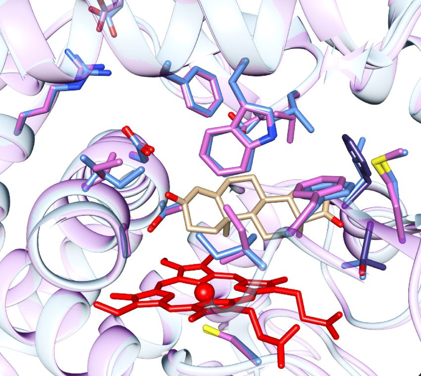
Int. J. Mol. Sci. 2021, 22, 631                                                                                            2 of 16

                                        The enzyme is present in all vertebrates as the product of expression of a single gene,
                                  with some exceptions represented by pigs and teleosts, where duplication events have pro-
                                  duced three and two isoforms, respectively [10–13]. Furthermore, the protein is expressed
                                  in different tissues in vertebrates, where it plays an essential role in reproductive biology
                                  as estrogens are responsible for ovarian differentiation, development of the reproductive
                                  system, sex differentiation, and reproduction [14]. Moreover, a critical role of estrogens
                                  has also been demonstrated in brain, bone, skin, fat, and cardiovascular tissues [15–20]. In
                                  humans, tissue-specific regulation of aromatase gene expression is allowed by the presence
                                  of eleven promoters and alternative first exons [21]. However, a wide tissue distribution
                                  of the aromatase protein and a complex regulatory region in its gene is already present in
                                  fishes [22].
                                        Vertebrates have been used as models to understand the roles of aromatase and es-
                                  trogens in the different tissues where it is expressed. For example, in birds and mammals,
                                  it has been demonstrated that in the brain, there is a rapid modulation of aromatase
                                  activity through phosphorylation and that estrogens can be considered neurotransmit-
                                  ters [23]. Moreover, estrogens are involved in different processes, such as neurogenesis,
                                  neuroprotection, and cognition [22,24].
                                        In reptiles and amphibians, temperature regulates aromatase expression and is re-
                                  sponsible for temperature-dependent sex determination [25–27]. In some hermaphrodite
                                  fishes, sex changes occur in response to environmental cues related to social interactions,
                                  and aromatase is involved in the remodeling of the gonads during this process [28,29]. Due
                                  to the phenotypic effects as a consequence of androgen/estrogen unbalance, amphibians
                                  and fishes are widely used as model organisms to understand the possible effect of many
                                  compounds that also target human aromatase [30,31], known as endocrine-disrupting
                                  chemicals (EDCs) [32,33].
                                        Among fishes, teleosts represent the only case where two isoforms are present (CYP19A1
                                  and CYP19B1), and they are preferentially expressed in the gonads and brain, respectively.
                                  Interestingly, these isoforms have also been reported to have different catalytic activity
                                  in comparison to the human enzyme [34,35], indicating that functional differences can be
                                  present. Thus, it is interesting to understand the phylogenetic origins of these differences.
                                        In this work, comparative sequence and structural analysis are used to investigate if
                                  and how the substrate-selective nature of aromatase has evolved, both in structural and
                                  functional terms. Its highly substrate-selective nature, calibrated for catalysis on androgens,
                                  makes it an optimal candidate for evolutionary studies, with the aim of (1) understanding
                                  if and how molecular evolution has structurally optimized this enzyme in order to make it
                                  more efficient and (2) determining what the conserved structural scaffold is and which are
                                  the amino acids that are essential for its function. Moreover, by identifying the functional
                                  amino acids that have not changed during evolution and excluding the ones shared with
                                  the other P450s, it is possible to obtain the fingerprint sequences of this enzyme. Structural
                                  analysis also allows us to identify a possible role for these residues and the rational basis
                                  for conservation. The most different aromatase sequences were also subjected to homology
                                  modeling to visualize where evolution has structurally modified the enzyme.

                                  2. Results
                                  2.1. Multiple Sequence Alignment
                                  2.1.1. Structural Conservation
                                       In order to identify the most conserved structural elements in aromatase, 365 se-
                                  quences, ranging from invertebrates to mammals, were used for multiple sequence align-
                                  ment. Out of the 365 sequences aligned, 66 were from mammals, 8 from birds, 12 from
                                  reptiles, 18 from amphibians, 259 from fishes, and 2 from the invertebrates of the genus
                                  Branchiostoma.
                                       For all the analyses performed in this work, the residue numbers refer to the sequence
                                  of human aromatase (CYP19A1, Uniprot ID P11511).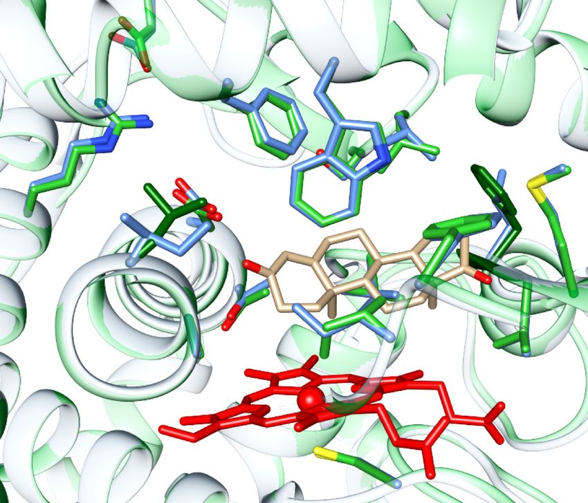
solvent. Thus, the conserved structural core in cytochrome P450 is formed by a four-helix
bundle formed by helices D, E, I, and L that is conserved among aromatase sequences; an
exception is made for the residues of helix D, exposed to the solvent (Figure 1) [36]. Helix
G is not conserved, whereas the F-G loop and the first part of helix F, known to be im-
Int. J. Mol. Sci. 2021, 22, 631
portant for opening the access channel in cytochrome P450, are conserved. 3 of 16
The key cysteine residue coordinating heme iron is obviously conserved in all the
sequences, and it is within a consensus sequence formed by FGFGPRX1CX2GK/R, where
X1 is variable (G, A, S, T, the
When or N) whereas
positions ofXthe
2 is A, V, L, or I. This consensus sequence is also
most conserved regions were analysed in the crystal struc-
well-conserved in cytochrome P450 (FXXGX(H/R)XCXG),
ture of the human enzyme, they resulted as part together
of helix with the meander
A (65–78), re- formed by
the β-sheet
gion, a loop strands
preceding the cysteine residue [36], which is also well-conserved in most
β1 (83–88) and β2 (93–97), helix E (187–205), part of helix F (221–224), the central
aromatase sequences.
part of helix I (residues 302–318 in human aromatase), helix K (354–366), the K-β3 loop
The three Arg
and theresidues
β3 strandinvolved in salt
(368–376), the bridges
β6 strandwith heme propionyl
(393–396), and helix groups (R115,
L and part of the L-K” loop
R145, and R435 in human aromatase) are also present in all the sequences, together
(427–448) (Figure 1). Helices C, D, F, and H carry conserved amino acids oriented toward with
Trp141, and aretheinvolved
core of thein protein
an H-bondandwith the heme propionyl
nonconserved amino acids group.
exposed to the solvent. Thus, the
A highlyconserved
conservedstructural
motif in cytochrome P450 is the EX
core in cytochrome P450 is formed1 X2 R motifbylocated in helix
a four-helix K formed by
bundle
and involvedhelices
in saltD, bridge interactions that are important for its tertiary structure
E, I, and L that is conserved among aromatase sequences; an exception is madeand
the correct incorporation
for the residues of of
the heme
helix cofactor to
D, exposed [36].
the This
solventmotif is conserved
(Figure in all
1) [36]. Helix G isse-
not conserved,
quences; X1 iswhereas
a serine the
residue, whereas
F-G loop X2 isfirst
and the L or M in
part of most
helix aromatase
F, known to sequences.
be important for opening the
access channel in cytochrome P450, are conserved.
(a) (b)
Figure
Figure 1. Crystal 1. Crystal
structure structure
of human of human(PDB
aromatase aromatase (PDBcolored
ID 4KQ8), ID 4KQ8), coloredtoaccording
according to the conser-
the conservation. The violet areas
vation. The violet areas correspond to the more conserved regions, whereas the dark green ones
correspond to the more conserved regions, whereas the dark green ones correspond to the most variable. Heme is shown in
correspond
red and the substrate to the most variable.
androstenedione in lightHeme
brown.is (a)
shown in red
Overall and theofsubstrate
structure androstenedione
human aromatase. (b) Theincore
light
structure of
brown. (a) Overall structure of human aromatase. (b) The core structure of aromatase, carrying the
aromatase, carrying the most conserved regions. The residues important for substrate binding are also shown.
most conserved regions. The residues important for substrate binding are also shown.
The key cysteine residue coordinating heme iron is obviously conserved in all the
2.1.2. Functional Conservation
sequences, and it is within a consensus sequence formed by FGFGPRX1 CX2 GK/R, where
The levelX1ofisconservation
variable (G, ofA,amino acids
S, T, or that are relevant
N) whereas X2 is A, for substrate
V, L, binding
or I. This and sequence is
consensus
catalysis wasalso
thenwell-conserved
verified in theinmultiple alignments.
cytochrome A highly conservedtogether
P450 (FXXGX(H/R)XCXG), alcohol–acid
with the meander
pair is present on helix
region, I in preceding
a loop cytochrome theP450, and residue
cysteine it is part[36],
of the proton
which relay
is also network
well-conserved in most
that allows the formation
aromatase of the reactive intermediate (Compound I) in the catalytic cycle.
sequences.
The three Arg residues involved in salt bridges with heme propionyl groups (R115,
R145, and R435 in human aromatase) are also present in all the sequences, together with
Trp141, and are involved in an H-bond with the heme propionyl group.
A highly conserved motif in cytochrome P450 is the EX1 X2 R motif located in helix K
and involved in salt bridge interactions that are important for its tertiary structure and the
correct incorporation of the heme cofactor [36]. This motif is conserved in all sequences; X1
is a serine residue, whereas X2 is L or M in most aromatase sequences.
2.1.2. Functional Conservation
The level of conservation of amino acids that are relevant for substrate binding and
catalysis was then verified in the multiple alignments. A highly conserved alcohol–acid
pair is present on helix I in cytochrome P450, and it is part of the proton relay network that
allows the formation of the reactive intermediate (Compound I) in the catalytic cycle. In
aromatase, the alcohol–acid pair is formed by an aspartic acid residue (D309 in human
aromatase) and a threonine residue (T310) that are conserved (exception is made for two
fish sequences), and they are preceded by a proline residue (P308) in all the sequences
analyzed. When compared to other P450s, this proline residue is unique to aromatase,
and it is responsible for the shift of the I-helix axis observed in the crystal structure of
the human enzyme [37]. Such a shift is important as it allows the 3-keto moiety of the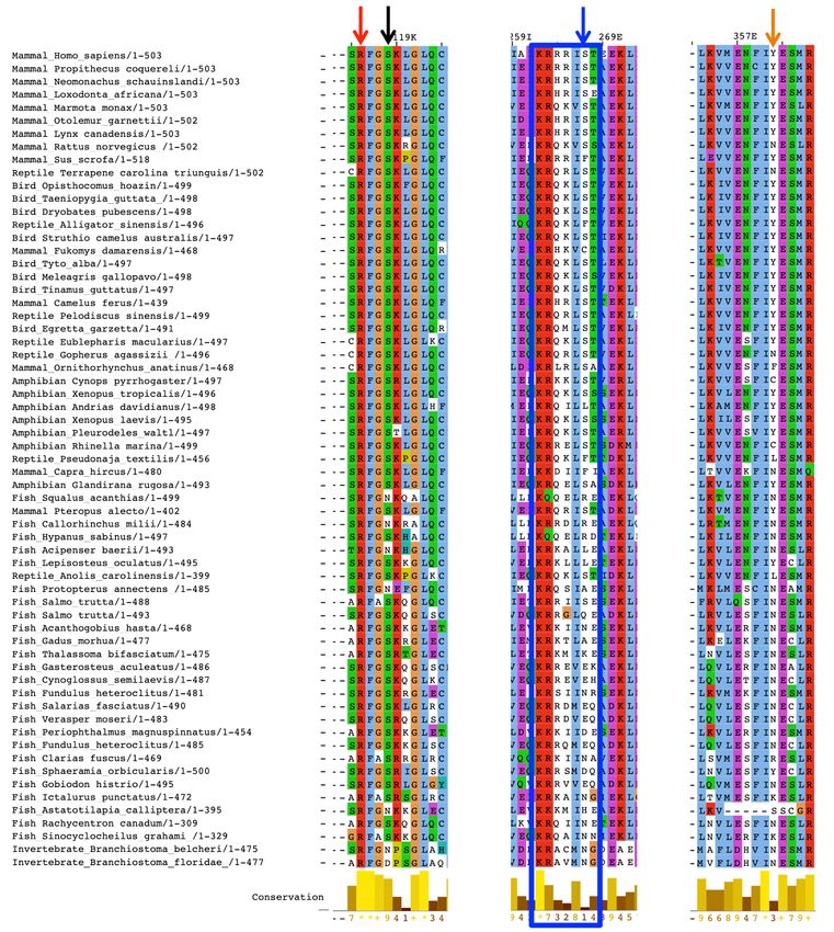
Int. J. Mol. Sci. 2021, 22, 631                                                                                                          4 of 16

                                          substrate androstenedione to be accommodated near the fifth turn of the I-helix that is
                                          formed by M303 and A307. These two residues are conserved, with some exceptions. The
                                          methionine is substituted by an isoleucine in five fish sequences, one amphibian sequence,
                                          and one mammal sequence; there is an alanine residue that is a glycine residue in 4.5%
                                          of fish sequences and in two invertebrates. Moreover, the shift of the I-helix allows the
                                          formation of a hydrogen bond between D309 and the 3-keto oxygen of the substrate. Such
                                          an aspartic acid residue has never been changed into a glutamic acid during evolution due
                                          to its important role in substrate binding and catalysis [38]. All these residues (303–310) are
                                          located on helix I, and they are part of one of six substrate recognition sites (SRSs), namely,
                                          SRS-4. The residues involved in androstenedione binding are highly conserved, with some
                                          exceptions represented by few fish sequences (Table 1).

      Table 1. Conservation of the residues involved in substrate binding and catalysis in human aromatase. The scores are
      normalized so that the average score for all residues is zero and the standard deviation is one. The lowest score represents
      the most conserved position in a protein. For reference, the lowest score associated with a fully conserved residue was
      −1.103, whereas the highest score obtained for a nonconserved residue in human aromatase was +2.844.

       Residue                       Location            Conservation Score                                Notes
         C437                     K”-L helix loop              −1.095
          I305                        I-helix                  −0.936                     L/V only in invertebrate Branchiostoma
         A306                         I-helix                  −1.002                         T in the mammal Capra hircus
         D309                         I-helix                  −1.058                 Q in CYP19B1 of the fish Halichoeres tenuispinis
         T310                         I-helix                  −1.011                           I in the fish Maylandia zebra
         F221                         F-helix                  −0.805
         W224                         F-helix                  −0.896
          I133                       B-C loop                  −1.038                         M in pig aromatase isoform 3
         F134                        B-C loop                  −1.073
         V370                 K-helix—β3 loop                  −1.001
         L372                 K-helix—β3 loop                  −0.202                                  Phe in fishes
                                                                                     S/ T in most fishes and in CYP19A1 of zebrafish
         V373                 K-helix—β3 loop                  −0.583
                                                                                                       and goldfish
         M374                           β3                     −1.031
         L477                      β8–β9 loop                  −1.011
                                                                                     A in many sequences, starting from mammals to
          S478                     β8–β9 loop                  −0.828
                                                                                                amphibians. S in fishes.
                                                                                   C or H in some mammals, birds and fishes, including
         R192                        Helix E                   −0.974
                                                                                               the two isoforms of zebrafish
         E483                      β9–β10 loop                 −0.761             Conserved in the two isoforms of zebrafish and goldfish

                                               Two other residues are important for aromatase function; they are predicted to be part
                                          of the proton relay network that allows the formation of the reactive Compound I in the
                                          typical P450 catalytic cycle: R192 and E483. These residues form a salt bridge in the same
                                          position as the one found in the crystal structure of the bacterial cytochrome P450cam [39].
                                          The residues R192 and E483 are highly conserved, starting from the sequences of aromatase
                                          from invertebrates. The crystal structure of the bacterial camphor-hydroxylating P450cam
                                          from Pseudomonas putida shows that this salt bridge is broken when the P450cam interacts
                                          with the redox partner that stabilizes the open conformation of the enzyme, exerting an
                                          effector role [39–41]. For human aromatase, the redox partner cytochrome P450 reductase
                                          (CPR) has been shown to promote substrate binding, acting as an effector [42], and the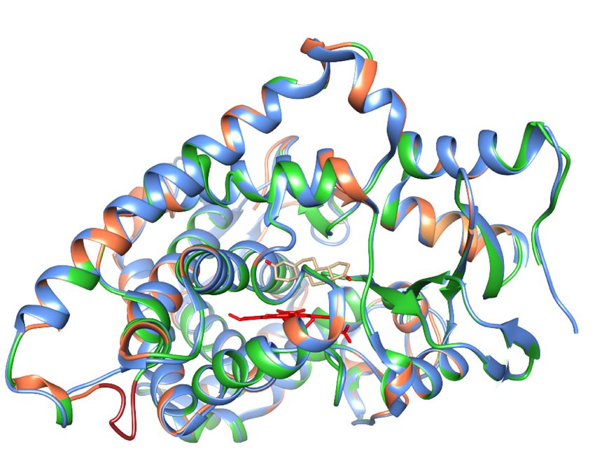
Int. J. Mol. Sci. 2021, 22, 631 5 of 16
presence of the R192-E483 salt bridge in the same structural position as P450cam suggests
that a similar effect can be exerted by its redox partner CPR.
2.1.3. Conservation of the Substrate Recognition Sites (SRSs)
Six regions have been identified to be important for substrate recognition and binding
in P450s: these are the so-called substrate recognition sites (SRSs). They are considered to
be the most variable regions among cytochrome P450 as their variation during evolution is
associated with new substrate selectivity. According to this idea, it is expected that the SRSs
of aromatase, a nonpromiscuous enzyme that is highly selective for androgen substrates,
have been highly conserved during evolution. Thus, the level of conservation of the six
SRSs was checked and is shown in Table S1. As it can be seen, SRS-4 is the most highly
conserved one (69.7% of the amino acids are conserved) as it carries amino acids crucial for
catalysis, whereas SRS-3 has been highly variable during aromatase evolution (15.4% of
conserved amino acids). In the other SRSs, about 40% of the amino acids are conserved.
As mentioned before, some residues in SRSs are shared in all P450s as they are essential
for their catalysis. For example, in SRS-4, the acid–alcohol pair is not unique for aromatase
as it is part of the proton relay network that allows the formation of reactive intermediates.
Thus, in order to identify the residues that are conserved and unique for aromatase in
the SRSs, multiple structural alignments of the 57 human P450s were performed using
the server PROMALS3D. For structural alignment, the server uses the crystal structures
available; their PDB IDs are used as input. When the structures are not available, the input
sequences are aligned after secondary structure prediction, and 3D structure constraints are
assigned based on homolog structures [43]. The multiple alignments obtained were then
evaluated by the ConSurf server to assign a conservation score for each amino acid position.
Table S2 shows the residues belonging to the six SRSs in aromatase and the corre-
sponding conservation score obtained from the alignment of the 363 sequences analyzed.
Moreover, it shows the conservation score for the same positions obtained from the align-
ment with all the other human P450s. This comparison was performed to identify the
residues conserved in the SRSs of all the human enzymes (shown in green in Table S2) and
the ones specific for aromatase (shown in red in Table S2).
In SRS-1, helix C carries a Trp residue (W141 in aromatase) that is an aromatic amino
acid in all P450s, important for heme binding. In many of them, it is followed by a positively
charged residue (present in all CYP2, CYP3, and CYP26 members). R145 is conserved
in most P450s as it is involved in heme binding, and the last two residues are small
hydrophobics in many of them. K150/A151 are conserved and specific for aromatase. The
helix B region is highly variable in human P450s. In aromatase, M127 is conserved as it
delineates the active site cavity, whereas N135 is part of an H-bond network also involving
R435, important for heme binding. The role of N135 is important as it bridges G131 and
N137, keeping the B-C loop in a conformation that allows the highly conserved I133 and
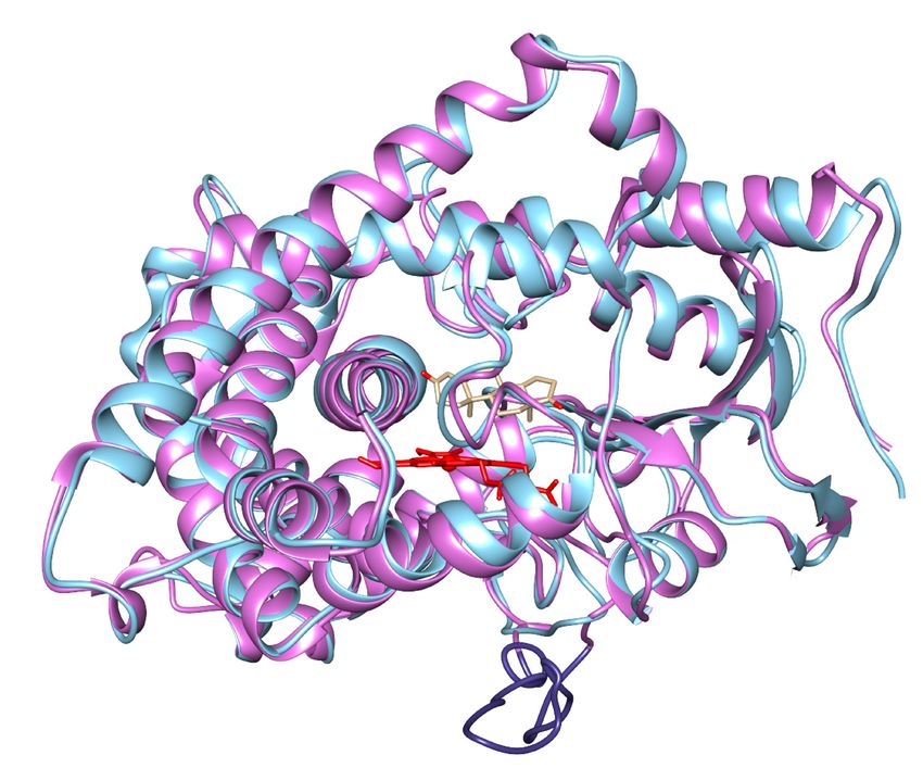
F134 to be part of the active site and to contact the substrate (Figure 2a).
SRS-2 is highly variable in P450s, and it carries conserved residues in aromatase. They
are located on helix F and on the F-G loop. They are important flexible elements in P450s,
including aromatase [44], as they are involved in the conformational changes that allow
ligand access to the active site [45]. Out of them, Tyr220 forms an important H-bond with
N295 that is part of SRS-4 and, with I125, defines the substrate access channel (Figure 2b).
In SRS-3, the cluster of three basic residues is not specific for aromatase as it is present
in all CYP4F members, CYP46A1, and, within the same helix (helix G), CYP51. Interestingly,
a glutamic acid is present before the cluster in all CYP4F members. EK is also present
in some CYP26/27 members. Interesting, all these P450 families are involved in steroid,
leukotriene, and retinoic and fatty acid processing [46–50].
SRS-4 and SRS-5 are the most conserved in human P450s. However, there are residues
specific for aromatase, including I305 and M374, that are involved in substrate binding. In
SRS5, the consensus sequence XEXXR is well conserved.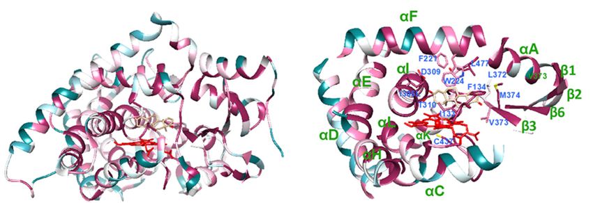
Int. J. Mol. Sci. 2021, 22, x FOR PEER REVIEW 6 of 17
Int. J. Mol. Sci. 2021, 22, 631 6 of 16
(a) (b)
Figure
Figure 2.
2. Role
Roleofofthe
thehighly
highlyconserved
conservedresidues
residuesin in
human
human aromatase
aromatase(PDB
(PDBID ID
4KQ8).
4KQ8).(a) Involvement of the
(a) Involvement ofhighly con-
the highly
served N135
conserved in bridging
N135 G131
in bridging and
G131 N137
and N137viavia
H-bonds
H-bonds (shown
(shownininorange),
orange),which
whichisisimportant
importantto tomaintain
maintain the
the B-C
B-C loop
loop
(blue) conformation and provide M127 and F134 to the active site of the protein. The H-bond network is shown in black.
(blue) conformation and provide M127 and F134 to the active site of the protein. The H-bond network is shown in black. (b)
(b) Involvement of the highly conserved Y220 in H-bonds that connect N295 and I125. N295 is part of SRS-4, shown in
Involvement of the highly conserved Y220 in H-bonds that connect N295 and I125. N295 is part of SRS-4, shown in orange,
orange, Y220 is part of SRS-2, shown in yellow, and SRS-3 is shown in magenta.
Y220 is part of SRS-2, shown in yellow, and SRS-3 is shown in magenta.
SRS-2 is highly variable in P450s, and it carries conserved residues in aromatase.
SRS-6 carries two His residues that are conserved in aromatase sequences that are part
They are located
of a β-hairpin, on helix
whereas F other
the and on the F-Gare
residues loop.
notThey are important flexible elements in
conserved.
P450s, including aromatase [44], as they are involved in the conformational changes that
allow ligand access
2.1.4. Consensus to thefor
Sequence active site [45]. OutModifications
Post-Translational of them, Tyr220 forms an important
H-bond with N295 that is part of SRS-4
Post-translational modifications on human aromatase and, with I125,havedefines
beenthe substrate
reported access
to alter its
channel (Figure
activity [51–54]. 2b).
In
TheSRS-3,
regionthebetween
cluster ofaminothreeacids
basic262residues
and 268 is not
is a specific
consensus for sequence
aromatasefor as different
it is pre-
sent in all CYP4F members, CYP46A1, and, within the same
kinases such as PKA (R-X1–2 -S/T-X) and PKG ((R/K)2–3 -X-S/T-X). In human aromatase, helix (helix G), CYP51. In-
terestingly, a glutamic acid is present before the cluster in all CYP4F
the sequence is KRRRIST, where a cluster of four basic residues gives a positively charged members. EK is also
present
patch oninthe some CYP26/27
surface that canmembers. Interesting,
attract opposite all these
charges. P450 families
However, only Kare andinvolved
the first in R
steroid, leukotriene, and retinoic and fatty acid processing [46–50].
are highly conserved, whereas the S and T residues are not conserved in fishes and the
SRS-4 and SRS-5
two invertebrate are the
sequences most 3).
(Figure conserved
This meansin human
that theP450s. However,
consensus there
sequence forare
PKA resi-
is
dues
presentspecific
starting forfrom
aromatase,
amphibians. including
On theI305otherandhand, M374, that are involved
the consensus for PKG thatin substrate
includes
binding.
two or threeIn SRS5,
basicthe consensus
residues sequence
is present XEXXR
in only 15% of is well
mammal conserved.
aromatase sequences.
SRS-6
The other residue reported to be phosphorylated is S118,aromatase
carries two His residues that are conserved in which is very sequences that are
well conserved,
part of a β-hairpin, whereas the other residues are not conserved.
together with an arginine residue presenting two amino acids before (R116). The only
exceptions are represented by six aromatase sequences from fishes and the two from
2.1.4. Consensus
invertebrates Sequence
where serinefor is Post-Translational
substituted by N Modifications
or D (Figure 3). Thus, this consensus
sequence for PKA is present starting from vertebrates.
Post-translational modifications on human aromatase have been reported to alter its
The[51–54].
activity other important residue known to be phosphorylated is Y361. This residue is
present in most mammal
The region between sequences
amino acids (83%)
262 and
and appears
268 is a in amphibians,
consensus wherefor
sequence it isdifferent
present
in 75% of
kinases suchtheassequences.
PKA (R-X1–2 In-S/T-X)
mammals,and PKGwhere it is not
((R/K) present, In
2–3-X-S/T-X). it is substituted
human by N,the
aromatase, as
in most fishes,
sequence where awhere
is KRRRIST, tyrosine residueofisfour
a cluster found onlyresidues
basic in 2.8% gives
of theasequences
positivelyanalyzed
charged
(Figure
patch on3).the surface that can attract opposite charges. However, only K and the first R are
highly conserved, whereas the S and T residues are not conserved in fishes and the two
2.1.5. Interaction with the Redox Partner
invertebrate sequences (Figure 3). This means that the consensus sequence for PKA is
Thestarting
present interaction
fromofamphibians.
P450s with On
theirthe
redox
otherpartner
hand, is
thecrucial for their
consensus function
for PKG thatand
in-
catalytic
cludes efficiency.
two or threeThe docking
basic siteisofpresent
residues cytochrome P450
in only reductase
15% (CPR)aromatase
of mammal and the P450
se-
enzyme is the proximal side, and it is mainly triggered by electrostatic interactions between
quences.
the positively charged surface of P450s and the negatively charged surface of CPR [55–57].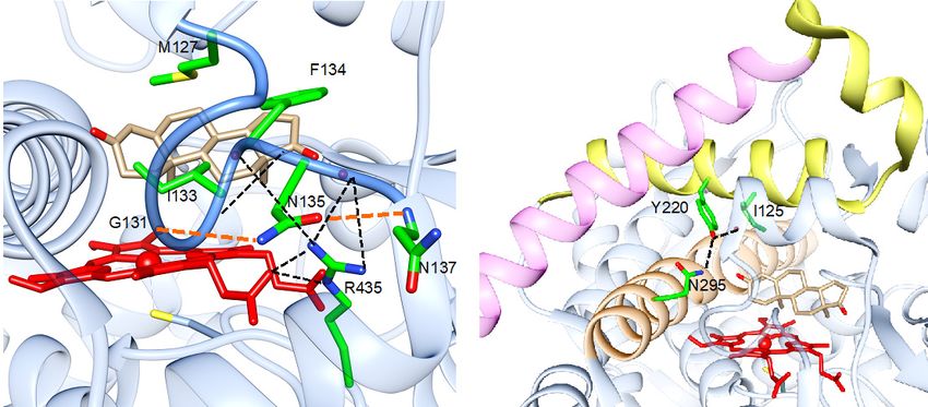
Int. J. Mol. Sci. 2021, 22, x FOR PEER REVIEW                                                                                           7 of 17

                                          The other residue reported to be phosphorylated is S118, which is very well con-
Int. J. Mol. Sci. 2021, 22, 631                                                                                                      7 of 16
                                    served, together with an arginine residue presenting two amino acids before (R116).                 The
                                    only exceptions are represented by six aromatase sequences from fishes and the two from
                                    invertebrates where serine is substituted by N or D (Figure 3). Thus, this consensus se-
                                    quence
                                    For      for PKAmany
                                        aromatase,      is present
                                                             basic starting
                                                                     residuesfrom
                                                                               havevertebrates.
                                                                                    been identified and suggested to be involved in
                                          The   other  important    residue  known    to be phosphorylated
                                    the interaction with CPR by site-directed mutagenesis         experiments is    Y361.
                                                                                                                  [58] and This  residue is
                                                                                                                           computational
                                    present   in most   mammal    sequences    (83%)  and  appears   in amphibians,     where it
                                    studies [59–61]. The conservation of these residues was checked in the multiple alignments,  is present
                                    in 75%
                                    and  the of the sequences.
                                             results  are shownIn  inmammals,     where
                                                                       Table 2. The      it is not present,
                                                                                    conservation    score is it is substituted
                                                                                                             included          byposition,
                                                                                                                        for each   N, as in
                                    most fishes,
                                    together   withwhere    a tyrosine
                                                    the result           residue
                                                               of the visual      is found
                                                                              analysis       only inus2.8%
                                                                                       that allows           of the the
                                                                                                        to identify  sequences
                                                                                                                         sequencesanalyzed
                                                                                                                                     where
                                    (Figure  3).
                                    the amino acids are not conserved.

       Figure3.3. Multiple
      Figure      Multiple sequence
                            sequence alignments
                                     alignments showing
                                                  showing only
                                                             only representative
                                                                   representative aromatase
                                                                                  aromatase sequences
                                                                                             sequences from
                                                                                                        from the
                                                                                                              the different
                                                                                                                  different classes
                                                                                                                             classes of
                                                                                                                                     of
       vertebrates and
      vertebrates  and the
                        thetwo
                             twoinvertebrates.
                                 invertebrates. The
                                                The three
                                                     three regions
                                                            regions shown
                                                                     shown are
                                                                            arethe
                                                                                theones
                                                                                    onescarrying
                                                                                         carryingthe
                                                                                                  thephosphorylation
                                                                                                      phosphorylationsites:
                                                                                                                         sites: S118
                                                                                                                                S118 isis
       indicated by the black arrow, and R116, which forms the consensus for PKA, is indicated by the red arrow. S267 is indi-
      indicated by the black arrow, and R116, which forms the consensus for PKA, is indicated by the red arrow. S267 is indicated
      by the blue arrow, and the cluster KRRIST, present on human aromatase, is shown in the blue box. Y361 is indicated by the
      orange arrow.
Int. J. Mol. Sci. 2021, 22, 631 8 of 16
Table 2. Conservation of the residues involved in the interaction with the redox partner in human aromatase. The scores are
normalized, so that the average score for all residues is zero and the standard deviation is one. The lowest score represents
the most conserved position in a protein. For reference, the lowest score associated with a fully conserved residue was
−1.103, whereas the highest score obtained for a nonconserved residue in human aromatase was +2.844.
Residue Conservation Score Notes
R in most fishes and Branchiostoma
K99 0.287 floridae, not conserved in 1 amphibian, 1 reptile, in 15% of fishes and
Branchiostoma belcheri (S)
K108 −0.024 Always substituted by R
R145 −0.972 Well conserved
K352 1.293 Conserved only in mammals
Not conserved in invertebrates (P) and 70% of fishes (including
K389 0.767
only isoform CYP19A1 in zebrafish)
K390 −0.231 K or R
Not conserved in two mammals, 20% of fishes (including CYP19B1
K420 0.472
of zebrafish) and E in invertebrates
Well conserved with some exceptions in fishes and the
R425 −0.881
invertebrates (T)
K440 −0.897 R in invertebrates
S153 −0.533 T in invertebrates and most fishes
Q351 0.799 Conserved in 90% of mammals
Y424 0.308 Conserved in mammals
Conserved in mammals and amphibians, H in 97% of fishes and T
Y441 −0.553
in invertebrates
Out of the nine basic residues that form the positively charged proximal site (Figure S1),
six are conserved as their mutation, when present, is conservative. The other three residues
appear during evolution at different times, as K352 is conserved in mammals and K389 and
K420 are well-conserved starting from amphibians. Concerning the four residues predicted
to form hydrogen bonds with CPR, two of them are conserved, and, interestingly, Q351
and Y424 are conserved only in mammals.
These data indicate that a patch of basic amino acids had already appeared in inver-
tebrates, and it has been highly conserved during evolution. However, other residues
were introduced later; these comprise the amino acids that reinforce the positively charged
proximal site as well as two residues that protrude from the proximal site of the enzyme
(Figure S1) to form H-bonds with the redox partner. These data suggest that the interaction
with the redox partner has been one of the driving forces for evolution in aromatase.
2.2. Homology Modeling of Evolutionarily Old Aromatase
Based on the sequence alignment, homology modeling was applied to two aromatase
sequences as it was found that they carry significant insertions, in addition to mutations, in
key positions.
The invertebrate aromatase sequence from Branchiostoma floridae was selected as it
shows an amino acid insertion, 40% of identity, and 60% of homology with the human one.
Thus, a homology model was built to study where the main differences between the two
aromatase enzymes are located.
A six-amino-acid insertion is present in the invertebrate sequence compared to all
the other sequences analyzed (between M276 and D277 in human aromatase), and the
model shows that such an insertion elongates the loop connecting helices H’ and the H
loop (Figure 4). Moreover, the analysis of the location of the substitutions shows that they
are all on the protein surface and on structural elements such as helix G, which are the leastone. Thus, a homology model was built to study where the main differences between the
two aromatase enzymes are located.
A six-amino-acid insertion is present in the invertebrate sequence compared to all
the other sequences analyzed (between M276 and D277 in human aromatase), and the
Int. J. Mol. Sci. 2021, 22, 631 model shows that such an insertion elongates the loop connecting helices H’ and the 9Hof 16
loop (Figure 4). Moreover, the analysis of the location of the substitutions shows that they
are all on the protein surface and on structural elements such as helix G, which are the
least conserved ones in aromatase. There are no mutations in the core structure of the
conserved ones in aromatase. There are no mutations in the core structure of the protein
protein and the active site, indicating that the main structural scaffold of aromatase was
and the active site, indicating that the main structural scaffold of aromatase was already
already present in this old protein. Moreover, many mutations are located in the SRSs,
present in this old protein. Moreover, many mutations are located in the SRSs, indicating
indicating that these areas have evolved in vertebrates.
that these areas have evolved in vertebrates.
(a) (b)
E483
F221
W224
R192 L477
L372F
D309
M374
F134
I305L
T310 I133
V373
E483
F221
R192 W224
L477
D309 L372F
I305 F134 M374
T310
I133
V373T
(c) (d)
Figure 4. Homology models of evolutionarily old aromatase. (a) Homology model for aromatase from the invertebrate
Branchiostoma floridae (green) superimposed onto the crystal structure of human aromatase (blue). The nonconserved
regions are shown in orange, and the grey shadow shows the location of the insertion. (b) Zoomed-in view of the active
site showing the conserved (green) and nonconserved residues (dark green) involved in substrate binding and catalysis.
(c) Homology model for aromatase from the pufferfish Takifugu rubripes (magenta) superimposed to the crystal structure of
human aromatase (blue). The grey shadow shows the location of the long insertion (violet). (d) Zoomed-in view of the
active site showing the conserved (magenta) and nonconserved residues (dark purple) involved in substrate binding and
catalysis. Heme is shown in red and the substrate androstenedione in light brown.
The multiple sequence alignment also shows the presence of some important muta-
tions together with a long insertion in aromatase from some fish species, including the
one from pufferfish Takifugu rubripes. In this case, the fish sequence shares 52% of identity
and 70% of homology with the human one. A homology model was built in order to
predict the possible effect of the substitutions found in the active site. Figure 4 shows the
model carrying a long insertion between N421 and V422, which corresponds to the loop
connecting helix K” and helix L.
Since this long insertion is modeled as a long loop, secondary structure prediction
tools were used to verify a possible elongation of the K” helix. However, both PsiPred
and I-Tasser servers did not predict any secondary structure formation for the amino acids
present in that loop. Such a result justifies the absence of such a long and not-necessary
loop in the other aromatase sequences.Int. J. Mol. Sci. 2021, 22, 631 10 of 16
Concerning the active site, while the substitution of L372 with a phenylalanine does
not seem to affect the polarity and dimensions of the catalytic pocket, the substitution of
V373 with the polar threonine residue, which in some species is a serine, can be predicted
to affect the polarity of the active site (Figure 4). As the substrate carries at least two
keto- (as in androstenedione) groups or one keto- group and one hydroxyl group (as in
testosterone), the presence of a serine/threonine residue can be predicted to possibly affect
the orientation and positioning of the substrate in the active site of the enzyme. Indeed, the
Thr/Ser residue could form a hydrogen bond with the substrate. Thus, this substitution
seems to be important to properly orient the substrate in the active site for efficient catalysis.
3. Discussion
Aromatase is a unique enzyme carrying out a three-step reaction on the androgen sub-
strate, with the third step leading to the aromatization of the A-ring of the steroid molecule.
This intriguing reaction has been the subject of many studies aimed at understanding
the mechanism of the third aromatization step [62,63]. Moreover, the crystal structure
of the human enzyme has indicated the amino acids within the protein matrix involved
in substrate binding and catalysis, and their role has been confirmed by site-directed
mutagenesis [64,65].
In this work, sequence and structural alignments were performed with aromatase
sequences available on databases. Unfortunately, the number of sequences for the different
classes of vertebrates is very different as most of the sequences are available from fishes
and mammals and, therefore, a bias is introduced in the conservation score. However, we
performed a qualitative analysis in order to see the effect of mutations in key positions
using the conservation score as an indicator for the level of conservation.
The multiple alignment shows that the enzyme structural scaffold and the key func-
tional residues have been highly conserved during evolution, with only few exceptions in
the aromatase sequences from fishes and invertebrates. Thus, the structural core elements
of the protein carrying the residues involved in substrate binding are evolutionarily old
and this is reasonable as they guarantee the specific function that aromatase has in species
conservation. On the other hand, while some SRSs have also been well-conserved during
evolution, SRS-3 has shown the lowest level of conservation (15% of the residues are highly
conserved). SRS-3 is located on helix G, a flexible element, which, together with helix F
and the F-G loop, is known to be involved in the opening and closure of the access channel
for the substrate. Interestingly, helix F and the F-G loop are much more conserved as they
belong to SRS-2, which shows 40% of the residues to be highly conserved. Out of the
conserved residues, we could identify the ones unique to aromatase, thanks to a structural
alignment with the other human P450 enzymes. The data show that some conserved and
unique amino acids, such as N135 and Y220, are involved in H-bond networks and have a
structural role that supports the positioning of the residues involved in substrate binding
in the active site.
A lower level of conservation is found in some of the amino acids that form the posi-
tively charged proximal side and in some other residues that are involved in the interaction
with the redox partner through the formation of H-bonds. This finding is very interesting
as CPR is shared between many P450 enzymes within the same organism. Moreover, we
have recently demonstrated that human CPR has an effector role as it facilitates substrate
binding by stabilizing the aromatase open conformation, which is optimal for substrate
access to the active site [42]. Thus, the data suggest that one of the driving forces for evo-
lution has been the optimization of the interface between aromatase and CPR in order to
make aromatase more competitive for the same shared redox partner. Such an optimization
involves the introduction of positively charged residues as well as amino acids that form
H-bonds and facilitate CPR binding, which, in turn, promotes catalysis.
The other interesting finding is the poor conservation of some residues known to be
involved in post-translational modifications. Phosphorylation is a rapid way to modulate
enzyme activity compared to regulation at the gene level. Aromatase activity is affectedInt. J. Mol. Sci. 2021, 22, 631 11 of 16
by phosphorylation, and some of the residues that can undergo this post-translational
modification have been identified [51–54]. Phosphorylation of S118 has been reported
to decrease aromatase activity in human cell lines [54]. The residue S118 is highly con-
served in aromatase sequences from vertebrates, together with R115, which forms the
consensus sequence for PKA. This consensus is missing in invertebrates and in few fish
sequences (3%).
Another consensus sequence for PKA, as well as for PKG, involves S267 and/or
Thr268. These residues are not present in fishes, whereas the consensus sequence for PKA
is present in amphibians. On the other hand, the consensus for PKG, which includes two
or three basic residues, has appeared late during evolution as it is present in only 15% of
the mammal aromatase sequences. Interestingly, this consensus sequence includes R264 in
human aromatase that is mutated into a Cys or His in some polymorphisms that are also
reported to alter aromatase activity when used in combination with polymorphic variants
of CPR [66]. Moreover, they have been associated with an increased risk for estrogen-
dependent pathologies such as breast cancer and polycystic ovary syndrome [67–70].
The other residue known to be phosphorylated is Y361, which appears in amphibians
but is not fully conserved even within mammals. Aromatase phosphorylation in this po-
sition has been associated with tumor progression in breast cancer cell lines [52]. Indeed,
short exposure to estradiol was found to increase aromatase activity through phosphoryla-
tion of a tyrosine residue (Y361) by c-Src kinase in estrogen-dependent MCF-7 breast cancer
epithelial cells. The authors hypothesized the presence of a positive nongenomic autocrine
loop between estradiol and aromatase in MCF-7 breast cancer cells [52]. Moreover, it was
also demonstrated that estradiol impairs the ability of the tyrosine phosphatase PTP1B to
dephosphorylate aromatase, resulting in increased aromatase activity and estrogen produc-
tion [71]. The multiple sequence alignment shows that the tyrosine residue in position 361,
located on helix K, which is one of the most conserved structural elements in aromatase,
appears in few fish species, but it is poorly conserved even among mammals, where it is
substituted by an asparagine residue, as in most fishes.
Taken together, the results of the conservation of the phosphorylation sites show that
evolution has introduced and is still introducing amino acids in key surface positions
that can be phosphorylated and consensus sequences in order to modulate aromatase
activity. Thus, the need for quickly and locally altering the estrogen concentration in
cells seems to be the other driving force for the evolution of this enzyme. This finding is
supported by the fact that a rapid regulation of aromatase activity is known to occur in
neurons [72,73] and teleost fishes express aromatase only in glial cells, indicating that the
ability to synthesize estrogens in neurons has been acquired during evolution [74,75]. In
the brain, the acquisition of phosphorylable sites may be explained by the need to modulate
estrogen production in higher vertebrate neurons, where rapid changes in estrogen levels,
as a consequence of aromatase phosphorylation, have been associated with important
physiological and behavioral responses [73].
It is interesting to note that if, on the one hand, the introduction of phosphorylation
sites can be evolutionarily beneficial, as in the case of brain aromatase, on the other hand,
phosphorylation of residues that increases aromatase activity can strengthen the negative
effects of estrogens, as in the case of breast cancer.
In conclusion, this study on aromatase shows that molecular evolution has worked
to maintain a high selectivity for a substrate-specific human cytochrome P450 such as
aromatase. However, based on the mutations introduced in key sites, it has been observed
that evolution has introduced residues that optimize the interaction with the redox part-
ner and phosphorylation sites that give the possibility of rapidly modulating its activity
through phosphorylation. It will be interesting to extend the study to other P450s that
are highly substrate-selective to understand how molecular evolution has worked for this
group of P450s.Int. J. Mol. Sci. 2021, 22, 631 12 of 16
4. Materials and Methods
4.1. Multiple Sequence and Structural Alignments
A total of 365 aromatase sequences from vertebrates and the one available from the
cephalochordate Branchiostoma were retrieved from the Uniprot database [76] using the
ConfSurf server [77] in two different searches. The first one included up to 500 sequences
closest to the human aromatase sequence, with at least 40% of identity from the reference
database “Clean Uniprot”. The second search was performed by searching for up to 500
sequences that sample the list of homologs to the query that was the sequence of human
aromatase. In this case, the minimal percentage of identity was 40%. The sequence was
extrapolated from the crystal structure (PDB ID 3S79) so that the server could automatically
calculate evolutionary conservation scores and map them on the aromatase structure [78].
These parameters were chosen on the basis that they allowed the retrieval of only aromatase
sequences that were manually verified.
Out of the 365 sequences aligned, 66 were from mammals, 8 from birds, 12 from
reptiles, 18 from amphibians, 259 from fishes, and 2 from the invertebrates of the genus
Branchiostoma.
The sequences were aligned through the HMMER algorithm [79] and visualized and
analyzed with Jalview software [80]. Position-specific conservation scores were computed
using the empirical Bayesian algorithm [81]. The scores were normalized so that the average
score for all residues was zero and the standard deviation was one. In aromatase, the lowest
score associated with a fully conserved residue was −1.103 (N135), whereas the highest
score obtained for a nonconserved residue was +2.844 (E181). The amino acid conservation
output, together with the structural conservation from ConSurf server, was checked by
visual inspection. Visual inspection is always needed to check correct alignment.
The substrate recognition sites (SRSs) in human aromatase were identified from a
structural alignment with the crystal structure of CYP2C8 (PDB ID 2NNJ) [82] performed
using the software UCSF Chimera [83]. Indeed, the SRSs were annotated [84] based on the
CYP2C family [85].
Structural alignments between aromatase and all the other human P450s were per-
formed using PROMALD3D, a multiple-structure-based alignment refined in combination
with sequence constraints [43]. The alignment took into account the crystal structures
available and the prediction of secondary structure elements for the unknown structures.
Once structurally aligned, the conservation score was assigned using the ConSurf server.
The structural analysis of the conserved amino acids was performed using UCSF
Chimera software that was also used for figure preparation [83].
4.2. Homology Modeling
Homology models were built using the software Modeller 9.25 [86], I-tasser [87],
and the crystal structure of human aromatase (PDB ID 3S79, 3EQM) as a template. The
best model was selected according to the Z-DOPE score, with energy minimized using
Amberff14SB forcefield [88] and subjected to validation using Molprobity [89], ProSA [90],
and QMEAN [91].
The homology model of aromatase from Branchiostoma floridae was obtained from
Modeller with a Z-DOPE score of −1.0. The validation from the ProSA server showed
a Z-score of −9.44 that is within the values of known 3D structures of similar length.
The QMEN4 value was −2.89, and the Ramachandran plot showed that 94% were in the
favored regions.
The homology model of pufferfish was first obtained from Modeller (Z-DOPE score
−1.23). The long insertion was modeled as a long loop, as expected. Thus, a secondary
structure prediction was carried out using the PSIPred server [92] and I-Tasser [87]. The
validation from the ProSA server showed a Z-score of −7.86, which is within the values
of known 3D structures of similar length, whereas the QMEAN4 value was −3.05. The
Ramachandran plot showed that 94.57% of the residues were in the favored regions.Int. J. Mol. Sci. 2021, 22, 631 13 of 16
Supplementary Materials: Supplementary materials can be found at https://www.mdpi.com/1422
-0067/22/2/631/s1.
Author Contributions: Conceptualization, G.D.N. and G.G.; methodology, G.D.N. and G.G.; soft-
ware, G.D.N., C.Z., and A.G.M.; validation, G.D.N., C.Z., and A.G.M.; formal analysis, G.D.N., C.Z.,
and A.G.M.; investigation, G.D.N., C.Z., and A.G.M.; resources, G.D.N. and G.G.; data curation, G.D.
and C.Z.; writing—original draft preparation, G.D.N.; writing—review and editing, G.D.N., C.Z., and
G.G.; visualization, G.D.N., C.Z., and A.G.M.; supervision, G.D.N. and G.G.; project administration,
G.D.N. and G.G.; funding acquisition, G.D.N. and G.G. All authors have read and agreed to the
published version of the manuscript.
Funding: This research received no external funding.
Institutional Review Board Statement: Not applicable.
Informed Consent Statement: Not applicable.
Data Availability Statement: The data presented in this study are available on request from the
corresponding author.
Conflicts of Interest: The authors declare no conflict of interest.
References
1. Thompson, E.A.; Siiteri, P.K. Utilization of oxygen and reduced nicotinamide adenine dinucleotide phosphate by human placental
microsomes during aromatization of androstenedione. J. Biol. Chem. 1974, 249, 5364–5372. [CrossRef]
2. Simpson, E.R.; Mahendroo, M.S.; Means, G.D.; Kilgore, M.W.; Hinshelwood, M.M.; Graham-Lorence, S.; Amarneh, B.; Ito, Y.;
Fisher, C.R.; Michael, M.D.; et al. Aromatase cytochrome P450, the enzyme responsible for estrogen biosynthesis. Endocr. Rev.
1994, 15, 342–355. [CrossRef] [PubMed]
3. Coon, M.J. Cytochrome P450: Nature’s most versatile biological catalyst. Annu. Rev. Pharmacol. Toxicol. 2005, 45, 1–25. [CrossRef]
[PubMed]
4. Guengerich, F.P. Cytochrome p450 enzymes in the generation of commercial products. Nat. Rev. Drug. Discov. 2002, 1, 359–366.
[CrossRef] [PubMed]
5. Di Nardo, G.; Gilardi, G. Natural compounds as pharmaceuticals: The key role of cytochromes p450 reactivity. Trends Biochem.
Sci. 2020, 45, 511–525. [CrossRef]
6. Nelson, D.R.; Goldstone, J.V.; Stegeman, J.J. The Cytochrome P450 Genesis Locus: The Origin and Evolution of Animal
Cytochrome P450s. Philos. Trans. R. Soc. Lond B Biol. Sci. 2013, 368, 20120474. [CrossRef]
7. Callard, G.V.; Tarrant, A.M.; Novillo, A.; Yacci, P.; Ciaccia, L.; Vajda, S.; Chuang, G.-Y.; Kozakov, D.; Greytak, S.R.; Sawyer, S.; et al.
Evolutionary origins of the estrogen signaling system: Insights from amphioxus. J. Steroid Biochem. Mol. Biol. 2011, 127, 176–188.
[CrossRef]
8. Callard, G.V.; Pudney, J.A.; Kendall, S.L.; Reinboth, R. In vitro conversion of androgen to estrogen in amphioxus gonadal tissues.
Gen. Comp. Endocrinol. 1984, 56, 53–58. [CrossRef]
9. Mizuta, T.; Kubokawa, K. Presence of sex steroids and cytochrome P450 genes in amphioxus. Endocrinology 2007, 148, 3554–3565.
[CrossRef]
10. Kishida, M.; Callard, G.V. Distinct cytochrome P450 aromatase isoforms in zebrafish (Danio rerio) brain and ovary are differentially
programmed and estrogen regulated during early development. Endocrinology 2001, 142, 740–750. [CrossRef]
11. Tchoudakova, A.; Kishida, M.; Wood, E.; Callard, G.V. Promoter characteristics of two cyp19 genes differentially expressed in the
brain and ovary of teleost fish. J. Steroid Biochem. Mol. Biol. 2001, 78, 427–439. [CrossRef]
12. Conley, A.J.; Corbin, C.J.; Hughes, A.L. Adaptive evolution of mammalian aromatases: Lessons from Suiformes. J. Exp. Zool. Part
A: Ecol. Genet. Physiol. 2009, 311, 346–357. [CrossRef] [PubMed]
13. Corbin, C.J.; Hughes, A.L.; Heffelfinger, J.R.; Berger, T.; Waltzek, T.B.; Roser, J.F.; Santos, T.C.; Miglino, M.A.; Oliveira, M.F.; Braga,
F.C.; et al. Evolution of suiform aromatases: Ancestral duplication with conservation of tissue-specific expression in the collared
peccary (Pecari tayassu). J. Mol. Evol. 2007, 65, 403–412. [CrossRef] [PubMed]
14. Hamilton, K.J.; Hewitt, S.C.; Arao, Y.; Korach, K.S. Estrogen hormone biology. Curr. Top. Dev. Biol. 2017, 125, 109–146. [CrossRef]
15. McEwen, B.S.; Milner, T.A. Understanding the broad influence of sex hormones and sex differences in the brain. J. Neurosci. Res.
2017, 95, 24–39. [CrossRef]
16. Galea, L.A.M.; Frick, K.M.; Hampson, E.; Sohrabji, F.; Choleris, E. Why estrogens matter for behavior and brain health. Neurosci.
Biobehav. Rev. 2017, 76, 363–379. [CrossRef]
17. Almeida, M.; Laurent, M.R.; Dubois, V.; Claessens, F.; O’Brien, C.A.; Bouillon, R.; Vanderschueren, D.; Manolagas, S.C. Estrogens
and androgens in skeletal physiology and pathophysiology. Physiol. Rev. 2017, 97, 135–187. [CrossRef]
18. Brincat, M.P.; Baron, Y.M.; Galea, R. Estrogens and the skin. Climacteric 2005, 8, 110–123. [CrossRef]
19. Knowlton, A.A.; Lee, A.R. Estrogen and the cardiovascular system. Pharmacol. Ther. 2012, 135, 54–70. [CrossRef]Int. J. Mol. Sci. 2021, 22, 631 14 of 16
20. Bernasochi, G.B.; Rupasinghe, T.T.W.; Bell, J.R.; Roessner, U.; Boon, W.C.; Delbridge, L.M.D. A novel mass spectrometric
methodology facilitates quantification of testosterone and progesterone, but not estrogens in cardiac and adipose tissues. J. Mol.
Cell. Cardiol. 2020, 140, 11–12. [CrossRef]
21. Wang, H.; Li, R.; Hu, Y. The alternative noncoding exons 1 of aromatase (Cyp19) gene modulate gene expression in a posttran-
scriptional manner. Endocrinology 2009, 150, 3301–3307. [CrossRef] [PubMed]
22. Piferrer, F.; Blázquez, M. Aromatase distribution and regulation in fish. Fish Physiol. Biochem. 2005, 31, 215–226. [CrossRef]
[PubMed]
23. Cornil, C.A.; Ball, G.F.; Balthazart, J. The dual action of estrogen hypothesis. Trends Neurosci. 2015, 38, 408–416. [CrossRef]
[PubMed]
24. Coumailleau, P.; Pellegrini, E.; Adrio, F.; Diotel, N.; Cano-Nicolau, J.; Nasri, A.; Vaillant, C.; Kah, O. Aromatase, estrogen receptors
and brain development in fish and amphibians. Biochim. Biophys. Acta 2015, 1849, 152–162. [CrossRef] [PubMed]
25. Pieau, C.; Dorizzi, M. Oestrogens and temperature-dependent sex determination in reptiles: All is in the gonads. J. Endocrinol.
2004, 181, 367–377. [CrossRef]
26. Matsumoto, Y.; Buemio, A.; Chu, R.; Vafaee, M.; Crews, D. Epigenetic control of gonadal aromatase (cyp19a1) in temperature-
dependent sex determination of red-eared slider turtles. PLoS ONE 2013, 8, e63599. [CrossRef]
27. Flament, S. Sex Reversal in Amphibians. Sex Dev. 2016, 10, 267–278. [CrossRef]
28. Nakamura, M.; Kobayashi, T.; Chang, X.-T.; Nagahama, Y. Gonadal Sex Differentiation in Teleost Fish. J. Exp. Zool. 1998, 281,
362–372. [CrossRef]
29. Sunobe, T.; Nakamura, M.; Kobayashi, Y.; Kobayashi, T.; Nagahama, Y. Aromatase immunoreactivity and the role of enzymes in
steroid pathways for inducing sex change in the hermaphrodite gobiid fish Trimma Okinawae. Comp. Biochem. Physiol. 2005, 141,
54–59. [CrossRef]
30. Baravalle, R.; Ciaramella, A.; Baj, F.; Di Nardo, G.; Gilardi, G. Identification of endocrine disrupting chemicals acting on human
aromatase. Biochim. Biophys. Acta Proteins Proteom. 2018, 1866, 88–96. [CrossRef]
31. Zhang, C.; Schilirò, T.; Gea, M.; Bianchi, S.; Spinello, A.; Magistrato, A.; Gilardi, G.; Di Nardo, G. Molecular basis for endocrine
disruption by pesticides targeting aromatase and estrogen receptor. Int. J. Environ. Res. Public Health 2020, 17, 5664. [CrossRef]
[PubMed]
32. Kloas, W. Amphibians as a model for the study of endocrine disruptors. Int. Rev. Cytol. 2002, 216, 1–57. [CrossRef] [PubMed]
33. Scholz, S.; Renner, P.; Belanger, S.E.; Busquet, F.; Davi, R.; Demeneix, B.A.; Denny, J.S.; Léonard, M.; McMaster, M.E.; Villeneuve,
D.L.; et al. Alternatives to in vivo tests to detect endocrine disrupting chemicals (edcs) in fish and amphibians—Screening for
estrogen, androgen and thyroid hormone disruption. Crit. Rev. Toxicol. 2013, 43, 45–72. [CrossRef] [PubMed]
34. Zhao, J.; Mak, P.; Tchoudakova, A.; Callard, G.; Chen, S. Different catalytic properties and inhibitor responses of the goldfish
brain and ovary aromatase isozymes. Gen. Comp. Endocrinol. 2001, 123, 180–191. [CrossRef] [PubMed]
35. Tong, S.K.; Chiang, E.F.; Hsiao, P.H.; Chung, B. Phylogeny, expression and enzyme activity of zebrafish cyp19 (P450 aromatase)
genes. J. Steroid Biochem. Mol. Biol. 2001, 79, 299–303. [CrossRef]
36. Hasemann, C.A.; Kurumbail, R.G.; Boddupalli, S.S.; Peterson, J.A.; Deisenhofer, J. Structure and function of cytochromes P450: A
comparative analysis of three crystal structures. Structure 1995, 3, 41–62. [CrossRef]
37. Ghosh, D.; Griswold, J.; Erman, M.; Pangborn, W. Structural basis for androgen specificity and oestrogen synthesis in human
aromatase. Nature 2009, 457, 219–223. [CrossRef]
38. Di Nardo, G.; Breitner, M.; Bandino, A.; Ghosh, D.; Jennings, G.K.; Hackett, J.C.; Gilardi, G. Evidence for an elevated aspartate
pKa in the active site of human aromatase. J. Biol. Chem. 2015, 290, 1186–1196. [CrossRef]
39. Tripathi, S.; Li, H.; Poulos, T.L. Structural basis for effect or control and redox partner recognition in cytochrome P450. Science
2013, 340, 1227–1230. [CrossRef]
40. Liou, S.-H.; Mahomed, M.; Lee, Y.-T.; Goodin, D.B. Effector roles of putidaredoxin on cytochrome P450cam conformational states.
J. Am. Chem. Soc. 2016, 138, 10163–10172. [CrossRef]
41. Hollingsworth, S.A.; Batabyal, D.; Nguyen, B.D.; Poulos, T.L. Conformational selectivity in cytochrome P450 redox partner
interactions. Proc. Natl. Acad. Sci. USA 2016, 113, 8723–8728. [CrossRef] [PubMed]
42. Zhang, C.; Catucci, G.; Di Nardo, G.; Gilardi, G. Effector role of cytochrome P450 reductase for androstenedione binding to
human aromatase. Int. J. Biol. Macromol. 2020, 164, 510–517. [CrossRef] [PubMed]
43. Pei, J.; Kim, B.H.; Grishin, N.V. PROMALS3D: A tool for multiple protein sequence and structure alignments. Nucleic Acids Res.
2008, 36, 2295–2300. [CrossRef] [PubMed]
44. Di Nardo, G.; Breitner, M.; Sadeghi, S.J.; Castrignanò, S.; Mei, G.; Di Venere, A.; Nicolai, E.; Allegra, P.; Gilardi, G. Dynamics
and flexibility of human aromatase probed by FTIR and time resolved fluorescence spectroscopy. PLoS ONE 2013, 8, e82118.
[CrossRef] [PubMed]
45. Poulos, T.L. Cytochrome P450 flexibility. Proc. Natl. Acad. Sci. USA 2003, 100, 13121–13122. [CrossRef]
46. Kalsotra, A.; Strobel, H.W. Cytochrome P450 4F subfamily: At the crossroads of eicosanoid and drug metabolism. Pharm. Ther.
2006, 112, 589–611. [CrossRef]
47. Mast, N.; Norcross, R.; Andersson, U.; Shou, M.; Nakayama, K.; Bjorkhem, I.; Pikuleva, I.A. Broad substrate specificity of human
cytochrome P450 46A1 which initiates cholesterol degradation in the brain. Biochemistry 2003, 42, 14284–14292. [CrossRef]You can also read