Listeria monocytogenes: The Impact of Cell Death on Infection and Immunity - MDPI
←
→
Page content transcription
If your browser does not render page correctly, please read the page content below
pathogens
Review
Listeria monocytogenes: The Impact of Cell Death
on Infection and Immunity
Courtney E. McDougal and John-Demian Sauer *          ID

 Department of Medical Microbiology and Immunology, School of Medicine and Public Health,
 University of Wisconsin-Madison, Madison, WI 53706, USA; cmcdougal@wisc.edu
 * Correspondence: sauer3@wisc.edu; Tel.: +608-263-1529; Fax: +608-262-8418

 Received: 30 November 2017; Accepted: 3 January 2018; Published: 11 January 2018

 Abstract: Listeria monocytogenes has evolved exquisite mechanisms for invading host cells and
 spreading from cell-to-cell to ensure maintenance of its intracellular lifecycle. As such, it is not
 surprising that loss of the intracellular replication niche through induction of host cell death has
 significant implications on the development of disease and the subsequent immune response.
 Although L. monocytogenes can activate multiple pathways of host cell death, including necrosis,
 apoptosis, and pyroptosis, like most intracellular pathogens L. monocytogenes has evolved a series
 of adaptations that minimize host cell death to promote its virulence. Understanding how
 L. monocytogenes modulates cell death during infection could lead to novel therapeutic approaches.
 In addition, as L. monocytogenes is currently being developed as a tumor immunotherapy platform,
 understanding how cell death pathways influence the priming and quality of cell-mediated immunity
 is critical. This review will focus on the mechanisms by which L. monocytogenes modulates cell death,
 as well as the implications of cell death on acute infection and the generation of adaptive immunity.

 Keywords: Listeria monocytogenes; cell death; necrosis; apoptosis; pyroptosis; cell-mediated immunity;
 CD8+ T-cells

1. Introduction
     Listeria monocytogenes is a Gram-positive facultative intracellular pathogen. Due in large part
to its ability to survive in both cold and high-salt conditions, it enters the food chain and can
lead to the severe disseminated infection Listeriosis [1]. Following ingestion, L. monocytogenes can
invade intestinal epithelial cells, gaining access to the lymphatic system and blood stream, ultimately
resulting in dissemination to the liver, spleen, central nervous system, and, in pregnant women,
the placenta. Infection causes symptoms ranging from mild gastroenteritis to more severe meningitis
and spontaneous miscarriage in the context of disseminated infections [2]. Disseminated listeriosis can
result in mortality rates as high as 30% despite antibiotic treatment [2].
     Following ingestion and upon entry into a host cell, either through phagocytosis
or internalin-dependent receptor mediated endocytosis [3,4], L. monocytogenes utilizes the
cholesterol-dependent cytolysin (CDC) listeriolysin O (LLO) to escape the phagosome into the
cytosol [5,6]. Once in the cytosol, L. monocytogenes expresses the protein ActA to hijack host actin,
thus facilitating cell-to-cell spread [7]. The combination of LLO and ActA results in an almost
exclusively intracellular lifecycle during infection, thereby avoiding extracellular host defenses,
including complement and neutrophils [8–10]. Indeed, loss of either LLO or ActA leads to full
attenuation of virulence demonstrating the importance of accessing and maintaining its intracellular
niche [5,7]. Furthermore, as discussed throughout this review, L. monocytogenes has developed multiple
strategies to maintain host cell viability and avoid triggering both programmed and non-programmed
host cell death pathways to promote its virulence.

Pathogens 2018, 7, 8; doi:10.3390/pathogens7010008                         www.mdpi.com/journal/pathogens
Pathogens 2018, 7, 8 2 of 17
In addition to being an important human and animal pathogen, L. monocytogenes is also being
developed as a novel vaccine platform, particularly in the context of tumor immunotherapy [11].
Due in large part to its constitutive intracellular lifecycle, L. monocytogenes infection naturally triggers
robust CD8+ T-cell responses [12]. While the exact mechanisms by which L. monocytogenes triggers
cell-mediated immunity remain unclear, its promise as an immunotherapy platform is illustrated
by the >15 active or completed clinical trials using attenuated L. monocytogenes for the treatment of
a variety of cancers (http://clinicaltrials.gov). L. monocytogenes naturally targets antigen-presenting
cells during infection and, due to it cytosolic localization, delivers antigens directly to the class
I major histocompatibility complex (MHC) presentation pathway. L. monocytogenes is also highly
genetically tractable, facilitating both pathogen attenuation for clinical safety and the ability to
engineer the pathogen to express tumor antigens of interest [11]. Two different L. monocytogenes-based
immunotherapeutic platforms from Advaxis and Aduro Biotech take advantage of the long standing
observation that while cytosolic access is necessary for triggering cell mediated immunity, cell-to-cell
spread of the pathogen is not, thereby ensuring vaccine safety [13,14]. Understanding how cell
death influences immunity in the context of L. monocytogenes infection is important to optimize these
platforms for the generation of robust cell-mediated immune responses.
L. monocytogenes infection impacts a variety of different host cell death pathways, including both
programmed and non-programmed cell death. In this review, we will discuss the influences of host
cell death pathways, including necrosis and necroptosis, apoptosis, and inflammasome-mediated
pyroptosis on both L. monocytogenes virulence as well as L. monocytogenes-induced immunity. We will
highlight the ways in which these responses are triggered and the mechanisms used by L. monocytogenes
to manipulate activation of cell death. Understanding how cell death influences both acute
infection and L. monocytogenes triggered cell-mediated immunity could provide critical insights into
novel therapeutics for the treatment of infection, as well as the development of vaccine strains as
cancer immunotherapies.
2. Necrosis and Necroptosis
Traditionally, necrotic cell death was thought to be an accidental, uncontrolled, lytic, and
inflammatory cell death. However, more recently it has become clear that necrosis can also be
programmed, most notably in the cell-death pathway called necroptosis, potentially as an antimicrobial
defense against intracellular pathogens [15]. Traditional necrosis is triggered by osmotic imbalances
and/or the activity of pore forming toxins, whereas the necroptosis pathway is a tightly regulated
programmed cell death pathway activated through multiple different signaling cascades ultimately
leading to the activation of Receptor Interacting Serine/Threonine-Protein Kinase 3 (RIPK3) kinase
and the pseudokinase mixed lineage kinase domain-like, MLKL, the executioner of necroptosis [15,16].
Importantly, necroptosis and apoptosis signaling cascades intimately interact such that inhibition of
apoptosis potentiates the necroptosis pathway, potentially as a host defense strategy for pathogens
that manipulate apoptotic pathways to promote their virulence [17]. Necrotic death, traditional or
programmed, is characterized by organelle damage, pore formation, cellular swelling, and osmotic
lysis, ultimately releasing cellular content to the extracellular space, including danger-associated
molecular patterns (DAMPs), such as HMGB1 [18–20]. Due to the downstream effects of DAMP
release, necrosis was originally hypothesized to be an inflammatory and immune-stimulating form of
cell death.
The essential L. monocytogenes virulence factor listeriolysin O (LLO) is a member of the cholesterol
dependent cytolysin family that includes many other important pore forming toxins including
pneumolysin and streptolysin O, among others [21]. As such, it is not surprising that LLO has the
capacity to induce traditional necrosis; however, the relevance of this to infection is less clear [22–24].
L. monocytogenes has evolved multiple regulatory mechanisms, including transcriptional, translational
and posttranslational regulation, to limit the toxicity of LLO in vivo, thereby ensuring survival of host
cells and maintenance of intracellular niches [25–27]. Perhaps most notably, LLO contains a seriesPathogens 2018, 7, 8 3 of 17
of residues known as the acidic triad that ensures stability at low pH but results in instability at
neutral pH. This adaptation ensures limited activity of LLO in the neutral pH of the cytosol, thereby
limiting toxicity, but allowing activity in the acidifying environment of the maturing phagosome [28].
Importantly, bacteria with mutations that result in increased production and/or activity of LLO
trigger traditional necrosis and are severely attenuated in vivo [25,26,29]. In this context, attenuation is
mediated by neutrophils, as the virulence of L. monocytogenes expressing toxic LLO is rescued following
depletion of neutrophils [25]. Nevertheless, despite the multiple redundant mechanisms for controlling
the expression and activity of LLO, and their demonstrated role in promoting virulence, other studies
have demonstrated that LLO can still be active at a neutral pH depending on the relative concentration
of cholesterol in the target membrane [30]. As such, the potential importance of LLO-mediated necrosis
under physiologic LLO expression conditions in vivo is not yet fully understood, in part due to the
limited tools available to study non-programmed cell death in vivo.
The role of programmed necrosis during L. monocytogenes infection is significantly less clear,
as necroptosis is a relatively new field and studies are just beginning to uncover potential roles
for programmed necrosis in response to bacterial infections. Recent work from the Orihuela Lab
demonstrated that L. monocytogenes and other pore-forming, toxin-producing bacteria could induce
a RIPK3/MLKL dependent necroptosis in macrophages, although in the case of L. monocytogenes
whether this was due to extracellular LLO, phagosomal escape, or even the presence of LLO was
not assessed [23]. Additionally, the Lecuit Lab recently reported that L. monocytogenes infection
triggers massive RIPK1-mediated necroptotic depletion of Kupffer Cells in the livers of infected
mice [31]. In this model, the induction of Kupffer Cell necroptosis recruits monocytes that more robustly
control L. monocytogenes infection and then go on to reseed the liver as tissue resident macrophages,
demonstrating that in this context the necroptosis pathway is a host protective response [31].
That said, the true impact of L. monocytogenes-associated necroptosis on in vivo infection is yet to
be determined, as challenge of MLKL-deficient mice with L. monocytogenes has not yet been reported.
Interestingly, an alternative RIPK3-independent, IRF3-dependent pathway of L. monocytogenes-induced,
programmed necrosis has recently been reported [32]. The mechanism by which IRF3-dependent
necrosis is induced during infection is unknown, though the transcriptional activity of IRF3 itself is
not necessary to elicit death. The authors of this study interpreted the increased resistance of IRF3−/−
mice to indicate that in this context, programmed necrosis was beneficial to L. monocytogenes; however,
it is more likely that the resistance of IRF3−/− mice is associated with its role in type I interferon
activation, as has also been observed in IFNAR−/− mice [33,34]. Additionally, IRF3−/− mice have
been shown to also be deficient in a Bcl-2 family protein [35]. As Bcl-2 family members have known
roles in cell death processes, it is likely that deficiency of this molecule contributes to the phenotype.
Both traditional and programmed necrosis are highly inflammatory processes that, in contrast
to apoptosis, are thought to be pro-immunogenic. Injection of necrotic cells leads to recruitment of
innate immune cells and upregulation of costimulatory molecule expression on antigen-presenting
cells [36–38], ultimately leading to heightened T cell stimulation and proliferation [38]. In contrast,
in the context of L. monocytogenes immunization, necrosis inhibits optimal T-cell priming and,
ultimately, protective immunity. Immunization with a strain of L. monocytogenes engineered
to express mis-regulated LLO led to decreased primary and recall CD8+ T-cell responses [39].
Consistent with necrosis being an inflammatory process, however, L. monocytogenes-induced necrosis
was able to boost immunity induced by primary dendritic cell immunization [39]. The mechanism
by which necrosis impairs L. monocytogenes stimulated cell-mediated immunity is incompletely
understood, though Theisen et al. suggested that hyperactivation of necrosis led to both a loss
of cross-presenting dendritic cells as well as suboptimal expression of costimulatory molecules [39].
Cross-priming from CD11c+ CD8α+ expressing dendritic cells is critical for T cell cytotoxic ability
and proliferation during L. monocytogenes infection [40,41]. Loss of these cell subsets could lead to
the diminished protective immune response seen during infection with necrosis inducing strains.
Additionally, L. monocytogenes-induced necrosis resulted in a loss of CD86 expression on dendriticPathogens 2018, 7, 8 4 of 17
cells 48 h post infection
Pathogens 2018, 7, 8 relative to wild type immunizations [39]. Though still showing 4 oflarger
17 CD86
expression than PBS immunized mice, deficiencies in co-stimulatory molecule expression compared
infection relative to wild type immunizations [39]. Though still showing larger CD86 expression than
to wild typePBSimmunization
immunized mice,could contribute
deficiencies to impaired
in co-stimulatory protective
molecule immunity
expression comparedduring
to wildinfection
type with
necrosis-inducing
immunization L. monocytogenes.
could contribute Nevertheless, contrary
to impaired protective to the
immunity proimmunogenic
during role of necrosis
infection with necrosis-
inducing
in the context L. monocytogenes.
of sterile immunity, Nevertheless,
activationcontrary to the in
of necrosis proimmunogenic
the context of role
L. of necrosis in the infection
monocytogenes
context of sterile immunity, activation of necrosis in the context of L. monocytogenes infection results
results in impaired host cell-mediated immunity.
in impaired host cell-mediated immunity.
Taken together (Figure
Taken together 1), activation
(Figure 1), activation of traditional
of traditional necrosis
necrosis due todue to LLO-mediated
LLO-mediated toxicity is toxicity
is associated withwith
associated both bothdecreased virulence
decreased virulence and decreased
and decreased activation ofactivation of adaptive
adaptive immunity. Consistentimmunity.
Consistentwithwiththese
these observations, L. monocytogenes
observations, has evolved
L. monocytogenes has multiple
evolvedmechanisms to regulate LLO
multiple mechanisms to regulate
expression, translation, and activity to minimize host toxicity. The role of programmed necrosis is
LLO expression, translation, and activity to minimize host toxicity. The role of programmed necrosis
less clear, although the preponderance of data suggests that this is a host protective response that L.
is less clear, although the
monocytogenes mustpreponderance
avoid to promote its ofpathogenesis,
data suggests whilethat
the this isprogrammed
role of a host protective
necrosis response
in L. that
L. monocytogenes must avoid to
monocytogenes-triggered promote
immunity its pathogenesis, while the role of programmed necrosis in
is unknown.
L. monocytogenes-triggered immunity is unknown.
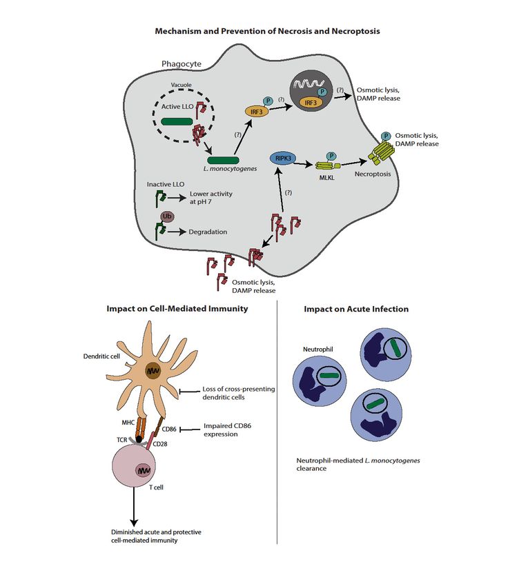
Figure 1. Induction of necrosis by L. monocytogenes and implications on immunity and virulence.
LLO can induce traditional necrosis; as such, L. monocytogenes has evolved mechanisms to avoid
Figure 1. Induction of necrosis by L. monocytogenes and implications on immunity and virulence. LLO
lytic activity of LLO outside the vacuole, including an acidic pH optimum and ubiquitin-mediated
can induce traditional necrosis; as such, L. monocytogenes has evolved mechanisms to avoid lytic
degradation. Strains of L. monocytogenes-induced to express active LLO in the cytosol lead to membrane
pore formation and osmotic lysis. In addition, L. monocytogenes induces programmed necrosis.
Multiple proposed pathways exist, including a RIPK3-mediated pathway leading to pore formation
by MLKL and an IRF3 dependent pathway that occurs by a yet undefined mechanism. Induction of
necrosis ultimately leads to a host-protective neutrophil-mediated clearance of L. monocytogenes and
an impaired cell-mediated immune response. Impaired immunity is at least in part mediated by
diminished numbers of cross-presenting dendritic cells, as well as lower CD86 expression.Pathogens 2018, 7, 8 5 of 17
3. Pyroptosis
Pyroptosis, an alternative form of lytic cell death, is mediated by cytosolic innate immune sensing
complexes called inflammasomes [42,43]. Canonical inflammasomes are multiprotein complexes made
up of receptors (nucleotide binding domain and leucine-rich repeat containing receptors (NLRs)
or absent in melanoma (AIM2)-like receptors (ALRs)), the adaptor protein apoptosis-associated
spec-like protein containing a caspase recruitment domain (ASC), and the pyroptosis executioner
caspase-1 [44–48]. Multiple types of inflammasomes respond to a variety of pathogen-associated or
danger-associated molecular patterns (PAMPs and DAMPs). For example, the NLRC4 inflammasome,
which requires neuronal apoptosis inhibitory proteins (NAIPs) as adaptor molecules, recognizes
flagellin and type III secretion system components [49], whereas the AIM2 inflammasome recognizes
double stranded DNA in the cytosol [50,51]. Despite intense study, the molecular mechanism by
which NLRP3 is activated by a diverse array of molecules, including uric acid, ATP, and pore forming
toxins, is less well understood [52]. Independent of specific PAMPs/DAMPs and the NLRs/ALRs they
activate, downstream signaling is conserved such that ASC is recruited to the receptor, resulting in
the recruitment, autoproteolysis, and activation of caspase-1. Caspase-1 activation leads to several
downstream effects including maturation and secretion of inflammatory cytokines IL-1β and IL-18,
modulation of lipid mediators called eicosanoids, and induction of gasdermin D (GSDMD)-dependent
pyroptosis [53–56]. Pyroptosis shares characteristics of both apoptosis and necrosis; like apoptosis,
pyroptosis is characterized by DNA cleavage, nuclear condensation, and caspase dependence [18],
whereas similar to necrosis, pyroptosis results in membrane pore formation, subsequent water
influx, cellular swelling, and eventual release of cytoplasmic content [57,58]. Additionally, similar to
programmed necrosis, there appears to be crosstalk between pyroptosis and apoptosis such that
suppression of caspase-1 or GSDMD results in activation of an alternative caspase-8 dependent
pyroptosis [59].
Inflammasome activation is a potently host-protective response in the context of a variety of
bacterial infections [60], including Listeriosis [39,61–63]. The mechanisms by which inflammasome
activation protects the host however are not clear, particularly in the context of L. monocytogenes
infection. As is the case with necrosis, it has been proposed that loss of the intracellular
niche and exposure to extracellular neutrophils may result in killing of intracellular pathogens,
potentially through the creation of pore-induced intracellular traps (PITs) [61]. However, unlike
in the context of necrosis, attenuated inflammasome-activating L. monocytogenes are not rescued
in the absence of neutrophils [63], suggesting that host protection by inflammasome activation is
multifactorial. For example, activation of caspase-1 may be directly antimicrobial by activating GSDMD,
which can then bind to cardiolipin in bacterial membranes and cause subsequent pore formation [64].
Consistent with this, supernatants from pyroptotic cells directly reduced colony forming units (CFU)
of both Gram-negative and Gram-positive pathogens, including L. monocytogenes [64]. Regardless of
the mechanism, activation of the inflammasome can potently attenuate virulence, potentially through
multiple redundant mechanisms; as such, L. monocytogenes has developed strategies to avoid activation
of the inflammasome.
L. monocytogenes represses flagellin expression in vivo, at least in part via the transcriptional
regulator MogR, limiting activation of the NLRC4 inflammasome [65,66]. Misregulation of
flagellin expression or forced ectopic expression of flagellin leads to potent virulence attenuation
in an NLRC4-dependent manner, highlighting the importance of avoiding inflammasome activation
to promote virulence [63,67,68]. Additionally, as bacteriolysis in the cytosol triggers activation of
the AIM2 inflammasome [50,51,69,70], L. monocytogenes has evolved a variety of mechanisms to
ensure cytosolic survival, thus avoiding inflammasome activation and promoting virulence [69,71,72].
Specifically, the protein of unknown function YvcK and its regulatory kinase, PrkA, are required
for resistance to cell wall stress, cytosolic survival, AIM2 avoidance, and, ultimately, virulence
in vivo [69,71]. Similarly, peptidoglycan modification enzymes that are necessary for lysozyme
resistance promote cytosolic survival and AIM2 avoidance [73]. How the host cell targetsPathogens 2018, 7, 8 6 of 17
cytosolic bacteria for killing is largely unknown; however, it is clear that adaptations to protect
against these host defenses are essential for AIM2 inflammasome evasion and ultimately virulence.
Finally, although it has been demonstrated that pore-forming toxins including LLO can trigger
NLRP3 activation [74–76], as mentioned above, L. monocytogenes regulates LLO transcriptionally,
translationally, and post-translationally, to minimize activity on the plasma membrane and therefore
minimize activation of the NLRP3 inflammasome in vivo [26–29].
Similar to necrosis, it was hypothesized that proinflammatory pyroptosis would promote
immunity. As L. monocytogenes largely avoids inflammasome activation during infection, multiple groups
engineered L. monocytogenes to hyperactivate the inflammasome in the hopes of promoting increased
cell-mediated immune responses [63,68]. Somewhat surprisingly, however, hyper-inflammasome
activation significantly reduced cell-mediated immunity [39,63,77]. Importantly, inhibition of immunity
due to inflammasome activation is not simply an artifact of hyperactivation, as immunization
of caspase-1/11 deficient mice with wild-type L. monocytogenes resulted in improved immunity
(unpublished observations Erin Theisen, Courtney McDougal, and John-Demian Sauer). This suggests
that even the small amount of inflammasome activation during wild type infection inhibits
cell-mediated immunity. How inflammasome activation negatively affects L. monocytogenes-stimulated
immunity is less clear. While the maturation and secretion of proinflammatory cytokines IL-1β
and IL-18 does not contribute to the inflammasome-impaired immune response (unpublished
observations Erin Theisen, Courtney McDougal, and John-Demian Sauer), it is clear that some other
component of the inflammatory milieu inhibits priming of an optimal CD8+ T-cell response [39].
One hypothesis is that the earlier, more robust inflammatory cytokine response associated with
inflammasome activation is detrimental to optimal T-cell priming [39,77], consistent with the model
that both too much and too little inflammation is detrimental for priming an optimal T-cell response.
Alternatively, inflammasome-mediated eicosanoid modulation may negatively impact the generation
of cell-mediated immunity in the context of L. monocytogenes infection. von Moltke et al. highlighted
a COX-dependent hypothermia and vascular leakage in mice after delivery of a potent inflammasome
agonist [54]. Additionally, eicosanoids have been implicated in regulating CD8+ T-cell responses
in the context of Mycobacterial and LCMV-associated immunity [78,79]. Specifically, PGE2 has been
reported to inhibit CD8+ T-cell proliferation in some contexts, offering another potential mechanism
by which inflammasome activation could inhibit cell mediated immunity [79,80]. Whether or not
inflammasome-dependent eicosanoid modulation impacts L. monocytogenes-stimulated immunity
remains to be determined. Finally, the role of cellular lysis during pyroptosis on immunity is also
incompletely understood. Pore formation and lysis can result in release of DAMPs such as HMGB1
modulating the inflammatory milieu associated with inflammasome activation. Previously, separating
the impact of pyroptosis from other inflammasome consequences was difficult; however, the discovery
of GSDMD as the mediator of pore formation during pyroptosis [55,56] should allow for the assessment
of the specific role of pyroptosis in cell-mediated immunity.
Taken together (Figure 2), similar to necrosis, albeit by different mechanisms, it is clear that
inflammasome-mediated pyroptosis negatively impacts both virulence and priming of cell-mediated
immunity in the context of L. monocytogenes infection. As such, L. monocytogenes, like other professional
intracellular pathogens [81], has evolved mechanisms to avoid detection by this potent, host-protective,
innate immune defense.Pathogens 2018, 7, 8 7 of 17
Pathogens 2018, 7, 8 7 of 17
Mechanism and Prevention of Pyroptosis
Active LLO Inactive LLO
Lower
activity
yvck, oat, pgd Ub at pH 7
Mog-mediated Degradation
regulation
Phagocyte
NLRP3
ASC
AIM2 Caspase-1
NAIP ASC
Caspase-1
NLRC4
Caspase-1 activation Bacteriolysis
ASC
Caspase-1
(?)
GSDMD
IL-1β, IL-18 Eicosanoids
Pyroptosis,
DAMPrelease
Impact on Cell-Mediated Immunity Impact on Acute Infection
Excessive cytokine production (?)
Bacteriolysis via GSDMD binding to cardiolipin
and subsequent pore formation
Diminished cell-mediated immunity due to
Trapping of bacteria in pore-induced intracellular traps
HMGB1
Immune modulating Release of DAMPs through
eicosanoid production (?) GSDMD-induced pores (?)
Other redundant mechanisms leading to
extracellular bacterial killing (?)
Neutrophil
Secondary phagocyte
Figure 2. Mechanism and effects of pyroptosis during L. monocytogenes infection. L. monocytogenes can
Figure 2. Mechanism and effects of pyroptosis during L. monocytogenes infection. L. monocytogenes can
activate the
activate the NLRC4,
NLRC4,AIM2,
AIM2,and andNLRP3
NLRP3 inflammasomes
inflammasomes through
throughflagellin, bacterial
flagellin, DNA
bacterial DNArelease, and
release,
LLO-mediated pore formation, respectively. Inflammasome activation
and LLO-mediated pore formation, respectively. Inflammasome activation results in caspase-1 results in caspase-1
autoproteolysis followed
autoproteolysis followed by by cytokine
cytokine and
andeicosanoid
eicosanoidrelease,
release,and
andthe theinduction
inductionofof thethelytic form
lytic formof
cell death called pyroptosis. Importantly, as this leads to the loss of the intracellular
of cell death called pyroptosis. Importantly, as this leads to the loss of the intracellular niche, niche, L.
monocytogenes has evolved strategies to avoid inflammasome activation. It represses
L. monocytogenes has evolved strategies to avoid inflammasome activation. It represses flagellin flagellin during
infection
during throughthrough
infection MogR, regulates expression
MogR, regulates and activity
expression and of LLO, of
activity andLLO,prevents release ofrelease
and prevents bacterial
of
DNA through
bacterial genes designed
DNA through to combat
genes designed cell cell
to combat wallwall
stress
stress(yvcK,
(yvcK,oat, oat, pgd).
pgd). Activation
Activation of of
inflammasomes results in decreased bacterial virulence, potentially through
inflammasomes results in decreased bacterial virulence, potentially through direct bacterial poredirect bacterial pore
formation by
formation by GSDMD,
GSDMD, bacterial
bacterial trapping
trapping inin pore-induced
pore-induced intracellular
intracellular traps,
traps, and
and potential
potential redundant
redundant
mechanisms of extracellular killing. Additionally, inflammasome activation impairs
mechanisms of extracellular killing. Additionally, inflammasome activation impairs cell-mediated cell-mediated
immunity through
immunity through aa suboptimal
suboptimal inflammatory
inflammatory milieu
milieu associated
associated with
with proinflammatory
proinflammatory cytokines,
cytokines,
eicosanoids, and pyroptosis-released
eicosanoids, and pyroptosis-released DAMPS.DAMPS.
4. Apoptosis
Apoptosis is a non-lytic cell death characterized by cell shrinkage, chromatin condensation,
and extensive membrane blebbing that produces apoptotic bodies [82]. Apoptosis is traditionallyPathogens 2018, 7, 8 8 of 17
subdivided into two pathways: (1) the extrinsic pathway, triggered by ligands such as tumor
necrosis factor family molecules which trigger death receptor oligomerization and activation of
caspase-8 [83]; or (2) the intrinsic pathway, initiated by signals such as oxidative stress or DNA
damage, which ultimately lead to mitochondrial permeabilization by pro-apoptotic Bcl-2 family
proteins, release of cytochrome c and, ultimately, formation of the apoptosome resulting in the
activation of caspase-9 [84]. Both pathways require the activation of downstream caspases, the most
important of which is the caspase-3, which leads to a substantial reorganization of the cytoskeleton
as well as degradation of chromosomal DNA, inactivation of inflammatory DAMPs, and, ultimately,
culminates in the ordered disassembly of the cell into apoptotic bodies [85]. As a natural process during
development, apoptosis is not only non-inflammatory but in many cases is actively anti-inflammatory,
which has led to the idea that some pathogens have hijacked this system to enhance and perpetuate
infection [36,86].
Over 20 years ago it was first reported by Unanue and colleagues that infected hepatocytes
undergo apoptosis during L. monocytogenes infection. This study suggested that hepatocyte
apoptosis was host-protective by recruiting neutrophils to the sites of infection, in contrast to
the canonical view of apoptosis as being anti-inflammatory [87]. Since these early studies it has
been demonstrated by other groups that infected hepatocytes undergo apoptosis, likely in a TNFα
dependent manner [88,89], although whether this is host protective or beneficial to the bacterium
remains unclear. Both TRAIL-deficient mice, as well as mice deficient in a variety of proapoptotic
BH3-only Bcl-2 family members, were significantly more resistant to L. monocytogenes challenge,
although this was not definitively localized to the hepatocyte compartment [90,91]. While it is clear
that hepatocytes can undergo apoptosis during infection, the mechanism by which this is mediated
and the consequences of hepatocyte apoptosis on the outcome of L. monocytogenes infection remains to
be fully defined.
While hepatocyte apoptosis in the context of L. monocytogenes infections has been only sporadically
documented, apoptosis of lymphocytes early during infection is a well-established consequence
of L. monocytogenes infection. It has been long understood that infection leads to a depletion of
lymphocytes within the periarteriolar lymphoid sheaths [92], seen as early as 24 h post infection in
a dose-dependent manner [93]. In contrast to apoptosis of infected hepatocytes, lymphocyte apoptosis
is not associated with direct infection of these cells, but rather, infected phagocytes are in close proximity
to the uninfected apoptotic lymphocytes [93,94]. Two, non-mutually exclusive pathways have been
implicated in triggering lymphocyte apoptosis during L. monocytogenes infection: (1) the direct activity
of sub-lytic concentrations of LLO on lymphocytes and (2) the copious amounts of type I interferon
produced in the context of L. monocytogenes infection. One model for LLO-mediated apoptosis is
that it acts as a perforin alternative for the delivery of host granzyme to target lymphocytes [95–97].
Ex vivo isolation of CD4+ T cells treated with exogenous LLO led to activation of apoptotic caspases-3,
-6, and -9, and subsequently 75% T cell apoptosis by 8 h post treatment [95], whereas apoptosis of
granzyme-deficient T cells was reduced by approximately 50%. Importantly, this seminal work
was done using CD4+ T cells that only had ~30% staining positive for granzymes, suggesting
an even larger potential role for granzyme in lymphocyte apoptosis [95]. Consistent with this model,
granzyme-deficient mice demonstrated less lymphocyte apoptosis and were 15-fold more resistant to
acute L. monocytogenes infections [95], suggesting that activation of lymphocyte apoptosis is a pathogen
beneficial virulence strategy.
In addition to a direct role for extracellular LLO, type I interferons (IFN) have been implicated in
lymphocyte apoptosis during L. monocytogenes infections. L. monocytogenes infection results in robust
production of type I interferon due to activation of STING by both c-di-AMP and less frequently
cytosolic bacterial DNA [98–100]. Type I IFN subsequently has been shown to antigen-independently
preactivate T cells, potentially increasing T cell sensitivity to apoptosis [101]. Following systemic
infection with L. monocytogenes, type I IFN receptor (IFNAR)-deficient mice demonstrate both
decreased lymphocyte apoptosis and increased resistance to infection, consistent with a model wherebyPathogens 2018, 7, 8 9 of 17
lymphocyte apoptosis is a pathogen-driven process to promote virulence [33,34,101]. Independent of
the mechanism of apoptosis induction, it is thought that the increased virulence of L. monocytogenes
associated with lymphocyte apoptosis is mediated by the activation of an immunosuppressive
state. Uptake of apoptotic bodies results in production of anti-inflammatory cytokine production,
notably IL-10, by phagocytes [102]. IL-10 antagonizes production of proinflammatory IFN-γ, which is
required for optimal acute defense against L. monocytogenes. Consistent with this model, IL-10-deficient
mice showed increased resistance to L. monocytogenes infection despite having equivalent levels of
apoptotic splenic cells, suggesting the susceptibility associated with lymphocyte apoptosis could
be mediated by IL-10 [94,103]. Finally, mice lacking lymphocytes altogether are acutely resistant
to L. monocytogenes, further suggesting that increased lymphocyte apoptosis results in decreased
resistance to infection [103–105].
In addition to effects on acute virulence, induction of apoptosis during L. monocytogenes infection
influences the priming of adaptive immune responses. Perhaps not surprisingly, given the known
detrimental role of IL-10 in the generation of cell mediated immunity [103], L. monocytogenes engineered
to induce apoptosis generated worse T-cell responses following immunization compared to wild
type L. monocytogenes [39]. Somewhat surprisingly, however, in this context, increased apoptosis
was not associated with increased levels of IL-10 suggesting additional, apoptosis-dependent,
IL-10 independent detrimental effects on cell mediated immunity. How, mechanistically speaking,
apoptosis of either infected cells or bystander lymphocytes might negatively influence the generation
of cell-mediated immunity independent of IL-10 is currently unknown. Also consistent with
a negative impact of increased apoptosis on generation of adaptive immunity was the observation that
hyperinduction of type I interferons is detrimental to CD8+ T-cell priming, although this was never
explicitly tied to levels of lymphocyte apoptosis [106].
In summary (Figure 3), L. monocytogenes-induced lymphocyte apoptosis, whether mediated by
LLO, type I interferons, or a combination of the two, potentiates acute infection, likely through the
induction of an anti-inflammatory state induced by uptake of apoptotic bodies. Indeed, Ucker and
colleagues demonstrated that direct injection of apoptotic cells, but not necrotic cells, increases
virulence of L. monocytogenes by a mechanism they coined “innate apoptotic immunity” [107]. It is
exciting to speculate that this is an adaptation evolved by L. monocytogenes to promote its virulence.
Promoting apoptosis to potentiate virulence is a strategy used by some other pathogens, including
HIV, whereby CD4+ T cell apoptosis inhibits clearance of the virus [108], while Mycobacterium
avium-induced apoptosis mediates bacterial spread [109]. The mechanism of induction of hepatocyte
apoptosis is significantly less well understood, as is its impact on acute infection or immunity.
Finally, the detrimental effect of apoptosis on L. monocytogenes-stimulated immunity suggests
that mechanisms that might inhibit apoptosis could result in more robust L. monocytogenes-based
immunotherapy platforms.Pathogens 2018, 7, 8 10 of 17
Pathogens 2018, 7, 8 10 of 17
(A)
Figure 3. Cont.Pathogens 2018, 7, 8 11 of 17
Pathogens 2018, 7, 8 11 of 17
(B)
Figure
Figure3. Mechanism
3. Mechanism and impact of apoptosis
and impact during L.
of apoptosis monocytogenes
during infection. infection.
L. monocytogenes (A) Direct infection
(A) Direct
of infection
hepatocytes with L. monocytogenes leads to apoptosis, potentially
of hepatocytes with L. monocytogenes leads to apoptosis, potentially through a TNFα and/or aBcl-2
through TNFα
family member-mediated mechanism. Induction of apoptosis through either
and/or Bcl-2 family member-mediated mechanism. Induction of apoptosis through either pathway pathway ultimately
leads to caspase-3
ultimately activation.activation.
leads to caspase-3 Hepatocyte apoptosis
Hepatocyte is host-protective
apoptosis through
is host-protective release
through of of
release
chemoattractants and subsequent neutrophil-mediated L. monocytogenes killing. The
chemoattractants and subsequent neutrophil-mediated L. monocytogenes killing. The complete impact on complete impact
on cell-mediated
cell-mediatedimmunity,
immunity, if any,
if any, is unknown.
is unknown. (B)contrast
(B) In In contrast to hepatocytes,
to hepatocytes, L. monocytogenes-
L. monocytogenes-mediated
mediated
lymphocytelymphocyte
killing iskilling is independent
independent of direct of infection.
direct infection. Lymphocyte
Lymphocyte apoptosis
apoptosis is instead
is instead induced
induced through sublytic concentrations of LLO and/or type I IFN from infected
through sublytic concentrations of LLO and/or type I IFN from infected phagocytes. Type I IFN phagocytes. Type I
IFN produced after STING activation leads to antigen-independent preactivation
produced after STING activation leads to antigen-independent preactivation of T cells as indicated by of T cells as
indicated by CD69 expression,
CD69 expression, potentiallypotentially
sensitizingsensitizing them to capabilities
them to apoptotic apoptotic capabilities of LLO.
of LLO. Uptake of Uptake
apoptotic
of bodies
apoptotic by bodies
secondaryby secondary
phagocytesphagocytes leads to increased
leads to increased bacterial virulence
bacterial virulence though IL-10 though IL-10
production,
production,
as well asasdiminished
well as diminished
cell-mediatedcell-mediated
immunity,immunity,
through boththrough both IL-10-dependent
IL-10-dependent and
and potentially
potentially IL-10-independent
IL-10-independent mechanisms. mechanisms.
5. Conclusions and Future Directions
In summary, cell death has potent impacts on the outcomes of infections, particularly in the
context of exquisitely adapted intracellular pathogens such as L. monocytogenes. In most cases, hostPathogens 2018, 7, 8 12 of 17
5. Conclusions and Future Directions
In summary, cell death has potent impacts on the outcomes of infections, particularly in the context
of exquisitely adapted intracellular pathogens such as L. monocytogenes. In most cases, host cell death
is protective and L. monocytogenes has evolved ways to avoid activation of these pathways through
regulation of their virulence factors. Whether L. monocytogenes has specific virulence factors to directly
modulate cell death pathways is an outstanding question, but as more cell death pathways, such as
necroptosis, are discovered, that possibility becomes more and more likely. The exception to the cell
death avoidance model is the activation of bystander apoptosis of lymphocytes. Importantly, this form
of cell death does not directly eliminate a replication niche and in fact appears to be detrimental to the
host and beneficial to the pathogen, fueling the longstanding, speculative, but exciting hypothesis that
L. monocytogenes promotes type I interferon responses to create an immunosuppressive environment to
enhance virulence. Finally, our understanding of how cell death influences the generation of adaptive
immune responses is still in its infancy. The classic dogma of immunogenic necrosis and tolerogenic
apoptosis has been turned on its head, even in the context of sterile immunity. How these pathways
influence immunity in the context of infection is a future frontier. Furthermore, as more novel cell
death pathways continue to be discovered, their interactions with the innate and adaptive immune
systems will have to be understood in order to refine our use of bacteria such as L. monocytogenes as
beneficial microbes in the context of vaccines and tumor immunotherapy.
Acknowledgments: This work was supported by National Institutes of Health National Cancer Institute
(R01CA188034).
Author Contributions: CEM and JDS equally contributed to the conceptualization and execution of this review.
Conflicts of Interest: The authors declare no conflict of interests.
References
1. Ferreira, V.; Wiedmann, M.; Teixeira, P.; Stasiewicz, M.J. Listeria monocytogenes Persistence in Food-Associated
Environments: Epidemiology, Strain Characteristics, and Implications for Public Health. J. Food Prot. 2014,
77, 150–170. [CrossRef] [PubMed]
2. Swaminathan, B.; Gerner-Smidt, P. The epidemiology of human listeriosis. Microbes Infect. 2007, 9, 1236–1243.
[CrossRef] [PubMed]
3. Mengaud, J.; Ohayon, H.; Gounon, P.; Mege, R.-M.; Cossart, P. E-cadherin is the receptor for internalin,
a surface protein required for entry of L. monocytogenes into epithelial cells. Cell 1996, 84, 923–932. [CrossRef]
4. Shen, Y.; Naujokas, M.; Park, M.; Ireton, K. InIB-dependent internalization of Listeria is mediated by the Met
receptor tyrosine kinase. Cell 2000, 103, 501–510. [CrossRef]
5. Portnoy, D.A.; Jacks, P.S.; Hinrichs, D.J. Role of hemolysin for the intracellular growth of Listeria monocytogenes.
J. Exp. Med. 1988, 167, 1459–1471. [CrossRef] [PubMed]
6. Hamon, M.A.; Ribet, D.; Stavru, F.; Cossart, P. Listeriolysin O: The Swiss army knife of Listeria.
Trends Microbiol. 2012, 20, 360–368. [CrossRef] [PubMed]
7. Kocks, C.; Gouin, E.; Tabouret, M.; Berche, P.; Ohayon, H.; Cossart, P. L. monocytogenes-induced actin assembly
requires the actA gene product, a surface protein. Cell 1992, 68, 521–531. [CrossRef]
8. Carr, K.D.; Sieve, A.N.; Indramohan, M.; Break, T.J.; Lee, S.; Berg, R.E. Specific depletion reveals a novel role
for neutrophil-mediated protection in the liver during Listeria monocytogenes infection. Eur. J. Immunol. 2011,
41, 2666–2676. [CrossRef] [PubMed]
9. Baker, L.A.; Campbell, P.A.; Hollister, J.R. Chemotaxigenesis and complement fixation by Listeria
monocytogenes cell wall fractions. J. Immunol. 1977, 119, 1723–1726. [PubMed]
10. Shaughnessy, L.M.; Swanson, J.A. The role of the activated macrophage in clearing Listeria monocytogenes
infection. Front. Biosci. 2007, 12, 2683–2692. [CrossRef] [PubMed]
11. Le, D.T.; Dubenksy, T.W.; Brockstedt, D.G. Clinical development of Listeria monocytogenes-based
immunotherapies. Semin. Oncol. 2012, 39, 311–322. [CrossRef] [PubMed]Pathogens 2018, 7, 8 13 of 17
12. Bahjat, K.S.; Liu, W.; Lemmens, E.E.; Schoenberger, S.P.; Portnoy, D.A.; Dubensky, T.W., Jr.; Brockstedt, D.G.
Cytosolic entry controls CD8+-T-cell potency during bacterial infection. Infect. Immun. 2006, 74, 6387–6397.
[CrossRef] [PubMed]
13. Aduro BioTech. LADD Engineering Listeria Mononcytogenes Bacteria. 2017. Available online:
http://www.aduro.com/technology/ladd/ (accessed on 17 May 2017).
14. Lm Technology—Advaxis. 2017. Available online: https://www.advaxis.com/lm-technology/ (accessed on
15 November 2017).
15. Blériot, C.; Lecuit, M. The interplay between regulated necrosis and bacterial infection. Cell. Mol. Life Sci.
2016, 73, 2369–2378. [CrossRef] [PubMed]
16. Pasparakis, M.; Vandenabeele, P. Necroptosis and its role in inflammation. Nature 2015, 517, 311–320.
[CrossRef] [PubMed]
17. Vanden Berghe, T.; Kaiser, W.J.; Bertrand, M.J.; Vandenabeele, P. Molecular crosstalk between apoptosis,
necroptosis, and survival signaling. Mol. Cell. Oncol. 2015, 2, e975093. [CrossRef] [PubMed]
18. Kroemer, G.; Galluzzi, L.; Vandenabeele, P.; Abrams, J.; Alnemri, E.S.; Baehrecke, E.H.; Blagosklonny, M.V.;
El-Deiry, W.S.; Golstein, P.; Green, D.R.; et al. Classification of cell death: Recommendations of the
Nomenclature Committee on Cell Death 2009. Cell Death Differ. 2009, 16, 3–11. [CrossRef] [PubMed]
19. Kokkola, R.; Andersson, A.; Mullins, G.; Ostberg, T.; Treutiger, C.-J.; Arnold, B.; Nawroth, P.; Andersson, U.;
Harris, R.A.; Harris, H.E. RAGE is the Major Receptor for the Proinflammatory Activity of HMGB1 in Rodent
Macrophages. Scand. J. Immunol. 2005, 61, 1–9. [CrossRef] [PubMed]
20. Scaffidi, P.; Misteli, T.; Bianchi, M.E. Release of chromatin protein HMGB1 by necrotic cells triggers
inflammation. Nature 2002, 418, 191–195. [CrossRef] [PubMed]
21. Seveau, S. Multifaceted activity of listeriolysin O, the cholesterol-dependent cytolysin of Listeria
monocytogenes. Subcell. Biochem. 2014, 80, 161–195. [PubMed]
22. Barsig, J.; Kaufmann, S.H. The mechanism of cell death in Listeria monocytogenes-infected murine
macrophages is distinct from apoptosis. Infect. Immun. 1997, 65, 4075–4081. [PubMed]
23. González-Juarbe, N.; Gilley, R.P.; Hinojosa, C.A.; Bradley, K.M.; Kamei, A.; Gao, G.; Dube, P.H.;
Bergman, M.A.; Orihuela, C.J. Pore-Forming Toxins Induce Macrophage Necroptosis during Acute Bacterial
Pneumonia. PLoS Pathog. 2015, 11, e1005337. [CrossRef] [PubMed]
24. Geoffroy, C.; Gaillard, J.-L.; Alouf, J.E.; Berche, P. Purification, Characterization, and Toxicity of the
Sulfhydryl-Activated Hemolysin Listeriolysin 0 from Listeria monocytogenes. Infect. Immun. 1987, 55,
1641–1646. [PubMed]
25. Glomski, I.J.; Decatur, A.L.; Portnoy, D.A. Listeria monocytogenes mutants that fail to compartmentalize
listerolysin O activity are cytotoxic, avirulent, and unable to evade host extracellular defenses. Infect. Immun.
2003, 71, 6754–6765. [CrossRef] [PubMed]
26. Glomski, I.J.; Gedde, M.M.; Tsang, A.W.; Swanson, J.A.; Portnoy, D.A. The Listeria monocytogenes hemolysin
has an acidic pH optimum to compartmentalize activity and prevent damage to infected host cells. J. Cell Biol.
2002, 156, 1029–1038. [CrossRef] [PubMed]
27. Schnupf, P.; Portnoy, D.A.; Decatur, A.L. Phosphorylation, ubiquitination and degradation of listeriolysin O
in mammalian cells: Role of the PEST-like sequence. Cell Microbiol. 2006, 8, 353–364. [CrossRef] [PubMed]
28. Schuerch, D.W.; Wilson-Kubalek, E.M.; Tweten, R.K. Molecular basis of listeriolysin O pH dependence.
Proc. Natl. Acad. Sci. USA 2005, 102, 12537–12542. [CrossRef] [PubMed]
29. Schnupf, P.; Hofmann, J.; Norseen, J.; Glomski, I.J.; Schwartzstein, H.; Decatur, A.L. Regulated translation of
listeriolysin O controls virulence of Listeria monocytogenes. Mol. Microbiol. 2006, 61, 999–1012. [CrossRef]
[PubMed]
30. Bavdek, A.; Gekara, N.O.; Priselac, D.; Gutiérrez Aguirre, I.; Darji, A.; Chakraborty, T.; Maček, P.; Lakey, J.H.;
Weiss, S.; Anderluh, G. Sterol and pH Interdependence in the Binding, Oligomerization, and Pore Formation
of Listeriolysin O. Biochemistry 2007, 46, 4425–4437. [CrossRef] [PubMed]
31. Blériot, C.; Dupuis, T.; Jouvion, G.; Eberl, G.; Disson, O.; Lecuit, M. Liver-resident macrophage necroptosis
orchestrates type 1 microbicidal inflammation and type-2-mediated tissue repair during bacterial infection.
Immunity 2015, 42, 145–158. [CrossRef] [PubMed]
32. Di Paolo, N.C.; Doronin, K.; Baldwin, L.K.; Papayannopoulou, T.; Shayakhmetov, D.M. The transcription
factor IRF3 triggers defensive suicide necrosis in response to viral and bacterial pathogens. Cell Rep. 2013, 3,
1840–1846. [CrossRef] [PubMed]Pathogens 2018, 7, 8 14 of 17
33. O’Connell, R.M.; Saha, S.K.; Vaidya, S.A.; Bruhn, K.W.; Miranda, G.A.; Zarnegar, B.; Perry, A.K.; Nguyen, B.O.;
Lane, T.F.; Taniguchi, T.; et al. Type I Interferon Production Enhances Susceptibility to Listeria monocytogenes
Infection. J. Exp. Med. 2004, 200, 437–445. [CrossRef] [PubMed]
34. Auerbuch, V.; Brockstedt, D.G.; Meyer-Morse, N.; O’Riordan, M.; Portnoy, D.A. Mice lacking the type
I interferon receptor are resistant to Listeria monocytogenes. J. Exp. Med. 2004, 200, 527–533. [CrossRef]
[PubMed]
35. Nakajima, A.; Nishimura, K.; Nakaima, Y.; Oh, T.; Noguchi, S.; Taniguchi, T.; Tamura, T. Cell type-dependent
proapoptotic role of Bcl2L12 revealed by a mutation concomitant with the disruption of the juxtaposed Irf3
gene. Proc. Natl. Acad. Sci. USA 2009, 106, 12448–12452. [CrossRef] [PubMed]
36. Barker, R.N.; Erwig, L.-P.; Pearce, W.P.; Devine, A.; Rees, A.J. Differential Effects of Necrotic or Apoptotic
Cell Uptake on Antigen Presentation by Macrophages. Pathobiology 1999, 67, 302–305. [CrossRef] [PubMed]
37. Festjens, N.; Vanden Berghe, T.; Vandenabeele, P. Necrosis, a well-orchestrated form of cell demise: Signalling
cascades, important mediators and concomitant immune response. Biochim. Biophys. Acta Bioenerg. 2006,
1757, 1371–1387. [CrossRef] [PubMed]
38. Sauter, B.; Albert, M.L.; Francisco, L.; Larsson, M.; Somersan, S.; Bhardwaj, N. Consequences of cell death:
exposure to necrotic tumor cells, but not primary tissue cells or apoptotic cells, induces the maturation of
immunostimulatory dendritic cells. J. Exp. Med. 2000, 191, 423–434. [CrossRef] [PubMed]
39. Theisen, E.; Sauer, J.-D. Listeria monocytogenes-Induced Cell Death Inhibits the Generation of Cell-Mediated
Immunity. Infect. Immun. 2017, 85, e00733-16. [CrossRef] [PubMed]
40. Janda, J.; Schoneberger, P.; Skoberne, M.; Messerle, M.; Russmann, H.; Geginat, G. Cross-presentation of
Listeria-derived CD8 T cell epitopes requires unstable bacterial translation products. J. Immunol. 2004, 173,
5644–5651. [CrossRef] [PubMed]
41. Reinicke, A.T.; Omilusik, K.D.; Basha, G.; Jefferies, W.A. Dendritic cell cross-priming is essential for immune
responses to Listeria monocytogenes. PLoS ONE 2009, 4, e7210. [CrossRef] [PubMed]
42. Von Moltke, J.; Ayres, J.S.; Kofoed, E.M.; Chavarria-Smith, J.; Vance, R.E. Recognition of bacteria by
inflammasomes. Annu. Rev. Immunol. 2013, 31, 73–106. [CrossRef] [PubMed]
43. Martinon, F.; Mayor, A.; Tschopp, J. The inflammasomes: Guardians of the body. Annu. Rev. Immunol. 2009,
27, 229–265. [CrossRef] [PubMed]
44. DeYoung, K.L.; Ray, M.E.; Su, Y.A.; Anzick, S.L.; Johnstone, R.W.; Trapani, J.A.; Meltzer, P.S.; Trent, J.M.
Cloning a novel member of the human interferon-inducible gene family associated with control of
tumorigenicity in a model of human melanoma. Oncogene 1997, 15, 453–457. [CrossRef] [PubMed]
45. Inohara; Chamaillard; McDonald, C.; Nunez, G.; Inohara, N.; Chamaillard, M.; McDonald, C.; Nunez, G.
NOD-LRR proteins: role in host-microbial interactions and inflammatory disease. Annu. Rev. Biochem. 2004,
74, 355–383. [CrossRef] [PubMed]
46. Martinon, F.; Hofmann, K.; Tschopp, J. The pyrin domain: A possible member of the death domain-fold
family implicated in apoptosis and inflammation. Curr. Biol. 2001, 11, R118–R120. [CrossRef]
47. Masumoto, J.; Taniguchi, S.; Ayukawa, K.; Sarvotham, H.; Kishino, T.; Niikawa, N.; Hidaka, E.; Katsuyama, T.;
Higuchi, T.; Sagara, J. ASC, a novel 22-kDa protein, aggregates during apoptosis of human promyelocytic
leukemia HL-60 cells. J. Biol. Chem. 1999, 274, 33835–33838. [CrossRef] [PubMed]
48. Lu, A.; Magupalli, V.G.; Ruan, J.; Yin, Q.; Atianand, M.K.; Vos, M.R.; Schröder, G.F.; Fitzgerald, K.A.; Wu, H.;
Egelman, E.H. Unified Polymerization Mechanism for the Assembly of ASC-Dependent Inflammasomes.
Cell 2014, 156, 1193–1206. [CrossRef] [PubMed]
49. Zhao, Y.; Yang, J.; Shi, J.; Gong, Y.-N.; Lu, Q.; Xu, H.; Liu, L.; Shao, F. The NLRC4 inflammasome receptors
for bacterial flagellin and type III secretion apparatus. Nature 2011, 477, 596–600. [CrossRef] [PubMed]
50. Hornung, V.; Ablasser, A.; Charrel-Dennis, M.; Bauernfeind, F.; Horvath, G.; Caffrey, D.R.; Latz, E.;
Fitzgerald, K.A. AIM2 recognizes cytosolic dsDNA and forms a caspase-1-activating inflammasome with
ASC. Nature 2009, 458, 514–518. [CrossRef] [PubMed]
51. Fernandes-Alnemri, T.; Yu, J.W.; Datta, P.; Wu, J.; Alnemri, E.S. AIM2 activates the inflammasome and cell
death in response to cytoplasmic DNA. Nature 2009, 458, 509–513. [CrossRef] [PubMed]
52. Mariathasan, S.; Weiss, D.S.; Newton, K.; McBride, J.; O’Rourke, K.; Roose-Girma, M.; Lee, W.P.; Weinrauch, Y.;
Monack, D.M.; Dixit, V.M. Cryopyrin activates the inflammasome in response to toxins and ATP. Nature
2006, 440, 228–232. [CrossRef] [PubMed]Pathogens 2018, 7, 8 15 of 17
53. Martinon, F.; Burns, K.; Tschopp, J. The inflammasome: a molecular platform triggering activation of
inflammatory caspases and processing of proIL-beta. Mol. Cell 2002, 10, 417–426. [CrossRef]
54. Von Moltke, J.; Trinidad, N.J.; Moayeri, M.; Kintzer, A.F.; Wang, S.B.; van Rooijen, N.; Brown, C.R.;
Krantz, B.A.; Leppla, S.H.; Gronert, K.; et al. Rapid induction of inflammatory lipid mediators by the
inflammasome in vivo. Nature 2012, 490, 107–111. [CrossRef] [PubMed]
55. Shi, J.; Zhao, Y.; Wang, K.; Shi, X.; Wang, Y.; Huang, H.; Zhuang, Y.; Cai, T.; Wang, F.; Shao, F. Cleavage of
GSDMD by inflammatory caspases determines pyroptotic cell death. Nature 2015, 526, 660–665. [CrossRef]
[PubMed]
56. Kayagaki, N.; Stowe, I.B.; Lee, B.L.; O’Rourke, K.; Anderson, K.; Warming, S.; Cuellar, T.; Haley, B.;
Roose-Girma, M.; Phung, Q.T.; et al. Caspase-11 cleaves gasdermin D for non-canonical inflammasome
signalling. Nature 2015, 526, 666–671. [CrossRef] [PubMed]
57. Lamkanfi, M.; Sarkar, A.; Vande Walle, L.; Vitari, A.C.; Amer, A.O.; Wewers, M.D.; Tracey, K.J.;
Kanneganti, T.-D.; Dixit, V.M. Inflammasome-dependent release of the alarmin HMGB1 in endotoxemia.
J. Immunol. 2010, 185, 4385–4392. [CrossRef] [PubMed]
58. Fink, S.L.; Cookson, B.T. Caspase-1-dependent pore formation during pyroptosis leads to osmotic lysis of
infected host macrophages. Cell. Microbiol. 2006, 8, 1812–1825. [CrossRef] [PubMed]
59. Mascarenhas, D.P.A.; Cerqueira, D.M.; Pereira, M.S.F.; Castanheira, F.V.S.; Fernandes, T.D.; Manin, G.Z.;
Cunha, L.D.; Zamboni, D.S. Inhibition of caspase-1 or gasdermin-D enable caspase-8 activation in the
Naip5/NLRC4/ASC inflammasome. PLOS Pathog. 2017, 13, e1006502. [CrossRef] [PubMed]
60. Lamkanfi, M.; Dixit, V.M. Mechanisms and Functions of Inflammasomes. Cell 2014, 157, 1013–1022.
[CrossRef] [PubMed]
61. Jorgensen, I.; Zhang, Y.; Krantz, B.A.; Miao, E.A. Pyroptosis triggers pore-induced intracellular traps (PITs)
that capture bacteria and lead to their clearance by efferocytosis. J. Exp. Med. 2016, 213, 2113–2128. [CrossRef]
[PubMed]
62. Miao, E.A.; Leaf, I.A.; Treuting, P.M.; Mao, D.P.; Dors, M.; Sarkar, A.; Warren, S.E.; Wewers, M.D.; Aderem, A.
Caspase-1-induced pyroptosis is an innate immune effector mechanism against intracellular bacteria.
Nat. Immunol. 2010, 11, 1136–1142. [CrossRef] [PubMed]
63. Sauer, J.; Pereyre, S.; Archer, K.A.; Burke, T.P.; Hanson, B.; Lauer, P.; Portnoy, D.A. Listeria monocytogenes
engineered to activate the Nlrc4 inflammasome are severely attenuated and are poor inducers of protective
immunity. Proc. Natl. Acad. Sci. USA 2011, 108, 12419–12424. [CrossRef] [PubMed]
64. Liu, X.; Zhang, Z.; Ruan, J.; Pan, Y.; Magupalli, V.G.; Wu, H.; Lieberman, J. Inflammasome-activated
gasdermin D causes pyroptosis by forming membrane pores. Nature 2016, 535, 153–158. [CrossRef] [PubMed]
65. Shen, A.; Higgins, D.E. The MogR transcriptional repressor regulates nonhierarchal expression of flagellar
motility genes and virulence in Listeria monocytogenes. PLoS Pathog. 2006, 2, e30. [CrossRef] [PubMed]
66. Peel, M.; Donachie, W.; Shaw, A. Temperature-dependent Expression of Flagella of Listeria manocytogenes
Studied by Electron Microscopy, SDS-PAGE and Western Blotting. Microbiology 1988, 134, 2171–2178.
[CrossRef] [PubMed]
67. Warren, S.E.; Mao, D.P.; Rodriguez, A.E.; Miao, E.A.; Aderem, A. Multiple Nod-like receptors activate
caspase 1 during Listeria monocytogenes infection. J. Immunol. 2008, 180, 7558–7564. [CrossRef] [PubMed]
68. Warren, S.E.; Duong, H.; Mao, D.P.; Armstrong, A.; Rajan, J.; Miao, E.A.; Aderem, A. Generation of a Listeria
vaccine strain by enhanced caspase-1 activation. Eur. J. Immunol. 2011, 41, 1934–1940. [CrossRef] [PubMed]
69. Sauer, J.D.; Witte, C.E.; Zemansky, J.; Hanson, B.; Lauer, P.; Portnoy, D.A. Listeria monocytogenes triggers
AIM2-mediated pyroptosis upon infrequent bacteriolysis in the macrophage cytosol. Cell Host Microbe 2010,
7, 412–419. [CrossRef] [PubMed]
70. Jones, J.W.; Kayagaki, N.; Broz, P.; Henry, T.; Newton, K.; O’Rourke, K.; Chan, S.; Dong, J.; Qu, Y.;
Roose-Girma, M.; et al. Absent in melanoma 2 is required for innate immune recognition of Francisella
tularensis. Proc. Natl. Acad. Sci. USA 2010, 107, 9771–9776. [CrossRef] [PubMed]
71. Pensinger, D.A.; Boldon, K.M.; Chen, G.Y.; Vincent, W.J.B.; Sherman, K.; Xiong, M.; Schaenzer, A.J.;
Forster, E.R.; Coers, J.; Striker, R.; et al. The Listeria monocytogenes PASTA Kinase PrkA and Its Substrate
YvcK Are Required for Cell Wall Homeostasis, Metabolism, and Virulence. PLOS Pathog. 2016, 12, e1006001.
[CrossRef] [PubMed]You can also read