SETD2 MUTATION IN RENAL CLEAR CELL CARCINOMA SUPPRESS AUTOPHAGY VIA REGULATION OF ATG12 - DIVA
←
→
Page content transcription
If your browser does not render page correctly, please read the page content below
González-Rodríguez et al. Cell Death and Disease (2020)11:69
                                    https://doi.org/10.1038/s41419-020-2266-x                                                                                                              Cell Death & Disease

                                     ARTICLE                                                                                                                                                    Open Access

                                    SETD2 mutation in renal clear cell carcinoma
                                    suppress autophagy via regulation of ATG12
                                    Patricia González-Rodríguez1, Pinelopi Engskog-Vlachos1, Hanzhao Zhang1,3, Adriana-Natalia Murgoci1,
                                    Ioannis Zerdes2 and Bertrand Joseph 1

                                      Abstract
                                      Inactivating mutations in the SETD2 gene, encoding for a nonredundant histone H3 methyltransferase and regulator of
                                      transcription, is a frequent molecular feature in clear cell renal cell carcinomas (ccRCC). SETD2 deficiency is associated
                                      with recurrence of ccRCC and bears low prognostic values. Targeting autophagy, a conserved catabolic process with
                                      critical functions in maintenance of cellular homeostasis and cell conservation under stress condition, is emerging as a
                                      potential therapeutic strategy to combat ccRCC. Epigenetics-based pathways are now appreciated as key components
                                      in the regulation of autophagy. However, whether loss of function in the SETD2 histone modifying enzyme occurring
                                      in ccRCC cells may impact on their ability to undergo autophagy remained to be explored. Here, we report that SETD2
                                      deficiency in RCC cells is associated with the aberrant accumulation of both free ATG12 and of an additional ATG12-
                                      containing complex, distinct from the ATG5–ATG12 complex. Rescue of SETD2 functions in the SETD2 deficiency in
                                      RCC cells, or reduction of SETD2 expression level in RCC cells wild type for this enzyme, demonstrates that SETD2
                                      deficiency in RCC is directly involved in the acquisition of these alterations in the autophagic process. Furthermore, we
                                      revealed that deficiency in SETD2, known regulator of alternative splicing, is associated with increased expression of a
                  1234567890():,;
1234567890():,;
                  1234567890():,;
1234567890():,;

                                      short ATG12 spliced isoform at the depend of the canonical long ATG12 isoform in RCC cells. The defect in the ATG12-
                                      dependent conjugation system was found to be associated with a decrease autophagic flux, in accord with the role
                                      for this ubiquitin-like protein conjugation system in autophagosome formation and expansion. Finally, we report that
                                      SETD2 and ATG12 gene expression levels are associated with favorable respective unfavorable prognosis in ccRCC
                                      patients. Collectively, our findings bring further argument for considering the SETD2 gene status of ccRCC tumors,
                                      when therapeutic interventions, such as targeting the autophagic process, are considered to combat these kidney
                                      cancers.

                                    Introduction                                                                                        urological tumor. RCC is a heterogeneous group of
                                      Renal cell carcinomas (RCC) are primary malignant                                                 malignancies with the definition of different histological
                                    adenocarcinomas that originates in the renal tubular                                                subtypes, including clear cell renal cell carcinomas
                                    epithelium. RCC accounts for around 90–95% of the                                                   (ccRCC), papillary RCC, and chromophobe RCC. How-
                                    neoplasms arising from the kidney1. Approximately 40%                                               ever, the ccRCC is predominant, and account for more
                                    of patients with RCC die because of the disease progres-                                            than 75% of the RCC. The most common feature of
                                    sion, making of RCC the most lethal and malignant                                                   ccRCC is the biallelic inactivation of the tumor suppressor
                                                                                                                                        gene von Hippel–Lindau (VHL) due to chromosome 3p
                                                                                                                                        deletion and gene mutation2,3. VHL encodes an E3 ubi-
                                    Correspondence: Bertrand Joseph (bertrand.joseph@ki.se)
                                    1
                                     Institute of Environmental Medicine, Toxicology Unit, Karolinska Institutet,                       quitin ligase that targets HIF1α and HIF2α for degrada-
                                    17177 Stockholm, Sweden
                                    2
                                                                                                                                        tion4. VHL loss results in uncontrolled activity of these
                                     Department of Oncology-Pathology, Karolinska Institutet, 17177 Stockholm,
                                                                                                                                        hypoxia-inducible transcription factors, and the con-
                                    Sweden
                                    Full list of author information is available at the end of the article.                             sequent activation of genes involved in the control of
                                    Edited by M. Piacentini

                                    © The Author(s) 2020
                                                        Open Access This article is licensed under a Creative Commons Attribution 4.0 International License, which permits use, sharing, adaptation, distribution and reproduction
                                                        in any medium or format, as long as you give appropriate credit to the original author(s) and the source, provide a link to the Creative Commons license, and indicate if
                                    changes were made. The images or other third party material in this article are included in the article’s Creative Commons license, unless indicated otherwise in a credit line to the material. If
                                    material is not included in the article’s Creative Commons license and your intended use is not permitted by statutory regulation or exceeds the permitted use, you will need to obtain
                                    permission directly from the copyright holder. To view a copy of this license, visit http://creativecommons.org/licenses/by/4.0/.

                                    Official journal of the Cell Death Differentiation Association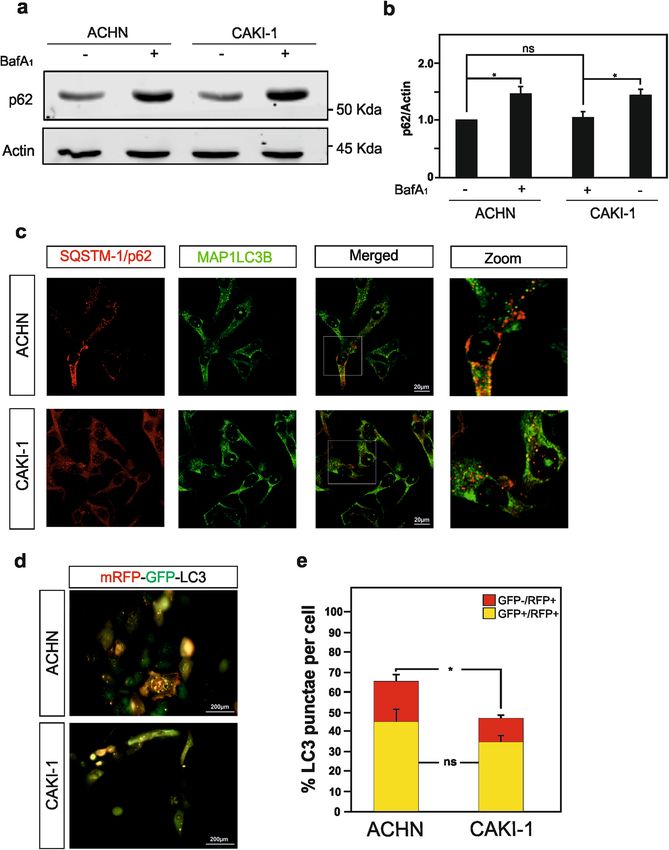
González-Rodríguez et al. Cell Death and Disease (2020)11:69                                                    Page 2 of 15

angiogenesis, glycolysis, lipogenesis, cell cycle, and apop-    stimulate and inhibit autophagy have been proposed as
tosis5. Large-scale cancer genomics high-throughput             cancer therapies19. Likewise, inhibition and induction of
sequencing efforts have brought extra level of molecular        autophagy have both been considered as therapeutic
complexity to ccRCC with the identification of additional        strategies to combat RCC20–24. Additional studies suggest
driver genes that might contribute to the disease, beyond       that autophagic gene polymorphisms are associated with
the VHL gene. Indeed, several genes regulating chromatin        ccRCC risk and patient outcome25,26. Despite the fact that
remodeling, located on chromosome 3p like VHL,                  autophagy is indisputable associated to cytoplasmic
including those of the SWI/SNF chromatin remodeling             events; nuclear events are nowadays considered of
complex (PBRM1 and BAP-1) and the histone modifying             importance for this process. Indeed, this process is tightly
enzyme SETD2 are reported to be frequently mutated in           regulated by epigenetic and associated transcriptional
ccRCC6,7.                                                       programs, with reported central role for several histone
  De facto, the prevalence of SETD2-inactivating muta-          modifying enzymes27–32. However, whether the deficiency
tions in cancer has the highest frequency in ccRCC8.            in the SETD2 histone methyltransferase observed in
SETD2 mutations are observed in ~10% of human ccRCC             ccRCC could impact the autophagic core machinery and
primary tumors, and the frequency dramatically increase         thereby this biological process is yet to be investigated.
to ~30% in metastatic ccRCC patient samples, thereby
suggesting a role for this genetic alteration in driving the    Results
metastatic progression of ccRCC2,3,7,9. The loss of SETD2       SETD2 deficiency in renal cell carcinoma cells is associated
functions correlates with aggressive clinicophatological        with reduced autophagy flux
features, increased risk of recurrence, and predicts a            In order to investigate the impact of SETD2 deficiency
reduced overall and progression-free survival of ccRCC          could have on the autophagic process in RCC cells, the
patients10–12. Collectively, these observations argue for a     ACHN cell line, i.e., SETD2-competent RCC cells, and
role of SETD2 inactivation not only in driving the devel-       the CAKI-1 cell line, i.e., SETD2-deficient RCC cells
opment of tumors, but as well in promoting progression          were selected (Table 1). Worth a note, these particular
of the disease. SETD2, which stands for Su(var), Enhancer       RCC cell lines were also selected based on the fact that
of zeste, Trithorax(SET)-domain containing 2, is a non-         they are wild type for the VHL gene, therefor avoiding
redundant methyltransferase responsible for the tri-            one additional gene deficiency, which could impact on
methylation on residues lysine 36 on histone H3 (i.e.,          the interpretation of the sole effect of SETD2 deficiency.
H3K36me3) in the gene body of actively transcribed              Furthermore, these cell lines are available from cell lines
genes13,14. SETD2-mediated H3K36me3 promotes tran-              bank depositories and are well characterized in term of
scriptional elongation and plays as well important roles in     their SETD2 and VHL genes mutational status33,34.
DNA double-stranded break repair, DNA methylation,              Therefore, first we confirmed by immunoblot analysis
and RNA splicing8. The loss of SETD2 may therefor cause         the presence of SETD2 protein expression in the ACHN
genomic instability, aberrant transcriptional program,          RCC cells, but its absence in the CAKI-1 RCC cells
widespread RNA processing defects, and impact on                (Fig. 1a). In addition, we checked H3K36me3 expression
multiple biological processes ranging from cell prolifera-      levels, the histone posttranslational modification that
tion, cell differentiation, and cell death15.                   requires the enzymatic activity of SETD2, were also
  In the recent years, another biological process, macro-       found to be robustly decreased in the SETD2-deficient
autophagy, referred to hereafter as autophagy, has              CAKI-1 cells as compare to the SETD2-competent
attracted attention in the field of RCC16. Autophagy is a        ACHN cells (Fig. 1b). Next, we monitored the occur-
catabolic process by which cytoplasmic components are           rence of baseline autophagy in these RCC cells, as
degraded by the lysosome, and is involved in both phy-          established by an increased lipidation of the main
siological and pathological conditions17. Autophagy             autophagic marker MAP1LC3B, referred as LC3, as it is
comprises a series of dynamic membrane rearrangements           responsible of substrate recognition to be degraded via
orchestrated by a core set of autophagy-related (ATG)           autophagy and autophagosome formation. Indeed,
proteins18. Autophagy involves the assembly of the pha-         occurrence of autophagy results in an increased ratio of
gophore, followed by the formation of the autophagosome         the lipidated form (LC3-II) to the unlipidated form
that contains the cargo to be degraded. Subsequently,           (LC3-I), known as LC3 conversion. Our results show
autophagosomes fuse with lysosomes, generating auto-            that RCC cells with a SETD2 loss of function exhibit a
lysosomes, breaking down the cargo by lysosomal                 decrease in LC3-II expression level as compared with
enzymes providing energy and macromolecules pre-                RCC cells competent for this enzyme when analyzing
cursors that can be reused. Although autophagy is pri-          the ratio between LC3-II/actin (Fig. 1c, d). Since SETD2
marily a protective process for the cell, it can also           is considered to contribute on the regulation of tran-
contribute to cell death. Hence, interventions to both          scription and expression of specific genes, we wondered

Official journal of the Cell Death Differentiation Association
González-Rodríguez et al. Cell Death and Disease (2020)11:69 Page 3 of 15
ATCC American Type Culture Collection, DZME German Collection of Microorganisms and Cell Culture; ECACC European Collection of Authenticated Cell Cultures, CLS Cell Line Service, COSMIC Catalog of Somatic Mutations
References
whether SETD2 lost-of-function promotes the decrease
found on LC3 at protein level in RCC. For this purpose,
33,55,56
33,56,57
33,56,57
33,57
58,59
60,61
we analyze by reverse transcription polymerase chain
reaction (RT-qPCR) the mRNA expression level of
VHL status; SETD2 status (COSMIC)
LC3B gene in both RCC cell lines, which revealed a
small significant increase in messenger expression
between the SETD2-competent and the SETD2-deficent
cell lines. This data support the idea that the observed
+ for wild-type gene
− for mutated gene
decrease in LC3 at protein level in CAKI-1 cells is not
the result of a differential transcriptional regulation of
VHL−; SETD2−
VHL−; SETD2−
VHL+; SETD2−
VHL−; SETD2+
VHL−; SETD2+
VHL+; SETD2+
MAP1LC3B gene in the SETD2-deficient RCC cells
(Fig. 1f). Moreover, in order to examine whether SETD2
deficiency has an impact on autophagic flux per se, LC3
conversion was assessed in ACHN and CAKI-1 cells in
the presence of a late autophagy inhibitor, Bafilomycin
derived from primary tumor site,
derived from primary tumor site,
derived from primary tumor site,
derived from primary tumor site,
A1 (BafA1), which inhibits the fusion of the autopha-
derived from metastatic site,
derived from metastatic site,
gosome with the lysosome and eventually LC3-II
degradation. LC3-II/LC3-I ratio upon BafA1 treatment
shows that the CAKI-1 cells that harbor a SETD2
mutation showed a decrease of autophagic flux, when
compared with the SETD2-competent AHCN cells
Renal cell carcinoma cell lines, inclusive their SETD2 and VHL status, used in this study.
(Fig. 1c, e).
The ubiquitin-associated protein p62 (also known as
Sequestosome-1), an adapter protein, which possesses a
Renal cell carcinoma,
Renal cell carcinoma,
Renal cell carcinoma,
Renal cell carcinoma,
Renal cell carcinoma,
Renal cell carcinoma,
short-LC3 interaction region that facilitates its direct
interaction with LC3, is also used to monitor autop-
female, 63Y
hagy35,36. p62 is an autophagosome cargo protein that
male, 22Y
male, 49Y
male, 69Y
male, 52Y
male, 77Y
Disease
targets other proteins that bind to it for selective degra-
dation via autophagy. Degradation of p62 is another
widely used readout to monitor autophagic activity as p62
ATCC® HTB-47™; DSMZ ACC-54; ECACC 93120819
directly binds to LC3 and is selectively degraded by
autophagy. Therefore, cells undergoing autophagy should
demonstrate the colocalization of p62 and LC3B. Immu-
ATCC® CRL-1611™; ECACC 88100508
noblot analysis reveals that p62 protein is found to be
ATCC® HTB-46™; DSMZ ACC-731
ATCC® CRL-1933™; CLS #300106
ATCC® HTB-44™; DSMZ ACC-55
expressed at similar level in ACHN and CAKI-1 RCC
cells, and that p62 accumulates in CAKI-1 cells when
autophagy was inhibited using BafA1 treatment (Fig. 2a, b).
Cell line collections
Moreover, immunofluorescence microscopy imaging for
both LC3B and p62 proteins exposed a significant
CLS #300249
decrease in autophagosome formation in CAKI-1 cells, as
compared with ACHN cells, as illustrated by the reduced
LC3B/p62 colocalization (Fig. 2c).
To further confirm the impact of SETD2 deficiency on
the autophagic flux in RCC cells, we took advantage of a
Research resource identifier
tandem reporter construct, mRFP-GFP-LC337. The green
fluorescence of this tandem reporter is attenuated in the
acidic pH lysosomal environment, whereas the mRFP is
RRID:CVCL_1067
RRID:CVCL_0234
RRID:CVCL_0235
RRID:CVCL_1056
RRID:CVCL_5873
RRID:CVCL_1050
not. Therefore, the green fluorescent component of the
composite yellow fluorescence from this mRFP-GFP-LC3
reporter is lost upon autophagosome fusion with a lyso-
some, whereas the red fluorescence remains detectable.
Thus, this probe allows distinction between autophago-
Table 1
RCC-FG2
in Cancer
Caki-1
Caki-2
ACHN
somes (GFP+/RFP+ yellow puncta) and autolysosomes
769-P
A498
(GFP−/RFP+ red puncta). Our results obtained show that
Official journal of the Cell Death Differentiation Association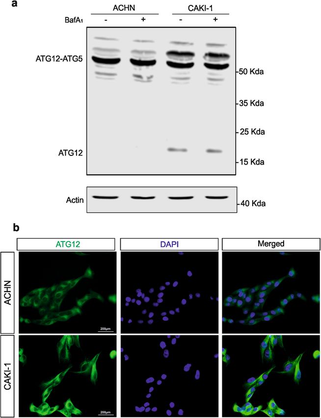
González-Rodríguez et al. Cell Death and Disease (2020)11:69                                                                             Page 4 of 15

  Fig. 1 Characterization of SETD2, H3K36me3, LC3-I, and LC3-II expression levels in ACHN and CAKI-1 RCC cells. Immunoblot analysis of the
  histone modifying enzyme SETD2 (a) and its histone target H3K36me3 (b) expression levels in ACHN and CAKI-1 renal clear carcinoma cells
  confirmed the SETD2 deficiency in the last named RCC cell line. c Immunoblot analysis of LC3 shows decreased expression level in SETD2-deficient
  CAKI-1 cells as compared with SETD2-wild-type ACHN cells. Treatment with Bafilomycin A1 (BafA1, 40 nM) for 4 h shows a significant decrease in LC3-
  II lipidation in CAKI-1 cells as compared with ACHN cells. d Quantification of LC3-II expression compared with the expression of the housekeeping
  gene, actin, with and without BafA1 treatment in both cell lines. e Quantification of LC3-II/LC3-I ratio to monitor autophagic flux in ACHN and CAKI-1
  cells with and without BafA1 treatment demonstrates that cells that lacks SETD2 expression exhibit a decreased autophagic flux. f Analysis of
  MAP1LC3B mRNA expression by qPCR in SETD2-deficient CAKI-1 cells and SETD2-competent ACHN cells. Bars display the mean of four (d, e) or three
  (f) experiments, error bars represent SEM; ***p ≤ 0.001; **p ≤ 0.01; *p ≤ 0.05.

as expected CAKI-1 cells present an overall lower number                      ATG12 complexes and free ATG12 accumulate in SETD2-
of LC3 punctae cells, as well as significantly less red-color-                 deficient renal cell carcinoma cells
labeled LC3 puncta, i.e., autolysosomes. In addition,                           Thereafter, in order to get insights on how SETD2
SETD2-dedicient CAKI-1 cells show a reduced autopha-                          deficiency could impact on the autophagic flux, we
gic flux in the SETD2-deficient cells, as compared with                         undertaken to look at key component of the autophagy
the SETD2-competent cells, were further confirm by the                         core machinery that contribute to the control of autop-
mRFP-GFP-LC3 autophagic flux assay (Fig. 2d, e).                               hagosome formation and expansion. Autophagy relies on
  Collectively, these data indicate a reduced autophagic                      the activities of two ubiquitin-like protein conjugation
flux in the SETD2-deficient CAKI-1 RCC cells, which is                          systems, which catalyze the covalent conjugation of
not due to a transcriptional decrease of LC3 expression, as                   ATG12–ATG5 and therefore addiction of the lipid
compared with the ACHN RCC cells, which carry a wild-                         phosphatidylethanolamine to LC3. A common E1-like
type version of this methyltransferase enzyme.                                enzyme, ATG7, facilitates these reactions. Thereafter,

Official journal of the Cell Death Differentiation Association
González-Rodríguez et al. Cell Death and Disease (2020)11:69                                                                            Page 5 of 15

  Fig. 2 RCC cells deficient for SETD2 expression shows a decrease in autophagic flux. a Immunoblot analysis, and its quantification in b, of
  SQSTM1/p62 expression level in SETD2-deficient CAKI-1 cells and SETD2-competent ACHN cells show a modest increase in p62 protein upon BafA1
  treatment (but not in untreated conditions) in the CAKI-1 cells as compared with ACHN cells. c Immunofluorescence analysis of SQSTM1/p62 and
  MAP1LC3B reveals decreased colocalization of these two proteins in the SETD2-deficient RCC cells. d, e Both RCC cell lines were transfected with the
  tandem reporter construct, mRFP-GFP-LC3 that allows distinguishing between autophagosomes (GFP+/RFP+ yellow puncta) and autolysosomes
  (GFP−/RFP+ red puncta). Representative immunofluorescence images for mRFP-GFP-LC3 transfected ACHN and CAKI-1 cells are depicted in d.
  e Quantification of GFP+/RFP+ yellow puncta and GFP−/RFP+ red puncta in at least 30 cells per experimental condition shows a significant
  decrease on autolysosomes number. Bars display the mean of three experiments, error bars represent SEM; ns nonsignificant; **p ≤ 0.01.

Official journal of the Cell Death Differentiation Association
González-Rodríguez et al. Cell Death and Disease (2020)11:69                                                           Page 6 of 15

ATG12–ATG5 conjugate proceeds to form an active
multimeric complex together with autophagy-related 16-
like 1 (ATG16L1) that localizes to sites of autophagosome
assembly and promote the conjugation of phosphatidy-
lethanolamine to LC3B38,39. In fact, deficiencies in the
Atg5, Atg7, and Atg12 core autophagy genes have all been
reported to impair induced and constitutive autophagy in
mouse models40–42. Hence, ATG7 expression, as well as
the formation of the ATG5–ATG12 covalent complexes
were investigated by immunobloting in ACHN and CAKI-
1 RCC cells. No major differences in ATG7 expression
level were observed in SETD2-deficient versus SETD2-
competent RCC cells (Supplementary Fig. 1a, b). How-
ever, striking differences were noted when ATG12
immunoblot was performed. Indeed, this experimental set
revealed that in the CAKI-1 cells, i.e., SETD2-deficient
RCC cells, exhibit a higher ATG12 expression level, and
detection of immunoreactive bands, in addition to the one
corresponding to the ATG5–ATG12 covalent complex,
for free ATG12 protein, as well as for an additional
ATG12-containing complex around 60 kDa. In contrast,
ACHN cells, SETD2-competent RCC cells, mainly
exhibited the ATG12-immunoreactive band correspond-
ing to the ATG5–ATG12 conjugate but not free ATG12
(Fig. 3a). Immunofluorescence imaging for ATG12 pro-
tein confirmed its higher expression level, in punctate
structure, in CAKI-1 cells as compared with ACHN cells           Fig. 3 Free ATG12 and ATG12-associated complexes expression
                                                                 is increased in SETD2-deficient cells. a Immunoblot analysis of
(Fig. 3b).
                                                                 ATG12 protein in SETD2-deficient CAKI-1 cells and SETD2-competent
   Moreover, to assess whether the accumulation of free          ACHN cells, reveal the accumulation of both free ATG12 and of an
ATG12 and the additional ATG12-containing complex is             additional ATG12-containing complex, distinct from the ATG5–ATG12
SETD2 dependent, four additional RCC cell lines, CAKI-2          complex, in the SETD2-deficient RCC cells. b Immunofluorescence
and 769-P both lines characterized by VHL gene mutation          analysis shows an increase expression for ATG12 in CAKI-1 cells as
                                                                 compared with ACHN cells.
but a wild-type SETD2 gene, as well as A498 and RCC-
FG2 both lines characterized by VHL and SETD2 (loss of
function) gene mutation were analyzed (Table 1).
Immunoblot analysis for SETD2 protein expression, as            Immunofluorescence imaging for ATG12 protein indi-
well as H3K36me3 expression levels for these additional         cated higher expression level in RCC-FG2 RCC cells as
RCC cell lines, which confirm the SETD2 loss of function         compared with 769-P RCC cells (Supplementary Fig. 3e).
in A498 and RCC-FG2 RCC cells are depicted in Sup-              A reduced autophagic flux, as illustrated by reduced LC3-
plementary Figs. 2a, b and 3a, b. We also explored whe-         II/LC3-I ratio upon BafA1 treatment, in the SETD2-
ther VHL gene mutation, the most prevalent reported             deficient A498 and RCC-FG2 RCC cells, as compared
gene deficiency in RCC cells, could impact on the accu-          with the CAKI-2 and 769-P RCC cells, which carry a wild-
mulation of free ATG12 and occurrence of ATG12                  type version of this methyltransferase enzyme was also
additional complex. Immunoblot analysis demonstrated            observed (Supplementary Figs. 2c, d and 3f, g).
that the aberrant accumulation of free ATG12 protein
and of the additional ATG12-containing complex can be           Occurrence of free ATG12 and aberrant ATG12-containing
observed in all RCC cells deficient for SETD2, i.e., A498,       complex in renal cell carcinoma cells is SETD2 dependent
Caki-1, and RCC-FG2 RCC cells, and that the VHL status            Hence, we report that SETD2 deficiency in RCC cells is
of the cells did not impact in the acquisition of these         associated with the aberrant accumulation of both free
characteristics suggesting that the phenotype observed is       ATG12 and of an additional ATG12-containing complex,
clearly due to SETD2 deficiency and VHL does not play            distinct from the ATG5–ATG12 complex, which also
role on ATG12 regulation (Fig. 4a and Supplementary             appears to be associated with reduced autophagic flux in
Fig. 3c, d). Of note, the VHL status of the RCC cells did       these cells. In order to gain evidence that SETD2 defi-
not either impact on ATG7 expression levels. (Fig. 4b, c).      ciency in RCC is directly involved in the acquisition of

Official journal of the Cell Death Differentiation Association
González-Rodríguez et al. Cell Death and Disease (2020)11:69 Page 7 of 15
(Fig. 5a). Remarkably, reintroducing wild-type SETD2 in
CAKI-1 that are otherwise deficient for the enzyme was
enough to significantly reduce the presence of both free
ATG12 and of the additional ATG12-containing complex
in these cells, as shown by immunoblotting analysis
(Fig. 5b). Similar reductions were also observed in CAKI-1
cells overexpressing SETD2 and treated with the late
inhibitor of autophagy BafA1. On the other hand, com-
plementary experiments were performed in which SETD2
gene expression was targeted using a pool of four small-
interfering RNAs (siRNAs) in the ACHN cells, wild type
for the enzyme. As expected, the SETD2 siRNAs pool
leads to robust downregulation of SETD2 protein
expression level in ACHN RCC cells (Fig. 6a). Decrease
SETD2 protein expression on its own was able to promote
the appearance of both free ATG12 and of the additional
ATG12-containing complex in ACHN RCC cells (Fig. 6b).
Blocking the autophagic flux with BafA1 treatment fur-
ther increase the accumulation of these ATG12-
immunoreactive bands in the immunoblots.
SETD2 deficiency in renal cell carcinoma cells is associated
with increase expression of a ATG12 short spliced isoform
Thereafter, we wanted to elucidate how SETD2 defi-
ciency in RCC cells could have an impact on the
expression of ATG12, and potentially control to the
expression of different ATG12 variants that could lead to
occurrence of free ATG12 and additional ATG12-
containing complexes. SETD2-mediated H3K36 tri-
methylation has been implicated in the regulation of
alternative splicing43–45. In fact, posttranslational mod-
ification of histone tails is closely associated with the
regulation of this process and H3K36me3 is of particular
interest as the levels of this specific histone mark differ
based on exon utilization, with alternatively spliced exons
having lower levels of H3K36me3 than those that are
Fig. 4 VHL mutation in A498 and CAKI-2 RCC cells does not
constitutively included44,46. Furthermore, altering SETD2
impact on the accumulation of free ATG12, and ATG12 forming
complexes. a Immunoblot analysis of ATG12 protein in CAKI-2 gene expression levels is enough to influence the inclusion
characterized by VHL gene mutation but a wild-type SETD2 gene, and of exons in genes known to be alternatively spliced43.
A498 characterized by both VHL and SETD2 (loss of function) gene Finally and of importance for the current investigation,
mutation revealed that the accumulation in free ATG12, and the H3K36me3-deficient ccRCC tumors have been reported
ATG12-containing complex, distinct from the ATG5–ATG12 complex,
to show alterations in splicing and evidence of intron
is independent of the VHL gene status. b Immunoblot analysis, and its
quantification in c, of ATG7 expression does not show striking retention15.
differences between those two RCC cell lines. Bars represent the mean In fact, UniProt, database of protein sequence and
of three independent experiments, error bars represent SEM. Ns functional information available at uniport.org, provide
nonsignificant. information about the human ATG12 protein (accession
number O94817). This entry describes the existence of
two isoforms, with respective length of 140 and 74 amino
these defects in the autophagic process, experiments acid residues, suggested to be generated by alternative
aiming at the gain and loss of function of SETD2 in RCC splicing. The isoform 1, the longest one, with identifier
cells were undertaken. O94817-1 is considered as the canonical sequence. The
Rescue of SETD2 functions in the CAKI-1 cells was shortest isoform 2 with identifier O94817-4, which lacks
achieved by the transient overexpression of an expression an exon in the coding region compared with the isoform
vector encoding for the enzyme as shown by RT-qPCR 1, present a shorter and distinct carboxyl-terminus.
Official journal of the Cell Death Differentiation AssociationGonzález-Rodríguez et al. Cell Death and Disease (2020)11:69 Page 8 of 15 Fig. 5 Rescue of SETD2 expression in SETD2-deficient CAKI-1 cells lead to decreased expression of free ATG12 and ATG12-containing complexes and increased of LC3-II lipidation. a–d SETD2-deficient CAKI-1 cells were transfected with an expression vector encoding for SETD2. a qPCR analysis of SETD2 mRNA expression level in SETD2-deficient CAKI-1 cells upon SETD2 overexpression. b Immunoblot analysis of ATG12 protein reveals that rescue of SETD2 expression in CAKI-1 lead to decrease in observed levels of free ATG12, and the ATG12-containing complex, distinct from the ATG5–ATG12 complex. c Immunoblot analysis, and its quantification in d, of LC3B expression upon those conditions shows increase LC3-II lipidation in CAKI-1 cells. Bars represent the mean of three independent experiments, error bars represent SEM. Ns nonsignificant; *p ≤ 0.05; ****p ≤ 0.0001. Whereas the transcript for ATG12 isoform 1 mRNA RCC cells. Taking advantage of the unique sequences originate from four distinct exons, the transcript for the present in their carboxyl-terminus of each ATG12 iso- shortest ATG12 isoform, i.e., isoform 2, only originate form, we selected primers allowing the individual quan- from three exons (Fig. 7a). De facto, the sequence for this tification of their messenger expression by qPCR variant differs from the canonical sequence as follows: an (Supplementary Fig. 4). This mRNA analysis revealed that alternative sequence for residues 56–74 (DILLK- SETD2-deficient RCC cells exhibited a significantly higher AVGDTPIMKTKKWA → YLCESVLCSFPRPRSWNSL), expression of the ATG12 short isoform as compared with and the sequence for residues 75–140 is missing (Fig. 7b). RCC cells carrying a wild-type version of SETD2. The Given the strong evidence for SETD2-dependent reg- overall ATG12 messenger expression, as well as the ratio ulation of alternative splicing, and the suggested existence between ATG12 short isoform versus ATG12 long iso- of ATG12 isoforms issue of alternative splicing for, we form were also found to be increased in the SETD2- speculated that dysregulation of SETD2 might impact on deficient RCC cells (Fig. 7c, d). Collectively, these data the differential expression of these ATG12 isoforms in indicate that deficiency in SETD2, known regulator of Official journal of the Cell Death Differentiation Association
González-Rodríguez et al. Cell Death and Disease (2020)11:69 Page 9 of 15
Fig. 6 Downregulation of SETD2 expression in SETD2-competent ACHN cells lead to increase of an accumulation of free ATG12 and ATG12
complexes and decrease in LC3-II lipidation. a–d Downregulation of SETD2 expression in the SETD2-competent ACHN cells was achieved by
small-interfering RNA targeting SETD2. a Immunoblot analysis for SETD2 confirm the reduction in SETD2 expression in ACHN cells upon treatment
with a siRNA directed against SETD2 for 48 h. b Immunoblot analysis of ATG12 protein reveals that the downregulation of SETD2 expression in the
ACHN SETD2-competent cells promotes the increase in free ATG12 and ATG12-associated complexes. c Analysis of LC3B expression in upon
downregulation of SETD2 expression. Bafilomycin A1 (40 nm) was used for 4 h treatment. d Quantification of LC3-II levels normalized to actin. Bars
represent the analysis of three independent experiments, error bars represent SEM; ***p ≤ 0.001.
alternative splicing, is associated with increased expres- directed against SETD2 leads to a significant decrease in
sion of the ATG12 short isoform to the depend of the LC3-II levels, which was more obvious in the cells treated
canonical ATG12 long isoform in RCC cells. with BafA1 (Fig. 7c, d).
Finally, with the purpose of exploring whether the above
Manipulation of ATG12 expression in renal cell carcinoma described deficiency in autophagy could impact on other
cells impact on their autophagy and cell migration cellular properties of RCC cells, we undertook to analysis
capability whether the manipulation of SETD2 could impact on the
Thereafter, since we could establish that SETD2 status migration capabilities of these cells. Indeed, autophagy
in RCC cells is a key determinant for the aberrant pre- has recently been described as a regulator of cell migra-
sence of free ATG12 and additional ATG12-containing tion48,49. Confluent RCC cell monolayers were subjected
complex in these cells, we decided to investigate how to a wound-healing assay to monitor cell motility. ACHN,
SETD2 manipulation in these cells could affect the SETD2-competent cells, were transfected with siRNAs
autophagic flux per se. The SETD2 rescue in CAKI-1 not targeting SETD2 or as control scramble siRNAs, whereas
only impacted on the presence of the additional ATG12- CAKI-1, SETD2-deficient cells, were transfected with an
immunoreactive bands in immunoblots, but as well on the expression vector encoding for SETD2, or an empty
autophaghic flux as demonstrated by the increased LC3 expression vector as control, 24 h before wounding.
conversion even observed in the presence of BafA1 Confluent cell cultures were scraped with a pipet tip to
treatment (Fig. 5c, d). When the autophagic flux was create a cell-free wound and images were captured at the
tested in ACHN cells, looking at LC3 conversion, it beginning and at regular intervals during cell migration to
appears that SETD2 downregulation using siRNA close the wound.
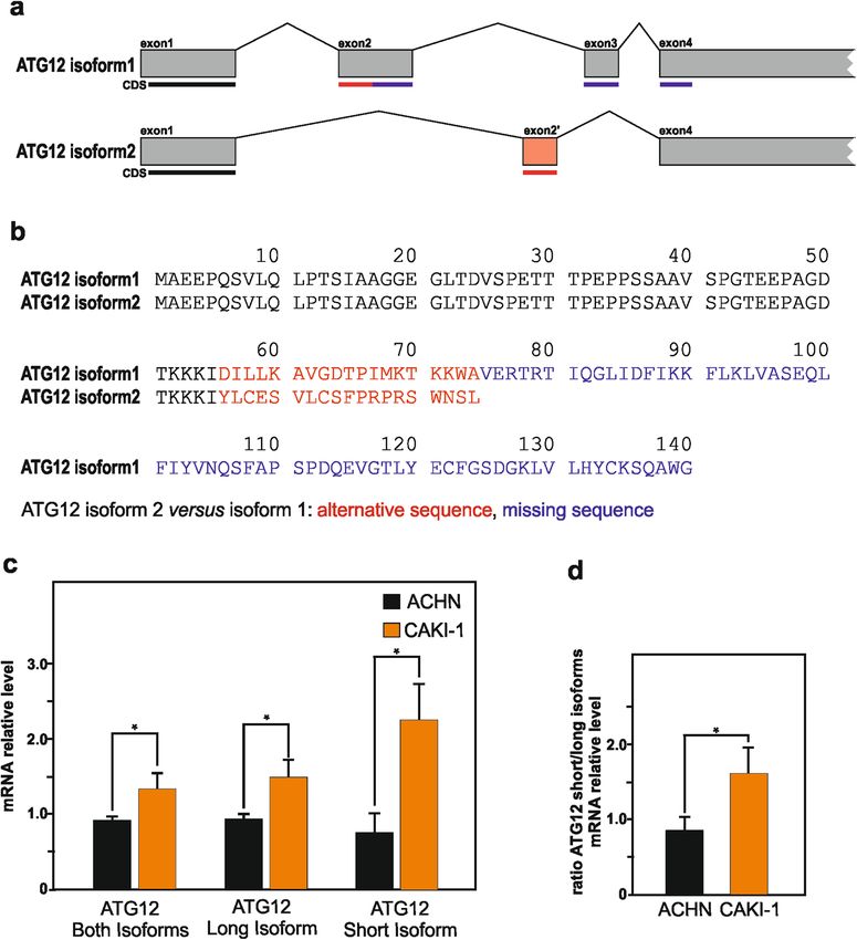
Official journal of the Cell Death Differentiation AssociationGonzález-Rodríguez et al. Cell Death and Disease (2020)11:69 Page 10 of 15 Fig. 7 SETD2 deficiency in RCC cells is associated with increase expression of a ATG12 short spliced isoform. a Illustration of ATG12 long isoform 1 and ATG12 short isoform exons and introns structures, as well as indication of the location of the coding sequences (CDS) in the exons. b Amino acid sequences for ATG12 long isoform 1 (with UniProt identifier O94817-1) and ATG12 short isoform 2 (with UniProt identifier O94817-4). For the two isoforms, shared amino acid sequences are reported in black color, alternative sequences are reported in red color, whereas the missing sequence in the short isoform is reported in blue color in the long isoform. c, d qPCR analysis of mRNA expression level for ATG12 short and long isoforms, or both of them, in SETD2-deficient CAKI-1 cells and SETD2-competent ACHN cells revealed significantly higher expression (c), as well as increased ratio between ATG12 short isoform versus ATG12 long isoform (d) in SETD2-deficient RCC cells. Bars represent the analysis of three independent experiments, error bars represent SEM; *p ≤ 0.05. A significant increase in wound-healing (cell motility) In clear cell RCC patients SETD2 and ATG12 gene activity was seen in ACHN cells in which the expression expression levels are associated with favorable respective of SETD2 had been targeted by an antisense approach unfavorable prognosis velocity, as compared with the ACHN cells control, Since we observe that SETD2 deficiency is associated transfected with a scramble siRNA (Supplementary with the appearance of free ATG12, as well as the Fig. 5a, b). Quite the opposite, CAKI-1 cells in which the expression additional ATG12-containing complexes, and expression of SETD2 had been restored by overexpression an overall increase in total ATG12 protein expression exhibited a substantial decrease in the migration rate levels, we decided to investigate whether ATG12 gene (Supplementary Fig. 5c, d). Thus, these data establish that expression levels, as well as the expression of the gene, i.e., SETD2 expression in RCC cells impact on their have cell SETD2, which product is causing the global increase in migration capability. ATG12 expression levels, could be used as survival Official journal of the Cell Death Differentiation Association
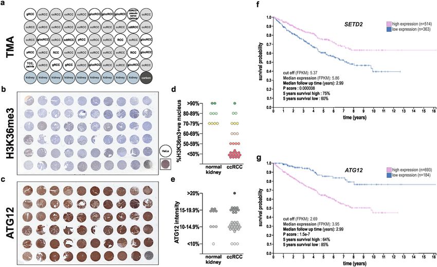
González-Rodríguez et al. Cell Death and Disease (2020)11:69                                                                                  Page 11 of 15

  Fig. 8 Low H3K36me3, but high ATG12 protein expression levels are observed in ccRCC tumors, and SETD2 and ATG12 gene expression
  levels have prognostic values for ccRCC patients. a Human kidney tissue microarrays including 30 tissues classified as ccRCC tumors (highlighted
  in light gray) were proceed for immunohistochemical staining for H3K36me3 (b) and ATG12 protein (c) and revealed low H3K36me3 (d) but high
  ATG12 (e) protein expressions. HeLa were used as a positive control for H3K36me3 protein expression in b. f, g SETD2 and ATG12 high versus low
  gene expression levels (expressed in FPKM and with a cut off of 5.37 for SETD2 and 4.66 for ATG12) in tumor tissue derived from ccRCC patients at the
  time of diagnosis and their correlation to patient survival, i.e., follow up after diagnosis (expressed in years) was extracted from the pathology atlas of
  the human cancer transcriptome available at the Human Protein Atlas (www.proteinatlas.org). Survival analysis, prognosis of each group of patients
  examined using Kaplan–Meier survival estimators, and survival outcomes of the groups compared by log-rank tests revealed that SETD2 should be
  considered as a favorable prognostic gene and ATG12 as an unfavorable prognostic gene for ccRCC patients.

prognostic factors for ccRCC patients. First, we took                            between expression level and patient survival was exam-
advantage of a human kidney TMA that include 30                                  ined. The prognosis of each group of patients was
ccRCC tumors (Fig. 8a). Immunohistochemistry analysis                            examined by the Human Protein Atlas using
of H3K36me3 expression levels, used as a readout for                             Kaplan–Meier survival estimators, and the survival out-
SETD2 enzymatic activity, as well as ATG12 protein                               comes of the two groups were compared by log-rank tests.
expression, confirmed low H3K36me3, but high ATG12                                Supporting our findings, the analysis of these gene
expression levels in most of the ccRCC tumors biopsies                           expression data set revealed that SETD2 should be con-
(Fig. 8b–e and Supplementary Fig. 6a–c). Thereafter, we                          sidered as a favorable prognostic gene whereas ATG12 as
looked at the available gene expression data sets from the                       an unfavorable prognostic gene for ccRCC patients
pathology atlas of the human cancer transcriptome47                              (Fig. 8f, g).
available at the Human Protein Atlas to explore whether
the expression levels of the SETD2 and ATG12 genes                               Discussion
could both be associated with a survival prognosis for                             In recent years, SETD2 has attracted interest as a potential
patients suffering from ccRCC. Based on the Fragments                            tumor suppressor gene, whose inactivation would therefor
Per Kilobase of transcript, per Million mapped reads                             participate to tumor initiation and progression for a wide
(FPKM) value of each gene, ccRCC patients were classi-                           range of human tumors, including epithelial, central nervous
fied into two expression groups, high and low expression                          system, and hematopoietic tumors6,50–53. However, SETD2-
(a FPKM of 5.37 and 4.66 were used as cut off for SETD2                          inactivating mutations distinguish themselves in the ccRCC
and ATG12 expression, respectively) and the correlation                          cancer, where they are most prevalent than in any other

Official journal of the Cell Death Differentiation Association
González-Rodríguez et al. Cell Death and Disease (2020)11:69                                                                      Page 12 of 15

Table 2 Antibody used in this study.

                                                     Research resource identifier                  Application             Sources

β-actin (mouse mAb)                                  RRID:AB_262137                               Western blot            Sigma Aldrich (A-3853)
ATG7 (rabbit mAb)                                    RRID:AB_10625656                             Western blot            Genetex (GTX61647)
ATG12 (rabbit pAb)                                   RRID:AB_10703973                             Western blot, IF        Abcam (ab155589)
G3PDH (rabbit pAb)                                   RRID:AB_2107456                              Western blot            Trevigen (2275-PC-100)
H3K36me3 (rabbit pAb)                                RRID:AB_1950412                              Western blot            Cell Signaling (4909)
LC3B (rabbit pAb)                                    RRID:AB_796155                               Western blot            Sigma Aldrich (L7543)
SETD2 (rabbit pAb)                                   RRID:AB_2811237                              Western blot            Genetex (GTX127905)
SQSTM1/p62 (mouse mAb)                               RRID:AB_945626                               Western blot, IF        Abcam (ab56416)

cancer type8,50. Furthermore, these mutations correlate with                       with the discovery that the prevalent SETD2 gene muta-
aggressive clinicophatological features, and are associated                        tion in ccRCC, directly impact on this autophagic core
with an unfavorable prognosis in patients with ccRCC10–12.                         machinery and thereby reduce the autophagic flux. We
Thus, given its frequency of inactivation in ccRCC, its cri-                       also expose that SETD2, as well as ATG12 gene expres-
tical role as a tumor suppressor, and its possible use as                          sion, can present prognostic value for ccRCC patients,
progression marker in ccRCC, effort have been placed to                            where low SETD2 expression but high ATG12 gene
elucidate the biological consequences of SETD2 loss of                             expressions are associated with an unfavorable prognosis.
function in ccRCC cells. Here, we report that SETD2-                               Thus, we argue that the SETD2 gene status of ccRCC
inactivating mutations have a significant impact on the                             tumors should be taken into account when therapeutic
autophagy. Mechanistically, we uncovered a SETD2 loss of                           intervention aiming at the autophagic process is under-
function-dependent occurrence of an aberrant ATG12-                                taken. This consideration could contribute to better
containing complex, in addition to the conventional ATG5/                          prognosis of such interventions, including in the clinics,
ATG12 covalent complex, as well as increased of free                               and provide more personalized cancer therapy for ccRCC
ATG12, in RCC cells carrying the SETD2 gene mutation.                              patients.
Furthermore, we confirmed the existence of two distinct
ATG12 isoforms that had been suggested in protein and                              Material and methods
gene databases, a canonical long isoform and a short isoform                       Cell culture
generated by alternative splicing, and revealed that SETD2                           ACHN, CAKI-1, CAKI-2, RCC-FG2, A498, and 769-P
deficiency in RCC cells promote a significant increase in the                        human ccRCC cell lines33,55–61, described in Table 1, were
expression levels of the short isoform. Whereas, the pre-                          cultured in Dulbecco’s modified Eagle’s Medium
sence of the short ATG12 isoform in SETD2-deficient cells                           (DMEM) with Glutamax®, supplemented with 10% fetal
is responsible for the presence of additional ATG12-                               bovine serum and 1% with Penicillin/Streptomycin
containing complex and free ATG12, which appears based                             (Gibco). The cells were grown in 75cm2-flasks under
on its molecular weight to be the long ATG12 isoform,                              standard conditions at 37 °C and 5% CO2.
potentially by competing it will require further investiga-
tions. The apparent defect in the ATG12-dependent con-                             Reagents and antibodies
jugation system was associated with decrease autophagic                              Bafilomycin A1 was purchased from Santa Cruz Bio-
flux, in accordance with the role for this ubiquitin-like                           technology (sc-201550). Information about antibodies
protein conjugation system in autophagosome formation                              used in this study are provided in Table 2.
and expansion38,39. Considering the impact autophagy can
have on cancer cells survival or dismiss54, targeting the                          Transfection of ACHN RCC cells with SETD2 siRNA
autophagy process has been considered as potential ther-                             ACHN human RCC cells were seeded in six-well-plate
apeutic strategy to combat various type of tumors, including                       dishes and transfected 24 h after platting either with
in patient with ccRCC20–24.                                                        nontargeting siRNA (used as control) or siRNA against
   Previous studies reported that polymorphisms in                                 SETD2 (50 nM) with 6 µL of lipofectamine 3000 (Invi-
autophagic gene are associated with ccRCC risk and                                 trogen), respectively. SETD2 (L-012448-00) with the fol-
patient outcome, indicating deficiencies in the autophagic                          lowing four validated siRNAs: UAAAGGAGGUAUAU
core machinery impact in ccRCC patients25,26. Our                                  CGAAU, GAGAGGUACUCGAUCAUAA, GCUCAGAG
investigation, bring an additional level of understanding,                         UUAACGUUUGA, and CCAAAGAUUCAGACAUAUA

Official journal of the Cell Death Differentiation Association
González-Rodríguez et al. Cell Death and Disease (2020)11:69 Page 13 of 15
and nontargeting ON-TARGET (D-001810) SMARTpool and quantification was performed using the ImageJ
siRNAs with the following four validated siRNAs: UGG software.
UUUACAUGUCGACUAA, UGGUUUACAUGUUGU-
GUGA, UGGUUUACAUGUUUUCUGA, and UGGUUU Immunofluorescence
ACAUGUUUUCCUA, were purchased from Dharmacon. ccRCC cells were seeded in six-well plates. The fol-
The cells were treated when indicated, harvested and lowing day cells were washed twice with PBS 1× and fix
analyzed 48 h after transfection. with 4% paraformaldehyde for 15 min at room tempera-
ture. Cells were then incubated with blocking/permeabi-
SETD2 overexpression in SETD2-deficient RCC cells lization (B/P) buffer (10 mM HEPES, 0.3% Tx-100, 3%
CAKI-1 and A498 RCC cells were seeded in six-well BSA) for 1 h at room temperature, following with the
plates. The following day, cells were transfected with 2 µg corresponding antibodies diluted in B/P buffer overnight
of SETD2 plasmid or pcDNA3.1, respectively, with lipo- at 4 °C. The following day, the samples were washed three
fectamine 3000 by following the manufacturer’s protocol. times for 5 min in PBS 1× and incubated with the sec-
24 h after transfection, cells were subjected to the corre- ondary antibody in second B/P buffer (0.2% Tween 20, 3%
sponding analysis. BSA) for 1 h at room temperature. Cells were washed two
times with 1× PBS and mounted with in situ mounting
mRFP-GFP-LC3 assay medium with DAPI (Duolink®). Secondary antibodies,
Cells were grown on coverslips in six-well dishes and Alexa Fluor® 488 Goat anti-Rabbit IgGs (1:500) and Alexa
after 24 h were transfected with lipofectamine 3000 Fluor® Goat anti-Mouse IgGs (1:500) (Invitrogen), were
(Invitrogen) and 2 µg of the mRFP-GFP-LC3 plasmid per used. Samples were analyzed with Axio Zoom V16, Stemi
well. After 24 h transfection, cells were fixed by incuba- 305, Zeiss Microscope.
tion in 4% paraformaldehyde for 7 min at room tem-
perature followed by nuclei staining using in situ Quantitative real time PCR
mounting medium with DAPI (Duolink®). Autophagy flux ACHN and CAKI-1 cells were seeded in six-well plates
was determined counting the number of LC3 positive and harvested after 24 h. Total RNA isolation was per-
cells. The mRFP-GFP-LC3 plasmid was a kind gift of Dr formed following the manufacturer’s instructions of
Tamotsu Yoshimori (Osaka University, Japan)37. Samples RNeasy mini kit (Qiagen). RNA quantifications of the
were analyzed with Axio Zoom V16, Stemi 305, Zeiss different samples were determined using a NanoDrop®
Microscope. spectrophotometer (Thermo Fisher Scientific). cDNA was
synthesized from 1 µg of RNA using Oligo (dT) primers,
Western blot analysis dNTP mix, and superscript IV (Invitrogen). qPCR was run
Cells were seeded in six-well plates and transfected on an ABI 7500 (Applied Biosystems) using SYBR™ Green
the day after (see above). The cells were harvested by Master Mix (Qiagen). GAPDH was used as a house-
using a cell scraper and lysed with Laemmli buffer keeping gene. QPCR statistical analysis was performed
(62.5 mM Tris-HCl pH 6.8, 2% SDS, 10% Glycerol, 5% using R. Primers used:
β-mercaptoethanol, and 0.02% Bromophenol Blue). MAP1LC3B_rev:
Obtained samples were sonicated (Bioruptor® Pico, 5′-CTGTAAGCGCCTTCTAATTATC-3′;
Diagenode) and boiled 8 min at 99 °C. Whole-cell lysate MAP1LC3B_fwd:
was resolved by SDS-PAGE gels (8 or 15% acrylamide) 5′-ATAGAACGATACAAGGGTGAG-3′;
and transferred onto nitrocellulose membranes (0.2 or SETD2_rev: 5′- CTCCTTTAGGTCTTTCCAAC-3′;
0.45 µm-pores) using wet transfer (Bio-Rad). The SETD2_fdw: 5′-AGAACAGCCAGATAAAACAG-3′;
membranes were blocked with 5% milk in PBS-Tween GAPDH_rev: 5′-TTTTTGGTTGAGCACAGG-3′;
20 0.1% for 1 h and incubated overnight at 4 °C with the GAPDH_fwd: 5′-ACAGTTGCCATGTAGACC-3′.
corresponding primary antibody. Furthermore, fol- Location and sequence of the primers used for quanti-
lowed by incubation with the appropriate IRDye® Sec- fication of the ATG12 long and short isoform, or both of
ondary Antibody (LI-COR, 1:10,000) for 1 h at room them are indicated in Supplementary Fig. 4.
temperature 20–25 °C. Immunoblot with anti-β-actin
or G3PDH antibody was used for standardization of Tissue microarray analysis
protein loading. Details about antibodies used in this The human kidney tissue microarray (TMA) consisted
study can be found in Table 2. Bands were visualized of 59 cores of patients with renal tumors among those 9
using an Odyssey CLx infrared imaging system (LI- cores correspond to normal tissue adjacent to the cancer.
COR Bioscience). All targeted proteins of interest were The TMA (Novus Biologicals; NBP2-30220) consists of
normalized to the selected housekeeping gene, intensity tissue sections of 4 μm thickness, prepared from the TMA
of the bands were verified within the same linear range formalin-fixed paraffin-embedded tissue blocks and
Official journal of the Cell Death Differentiation AssociationGonzález-Rodríguez et al. Cell Death and Disease (2020)11:69 Page 14 of 15
immunostaining was performed as following. First, slides SETD2: proteinatlas.org/ENSG00000145782-ATG12/
were dried for 1 h in an oven at 60°. For deparaffinization pathology/renal+cancer/KIRC.
process, slides were submerged in xylene for 20 min, ATG12: proteinatlas.org/ENSG00000181555-SETD2/
rehydration using descanting ethanol concentrations pathology/renal+cancer/KIRC.
(100–95–70%) to water (5 min, two times) and antigen
retrieval for 20 min under microwave treatment. The AR9 Statistical analysis
buffer (1:10 dilution in peroxidase-free water; Perkin For the in vitro analysis, statistical analysis was per-
Elmer, Boston, MA, USA) was used as the unmasking formed using GraphPad Prism (GraphPad Software,
buffer, while sections were then cooled in washing buffer Version 6.0), the threshold for statistical significance was
(PBS-Tween 20, 0.1%). Peroxide and protein block were considered when the p-value was equal or less than 0.05.
then performed for 10 and 5 min respectively with ready- For the survival analysis, the prognosis of each group of
to-use reagents (Ultravision LP Detection System HRP patients was examined by the Human Protein Atlas using
DAB, ThermoScientific, Waltham, MA, USA) before the Kaplan–Meier survival estimators, and the survival out-
application of primary antibody. Primary antibodies used comes of the two groups were compared by log-rank tests.
were directed against Tri-Methyl-Histone H3 (Lys36)
Acknowledgements
(1:100; (D5A7) XP® Rabbit monoclonal antibody; Cell We thank Dr. Andreas Lundqvist (Karolinska Institutet) and Dr Tamotsu
signaling (#4909)) and ATG12 (1:100; Rabbit polyclonal Yoshimori (Osaka University) for kindly providing us with the CAKI-1 cells, and
antibody, Abcam (ab155589)) and left overnight at 4°. The the mRFP-GFP-LC3 plasmid respectively. We are grateful to Dr. Daniel J.
Klionsky and Hana Popelka (University of Michigan) for helpful discussion. This
day after, primary antibody enhancer and secondary research is supported by the Swedish Research Council, the Swedish Brain
detection HRP-Polymer reagents applied were ready to Foundation, the Swedish Cancer Foundation, and the Swedish Cancer Society
use (Ultravision LP Detection System HRP DAB, Ther- (B.J.), the Karolinska Institutet Foundation (P.G.R. and B.J.). Open access funding
provided by Karolinska Institute.
moScientific, Waltham, MA, USA) and were combined
with DAB chromogenic substrate (ThermoScientific, Author details
Waltham, MA, USA). Counterstaining was performed 1
Institute of Environmental Medicine, Toxicology Unit, Karolinska Institutet,
using hematoxylin, sections were de-hydrated with 17177 Stockholm, Sweden. 2Department of Oncology-Pathology, Karolinska
Institutet, 17177 Stockholm, Sweden. 3Present address: Department of
ascending concentrations of ethanol (70–95–100%- Neuroscience, Uppsala University, Uppsala, Sweden
xylene) and slides were coversliped using a permanent
mounting media (Pertex, Histolab, Gothenburg, Sweden). Conflict of interest
The authors declare that they have no conflict of interest.
TMA slides were digitally scanned at an original
magnification of 20× using Glissando High-
Performance Desktop Scanner (Objective Imaging Ltd, Publisher’s note
Cambridge, UK) and immunohistochemical expression Springer Nature remains neutral with regard to jurisdictional claims in
published maps and institutional affiliations.
scoring was performed using ImageJ software v. 1.48
(NIH, Bethedsa, MD, USA) by two independent inves- Supplementary Information accompanies this paper at (https://doi.org/
tigators. For the tri-methyl-histone H3 (Lys36) antibody 10.1038/s41419-020-2266-x).
expression, the number of cells with nuclear marker
Received: 16 September 2019 Revised: 10 January 2020 Accepted: 13
positivity was estimated and their percentage out of the
January 2020
total cell number per TMA was calculated. For the
ATG12 expression, the median intensity score (in pix-
els) was measured for every TMA core. Healthy/normal
renal tissue was served as an internal control of IHC References
expression for both antibodies. HeLa cells were used as 1. Hsieh, J. J. et al. Renal cell carcinoma. Nat. Rev. Dis. Prim. 3, 17009 (2017).
2. Sato, Y. et al. Integrated molecular analysis of clear-cell renal cell carcinoma.
a positive control for tri-methyl-histone H3 (Lys36)
Nat. Genet. 45, 860–867 (2013).
antibody expression. 3. Gerlinger, M. et al. Genomic architecture and evolution of clear cell renal cell
carcinomas defined by multiregion sequencing. Nat. Genet. 46, 225–233
(2014).
Survival analysis
4. Maxwell, P. H. et al. The tumour suppressor protein VHL targets hypoxia-
SETD2 and ATG12 gene expression levels (expressed in inducible factors for oxygen-dependent proteolysis. Nature 399, 271–275
FPKM) in the tumor tissue at the time of diagnosis and (1999).
5. Yao, X. et al. VHL deficiency drives enhancer activation of oncogenes in clear
their correlation to patient survival, i.e., follow up after
cell renal cell carcinoma. Cancer Discov. 7, 1284–1305 (2017).
diagnosis (expressed in years) were extracted from the 6. Dalgliesh, G. L. et al. Systematic sequencing of renal carcinoma reveals inac-
pathology atlas of the human cancer transcriptome avail- tivation of histone modifying genes. Nature 463, 360–363 (2010).
7. Cancer Genome Atlas Research Network. Comprehensive molecular char-
able at the Human Protein Atlas (www.proteinatlas.org)47.
acterization of clear cell renal cell carcinoma. Nature 499, 43–49 (2013).
Raw data related to SETD2 and ATG12 for ccRCC tumors 8. Li, J. et al. SETD2: an epigenetic modifier with tumor suppressor functionality.
are available at the following URLs: Oncotarget 7, 50719–50734 (2016).
Official journal of the Cell Death Differentiation AssociationGonzález-Rodríguez et al. Cell Death and Disease (2020)11:69 Page 15 of 15
9. Hsieh, J. J. et al. Genomic biomarkers of a randomized trial comparing first-line 34. Feng, C. et al. PI3Kβ inhibitor TGX221 selectively inhibits renal cell carcinoma
everolimus and sunitinib in patients with metastatic renal cell carcinoma. Eur. cells with both VHL and SETD2 mutations and links multiple pathways. Sci.
Urol. 71, 405–414 (2017). Rep. 5, 9465 (2015).
10. Wang, J. et al. Prognostic value of SETD2 expression in patients with meta- 35. Pankiv, S. et al. p62/SQSTM1 binds directly to Atg8/LC3 to facilitate degra-
static renal cell carcinoma treated with tyrosine kinase inhibitors. J. Urol. 196, dation of ubiquitinated protein aggregates by autophagy. J. Biol. Chem. 282,
1363–1370 (2016). 24131–24145 (2007).
11. Hakimi, A. A. et al. Adverse outcomes in clear cell renal cell carcinoma 36. Komatsu, M. et al. Homeostatic levels of p62 control cytoplasmic inclusion
with mutations of 3p21 epigenetic regulators BAP1 and SETD2: a report body formation in autophagy-deficient mice. Cell 131, 1149–1163 (2007).
by MSKCC and the KIRC TCGA research network. Clin. Cancer Res. 19, 37. Kimura, S., Noda, T. & Yoshimori, T. Dissection of the autophagosome
3259–3267 (2013). maturation process by a novel reporter protein, tandem fluorescent-tagged
12. Liu, L. et al. Loss of SETD2, but not H3K36me3, correlates with aggressive LC3. Autophagy 3, 452–460 (2007).
clinicopathological features of clear cell renal cell carcinoma patients. Biosci. 38. Feng, Y., He, D., Yao, Z. & Klionsky, D. J. The machinery of macroautophagy. Cell
Trends 11, 214–220 (2017). Res. 24, 24–41 (2014).
13. Sun, X. J. et al. Identification and characterization of a novel human histone H3 39. Galluzzi, L. et al. Molecular definitions of autophagy and related processes.
lysine 36-specific methyltransferase. J. Biol. Chem. 280, 35261–3571 (2005). EMBO J. 36, 1811–1836 (2017).
14. Edmunds, J. W., Mahadevan, L. C. & Clayton, A. L. Dynamic histone H3 40. Kuma, A. et al. The role of autophagy during the early neonatal starvation
methylation during gene induction: HYPB/Setd2 mediates all H3K36 tri- period. Nature 432, 1032–1036 (2004).
methylation. EMBO J. 27, 406–420 (2008). 41. Komatsu, M. et al. Impairment of starvation-induced and constitutive autop-
15. Simon, J. M. et al. Variation in chromatin accessibility in human kidney cancer hagy in Atg7-deficient mice. J. Cell Biol. 169, 425–434 (2005).
links H3K36 methyltransferase loss with widespread RNA processing defects. 42. Malhotra, R., Warne, J. P., Salas, E., Xu, A. W. & Debnath, J. Loss of Atg12, but not
Genome Res. 24, 241–259 (2014). Atg5, in pro-opiomelanocortin neurons exacerbates diet-induced obesity.
16. Cao, Q. & Bai, P. Role of autophagy in renal cancer. J. Cancer 10, 2501–2509 Autophagy 11, 145–154 (2015).
(2019). 43. Luco, R. F. et al. Regulation of alternative splicing by histone modifications.
17. Levine, B. & Kroemer, G. Autophagy in the pathogenesis of disease. Cell 132, Science 327, 996–1000 (2010).
27–42 (2008). 44. de Almeida, S. F. et al. Splicing enhances recruitment of methyltransferase
18. Mizushima, N., Yoshimori, T. & Ohsumi, Y. The role of Atg proteins in autop- HYPB/Setd2 and methylation of histone H3 Lys36. Nat. Struct. Mol. Biol. 18,
hagosome formation. Annu. Rev. Cell Dev. Biol. 27, 107–132 (2011). 977–983 (2011).
19. Levy, J. M. M., Towers, C. G. & Thorburn, A. Targeting autophagy in cancer. Nat. 45. Zhu, K. et al. SPOP-containing complex regulates SETD2 stability and
Rev. Cancer 17, 528–542 (2017). H3K36me3-coupled alternative splicing. Nucleic Acids Res. 45, 92–105 (2014).
20. Russell, K. L. et al. Inhibiting autophagy in renal cell cancer and the associated 46. Kolasinska-Zwierz, P. et al. Differential chromatin marking of introns and
tumor endothelium. Cancer J. 25, 165–177 (2019). expressed exons by H3K36me3. Nat. Genet. 41, 376–381 (2009).
21. Li, M. L. et al. Chloroquine potentiates the anticancer effect of sunitinib on 47. Uhlen, M. et al. A pathology atlas of the human cancer transcriptome. Science
renal cell carcinoma by inhibiting autophagy and inducing apoptosis. Oncol. 357, eaan2507 (2017).
Lett. 15, 2839–2846 (2018). 48. Xu, Z. & Klionsky, D. J. Autophagy promotes cell motility by driving focal
22. Santoni, M. et al. Emerging strategies to overcome the resistance to current adhesion turnover. Autophagy 12, 1685–1686 (2016).
mTOR inhibitors in renal cell carcinoma. Biochim. Biophys. Acta 1845, 221–231 49. Kenific, C. M., Wittmann, T. & Debnath, J. Autophagy in adhesion and
(2014). migration. J. Cell Sci. 129, 3685–3693 (2016).
23. Zhang, Y. et al. Thymoquinone inhibits the metastasis of renal cell cancer cells 50. Fahey, C. C. & Davis, I. J. Setting the stage for cancer development: setd2 and
by inducing autophagy via AMPK/mTOR signaling pathway. Cancer Sci. 109, the consequences of lost methylation. Cold Spring Harb. Perspect. Med. 7,
3865–3873 (2018). a026468 (2017).
24. Serrano-Oviedo, L. et al. Autophagic cell death associated to Sorafenib in renal 51. Al Sarakbi, W. et al. The mRNA expression of SETD2 in human breast cancer:
cell carcinoma is mediated through Akt inhibition in an ERK1/2 independent correlation with clinico-pathological parameters. BMC Cancer 9, 290 (2009).
fashion. PLoS ONE 13, e0200878 (2018). 52. Zhang, J. et al. The genetic basis of early T-cell precursor acute lymphoblastic
25. Wang, Z. et al. Association of ATG7 polymorphisms and clear cell renal cell leukaemia. Nature 481, 157–163 (2012).
carcinoma risk. Curr. Mol. Med. 19, 40–47 (2019). 53. Zhu, X. et al. Identification of functional cooperative mutations of SETD2 in
26. Santoni, M. et al. Autophagic gene polymorphisms in liquid biopsies and human acute leukemia. Nat. Genet. 46, 287–293 (2014).
outcome of patients with metastatic clear cell renal cell carcinoma. Anticancer 54. Amaravadi, R., Kimmelman, A. C. & White, E. Recent insights into the function
Res. 38, 5773–5782 (2018). of autophagy in cancer. Genes Dev. 30, 1913–1930 (2016).
27. Füllgrabe, J. et al. The histone H4 lysine 16 acetyltransferase hMOF regulates 55. Kochevar., J. Blockage of autonomous growth of ACHN cells by anti-renal cell
the outcome of autophagy. Nature 500, 468–471 (2013). carcinoma monoclonal antibody 5F4. Cancer Res. 15, 2968–2972 (1990).
28. Artal-Martinez de Narvajas, A. et al. Epigenetic regulation of autophagy by the 56. Feng, C. et al. PI3Kβ inhibitor TGX221 selectively inhibits renal cell carcinoma
methyltransferase G9a. Mol. Cell Biol. 33, 3983–3993 (2013). cells with both VHL and SETD2 mutations and links multiple pathways. Sci.
29. Füllgrabe, J., Klionsky, D. J. & Joseph, B. The return of the nucleus: transcrip- Rep. 5, 9465 (2015).
tional and epigenetic control of autophagy. Nat. Rev. Mol. Cell Biol. 15, 65–74 57. Fogh, J., Fogh, J. M. & Orfeo, T. One hundred and twenty-seven cultured
(2014). human tumor cell lines producing tumors in nude mice. J. Natl. Cancer Inst.
30. Wei, F. Z. et al. Epigenetic regulation of autophagy by the methyltransferase 59, 221–226 (1977).
EZH2 through an MTOR-dependent pathway. Autophagy 11, 2309–2322 58. Högernann, I., Bock, S., Heppner, P. & Petrides, P. E. Cytogenetic and growth
(2015). factor gene analysis of a renal carcinoma cell line. Cancer Genet. Cytogenet. 78,
31. Shin, H. J. et al. AMPK-SKP2-CARM1 signalling cascade in transcriptional reg- 175–180 (1994).
ulation of autophagy. Nature 534, 553–557 (2016). 59. Gnarra, J. R. et al. Mutations of the VHL tumour suppressor gene in renal
32. Baek, S. H. & Kim, K. I. Epigenetic control of autophagy: nuclear events gain carcinoma. Nat. Genet. 7, 85–90 (1994).
more attention. Mol. Cell 65, 781–785 (2017). 60. Williams, R. D., Elliott, A. Y., Stein, N. & Fraley, E. E. In vitro cultivation of human
33. Brodaczewska, K. K., Szczylik, C., Fiedorowicz, M., Porta, C. & Czarnecka, A. M. renal cell cancer. II. Characterization of cell lines. In Vitro 14, 779–786 (1978).
Choosing the right cell line for renal cell cancer research. Mol. Cancer 15, 83 61. Williams, R. D. Human urologic cancer cell lines. Investig. Urol. 17, 359–363
(2016). (1980).
Official journal of the Cell Death Differentiation AssociationYou can also read