Nearest neighbor amino acids of specificity determining residues influence the activity of engineered Cre type recombinases - Nature
←
→
Page content transcription
If your browser does not render page correctly, please read the page content below
www.nature.com/scientificreports
OPEN Nearest‑neighbor amino acids
of specificity‑determining residues
influence the activity of engineered
Cre‑type recombinases
Anjali Soni1, Martina Augsburg2, Frank Buchholz2 & M. Teresa Pisabarro1*
The tyrosine-type site-specific DNA recombinase Cre recombines its target site, loxP, with high
activity and specificity without cross-recombining the target sites of highly related recombinases.
Understanding how Cre achieves this precision is key to be able to rationally engineer site-specific
recombinases (SSRs) for genome editing applications. Previous work has revealed key residues for
target site selectivity in the Cre/loxP and the related Dre/rox recombinase systems. However, enzymes
in which these residues were changed to the respective counterpart only showed weak activity on
the foreign target site. Here, we use molecular modeling and dynamics simulation techniques to
comprehensively explore the mechanisms by which these residues determine target recognition in
the context of their flanking regions in the protein–DNA interface, and we establish a structure-based
rationale for the design of improved recombination activities. Our theoretical models reveal that
nearest-neighbors to the specificity-determining residues are important players for enhancing SSR
activity on the foreign target site. Based on the established rationale, we design new Cre variants
with improved rox recombination activities, which we validate experimentally. Our work provides new
insights into the target recognition mechanisms of Cre-like recombinases and represents an important
step towards the rational design of SSRs for applied genome engineering.
Site-specific DNA recombinases (SSRs) are powerful tools for precise DNA rearrangements to allow inversions,
deletions and translocations in the genome of heterologous h osts1–4. The Cre/loxP recombinase system is a well-
validated and extensively studied member of the tyrosine SSRs protein f amily5,6. The Cre enzyme (Causes recom-
bination) from bacteriophage P1 is recognized as a prevalent tool for genetic alterations due to its efficiency and
specificity to recombine its native DNA target sequence (loxP) and because of its simplicity of use, i.e. no acces-
sory proteins are required for recombination c atalysis5,7. Cre specifically recombines loxP, which is composed of
two 13 base pair (bp) palindromic sequences parted by a spacer region of 8 bp (Fig. 1a)8. Each half-site of loxP is
recognized by one Cre monomer. Cre-mediated recombination requires the formation of a synapse comprising
a Cre tetramer recognizing two loxP sites. To start the recombination reaction, two Cre monomers in the tetra-
meric complex initiate the cleavage, whereas the other two are in an inactive or non-cleaving conformation1,5. The
multi-step recombination event proceeds through the formation of a Holliday junction intermediate undergoing
isomerization between the cleaving and non-cleaving conformations to complete reaction c atalysis4.
SSRs have become indispensable for complex DNA manipulation due to their precise and unique ability to
rearrange DNA both in vitro and in vivo, supporting many applications in biomedicine and biotechnology. To
extend the utility of SSRs, recent efforts have focused on finding additional naturally occurring Cre-like SSRs
and their respective target sites. The discovery of several such systems, including the Dre/rox9, VCre/VloxP10,
SCre/SloxP10, Vika/vox11, Nigri/nox12 and Panto/pox12 recombinase systems has greatly expanded the repertoire
of available SSRs that can be used alone or in combination to allow advanced genome e ngineering13–15, and to
build sophisticated synthetic biology c ircuits16,17. Importantly, while these enzymes, as well as their target sites,
share high sequence similarities, cross-recombination is typically not observed. For instance, Cre shares 41%
sequence identity with Dre, and their respective DNA target sites only differ in 3 out of the 13 bp per half-site
1
Structural Bioinformatics, BIOTEC, TU Dresden, Tatzberg 47‑51, 01307 Dresden, Germany. 2University Carl
Gustav Carus and Medical Faculty, UCC, Medical Systems Biology, TU Dresden, Fetscherstrasse 74, Dresden,
Germany. *email: maria_teresa.pisabarro@tu‑dresden.de
Scientific Reports | (2020) 10:13985 | https://doi.org/10.1038/s41598-020-70867-5 1
Vol.:(0123456789)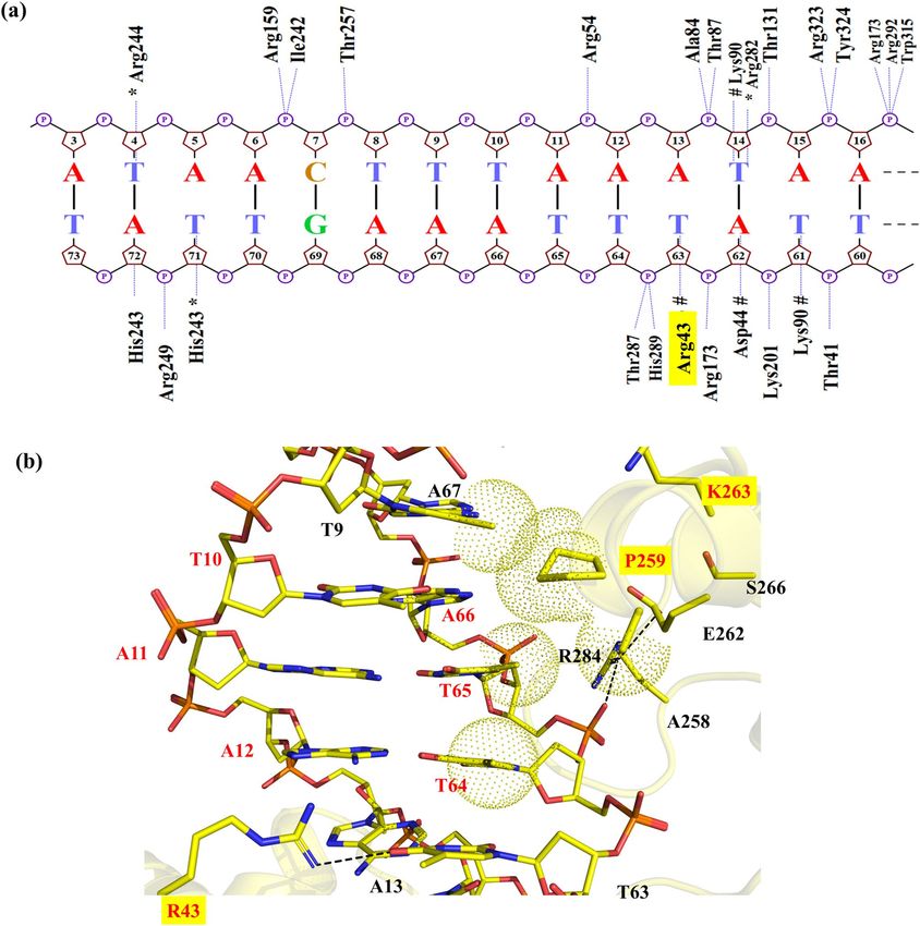
www.nature.com/scientificreports/
Figure 1. Nucleotides and amino acids at the protein-DNA interface of Cre/loxP and Dre/rox recombinase
systems. (a) Sequences of loxP and rox target sites. The three nucleotides that differ between loxP and rox are
highlighted in color. The numbering of the individual bases is provided for the upper and lower DNA strand,
respectively. (b) Protein amino acids facing the altered DNA bases of loxP and rox. (c) Snapshot of the cleaving
monomer of the Cre/loxP complex (PDB 1Q3U). The recognition regions at the protein-DNA interface are
displayed in magenta (helix B), orange (helix D), green (helix J) and purple (beta 4). The area of interest in this
study (i.e. PDIBJ area; the protein-DNA interface formed by amino acids of helix B and J and nucleotides 10/66,
11/65, and 12/64) is zoomed in, and relevant bases and amino acids are labeled. The numbering used is based on
the Cre/loxP crystal structure (for details see Fig. S1). Figure generated using Pymol (version 2.1, https://pymol
.org/).
(Fig. 1a). Nevertheless, Cre does not show activity on rox, and, similarly, Dre is inactive on loxP18. In previous
work, detailed comparative analyses of these recombinases have identified the amino acids K43, R259, and G263
of Cre as critical residues for the discrimination between the loxP and rox sites12. Indeed, the substitution of
these three amino acids in Cre by the corresponding Dre residues ( mCreK variant: K43R, R259P, and G263K)
(Fig. 1b) was sufficient to confer selectivity for rox, albeit at low activity. In order to establish a structure–func-
tion rationale, which could help in guiding further efforts for improving recombination properties, we decided
to investigate in detail the molecular recognition mechanisms of these specificity-determinant key residues at
positions 43, 259 and 263 in binding to loxP and rox sites.
Results and discussion
Based on the crystal structure of Cre bound to loxP in the synaptic state (PDB 1 Q3U19), we generated three-
dimensional (3D) molecular models of Dre/rox and m CreK in complex with loxP and rox target sites. Utilizing
structure-based modeling and molecular dynamics (MD) simulations, we explored the interactions involved
in protein-DNA binding in these complexes, particularly at the interface formed by the specificity-determining
amino acids at positions 43, 259 and 263, which lay on helix B and J, and the nucleotides at positions 10/66,
11/65, and 12/64 (Fig. 1b,c). Hereafter, we named this interfacial region as the P
DIBJ area.
MD‑based recognition analysis of Cre/loxP. In order to better understand the molecular recognition
mechanisms of the mCreK variant containing mutations at positions 43, 259 and 263 with respect to wild type
Cre, we decided to first perform a comprehensive comparative analysis of the recognition properties of the wild
type Cre/loxP and Dre/rox recombinase systems. For this purpose, we chose available structural information on
the Cre/loxP synaptic complex obtained by X-ray crystallography (see “Materials and methods” for details) and
performed MD simulations to examine at atomic detail the protein-DNA recognition and its binding energet-
Scientific Reports | (2020) 10:13985 | https://doi.org/10.1038/s41598-020-70867-5 2
Vol:.(1234567890)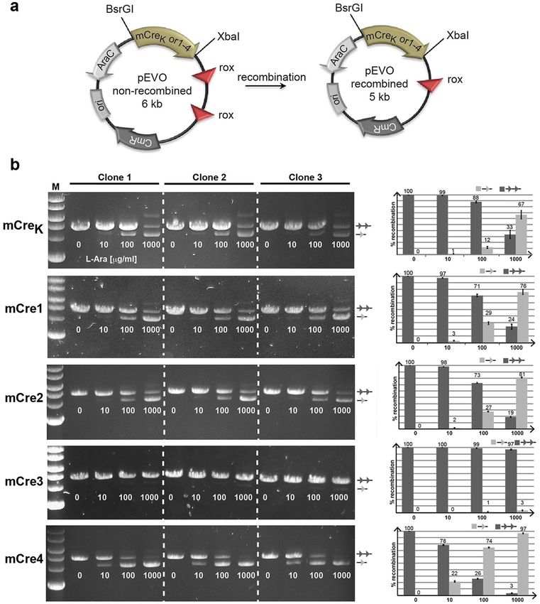
www.nature.com/scientificreports/
Figure 2. Analysis of protein-DNA interactions in Cre/loxP (a) Projection of hydrogen bond interactions
spotted in the last frame of the Cre/loxP MD trajectory. The base-specific interactions with the minor groove
are labeled with an asterisk (*). Hashes (#) denote the interactions with the major groove. Nucplot was used
to generate this image. (b) Detailed view of interactions observed in the Cre/loxP crystal structure and MD
simulation in the PDIBJ area. Hydrogen bonds are shown in black dashed lines. The specificity-determining
residues are highlighted with a yellow box. Water positions predicted by WaterMap (version 1.0, https://www.
schrodinger.com/) are shown in orange spheres, and water-mediated contacts are depicted by orange lines. For
comparison, a network of water molecules found in this area in another Cre/loxP crystal structure (PDB 3C29)
is shown superimposed with the Cre/loxP MD-refined structure (cyan spheres and lines). Pymol was used to
generate the image (version 2.1, https://pymol.org/).
ics. Results obtained from the MD simulations underlined the crucial interactions prevalent in the Cre/loxP
complex in the cleaving and non-cleaving conformers. The interactions observed for the cleaving conformer are
shown in Figs. 2a and S1. The protein-DNA hydrogen bonds observed in our simulations are in agreement with
those observed in the Cre/loxP crystal structure. Table S1 provides a comprehensive description of the hydrogen
bonds observed in the MD trajectory based on their frequency of occurrence. At the investigated PDIBJ area
(i.e. the interface formed by the specificity-determining amino acids at positions 43, 259, 263 of helix B and J,
and bases at positions 10/66, 11/65, and 12/64, Fig. 1c), the residues K43, R259 and G263 recognize the central
DNA base pairs C10/G66, G11/C65, T12/A64, and A13/T63 through direct hydrogen bonding (Fig. 2a). Residue
K43 acts as a hydrogen-bond donor interacting with G11(N7 atom) in the major groove of the DNA (appearing
in 73% of the simulation time, Fig. 2b and Table S1). Lysine may adopt different accessible rotamers in folded
states of p roteins20. As such, K43 can be found interacting dynamically with different bases in different crystal
Scientific Reports | (2020) 10:13985 | https://doi.org/10.1038/s41598-020-70867-5 3
Vol.:(0123456789)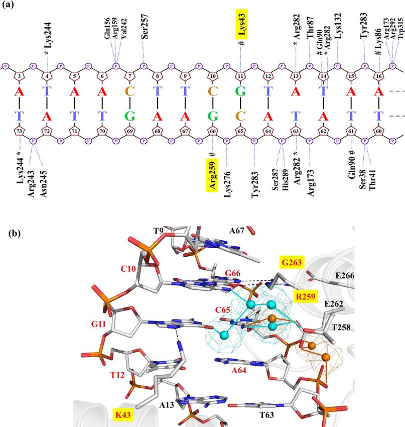
www.nature.com/scientificreports/
structures of Cre/loxP (i.e. with T63(O4) in PDB 1Q3U, and with G11(N7) in PDB 3C29). Our MD simulation
predominantly supports the interaction with G11(N7). Residue R259 forms a strong bidentate hydrogen bond
with the loxP bases G66(N7) (55% of the MD trajectory) and G66(O6) (40% of the MD trajectory) (Table S1).
This interaction is crucial for Cre/loxP recognition21. Position 259 is particularly interesting from the recogni-
tion perspective in SSRs, as it can accommodate a variety of mutations to maintain specific contacts of different
physico-chemical nature with the bases of the DNA target site22,23. Residue G263 provides stability to helix J
by allowing E266 to interact with R259 (through its free NH1 atom) and therefore applying constraints to its
orientation (Fig. 2b).
Because water molecules can be crucial in defining the structure, function and stability of protein-DNA
complexes24–26, we also analyzed water-mediated contacts in the Cre/loxP complex using WaterMap27. The exami-
nation of the interfacial solvent indicated the presence of a water molecule in the major groove bridging the
protein residue E262 to the base C65 (Fig. 2b). Interestingly, this predicted water-mediated contact coincides with
an equivalent observed in another synaptic crystal structure of Cre/loxP (PDB 3C2928; Fig. 2b). Residue E262
is regarded as “guardian for loxP selectivity” as it is responsible for modulating DNA binding and discriminat-
ing loxP from other substrates4,29. We also observed a water-mediated protein–DNA interaction between the
carboxylate group of E262 and the phosphates of the bases C65 and A64, which could point towards another
preferential water-mediated contact not observed in the available crystallographic data, probably due to resolu-
tion restrictions. In view of the ion distribution in the Cre/loxP complex, a high density of K + is observed in the
DNA major groove of the investigated PDIBJ area, which could be linked to the presence of a GC-rich region, and
in a lower extent in the loxP minor groove30,31 (Fig. S2a). Ions were also observed accumulating at the entrance
of the major groove, as their presence will minimize the repulsion between residue E262 and DNA phosphates
(see spatial location of E262 on helix J in Fig. 2b).
MD‑based recognition analysis of Dre/rox. To perform a comprehensive comparative analysis, we
investigated in detail the protein-DNA recognition properties in the Dre/rox recombinase system. For this pur-
pose, we built a 3D molecular model of Dre in complex with rox by using available structural data at the Protein
Data Bank (PDB32) on the Cre/loxP system as template and different software tools as validation for our mod-
eling (see “Materials and methods” and Supplementary Information for details). The comparison of the resulting
3D Dre/rox models obtained by different means (i.e. MODELLER/Discovery Studio (DS)33,34 and the SWISS-
MODEL35 and P HYRE236 webservers) with respect to the Cre/loxP structure showed similar RMSD’s values and,
therefore structural agreement substantiating our modeling (i.e. heavy-atom RMSD of 1.59, 1.56 and 1.60 Å
respectively, Fig. S3). For further studies, we chose the model obtained from DS.
The MD-based analysis of the obtained Dre/rox model indicated important amino acids involved in molecular
recognition through hydrogen bond interactions with the DNA minor groove (H243, R244, R282) and major
groove (R43, D44, K90) (Figs. 3a and S1). In comparison to Cre/loxP, the Dre/rox complex provides a modified
interaction profile with less base-specific and more non-specific contacts (i.e. DNA backbone) (Figs. 3a and S1).
A comprehensive description of the hydrogen bonds observed in the MD trajectory based on their frequency
of occurrence is provided in Table S2.
In the Dre/rox complex, residues R43, P259 and K263 were predicted to recognize the three bp T10/A66, A11/
T65, A12/T64 and A13/T63 through a combination of hydrogen bonds and van der Waals contacts (Fig. 3b).
Residue R43 was found to act as a hydrogen-bond donor interacting with O4 of T63 (at ~ 70% and 14% frequency
of occurrence for atoms R43(NH1) and R43(NH2), respectively) (Fig. 3b and Table S2). Residue P259 established
hydrophobic contacts with the methyl groups of T9 and T65 consequently forming a well-packed interface at the
major groove of rox. Alanine at position 258 was also found to be contributing partially to these hydrophobic
contacts. Residue K263 did not interact with any particular amino acid or base. With the presence of Proline at
position 259 and Serine at position 266, an intra-helical contact between these residues was not observed in the
Dre/rox complex. In comparison, in the Cre/loxP complex, residue E266 locks the orientation of R259 by forming
a hydrogen bond and thus contributing to stabilizing the helix (Figs. 2b, 3b). However, this stabilization of the
helix in the Dre/rox complex is provided by R284 (L284 in Cre), which interacts through hydrogen bonds with
E262 and with the phosphate of T65. Water-mediated interactions were not observed in the PDIBJ area of the
Dre/rox complex. Unlike Cre/loxP, the Dre/rox complex lacks the K + density at the major groove of the DNA.
As this complex also contains glutamic acid at position 262, the high K + density is observed at the entrance of
the major groove to minimize the repulsion between the respective amino acid and DNA phosphates (Fig. S2b).
MD‑based comparative recognition analyses of mCreK/loxP and mCreK/rox. We next modeled
the 3D structure of mCreK (K43R, R259P and G263K) in complex with loxP and with rox in order to establish a
structure–function rationale that could guide further engineering of mCreK with improved activity on rox. The
obtained mCreK/loxP and mCreK/rox complex structures were energy refined by MD simulations (see “Materials
and methods” section for details). In the m
CreK/loxP complex, R43 was observed to form bifurcated hydrogen
bonding with N7 of G11 and O4 of T12, whereas in the m CreK/rox complex R43 interacted with the DNA
backbone (Fig. 4a,b). Residue P259 formed compact van der Waals packing with the methyl groups of bases
T65 and T64 on rox, along with residue T258. This compactness is missing in the complex with loxP due to the
lack of those methyl groups in the altered bp C65 and A64 (Fig. 1a). Additionally, T258 was interacting with
E262 in the m CreK/rox complex via hydrogen bonding. Residue K263 was observed to establish interactions
with E266 in both m CreK/loxP and m CreK/rox complexes. However, a water-mediated contact was observed
in the m
CreK/loxP complex bridging E262 and the DNA backbone, while no such contact was detected in the
mCreK/rox complex (Fig. 4). Altogether, the m CreK/loxP complex exhibited fewer interactions in the P DIBJ area
than mCreK/rox, providing a rationale for the altered selectivity of mCreK on loxP and rox. The ion distributions
Scientific Reports | (2020) 10:13985 | https://doi.org/10.1038/s41598-020-70867-5 4
Vol:.(1234567890)
www.nature.com/scientificreports/
Figure 3. Analysis of protein-DNA interactions observed in the MD simulation of Dre/rox (a) Projection
of hydrogen bond interactions spotted in the last frame of the Dre/rox MD trajectory. The base-specific
interactions with the minor groove are labeled with an asterisk (*). Hashes (#) denote the interactions with
the DNA major groove. Nucplot was used to generate the image. (b) Detailed view of interactions observed
in the PDIBJ area of the Dre/rox complex based on MD simulations. The specificity-determining residues are
highlighted with a yellow box. Hydrogen bonds are shown in black dashed lines and van der Waals interactions
in dotted spheres. Pymol was used to generate the image (version 2.1, https://pymol.org/).
in these complexes are presented in Fig. S2c and d. In the mCreK/loxP complex, a high K+ density is observed
near the entrance of the major groove as mentioned above for the Cre/loxP complex, which could be associated
to the presence of a GC-rich area, whereas in the mCreK/rox complex K+ density is observed only between the
negatively charged groups of glutamic acid at position 266 and the DNA phosphates.
Rational engineering of new Cre variants with enhanced activities on rox. The detailed analysis
of the MD simulations suggested that amino acids on helix J other than those at positions 259 and 263 might
play an important role in molecular recognition in the mCreK/rox complex. Therefore, we investigated in detail
the possible implication in the recombination activity of residues in close proximity to specificity-determining
ones in the PDIBJ area.
Scientific Reports | (2020) 10:13985 | https://doi.org/10.1038/s41598-020-70867-5 5
Vol.:(0123456789)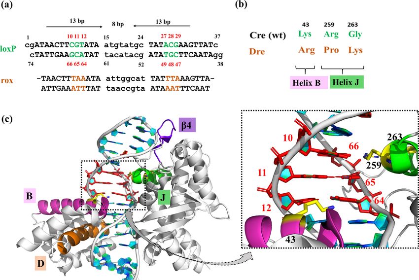
www.nature.com/scientificreports/
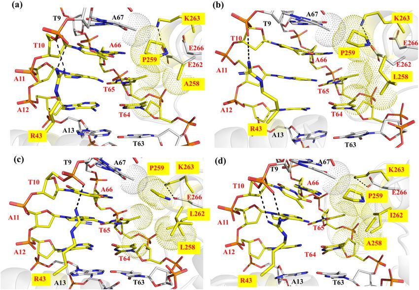
Figure 4. Detailed view of interactions observed in the (a) mCreK/loxP complex and (b) mCreK/rox complex
at the PDIBJ area based on MD simulations. The mutated amino acids with respect to wild type Cre are labeled
with a yellow box. The hydrogen bonds are shown in black dashed lines and the van der Waals interactions in
dotted spheres. A predicted water position using WaterMap (version 1.0, https://www.schrodinger.com/) is
represented by an orange sphere and its interactions in solid orange lines. Pymol was used to generate the image
(version 2.1, https://pymol.org/).
Based on the hypothesis that neighboring residues to those determining specificity could potentially be used
to tune activity, we focused on investigating in detail their recognition properties in order to select candidate
positions for the introduction of new functionalities that could help in the engineering of improved recombina-
tion activity on rox. In the mCreK/rox complex, amino acid T258 was observed to form a hydrogen bond with
the acceptor groups of E262, which also forced the latter to point towards the rox major groove (Fig. 4b). Thus,
we hypothesized that by breaking the hydrogen bond between residues T258 and E262, we could increase the
non-polar interactions at the PDIBJ area by the reorientation of T258 towards the major groove and pushing E262
away from the groove. This way, E262 could then participate in interactions with K263 and/or with the DNA
backbone and contribute towards stabilizing helix J. Hence, we decided to design several new variants of m CreK.
We designed a first new variant, mCre1, by introducing the mutation T258A in the mCreK structure, which
consists of a change to the Dre equivalent residue (mCre1; K43R, R259P, G263K, T258A) (see “Materials and
methods” for details). The MD-based analysis of the mCre1/rox complex showed that the side chain of A258
promotes hydrophobic interactions with the methyl group of DNA base T65 (Fig. 5a). In this mCre1 variant,
residues R43 and P259 were interacting with DNA bases T10 and A67, respectively, in a similar fashion as
observed in the m CreK/rox complex (Figs. 4b, 5a). As hypothesized from our structure-based rationale, residue
E262 was relocated pointing away from the groove and interacting with K263, and thus providing stability to
the helix J. This displacement of E262 created a little void in the groove, which allowed the incorporation of a
side chain bulkier than alanine at position 258. Therefore, we designed a second variant, mCre2, by introduc-
ing the mutation T258L (mCre2; K43R, R259P, G263K, T258L). The analysis of the results obtained in the MD
simulation of mCre2/rox showed better packing in the groove by filling the void and involving T65 and T64
bases of rox while maintaining all the above-mentioned interactions (Fig. 5b). Next, while keeping the mutation
T258L, and in order to further promote van der Waals and hydrophobic contacts, we designed another new
mutant variant, mCre3, including the mutation E262L (mCre3; K43R, R259P, G263K, T258L, E262L). Simulation
analysis of the mCre3/rox complex structure indicated that the mutations T258L and E262L had caused steric
hindrance with the DNA bases and other protein residues, which enforced residues L258 and L262 to reorient
and repack themselves with the adjacent hydrophobic residues L261, F265, I174 and A175 (Fig. 5c). Thereby,
mCre3 resulted in a loose packing at the interface (Fig. 5c). To relieve possible steric repulsions, we designed a
next variant, mCre4, in which position 262 was mutated to Isoleucine. Our molecular models indicated that the
best counterpart for this change would be Alanine at position 258 (mCre4; K43R, R259P, G263K, T258A, E262I).
Besides relieving the steric repulsions, we expected that these concurrent mutations in mCre4 would also provide
some flexibility to helix J as noticed in the mCre1 variant (having T258A). The results obtained from the MD
simulation analysis of the mCre4/rox complex showed the desired hydrophobic packing with the DNA bases
A67, T65, and T64 (Fig. 5d). Overall, the mCre4/rox complex exhibited the highest number of interactions and
the best packing complementarity. Water-mediated interactions were not observed in these newly engineered
complexes. The ion distributions in these variants are shown in Fig. S2e–h. These variants also displayed high
K+ densities between negatively charged E266 and DNA phosphates as observed in all the above complexes to
minimize charge repulsion.
Interestingly, in our simulations we observed that when E262 is mutated to hydrophobic residues (i.e. Leucine
and Isoleucine in mCre3 and mCre4, respectively), its role in binding to K263 is taken over by residue E266,
thereby providing stability to helix J. This interaction was also observed in the mCreK/rox complex. The observa-
tion of mutating-neighboring residues taking over the role of indispensable residues has also been previously
Scientific Reports | (2020) 10:13985 | https://doi.org/10.1038/s41598-020-70867-5 6
Vol:.(1234567890)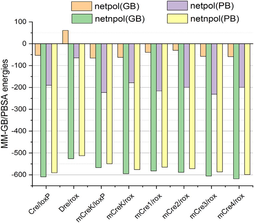
www.nature.com/scientificreports/
Figure 5. Detailed view of interactions observed at the PDIBJ area in the MD simulations of the newly designed
Cre variants with rox. (a) mCre1 (K43R, R259P, G263K, T258A) (b) mCre2 (K43R, R259P, G263K, T258L)
(c) mCre3 (K43R, R259P, G263K, T258L, E262L) and (d) mCre4 (K43R, R259P, G263K, T258A, E262I). The
mutated amino acids with respect to wild type Cre are labeled (yellow box). The hydrogen bonds are shown
in black dashed lines and van der Waals interactions in dotted spheres. Pymol was used to generate the image
(version 2.1, https://pymol.org/).
reported in evolved SSR systems37. Our MD-based analyses strongly emphasize on the relevance of having non-
polar residues at certain neighboring positions in the P DIBJ area, which could possibly affect activity.
In our rationale, when we hypothesized the breaking of the hydrogen bond between T258 and E262 in the
mCreK/rox complex, we proposed the simplest mutation of Threonine to Alanine at position 258 (as in mCre1
variant). As mentioned above, we then thoroughly investigated the impact of diverse amino acids on molecular
recognition by introducing the mutation at position 258 alone and in combination with position 262, which led
us to the use of hydrophobic residues i.e. Leucine and Isoleucine (as in mCre2, mCre3, and mCre4 variants). The
MD analyses showed that the inclusion of these hydrophobic residues enhances the complementary packing at
the DNA interface, mostly with the methyl groups of bases T65 and T66 in rox. The exception to this observation
was the mCre3 variant. Coincidently, the sequence alignment of Cre, Dre and other related naturally occurring
SSRs also revealed the presence of Alanine at position 258 in Dre and Panto, whereas the other SSRs present
Threonine, Proline or Glutamic acid at this position (Fig. S4). However, none of the naturally occurring SSRs
harbors bulky hydrophobic/non-polar residues at position 258 and/or 262. In fact, position 262 is occupied in
most cases by charged/polar residues. This observation was particularly interesting for Dre and Panto, as they
have Thymine as a base at positions 65 and 66 in their respective target sites. Our findings from the MD analysis
of the new Cre variants underscore the presence of bulky hydrophobic groups at positions 258 and 262 with
respect to rox. We further decided to investigate these findings energetically.
MD‑based energetic analyses. In order to gain a deeper understanding on the recognition properties
of the selected mutations and their potential effect on recombination activities, we estimated binding ener-
gies of all the respective protein-DNA complexes ( mCreK/loxP, mCreK/rox, mCre1/rox, mCre2/rox, mCre3/rox
and mCre4/rox). For this purpose, we performed a comparative energetic analysis of the mutant variants with
respect to the wild type complexes (Cre/loxP, Dre/rox) utilizing MM-GB/PBSA38. The predicted MM-GB/PBSA
binding energy of the Cre/loxP complex was higher than for the Dre/rox complex (− 662.61/− 781.01 versus
− 465.78/− 577.23 kcal/mol), which is due to its greater number of contacts (Table 1 and Fig. S1). The calculated
MM-GB/PBSA energies also reflected stronger binding of m CreK to rox than to loxP (− 657.54/− 785.05 and
Scientific Reports | (2020) 10:13985 | https://doi.org/10.1038/s41598-020-70867-5 7
Vol.:(0123456789)www.nature.com/scientificreports/
Complexes ΔEvdW ΔEele ΔGGB ΔGPB ΔGSA(GB) ΔGSA(PB) ΔGGBSA ΔGPBSA
Cre/loxP − 533.3 ± 16.6 − 28,163.6 ± 274.2 28,110.2 ± 266.9 27,973.5 ± 266.1 − 75.8 ± 1.4 − 57.5 ± 0.6 − 662.6 ± 31.4 − 781 ± 29.2
Dre/rox − 461.3 ± 15 − 21,026.5 ± 295 21,087.2 ± 282.1 20,962.0 ± 281.7 − 65.1 ± 1.6 − 51.4 ± 0.9 − 465.7 ± 33.8 − 577.2 ± 28.3
mCreK/loxP − 494.9 ± 14.3 − 27,939.3 ± 337.8 27,873.7 ± 336.2 27,716.1 ± 331.1 − 72.1 ± 1.5 − 54.7 ± 0.9 − 632.6 ± 26.6 − 772.9 ± 24.4
mCreK/rox − 519.3 ± 16.2 − 27,065.7 ± 286.6 27,003.1 ± 283.5 26,886.9 ± 288.9 − 75.6 ± 1.7 − 56.9 ± 0.8 − 657.5 ± 27.4 − 785.0 ± 26.1
mCre1/rox − 510.0 ± 25.5 − 27,428.3 ± 330.7 27,388.7 ± 331 27,212.2 ± 334.8 − 72.1 ± 2.3 − 55.4 ± 1.3 − 621.7 ± 24.8 − 781.5 ± 23.8
mCre2/rox − 515.7 ± 14.7 − 27,388.9 ± 290.4 27,358.3 ± 283 27,189.1 ± 288.1 − 72.8 ± 1.3 − 56.3 ± 0.8 − 619.1 ± 29.8 − 771.8 ± 33.4
mCre3/rox − 530.3 ± 16.5 − 28,623.0 ± 235.1 28,565.3 ± 223.5 28,392.1 ± 227.1 − 74.9 ± 1.5 − 56.6 ± 0.7 − 663.1 ± 34.8 − 818.0 ± 31.6
mCre4/rox − 542.3 ± 12.9 − 28,998.7 ± 289.4 28,939.4 ± 279.5 28,799.2 ± 281.9 − 76.1 ± 1.2 − 57.1 ± 0.6 − 677.8 ± 30.3 − 798.8 ± 26.1
Table 1. Calculated MM-GB/PBSA binding energies and corresponding standard deviations (kcal/mol) for
the studied SSRs complexes. ΔEvdW van der Waals energy, ΔEele columbic energy, ΔGGB Generalized-Born polar
solvation energy, ΔGPB Poisson–Boltzmann polar solvation energy, ΔGSA(GB/PB) non-polar solvation energy from
MM-GBSA/MM-PBSA.
Figure 6. Net free energy component analyses of the studied complexes; netpol(GB/PB): net polar contributions
from MM-GBSA/MM-PBSA ( netpol(GB/PB) = ΔEele + ΔGGB/PB), netnpol(GB/PB): net non-polar contributions from
MM-GBSA/MM-PBSA (netnpol(GB/PB) = ΔEvdW + ΔGSA(GB/PB)).
− 632.64/− 772.92 kcal/mol, respectively), as observed in the MD-based molecular analyses of the correspond-
ing complexes.
The interaction energies obtained for the newly engineered Cre variants indicated that all the introduced
mutations had a favorable effect on the molecular recognition of rox (mCre1: − 621.75/− 781.58, mCre2:
− 619.15/− 771.85, mCre3: − 663.06/818.02 and mCre4: − 677.83/− 798.86 kcal/mol) compared to the native
Dre/rox complex (Table 1). The binding energies of the new variants were not statistically very different from
mCreK/rox, and the strongest contributions in all the cases were obtained from the net non-polar (netnpol) compo-
nents (Table 1 and Fig. 6). We decided to perform the experimental validation of all the four variants against rox.
Experimental validation of rationally engineered new Cre variants. To test the newly designed
Cre variants (mCre1, mCre2, mCre3 and mCre4) experimentally, we introduced the corresponding mutations
into the m CreK coding sequence. The sequences were confirmed by sequencing, and the recombinase mutants
were cloned into the pEVOrox vector that allows regulated expression of the recombinase enzymes12. The vector
also harbored two rox sites in direct orientation as an excision substrate (Fig. 7a), making it possible to inves-
tigate recombinase activity by the growth of the plasmids in bacteria at different l-arabinose concentrations
followed by plasmid extraction, digestion and gel electrophoresis. Growing pEVOrox-mCreK at different l-ara-
binose concentrations confirmed that the three mutations introduced into Cre (K43R, R259P, G263K) conveyed
recombination activity on the rox target sites, although partial recombination was only visible at high l-arab-
inose concentrations (Fig. 7b). Changing the threonine at position 258 to alanine (mCre1) or leucine (mCre2)
slightly increased the recombination activity, but when the position 262 was changed to leucine in combination
to L258 (mCre3), the recombination activity was completely lost. The loss in activity could be associated to the
steric repulsions and poor interfacial interactions as observed through in silico analyses. Nevertheless, a remark-
Scientific Reports | (2020) 10:13985 | https://doi.org/10.1038/s41598-020-70867-5 8
Vol:.(1234567890)www.nature.com/scientificreports/
Figure 7. Rationally designed Cre mutants show increased recombination activity in E.coli. (a) Schematic
drawing of the plasmid assay. Important regions in the plasmids are indicated. Note the reduced size of
the plasmid after recombination. The restrictions sites (BsrGI and XbaI) used for cloning indicated Cre-
recombinase variants are depicted. The rox target sites are shown as red triangles. CmR, chloramphenicol
resistance gene; ori, origin of replication; AraC, arabinose operon regulatory gene. (b) Agarose gels of three
independently picked clones showing BsrGI and XbaI digested plasmids carrying indicated recombinases. The
amount of arabinose added to the growth medium is presented below each band in μg/ml l+-arabinose. The
line with two triangles indicates no recombination, whereas the line with one triangle marks the recombined
band. Quantifications of the ratios of band intensities (in percent of recombination) are shown to the right for
each mutant. The amount of arabinose added to the growth medium is shown on the X-axis. Error bars depict
standard deviation from the three independent experiments.
Scientific Reports | (2020) 10:13985 | https://doi.org/10.1038/s41598-020-70867-5 9
Vol.:(0123456789)www.nature.com/scientificreports/
able increase in recombination activity was observed when position 262 was changed to Isoleucine in conjunc-
tion with A258 (mCre4). Indeed, full recombination of the plasmid was observed at the highest l-arabinose
concentration for this variant (Fig. 7b), with increased recombination activity of up to 20-fold observed on the
rox site when compared to mCreK (Supplementary Fig. S5). These data experimentally validate our structure-
based rationale and MD simulation results, confirming that residues next to specificity-determining amino acids
influence the activity of Cre-type site-specific recombinases.
Conclusions
To better understand how Cre-type site-specific DNA recombinases may achieve better precision in terms of
recombination activity, we investigated in detail the protein-DNA recognition properties of such recombinase
systems in a comparative fashion by applying molecular modeling and dynamics simulations. Molecular dynam-
ics simulations and binding energy calculations were used to examine by molecular and energetic means the
mechanisms involved in DNA target recognition in the naturally occurring recombinase systems Cre/loxP and
Dre/rox and the engineered variant mCreK. Although being able to recombine rox, mCreK exhibited low recom-
bination activity, which made us consider a detailed analysis of the recognition properties of these recombinase
systems in the region in which the specificity-determining residues are located ( PDIBJ area) and to scrutinize at
atomic level any aspect that may affect activity. The analysis of our theoretical molecular models and MD simula-
tions pointed to neighbor amino acids of specificity-determining residues as relevant contributors to DNA target
recognition and, therefore, as promising candidate positions to be exploited for the rational design of improved
recombination activities. In particular, our MD-based analyses strongly emphasized on the relevance of having
non-polar substitutions at positions 258 and 262 in the P DIBJ area, a feature not observed in naturally occurring
SSRs. We established a rationale to account for the structure–function relationships, which we used to design
new Cre variants predicted to have improved recombination activity on rox. The experimental validation of
the newly designed Cre variants confirmed our predictions and supported the hypothesis that changes in the
nature of amino acids spatially close to the specificity-determining residues could lead to enhance activities of
engineered SSRs. This work demonstrates that computer-aided molecular modeling and simulation are valuable
tools to build up innovative rational strategies for the efficient engineering of SSR systems with desired properties
for applied site-specific recombination.
The results obtained should help for the future generation of designer-recombinases. Several amino acids
in particular regions in Cre-like designer recombinases have been classified as implicated in the specificity of
the enzymes28,37,39,40. DNA shuffling is currently used to combine beneficial mutations, thereby accelerating the
directed evolution process. However, because DNA shuffling relies on the homology of DNA fragments, this
method is not very efficient in combining residues that are close in sequence. Our results argue that amino acids
that flank specificity-determining residues have an important role in obtaining SSRs with the highest activity.
We, therefore, propose that targeted mutagenesis of nearest-neighbor amino acids of specificity-determining
residues should be performed to optimize the activity of engineered SSRs.
Materials and methods
Molecular modeling and MD simulations. The crystal structure of the Cre/loxP complex was obtained
from RCSB Protein Data Bank32 (PDB 1Q3U19, resolution 2.9 Å). This structure consists of four molecules of Cre
and two of the loxP target site. For simplicity, in our modeling and dynamics simulations we have used half of
the system (i.e. one loxP and two Cre molecules; cleaving and non-cleaving). This structure together with other
available structural homologs at the Protein Data Bank (PDBs: 5U91, 1KBU, 3CRX) were used as a template to
generate a 3D model of the Dre protein. For this, we used the comparative/homology modeling tool of Discovery
Studio (DS version 3.5, https://www.3dsbiovia.com/)26 and MODELLER33,34 as implemented in DS. The SWISS-
MODEL (version 1.0, https://swissmodel.expasy.org/)35 and PHYRE2 (version 2.0, https://www.sbg.bio.ic.ac.
uk/)36 webservers with default values were also used in order to generate further 3D models of Dre. Likewise, the
3D model of the DNA target rox was generated using the loxP structure as a template with the modeling tool of
DS. The Dre/rox complex was obtained by manual docking based on the superposition of the modeled Dre and
rox molecules with the Cre/loxP structure while keeping the catalytic tyrosine and phosphate in close proximity.
The 3D structures of all Cre mutant variants ( mCreK, mCre1, mCre2, mCre3, and mCre4) were also obtained
using the homology modeling tool of DS, and PDB 1Q3U was used as template. Similar manual docking pro-
cedures by superposition with the Cre/loxP structure were used to generate the 3D models of all Cre mutant
variants in complex with the DNA targets (mCreK/loxP, mCreK/rox, mCre1/rox, mCre2/rox, mCre3/rox, and
mCre4/rox). Hydrogen atoms were added to the complexes (including the wild type crystallographic structure
of Cre/loxP) using the leap module of AMBER14 (https://ambermd.org/)41, and force-field parameters were
assigned to the protein and DNA using ff14SB42 and parmbsc143 force-fields, respectively. Energy refinement of
all complexes was carried out by using molecular dynamics (MD) simulations adopting ABC (Ascona B-DNA
Consortium) protocols44,45.
MD simulations were performed on all the complexes with periodic boundary conditions in a truncated
octahedral cell using the AMBER14 software s uite41. The protein/DNA complexes were solvated with SPC/E46
water molecules, and charge neutrality was maintained by adding a sufficient number of potassium ions to the
system. Simulations were conducted with 0.15 M KCl concentration using parameters from D ang47. Counteri-
ons were randomly placed initially in a cell, but no less than 5 Å away from DNA and 3.5 Å from one another.
Electrostatics were handled using the Particle Mesh Ewald method48 with a cutoff of 10 Å. Lennard–Jones
interactions were truncated at 9 Å. Initial energy minimization of the solvent (2,500 steepest descent and 2,500
conjugate gradient) was performed with harmonic restraints of 25 kcal mol−1 Å−2 on the solute, and then the
minimization of solute–solvent was performed. Followed by minimization, equilibration was performed with
Scientific Reports | (2020) 10:13985 | https://doi.org/10.1038/s41598-020-70867-5 10
Vol:.(1234567890)www.nature.com/scientificreports/
slow heating of the solvent to 300 K at constant volume for a period of 200 ps, while restraining the solute atoms
by 25 kcal mol−1 Å−2. These positional restraints were gradually removed from 5 to 1 kcal mol−1 Å−2 during the
series of minimizations and equilibrations over a period of 1 ns. Finally, the production simulations were car-
ried out initially for 100 ns using an NPT ensemble and the Berendsen algorithm49, which were later extended
to 200 ns. All bonds involving hydrogen were constrained using S HAKE50.
Structural and energetic analyses of Protein–DNA molecular recognition. All the studied pro-
tein–DNA complexes remained structurally stable during the entire MD simulations as illustrated by the RMSD
values obtained through the simulation time (Fig. S6). The structure-based analysis of the direct and indirect
(water-mediated) hydrogen bonding established between the protein and DNA molecules in the studied com-
plexes was done using the cpptraj51 module of AMBER14 and the W aterMap27 tool of Schrodinger (version
1.0, WaterMap, Schrödinger, 2019; https://www.schrodinger.com/). WaterMap is based on the inhomogeneous
solvation theory, which analyses the results based on a short (2 ns) MD simulation. During MD, the complex is
held rigid and water molecules are allowed to move. The clustering analysis is then performed on the population
of water molecules to predict the location, and the free energy of each (favorable and unfavorable) water site
is calculated. As the predictions are done on the population density of water molecules, the residence times of
individual waters are not reported. Nucplot52 was also used for hydrogen bonding and van der Waals analyses.
VMD53 was used for trajectory analysis. Pymol was used for the generation of figures (version 2.1, https://pymol
.org/). The average K+ distribution analyses were performed using the Grid command of CPPTRAJ, AMBER14.
The energetic analysis consisted of binding enthalpies calculated from the last 50 ns of the initial MD simu-
lation using the MM-GB/PBSA38 module of AMBER (200 snapshots were used for calculations). The results
represented in Table 1 and Fig. 6 belong to the initial 100 ns MD trajectory. The free energy analyses on the
extended trajectory (100 ns to 200 ns) showed similar results (Table S3). The structural analyses performed for
the extended simulations confirmed the interaction details obtained with the frames extracted from the initial
MD trajectory and shown in Figs. 2, 3, 4 and 5.
Recombination assays in E. coli. For expression in E. coli, mCreK and variants thereof were cloned into
the pEVOrox vector12 utilizing the unique BsrGI and XbaI (NEB, Ipswich, MA, USA) restriction sites. To intro-
duce mutations into the mCreK coding sequence, site-directed mutagenesis was performed using the Q5 Site-
Directed Mutagenesis Kit (NEB, Ipswich, MA, USA) following the manufacturer’s instructions. The expression of
the recombinases from the pBAD promoter was induced with l-(+)-arabinose (Sigma-Aldrich Chemie GmbH).
Single colonies of XL1-blue E.coli (recA1 endA1 gyrA96 thi-1 hsdR17 supE44 relA1 lac [F′ proABlacIqZ∆M15
Tn10 (Tetr)]; Agilent, Santa Clara, CA, USA) containing pEVOrox plasmid with the recombinase and recombi-
nation target sites were cultured overnight in 5 ml Luria broth (LB) medium with 30 μg/ml Cm and 0, 10, 100 or
1,000 μg/ml l-(+)-arabinose at 37 °C and 200 rpm before plasmid extraction, digestion, and gel electrophoresis.
Gels were run to best separate the non-recombined forms and the recombined forms of the plasmids. The ca.
1 kb bands of the recombinase are therefore not visible in the gels in Fig. 7. Experiments were repeated with
two additional clones and bands corresponding in size to the non-recombined and the recombined vector were
quantified with Fiji–win64 software (ImageJ), respectively. The sum of non-recombined and recombined area
was regarded as 100% and the fraction of non-recombined and recombined areas were calculated in percent.
The standard deviations (STDDEV) of the 3 clones carrying the same mutations at identical l-(+)-arabinose
induction levels were calculated.
Received: 22 January 2020; Accepted: 3 August 2020
References
1. Grindley, N. D. F., Whiteson, K. L. & Rice, P. A. Mechanisms of Site-specific recombination. Annu. Rev. Biochem. 75, 567–605
(2006).
2. Jayaram, M. et al. An overview of tyrosine site-specific recombination: from an flp perspective. Microbiol Spectr. 3, 43-71. (2015).
3. Kilby, N. J., Snaith, M. R. & Murray, J. A. H. Site-specific recombinases: tools for genome engineering. Trends Genet. 9, 413–421
(1993).
4. Meinke, G., Bohm, A., Hauber, J., Pisabarro, M. T. & Buchholz, F. Cre recombinase and other tyrosine recombinases. Chem. Rev.
116, 12785–12820 (2016).
5. Van Duyne, G. D. A structural view of Cre- loxP site-specific recombination. Annu. Rev. Biophys. Biomol. Struct. 30, 87–104 (2001).
6. Sauer, B. Inducible gene targeting in mice using the Cre/lox system. Methods 14, 381–392 (1998).
7. Nagy, A. Cre recombinase: the universal reagent for genome tailoring. Genesis 26, 99–109 (2000).
8. Van Duyne, G. D. Cre recombinase. Microbiol. Spectr. 3, 119–138 (2014).
9. Anastassiadis, K. et al. Dre recombinase, like Cre, is a highly efficient site-specific recombinase in E. coli, mammalian cells and
mice. Dis. Model. Mech. 2, 508–515 (2009).
10. Suzuki, E. & Nakayama, M. VCre/VloxP and SCre/SloxP: new site-specific recombination systems for genome engineering. Nucleic
Acids Res. 39, e49–e49 (2011).
11. Karimova, M. et al. Vika/vox, a novel efficient and specific Cre/loxP-like site-specific recombination system. Nucleic Acids Res. 41,
e37–e37 (2013).
12. Karimova, M., Splith, V., Karpinski, J., Pisabarro, M. T. & Buchholz, F. Discovery of Nigri/nox and Panto/pox site-specific recom-
binase systems facilitates advanced genome engineering. Sci. Rep. 6, 30130 (2016).
13. Karimova, M. et al. A single reporter mouse line for Vika, Flp, Dre, and Cre-recombination. Sci. Rep. 8, 14453 (2018).
14. He, L. et al. Enhancing the precision of genetic lineage tracing using dual recombinases. Nat. Med. 23, 1488–1498 (2017).
Scientific Reports | (2020) 10:13985 | https://doi.org/10.1038/s41598-020-70867-5 11
Vol.:(0123456789)www.nature.com/scientificreports/
15. Yoshimura, Y. et al. Novel reporter and deleter mouse strains generated using VCre/VloxP and SCre/SloxP systems, and their
system specificity in mice. Transgenic Res. 27, 193–201 (2018).
16. Liu, W., Tuck, L. R., Wright, J. M. & Cai, Y. Using purified tyrosine site-specific recombinases in vitro to rapidly construct and
diversify metabolic pathways. Methods Mol. Biol. 1, 285–302 (2017).
17. Lin, Q., Qi, H., Wu, Y. & Yuan, Y. Robust orthogonal recombination system for versatile genomic elements rearrangement in yeast
Saccharomyces cerevisiae. Sci. Rep. 5, 15249 (2015).
18. Sauer, B. DNA recombination with a heterospecific Cre homolog identified from comparison of the pac-c1 regions of P1-related
phages. Nucleic Acids Res. 32, 6086–6095 (2004).
19. Ennifar, E. Crystal structure of a wild-type Cre recombinase-loxP synapse reveals a novel spacer conformation suggesting an
alternative mechanism for DNA cleavage activation. Nucleic Acids Res. 31, 5449–5460 (2003).
20. Berezovsky, I. N., Chen, W. W., Choi, P. J. & Shakhnovich, E. I. Entropic stabilization of proteins and its proteomic consequences.
PLoS Comput. Biol. 1, e47 (2005).
21. Kim, S., Kim, G., Lee, Y. & Park, J. Characterization of Cre-loxP interaction in the major groove: hint for structural distortion of
mutant Cre and possible strategy for HIV-1 therapy. J. Cell. Biochem. 80, 321–327 (2001).
22. Meinke, G., Karpinski, J., Buchholz, F. & Bohm, A. Crystal structure of an engineered, HIV-specific recombinase for removal of
integrated proviral DNA. Nucleic Acids Res. 45, 9726–9740 (2017).
23. Karpinski, J. et al. Directed evolution of a recombinase that excises the provirus of most HIV-1 primary isolates with high specific-
ity. Nat. Biotechnol. 34, 401–409 (2016).
24. Janin, J. Wet and dry interfaces: the role of solvent in protein–protein and protein–DNA recognition. Structure 7, R277–R279
(1999).
25. Reddy, C. K., Das, A. & Jayaram, B. Do water molecules mediate protein-DNA recognition?. J. Mol. Biol. 314, 619–632 (2001).
26. Discovery Studio 3.5, 2012. Accelrys Inc., San Diego, CA. https://www.accelr ys.com.
27. Young, T., Abel, R., Kim, B., Berne, B. J. & Friesner, R. A. Motifs for molecular recognition exploiting hydrophobic enclosure in
protein–ligand binding. Proc. Natl. Acad. Sci. 104, 808–813 (2007).
28. Baldwin, E. P. et al. A specificity switch in selected cre recombinase variants is mediated by macromolecular plasticity and water.
Chem. Biol. 10, 1085–1094 (2003).
29. Rufer, A. W. Non-contact positions impose site selectivity on Cre recombinase. Nucleic Acids Res. 30, 2764–2771 (2002).
30. Howerton, S. B., Sines, C. C., VanDerveer, D. & Williams, L. D. Locating monovalent cations in the grooves of B-DNA. Biochemistry
40(34), 10023–10031 (2001).
31. Lavery, R., Maddocks, J. H., Pasi, M. & Zakrzewska, K. Analyzing ion distributions around DNA. Nucleic Acids Res. 42, 8138–8149
(2014).
32. Berman, H. M. The protein data bank. Nucleic Acids Res. 28, 235–242 (2000).
33. Webb, B. & Sali, A. Comparative protein structure modeling using MODELLER. Curr. Protoc. Bioinform. 54, 5.6.1-5.6.37 (2016).
34. Šali, A. & Blundell, T. L. Comparative protein modelling by satisfaction of spatial restraints. J. Mol. Biol. 234, 779–815 (1993).
35. Waterhouse, A. et al. SWISS-MODEL: homology modelling of protein structures and complexes. Nucleic Acids Res. 46, W296–
W303 (2018).
36. Kelley, L. A., Mezulis, S., Yates, C. M., Wass, M. N. & Sternberg, M. J. E. The Phyre2 web portal for protein modeling, prediction
and analysis. Nat. Protoc. 10, 845–858 (2015).
37. Abi-Ghanem, J. et al. Engineering of a target site-specific recombinase by a combined evolution-and structure-guided approach.
Nucleic Acids Res. 41, 2394–2403 (2013).
38. Genheden, S. & Ryde, U. The MM/PBSA and MM/GBSA methods to estimate ligand-binding affinities. Expert Opin. Drug Discov.
10, 449–461 (2015).
39. Hartung, M. & Kisters-Woike, B. Cre mutants with altered DNA binding properties. J. Biol. Chem. 273, 22884–22891 (1998).
40. Santoro, S. W. & Schultz, P. G. Directed evolution of the site specificity of Cre recombinase. Proc. Natl. Acad. Sci. 99, 4185–4190
(2002).
41. Case, D. A. et al. The Amber biomolecular simulation programs. J. Comput. Chem. 26, 1668–1688 (2005).
42. Maier, J. A. et al. ff14SB: improving the accuracy of protein side chain and backbone parameters from ff99SB. J. Chem. Theory
Comput. 11, 3696–3713 (2015).
43. Ivani, I. et al. Parmbsc1: a refined force field for DNA simulations. Nat. Methods 13, 55–58 (2016).
44. Pasi, M. et al. μABC: a systematic microsecond molecular dynamics study of tetranucleotide sequence effects in B-DNA. Nucleic
Acids Res. 42, 12272–12283 (2014).
45. Lavery, R. et al. A systematic molecular dynamics study of nearest-neighbor effects on base pair and base pair step conformations
and fluctuations in B-DNA. Nucleic Acids Res. 38, 299–313 (2010).
46. Berendsen, H. J. C., Grigera, J. R. & Straatsma, T. P. The missing term in effective pair potentials. J. Phys. Chem. 91, 6269–6271
(1987).
47. Dang, L. X. Mechanism and thermodynamics of ion selectivity in aqueous solutions of 18-crown-6 ether: a molecular dynamics
study. J. Am. Chem. Soc. 117, 6954–6960 (1995).
48. Essmann, U. et al. A smooth particle mesh Ewald method. J. Chem. Phys. 103, 8577–8593 (1995).
49. Berendsen, H. J. C., Postma, J. P. M., Van Gunsteren, W. F., Dinola, A. & Haak, J. R. Molecular dynamics with coupling to an external
bath. J. Chem. Phys. 81, 3684 (1984).
50. Ryckaert, J.-P., Ciccotti, G. & Berendsen, H. J. C. Numerical integration of the cartesian equations of motion of a system with
constraints: molecular dynamics of n-alkanes. J. Comput. Phys. 23, 327–341 (1977).
51. Roe, D. R. & Cheatham, T. E. PTRAJ and CPPTRAJ: software for processing and analysis of molecular dynamics trajectory data.
J. Chem. Theory Comput. 9, 3084–3095 (2013).
52. Luscombe, N. NUCPLOT: a program to generate schematic diagrams of protein-nucleic acid interactions. Nucleic Acids Res. 25,
4940–4945 (1997).
53. Humphrey, W., Dalke, A. & Schulten, K. V. M. D. Visual molecular dynamics. J. Mol. Graph. 14, 33–38 (1996).
Acknowledgments
Authors gratefully acknowledge the technical support from Mario Hirt and Pedro Guillem Gloria, as well as the
computational facilities at ZIH, Technische Universität Dresden. We are thankful to Dr. Gloria Ruiz-Gómez for
valuable feedback and fruitful scientific discussions. Work in the Buchholz laboratory was supported by grants
from the European Union (ERC 742133, H2020 UPGRADE 825825), the German Research Foundation (DFG
BU 1400/7-1) and the BMBFGO-Bio 031B0633. Work in the Pisabarro group was partially funded by the Ger-
man Research Foundation (DFG PI 600/4-1). The authors would like to dedicate this work to our IT especialists,
health care professionals and many others on the frontlines whose great efforts are keeping all of us going during
the Corona pandemic.
Scientific Reports | (2020) 10:13985 | https://doi.org/10.1038/s41598-020-70867-5 12
Vol:.(1234567890)www.nature.com/scientificreports/
Author contributions
A.S., F.B., M.T.P conceived and designed the project. A.S. performed the computational studies. M.A. performed
the experiments. All authors analyzed the data and wrote the manuscript.
Funding
Open access funding provided by Projekt DEAL.
Competing interests
The authors declare no competing interests.
Additional information
Supplementary information is available for this paper at https://doi.org/10.1038/s41598-020-70867-5.
Correspondence and requests for materials should be addressed to M.T.P.
Reprints and permissions information is available at www.nature.com/reprints.
Publisher’s note Springer Nature remains neutral with regard to jurisdictional claims in published maps and
institutional affiliations.
Open Access This article is licensed under a Creative Commons Attribution 4.0 International
License, which permits use, sharing, adaptation, distribution and reproduction in any medium or
format, as long as you give appropriate credit to the original author(s) and the source, provide a link to the
Creative Commons license, and indicate if changes were made. The images or other third party material in this
article are included in the article’s Creative Commons license, unless indicated otherwise in a credit line to the
material. If material is not included in the article’s Creative Commons license and your intended use is not
permitted by statutory regulation or exceeds the permitted use, you will need to obtain permission directly from
the copyright holder. To view a copy of this license, visit http://creativecommons.org/licenses/by/4.0/.
© The Author(s) 2020
Scientific Reports | (2020) 10:13985 | https://doi.org/10.1038/s41598-020-70867-5 13
Vol.:(0123456789)You can also read


