Ube3a loss increases excitability and blunts orientation tuning in the visual cortex of Angelman syndrome model mice
←
→
Page content transcription
If your browser does not render page correctly, please read the page content below
J Neurophysiol 118: 634 – 646, 2017.
First published May 3, 2017; doi:10.1152/jn.00618.2016.

RESEARCH ARTICLE                        Sensory Processing

Ube3a loss increases excitability and blunts orientation tuning in the visual
cortex of Angelman syndrome model mice

              Michael L. Wallace,1 Geeske M. van Woerden,5 Ype Elgersma,5 Spencer L. Smith,1,2,3,4
              and Benjamin D. Philpot1,2,3,4
              1
               Curriculum in Neurobiology, University of North Carolina, Chapel Hill, North Carolina; 2Department of Cell Biology
              and Physiology, University of North Carolina, Chapel Hill, North Carolina; 3Neuroscience Center, University of North
              Carolina, Chapel Hill, North Carolina; 4Carolina Institute for Developmental Disabilities, University of North Carolina,
              Chapel Hill, North Carolina; and 5Department of Neuroscience, Erasmus MC University Medical Center, Rotterdam,
              The Netherlands
              Submitted 3 August 2016; accepted in final form 27 April 2017

   Wallace ML, van Woerden GM, Elgersma Y, Smith SL, Phil-                       tions of the 15q11– q13 chromosomal regions or mutations
pot BD. Ube3a loss increases excitability and blunts orientation                 specific to the UBE3A gene, which lies within that region,
tuning in the visual cortex of Angelman syndrome model mice. J                   cause loss of neuronal UBE3A and result in AS (Kishino et al.
Neurophysiol 118: 634 – 646, 2017. First published May 3, 2017;
doi:10.1152/jn.00618.2016.—Angelman syndrome (AS) is a neuro-                    1997). A mouse model of AS, harboring a Ube3a null allele on
developmental disorder caused by loss of the maternally inherited                the maternally inherited chromosome (Ube3am–/p⫹), recapitu-
allele of UBE3A. Ube3aSTOP/p⫹ mice recapitulate major features of                lates the major phenotypes seen in AS including ataxia, mi-
AS in humans and allow conditional reinstatement of maternal Ube3a               crocephaly, seizures, and cognitive disabilities (Jiang et al.
with the expression of Cre recombinase. We have recently shown that              1998).
AS model mice exhibit reduced inhibitory drive onto layer (L)2/3                    Layer (L)2/3 pyramidal neurons in visual cortex of Ube3am–/p⫹
pyramidal neurons of visual cortex, which contributes to a synaptic              mice have reduced inhibitory drive, which contributes to an
excitatory/inhibitory imbalance. However, it remains unclear how this            excitatory/inhibitory imbalance (Wallace et al. 2012). Loss of
loss of inhibitory drive affects neural circuits in vivo. Here we
                                                                                 inhibition is cell type specific, as pyramidal neurons, but not
examined visual cortical response properties in individual neurons to
explore the consequences of Ube3a loss on intact cortical circuits and           fast-spiking inhibitory interneurons, have reduced inhibitory
processing. Using in vivo patch-clamp electrophysiology, we mea-                 inputs. Pyramidal neurons in Ube3am–/p⫹ mice also display
sured the visually evoked responses to square-wave drifting gratings             increased intrinsic excitability, responding with more action
in L2/3 regular-spiking (RS) neurons in control mice, Ube3a-deficient            potentials to a given depolarizing current injection (Wallace et
mice, and mice in which Ube3a was conditionally reinstated in                    al. 2012). Deficits in synaptic inhibition and cellular excitabil-
GABAergic neurons. We found that Ube3a-deficient mice exhibited                  ity may underlie cognitive disabilities in Ube3am–/p⫹ mice,
enhanced pyramidal neuron excitability in vivo as well as weaker                 given that balanced excitation and inhibition are crucial for
orientation tuning. These observations are the first to show alterations         many stages of sensory processing, circuit excitability, and
in cortical computation in an AS model, and they suggest a basis for             neural computation (Isaacson and Scanziani 2011). Impor-
cortical dysfunction in AS.
                                                                                 tantly, investigation of visual cortical plasticity in vivo has
NEW & NOTEWORTHY Angelman syndrome (AS) is a severe                              demonstrated that loss of Ube3a severely blunts ocular-domi-
neurodevelopmental disorder caused by the loss of the gene UBE3A.                nance plasticity, a form of plasticity that is disrupted by
Using electrophysiological recording in vivo, we describe visual                 changes in inhibition (Hensch et al. 1998; Sato and Stryker
cortical dysfunctions in a mouse model of AS. Aberrant cellular                  2010; Yashiro et al. 2009). However, it remains unknown
properties in AS model mice could be improved by reinstating Ube3a
                                                                                 whether Ube3a loss disrupts cortical computations such as
in inhibitory neurons. These findings suggest that inhibitory neurons
play a substantial role in the pathogenesis of AS.                               orientation tuning or intrinsic excitability in vivo.
                                                                                    An intensively studied computation performed by the visual
Angelman syndrome; autism; Ube3a; visual cortex                                  cortex is orientation tuning, which is the preferential response
                                                                                 of a neuron to bar-shaped visual stimuli presented at a partic-
                                                                                 ular angle (Hubel and Wiesel 1962). Orientation tuning largely
ANGELMAN SYNDROME      (AS) is a severe neurodevelopmental
                                                                                 emerges through specific circuitry in visual cortex (Hubel and
disorder characterized by cognitive disability, seizures, ab-
                                                                                 Wiesel 1961; Lien and Scanziani 2013). Additionally, contrast
sence of speech, and high comorbidity with autism (Williams
                                                                                 sensitivity is a property of visual cortical neurons where
et al. 2006). The paternally inherited allele of UBE3A is
                                                                                 responses increase nonlinearly with increasing luminance con-
epigenetically silenced in neurons. Therefore maternal dele-
                                                                                 trast (Albrecht and Hamilton 1982). Optogenetic modulation of
   Address for reprint requests and other correspondence: B. D. Philpot, Univ.
                                                                                 local inhibitory neuron populations in cortex can alter orienta-
of North Carolina School of Medicine, 115 Mason Farm Rd., Chapel Hill, NC        tion tuning and contrast sensitivity, indicating the importance
27599-7545 (e-mail: bphilpot@med.unc.edu).                                       of inhibitory drive to these visually evoked properties (Atallah
634                                         0022-3077/17 Copyright © 2017 the American Physiological Society                            www.jn.org
                                  Downloaded from www.physiology.org/journal/jn (134.174.140.176) on January 23, 2020.
UBE3A LOSS IMPAIRS ORIENTATION TUNING 635
et al. 2012; Lee et al. 2012; Wilson et al. 2012). Therefore, we CaCl2 and 20 glucose bubbled with 95% O2-5% CO2 before applica-
hypothesized that loss of inhibition in AS model mice may tion) to prevent drying.
result in defective orientation tuning and contrast sensitivity. Whole cell recordings were made with the blind-patch method as
Here we describe the effects of Ube3a loss on visually described previously (Margrie et al. 2002). Patch pipettes were pulled
from thick-walled borosilicate glass (P2000; Sutter Instruments, No-
evoked responses, cellular excitability, and circuit excitability vato, CA). Open tip resistances were between 5 and 8 M⍀ when
(UP/DOWN states), using in vivo whole cell recordings. We pipettes were filled with the internal solution containing (in mM) 135
used a conditional Ube3a mouse line that restricts expression K-gluconate, 6 KCl, 10 HEPES, 0.1 EGTA, 4 Mg-ATP, 3 Na-GTP, 8
of Ube3a to cells expressing Cre, which mediates excision of Na-phosphocreatine, and 0.05% neurobiotin, with pH adjusted to 7.25
a STOP cassette (Ube3aSTOP/p⫹). This line exhibits behavioral with 1 M KOH and osmolarity adjusted to ~295 mosM by addition of
and synaptic phenotypes similar to the traditional Ube3am–/p⫹ sucrose. The micromanipulator (Luigs and Neumann, Germany) was
mouse model of AS (Jiang et al. 1998; Judson et al. 2016; arranged for electrodes to penetrate the brain perpendicular to the
Silva-Santos et al. 2015). We found that Ube3a loss has no cortical surface, and depth measurements were made from the pial
effect on the spontaneous activity of L2/3 regular-spiking (RS) surface to the recorded cell. All of the neurons included for analysis
neurons or on the local neural network as assayed by exami- were between 100 and 400 m from the pial surface. Current-clamp
recordings were performed with a Multiclamp 700A amplifier (Molecular
nation of UP/DOWN states. However, loss of Ube3a decreases Devices), and data were acquired at 20 kHz and Bessel filtered at 10 kHz.
the orientation tuning of L2/3 RS neurons without affecting Series resistance was 39 ⫾ 1.3 (n ⫽ 42), 39 ⫾ 1.4 (n ⫽ 32), and
contrast sensitivity. We show that reinstating Ube3a in 32 ⫾ 1.6 (n ⫽ 30) M⍀ for recordings in control, Ube3aSTOP/p⫹, and
GABAergic inhibitory neurons results in an intermediate effect Ube3aSTOP/p⫹:: Gad2-Cre mice, respectively. Data were discarded if
on orientation tuning, suggesting a role for Ube3a in inhibitory the series resistance changed ⬎30% during the course of the record-
interneurons and orientation tuning. Finally, increased excit- ing. Resting membrane potential was assessed immediately after
ability of RS neurons in Ube3aSTOP/p⫹ mice is rescued by break-in, and membrane resistance was measured as the steady-state
reinstating Ube3a in GABAergic inhibitory neurons. Together membrane potential in response to a 50-pA hyperpolarizing current
these data identify a specific cortical processing deficit in an step during a DOWN state. After break-in, we waited several minutes
AS model. to allow for synaptic activity (and UP/DOWN states) to return from
excess extracellular potassium before we conducted intrinsic excit-
ability experiments (Fig. 1). Reported voltages were not corrected for
MATERIALS AND METHODS junction potential. All analyses were performed in Clampfit 10.2 or
with custom routines written in MATLAB (MathWorks).
Animals
All studies were conducted with protocols approved by the Uni- Visual Stimulation
versity of North Carolina Animal Care and Use Committee. Visual stimulus presentation was controlled by routines written in
Ube3aSTOP mice were on the 129Sv/Pas background and generated by MATLAB (MathWorks) with the Psychophysics Toolbox extensions
the laboratory of Ype Elgersma (Silva-Santos et al. 2015). Gad2-Cre (Brainard 1997). Square-wave gratings (0.04 cycles/° at 2 Hz) were
mice (Taniguchi et al. 2011) on the C57BL/6J background were displayed on an LCD screen (Dell; 33 ⫻ 27 cm, 75 Hz refresh rate,
obtained from the Jackson Laboratory (JAX no. 010802). All exper- mean luminance ~46 cd/m2) centered 20 cm from the animal’s eyes.
iments were performed on mice obtained by crossing a female The screen was angled to stimulate the contralateral (left) eye for the
Ube3am⫹/STOP mouse [which has normal expression of Ube3a in the experiments, but the ipsilateral eye was not covered. Visual stimuli
brain (Silva-Santos et al. 2015)] with a male mouse heterozygous for were presented in a shuffled order. To acquire orientation tuning
Gad2-Cre. This cross produced four offspring genotypes: Ube3am⫹/p⫹ ⫾ curves, each of eight different orientations was presented at least six
Gad2-Cre mice (controls), Ube3aSTOP/p⫹ mice (AS model mice), and times. The contrast-response curves were obtained by showing the
Ube3aSTOP/p⫹::Gad2-Cre mice (inhibitory neuron Ube3a reinstate- preferred orientation at eight contrast levels logarithmically spanning
ment model). Experimental trios were littermates on a mixed 129Sv/ the range from 1% to 100% contrast.
Pas and C57BL/6J background, which had been backcrossed 2– 6
generations onto C57BL/6J from the original 129Sv/Pas background.
Mice of both sexes were used at postnatal day (P)70 –110 at equiva- Data Analysis
lent genotypic ratios and in strict compliance with animal protocols Spontaneous activity. Spontaneous spiking rates and UP/DOWN
approved by the Institutional Animal Care and Use Committee of the states were analyzed during presentation of a gray screen and calcu-
University of North Carolina at Chapel Hill. lated over a period of ~5 min. Only cells that had a clear bimodal
distribution of Vm were used for analysis (68 of 102 cells passed this
In Vivo Whole Cell Physiology criterion). For UP vs DOWN state detection, traces were low-pass
filtered at 3 Hz and the mean and standard deviation of the voltage
Mice were anesthetized with 5 mg/kg chlorprothixene followed by were calculated. UP state threshold was defined as the mean voltage
0.9 –1.2 g/kg urethane injected intraperitoneally (ip). Secretions were plus half the standard deviation of the voltage. UP states were detected
reduced by administration of atropine (0.3 mg/kg ip). Mice were whenever the membrane potential crossed this defined threshold and
stereotaxically secured after reaching a surgical plane of anesthesia remained above the threshold for at least 150 ms (Beltramo et al.
(~30 min), and then a uniform layer of water-based ophthalmic 2013; Gonçalves et al. 2013). Periods not detected as UP states were
ointment was applied to the cornea to prevent drying. A homeother- considered DOWN states. These parameters were empirically found
mic blanket (FHC, Bowdoinham, ME) maintained the animal’s body to detect UP and DOWN states accurately.
temperature within a physiological range (37 ⫾ 0.5°C). Incised tissue Spectral analysis. Recorded voltage signals were processed off-line
was locally anesthetized with bupivacaine (0.25% wt/vol), and a with custom-written scripts in MATLAB (MathWorks) (Sellers et al.
~1.5-mm2 craniotomy was performed over primary visual cortex (2.5 2013). Figures represent the average (⫾ SE) of the median power for
mm lateral to midline, 0.5 mm anterior to lambda). A durotomy was each frequency for an entire recording session from a single neuron.
then made (~0.2 mm2) and covered with artificial cerebrospinal fluid Time-dependent frequency content was determined by convolution of
(in mM: 124 NaCl, 3 KCl, 1.25 NaH2PO4, 26 NaHCO3, 1 MgCl2, 2 voltage signals with a family of Morlet wavelets (0.5–100 Hz, step
J Neurophysiol • doi:10.1152/jn.00618.2016 • www.jn.org
Downloaded from www.physiology.org/journal/jn (134.174.140.176) on January 23, 2020.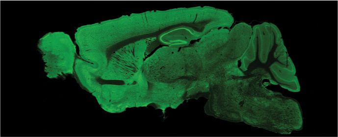
636 UBE3A LOSS IMPAIRS ORIENTATION TUNING
A Ube3am+/p+ :: ± Gad2-cre (Control) B C
L1
L2/3
Ube3aSTOP/p+
50 μm
D Control Ube3aSTOP/p+ Ube3aSTOP/p+ E
:: Gad2-cre 50 ***
Control **
Frequency (spikes/s)
STOP/p+
Ube3a :: Gad2-cre 40 **
160 pA Ube3aSTOP/p+ *
30
Ube3aSTOP/p+ *** *
*
20 :: Gad2-cre
240 pA
10
0
0 80 160 240 320 400
1.5 mm 320 pA
Current (pA)
F G H
* *
Resting Membrane Potential (mV)
Membrane Resistance (MΩ)
300 -90 500
Subpial Depth (μm) 400
-80
200
300
-70
200
100
-60 100
0 -50 0
Control Ube3aSTOP/p+ Ube3aSTOP/p+ Control Ube3aSTOP/p+ Ube3aSTOP/p+ Control Ube3aSTOP/p+ Ube3aSTOP/p+
:: Gad2-cre :: Gad2-cre :: Gad2-cre
Fig. 1. Reinstatement of Ube3a in Gad2-Cre⫹ inhibitory neurons normalizes intrinsic excitability and membrane resistance in L2/3 RS neurons of Ube3aSTOP/p⫹
mice. A: sagittal brain sections from control, Ube3aSTOP/p⫹, and Ube3aSTOP/p⫹::Gad2-Cre mice immunostained for UBE3A. B: schematic of in vivo whole cell
recording configuration. C: sample image of a L2/3 pyramidal neuron that was recorded, filled with neurobiocytin, and stained post hoc. D: sample recordings
from L2/3 regular-spiking (RS) pyramidal neuron in response to increasing current injections (scale bar 40 mV, 100 ms). E: average frequency vs. current curves
from whole cell recordings in control (n ⫽ 42 cells), Ube3aSTOP/p⫹ (n ⫽ 34), and Ube3aSTOP/p⫹:: Gad2-Cre (n ⫽ 31) mice. Note that all significance values
are post hoc comparisons between Ube3aSTOP/p⫹ group and either control (black asterisk) or Ube3aSTOP/p⫹::Gad2-Cre (green asterisk) groups. F: membrane
resistance measured during a DOWN state in RS neurons from control (n ⫽ 39), Ube3aSTOP/p⫹ (n ⫽ 31), and Ube3aSTOP/p⫹:: Gad2-Cre (n ⫽ 29) mice. G:
membrane potential during a DOWN state in L2/3 RS neurons from control (n ⫽ 42), Ube3aSTOP/p⫹ (n ⫽ 32), and Ube3aSTOP/p⫹:: Gad2-Cre (n ⫽ 30) mice.
H: depth from the pial surface of all RS cells recorded in L2/3 from control (n ⫽ 42), Ube3aSTOP/p⫹ (n ⫽ 31), and Ube3aSTOP/p⫹:: Gad2-Cre (n ⫽ 29) mice.
*P ⬍ 0.05, **P ⬍ 0.01 , ***P ⬍ 0.001 with post hoc test for significance.
width 0.5 Hz) with normalized amplitude, providing an optimal and recordings were filtered at 100 Hz to remove spikes. The F1
trade-off between time and frequency uncertainty (Goupillaud et al. (modulated) and F0 (mean) components of the subthreshold response
1984; Sohal et al. 2009). Total power for each frequency band was were calculated as shown in Fig. 7A. Orientation selectivity index
calculated by taking the median value across an epoch (i.e., UP state (OSI) was calculated as (1 ⫺ the circular variance) (Ringach et al.
or DOWN state) for the included frequencies (delta 0.5– 4 Hz, theta 1997). Orientation selectivity was also examined with peak-to-orthog-
4.5– 8 Hz, alpha 8.5–12 Hz, beta 12.5–29.5 Hz, gamma 30 – 80 Hz) onal ratios (Fig. 6H) (Rpref ⫺ Rortho)/(Rpref ⫹ Rortho), where Rpref is
(Sellers et al. 2013). In Fig. 4, analysis of the delta frequency band the response to the preferred direction and Rortho is the response 90°
was limited to 2– 4 Hz to avoid including edge artifacts from the away from the preferred direction. Direction selectivity index (DSI)
visual stimulation occurring for 1 s. was calculated as (Rpref ⫺ Rnull)/(Rpref ⫹ Rnull), where Rnull is the
Visually evoked responses. The spiking visual response to a given response 180° away from the preferred direction (Niell and Stryker
stimulus was the average rate over the stimulus duration (1 s). The 2008). The responses to the eight grating directions were fit with a
subthreshold (membrane potential) visual response to a given stimulus sum of two Gaussians (Fig. 6). The Gaussians were centered 180°
was measured as the “Area” (V ⫻ s) during the stimulus duration. For apart and had the same tuning sharpness (), but amplitudes for each
analysis of subthreshold responses, the “baseline” was calculated for of the two Gaussians were varied to fit the data. The fitting routine
each neuron as the average membrane potential during a DOWN state, used a least-squares method to minimize the Cartesian distance
J Neurophysiol • doi:10.1152/jn.00618.2016 • www.jn.org
Downloaded from www.physiology.org/journal/jn (134.174.140.176) on January 23, 2020.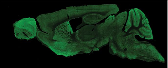
UBE3A LOSS IMPAIRS ORIENTATION TUNING 637
between the model and the data (Carandini and Ferster 2000). To We performed in vivo whole cell recordings from anesthe-
examine only robustly tuned neurons, we calculated the normalized tized mice to examine the contributions of UBE3A to intrinsic
(to the mean firing rate of the preferred direction) residuals of the fit. excitability and visually evoked responses of L2/3 cortical
We then applied a criterion of ⬍0.125 normalized residual to all the
cells and reanalyzed the data (Fig. 6G) (Cottam et al. 2013). The neurons in an intact cortical circuit (Fig. 1B). We chose to
tuning sharpness, or half-width at half height (HWHH), was measu- record from L2/3 neurons in the visual cortex as their responses
red as ⫻ [2 ln(2)]1/2. Contrast-response curves were fit with a to visual stimulation are well characterized (Niell and Stryker
hyperbolic ratio equation (Albrecht and Hamilton 1982): R共C兲 ⫽ 2008). Moreover, visual cortical deficits in synaptic function,
Rmaxcn⁄Cn50 ⫹ cn) ⫹ Roffset where c is contrast, C50 is the semisaturation anatomy, and critical period plasticity have been identified in
contrast, n is the fitting exponent that describes the shape of the curve, AS model mice (Wallace et al. 2012; Yashiro et al. 2009).
Rmax determines the gain, and Roffset is the baseline response. L2/3 pyramidal neurons were identified by cortical depth
and by their regular spiking characterized by an adapting firing
Immunohistochemistry
pattern to depolarizing current injections (Fig. 1D). A subset
(n ⫽ 6 cells) of these neurons were filled with neurobiocytin,
For a subset of recordings where the recorded neuron was recon- stained post hoc, and found to exhibit pyramidal morphology
structed, mice were killed by administration of pentobarbital (40 and spinous dendrites (Fig. 1C). All of the neurons included for
mg/kg) and subsequently intracardially perfused with ~80 ml of 4% analysis were between 100 and 400 m from the pial surface
paraformaldehyde (0.1 M, pH 6.8). Brains were then postfixed for (Fig. 1H). Given these parameters, it is likely that the vast
24 h and sliced coronally at 100 m. The slices were then permeabil- majority, if not all, of the neurons included in this study are
ized in 1% Triton X for 12 h and incubated at 4°C for 12 h in Alexa
488-conjugated streptavidin (1:1,000), 5% normal goat serum, and
L2/3 pyramidal neurons, which are referred to here as regular-
0.1% Triton X. For Fig. 1, sagittal sections were cut at 40 – 60 m spiking (RS) neurons.
and then washed in 0.1 M PBS, permeabilized in 0.2% Triton X, and Similar to in vitro results from Ube3am–/p⫹ mice (Wallace et
blocked in 5% normal goat serum. Primary antibody (mouse anti- al. 2012), we found that in vivo L2/3 RS neurons of
Ube3a, 1:750; Sigma) was incubated for 48 h at 4°C, and secondary Ube3aSTOP/p⫹ mice had increased spiking activity following
antibody (goat anti-mouse Alexa 488; A21131) was incubated at current injection compared with control mice (Fig. 1, D and E).
1:500 for 1 h at room temperature. Sections were imaged on a Zeiss Reinstatement of Ube3a in Gad2-Cre-positive (GABAergic)
LSM 710 confocal microscope. neurons in Ube3aSTOP/p⫹:: Gad2-Cre mice normalized intrin-
sic excitability to control levels (Fig. 1E), indicating that this
Statistics effect was non-cell-autonomous. Ube3aSTOP/p⫹ mice also
showed increased membrane resistance compared with control
The D’Agostino and Pearson omnibus normality test was used to mice, which was also normalized in Ube3aSTOP/p⫹:: Gad2-Cre
assess normality of data sets. If data were normally distributed, we mice (Fig. 1F). There were no apparent differences between
used a one- or two-way analysis of variance (ANOVA) with a groups in resting membrane potential (Fig. 1G). Thus, the
Tukey’s post hoc test to test for significance if an overall significant
effect was found. If data were not normally distributed, we used the
increase in intrinsic excitability observed in Ube3aSTOP/p⫹
Kruskal-Wallis test with Dunn’s post hoc test to test for significance. mice is likely due to increased membrane resistance.
The statistical measure and P value for each comparison are stated in
each figure legend. For sample sizes reported in figures, n represents Spontaneous Cortical Network Activity and Spiking Activity
number of neurons recorded. One to four neurons were recorded per in Ube3aSTOP/p⫹ Mice
animal. Graphs represent the mean, and error bars represent the SE.
For all figures significance values are post hoc comparisons. All The cortex of anesthetized mice commonly exhibits a slow
statistics were performed in GraphPad Prism 6. (⬍1 Hz) network oscillation (Steriade et al. 1993), which
consists of rhythmic cycles of synaptically mediated depolar-
izations and spiking activity (UP states) followed by reduced
RESULTS
synaptic input and termination of spiking activity (DOWN
Intrinsic Excitability of L2/3 Regular-Spiking Neurons states) (Haider and McCormick 2009). The slow oscillation
in Vivo requires balanced fluctuations of excitation and inhibition; thus
altered UP and DOWN states can indicate changes in excit-
To examine the role of UBE3A in cortical neurons in vivo ability of the local network (Sanchez-Vives and McCormick
we took advantage of Ube3aSTOP/p⫹ mice modeling AS. In 2000; Shu et al. 2003). We hypothesized that UP and DOWN
these mice, Ube3a can be conditionally reinstated by Cre- states may be altered given the excitatory/inhibitory imbalance
mediated removal of a STOP cassette inserted between exons we previously observed in vitro in AS model mice and that
3 and 4 of Ube3a (Silva-Santos et al. 2015). We used immu- such deficits have been observed in other models of neurode-
nocytochemistry to verify that UBE3A levels were high in velopmental disorders (Gibson et al. 2008; Hays et al. 2011;
control mice with intact Ube3a (Ube3am⫹/p⫹ ⫾ Gad2-Cre) but Paluszkiewicz et al. 2011).
was absent in neurons of Ube3aSTOP/p⫹ mice (Fig. 1A). Ube3a We measured network oscillations and spiking activity in
expression was effectively reinstated in forebrain inhibitory L2/3 RS neurons during presentation of a gray screen as a
interneurons, but not pyramidal neurons, in Ube3aSTOP/p⫹:: metric of spontaneous local network activity (Fig. 2, A and B).
Gad2-Cre mice. This is consistent with previous observations Spiking activity was very low in L2/3 RS neurons (Fig. 2C),
that the Gad2-Cre line expresses Cre in almost all GABAergic consistent with previous reports (de Kock et al. 2007; Wolfe et
neurons from mid- to late embryonic development (Taniguchi al. 2010). Average spontaneous firing rates did not differ
et al. 2011) and is also consistent with our previous studies in between experimental groups, and many (~50%) neurons did
this mouse line (Judson et al. 2016). not have appreciable spontaneous spiking events (Fig. 2C). We
J Neurophysiol • doi:10.1152/jn.00618.2016 • www.jn.org
Downloaded from www.physiology.org/journal/jn (134.174.140.176) on January 23, 2020.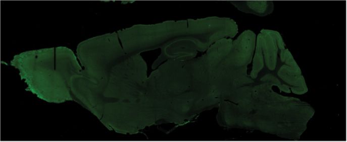
638 UBE3A LOSS IMPAIRS ORIENTATION TUNING
A B C
10
Spont. AP Freq.
(spikes/s)
1
0.1
0.01
-75 mV
0.001/0
UP Control Ube3aSTOP/p+ Ube3aSTOP/p+
:: Gad2-cre
DOWN
D E F
15 2.5 600
UP State Duration (ms)
UP State Freq. (events/s)
Vm Std. Dev. (mV)
2
500
10
1.5
400
1
5
300
0.5
0 0 200
Control Ube3aSTOP/p+ Ube3aSTOP/p+ Control Ube3aSTOP/p+ Ube3aSTOP/p+ Control Ube3aSTOP/p+ Ube3aSTOP/p+
:: Gad2-cre :: Gad2-cre :: Gad2-cre
Fig. 2. Global Ube3a deletion does not affect spontaneous spiking rates and oscillatory activity in L2/3 RS neurons. A: schematic of recording configuration
during spontaneous activity (note that animal is presented with a gray screen stimulus). B: sample recording of a spontaneously active L2/3 RS neuron (top) and
an example of automated detection of UP/DOWN states (bottom) (scale bars ⫽ 1 s, 25 mV). C: spontaneous spiking activity rates for all RS neurons recorded
in L2/3 of control (n ⫽ 41), Ube3aSTOP/p⫹ (n ⫽ 31), and Ube3aSTOP/p⫹::Gad2-Cre (n ⫽ 30) mice (note that points at “0.001/0” represent neurons that did not
exhibit spontaneous spiking activity during the recording session) (Kruskal-Wallis test, P ⫽ 0.315). D: standard deviation of the membrane voltage for all RS
neurons recorded in L2/3 for control (n ⫽ 28), Ube3aSTOP/p⫹ (n ⫽ 19), and Ube3aSTOP/p⫹::Gad2-Cre (n ⫽ 21) mice (ANOVA, P ⫽ 0.59). E: UP state frequency
for control (n ⫽ 28), Ube3aSTOP/p⫹ (n ⫽ 19), and Ube3aSTOP/p⫹::Gad2-Cre (n ⫽ 21) mice (Kruskal-Wallis test, P ⫽ 0.446). F: UP state duration for control
(n ⫽ 28), Ube3aSTOP/p⫹ (n ⫽ 19), and Ube3aSTOP/p⫹::Gad2-Cre (n ⫽ 21) mice (ANOVA, P ⫽ 0.161).
measured UP state frequency and duration, and they were not membrane potential (Fig. 3, D and E). Consistent with
different between groups (Fig. 2, E and F). The standard previous reports, UP states carried more power in the
deviation of the membrane voltage was also similar, indicating gamma bands than DOWN states; however, we did not find
that the voltage difference between UP and DOWN states was any changes in spectral power between the experimental
similar between groups (Fig. 2D). These data suggest that, groups for either UP states or DOWN states. Our data
despite an apparent excitatory/inhibitory imbalance in AS suggest that, at least in anesthetized mice, cortical oscilla-
model mice, spontaneous network activity and baseline firing tions are normal in Ube3aSTOP/p⫹ mice.
rates are not altered by the loss of Ube3a expression.
Spectral Analysis of Vm During Visual Stimulation
Spectral Analysis of Membrane Voltage During Spontaneous
Activity in Ube3aSTOP/p⫹ Mice The presentation of visual stimuli increases gamma (30 – 80
Hz) synchrony in visual cortex (Eckhorn et al. 1988). Addi-
EEG/ECoG recordings of cortical network oscillations are tionally, activating cortical parvalbumin-positive GABAergic
disrupted in AS individuals and model mice (Colas et al. 2005; neurons increases gamma band activity and improves behav-
Jiang et al. 1998; Thibert et al. 2013). As membrane potential ioral performance (Cardin et al. 2009). Disruptions in gamma
fluctuations in single neurons reflect local network synchrony synchrony have been observed in many psychiatric disorders,
and oscillations (Poulet and Petersen 2008), we performed a including autism (Orekhova et al. 2007). Therefore, we tested
spectral analysis of the membrane potential of L2/3 RS neurons whether increased gamma power induced by visual stimulation
during presentation of a gray screen to determine whether we was affected by loss of Ube3a (Fig. 4A). We measured the
could detect altered cortical oscillations in Ube3aSTOP/p⫹ mice spectral power preceding and during 1 s of visual stimulation
(Fig. 3B). We observed no significant changes in delta (0.5– 4 with drifting gratings and calculated the percent change in
Hz), theta (4.5– 8 Hz), alpha (8.5–12 Hz), beta (12.5–29.5 Hz), power with visual stimulation at each frequency. Consistent
or gamma (30 – 80 Hz) frequency bands in Ube3aSTOP/p⫹ mice with previous reports (Eckhorn et al. 1988; Sellers et al. 2013),
compared with control mice or Ube3aSTOP/p⫹:: Gad2-Cre mice we observed an increase in power in the gamma band with
(Fig. 3C). As UP and DOWN states have different biases for visual stimulation; however, we observed no differences be-
high- and low-frequency bands (Beltramo et al. 2013), we tween experimental groups (Fig. 4, B and C). Therefore,
performed a spectral analysis on the UP and DOWN states gamma oscillations induced by visual stimulation in anesthe-
separately in addition to the overall spectral analysis of tized mice are unaffected by Ube3a loss.
J Neurophysiol • doi:10.1152/jn.00618.2016 • www.jn.org
Downloaded from www.physiology.org/journal/jn (134.174.140.176) on January 23, 2020.UBE3A LOSS IMPAIRS ORIENTATION TUNING 639
A B
-65 mV
100
Frequency (Hz) 1
80 0.8
(mV2/ms)
0.6
60
0.4
40
0.2
20
0
Time 1s
C D E
16 All Spontaneous Activity 8 All UP States 5 All DOWN States
4
12 6
Power (V2)
3
8 4
2
4 2
1
0 0 0
Delta Theta Alpha Beta Gamma Delta Theta Alpha Beta Gamma Delta Theta Alpha Beta Gamma
STOP/p+
Ube3a
Control Ube3aSTOP/p+
:: Gad2-cre
Fig. 3. Spectral analysis of spontaneous UP/DOWN states in L2/3 RS neurons. A: schematic of recording configuration during spontaneous activity (note that
animal is presented with a gray screen stimulus). B: sample recording of a spontaneously active L2/3 RS neuron (top) and corresponding spectrogram (bottom)
(scale bar ⫽ 1 s, 20 mV). C: average power spectrum of spontaneous activity of individual L2/3 RS neurons for control (n ⫽ 24), Ube3aSTOP/p⫹ (n ⫽ 19), and
Ube3aSTOP/p⫹::Gad2-Cre (n ⫽ 18) mice. Frequency ranges are defined as delta (0.5– 4 Hz), theta (4.5– 8 Hz), alpha (8.5–12 Hz), beta (12.5–29.5 Hz), and gamma
(30 – 80 Hz) (2-way ANOVA, P ⫽ 0.462). D: average power spectrum of UP states of individual L2/3 RS neurons for control (n ⫽ 24), Ube3aSTOP/p⫹ (n ⫽
19), and Ube3aSTOP/p⫹::Gad2-Cre (n ⫽ 18) mice (2-way ANOVA, P ⫽ 0.58). E: average power spectrum of DOWN states of individual L2/3 RS neurons for
control (n ⫽ 24), Ube3aSTOP/p⫹ (n ⫽ 19), and Ube3aSTOP/p⫹::Gad2-Cre (n ⫽ 18) mice (2-way ANOVA, P ⫽ 0.53).
Effects of Ube3a Loss on Contrast Sensitivity in L2/3 (control, 28.9 ⫾ 1.6 mV; Ube3aSTOP/p⫹ 28.1 ⫾ 2.4 mV;
Regular-Spiking Neurons Ube3aSTOP/p⫹::Gad2-Cre 31.2 ⫾ 2.2 mV) or frequency of
spiking (Fig. 6C) of L2/3 neurons to the visual stimulus. First,
Contrast sensitivity is a property of L2/3 RS neurons where
we compared tuning sharpness of spiking tuning curves (Fig.
responses increase nonlinearly with increasing luminance con-
6D). Neurons in Ube3aSTOP/p⫹ mice had significantly broader
trast (Albrecht and Hamilton 1982). To examine whether the
loss of Ube3a altered contrast sensitivity, we performed whole tuning than in Ube3aSTOP/p⫹::Gad2-Cre mice (P ⬍ 0.05) and
cell recordings while presenting mice with drifting gratings of showed a trend for broader tuning compared with control mice
differing contrast shown at the neuron’s predetermined pre- (P ⫽ 0.16) (Fig. 6D). The OSI of spiking tuning curves was
ferred orientation (Fig. 5A). Spiking contrast responses did not significantly decreased in the Ube3aSTOP/p⫹ mice compared
differ between groups (Fig. 5, B–D). The same was true for with control mice (P ⬍ 0.05) (Fig. 6E). Ube3aSTOP/p⫹::Gad2-
subthreshold contrast response (Fig. 5, E–G). In conclusion, Cre mice showed an intermediate effect in OSI that was not
Ube3a loss does not affect the contrast response of L2/3 RS statistically different from control mice or Ube3aSTOP/p⫹ mice
neurons. This result confirms grossly normal function of visual (P ⫽ 0.48 and 0.29, respectively) (Fig. 6E). To investigate the
circuitry in Ube3a mice. OSI and tuning sharpness of robustly tuned neurons, we
examined robustness of the curve fit (sum of two Gaussians,
Effects of Ube3a Loss on Orientation Tuning in L2/3 see MATERIALS AND METHODS) by calculating the normalized (to
Regular-Spiking Neurons
the mean firing rate of the preferred direction) residuals of the
L2/3 RS neurons were recorded while drifting gratings were fit. We applied a criterion of ⬍0.125 normalized residual to all
presented in the visual field of the animal. There was no cells and analyzed neurons that passed this criterion (Fig. 6G)
difference in the average subthreshold response amplitude (Cottam et al. 2013). Robustly tuned Ube3aSTOP/p⫹ neurons
J Neurophysiol • doi:10.1152/jn.00618.2016 • www.jn.org
Downloaded from www.physiology.org/journal/jn (134.174.140.176) on January 23, 2020.640 UBE3A LOSS IMPAIRS ORIENTATION TUNING
A Visual Stimulus B
120
Change in Power (%)
80
40
0
Delta Theta Alpha Beta Gamma
20 mV
400 ms
C
100 1 120
Change in Power (%)
80 0.8 80
Frequency (Hz)
(mV2/ms)
60 0.6
40
0.4
40 0
2 4 8 16 32 64 128
0.2
Frequency (Hz)
20 -40
0
Ube3aSTOP/p+
Control Ube3aSTOP/p+
Time :: GAD2-cre
400 ms
Fig. 4. Spectral power changes induced with visual stimulation. A: sample recording of a L2/3 RS neuron during 1 s of visual stimulation (shaded region, top)
and corresponding spectrogram of recording (bottom). B: average change in power with visual stimulation at different frequency bands for control (n ⫽ 31),
Ube3aSTOP/p⫹ (n ⫽ 26), and Ube3aSTOP/p⫹::Gad2-Cre (n ⫽ 23) mice (2-way ANOVA, P ⫽ 0.542). Frequency ranges are defined as delta (2– 4 Hz), theta (4.5– 8
Hz), alpha (8.5–12 Hz), beta (12.5–29.5 Hz), and gamma (30 – 80 Hz). C: average change in power with visual stimulation for all frequencies for control (n ⫽
31), Ube3aSTOP/p⫹ (n ⫽ 26), and Ube3aSTOP/p⫹:: Gad2-Cre (n ⫽ 23) mice.
showed decreased OSI and increased tuning width compared during the stimulus is the F0 component. The F1 component
with control mice (P ⬍ 0.05) (Fig. 6, F and G). To examine has been shown to be more highly tuned for orientation than
robustly responsive neurons more closely, we performed the “Area” or the F0 measurement (Carandini and Ferster
ANOVA on the spiking responses to visual stimulation. Neu- 2000; Lien and Scanziani 2013; Niell and Stryker 2008). To
rons that did not show statistically distinguishable responsive- determine whether cells in each group were “simple” or “com-
ness to any particular orientation were excluded from subse- plex” we measured the F1-to-F0 ratio at each neuron’s pre-
quent analysis (Fig. 6H). Similarly to all neurons grouped ferred orientation (F1/F0Pref; Fig. 7C). Neurons that have a
together, robustly responsive Ube3aSTOP/p⫹ neurons also F1/F0 ⬎ 1 are typically considered “simple” cells and, sub-
showed decreased OSI compared with control mice (P ⬍ threshold OSI measurements using F1 values are most appro-
0.05)(Fig. 6H). As a final measure of orientation tuning we priate (Carandini and Ferster 2000; Niell and Stryker 2008).
calculated the preferred-to-orthogonal ratios for all cells and Almost all cells recorded had F1/F0Pref values ⬎1, and we
compared the groups using this metric. Surprisingly, there were calculated the subthreshold OSI using either Area (Fig. 7D) or
no statistically significant differences between groups for this F1 values (Fig. 7E) and compared between groups. Using F1
measure of orientation tuning (Fig. 6I). However, trends re- values gave more highly tuned subthreshold OSI for all groups
flected what we have observed with OSI (P ⫽ 0.15). Finally, compared with subthreshold OSI using Area, but the groups
we calculated the DSI for the spiking responses, and this metric were not statistically different with either measurement (Fig. 7,
was not measurably different between groups (Fig. 6J). To- D and E). Finally, we calculated the DSI for the subthreshold
gether these data strongly suggest that Ube3a loss results in responses (Area), and this measurement was not different
weaker orientation tuning in L2/3 RS neurons that are robustly between groups (Fig. 7F).
responsive and tuned to orientation. Together, these data indicate that Ube3a loss broadens
Subthreshold (i.e., membrane potential) responses recorded orientation tuning of the spiking responses in L2/3 RS neurons
in L2/3 RS neurons also showed orientation tuning, albeit less and has more subtle effects on subthreshold responses to
sharply tuned than spiking responses (Fig. 7, D and E) (Smith orientation.
et al. 2013). In response to drifting gratings the membrane
potential will fluctuate in amplitude at the same temporal DISCUSSION
frequency (2 Hz) of the stimulus (Fig. 7, A and B). The This work represents the first in vivo investigation into
difference between the peak and trough is the F1 (or frequency cellular excitability, orientation tuning, and contrast sensitivity
modulated) component, whereas the mean membrane potential in an AS model mouse line. We found that individual L2/3 RS
J Neurophysiol • doi:10.1152/jn.00618.2016 • www.jn.org
Downloaded from www.physiology.org/journal/jn (134.174.140.176) on January 23, 2020.UBE3A LOSS IMPAIRS ORIENTATION TUNING 641
A
0% 30% 50% 100%
Vm (mV)
Area (V*s)
B Contrast Sensitivity C Contrast Exponent D Semisaturation Contrast
2.5
(Spiking) (Spiking) (Spiking)
2
Frequency (Spikes/s)
100
10
Contrast Exponent
1.5
Contrast (%)
1 50
5
Control
0.5 Ube3aSTOP/p+
Ube3aSTOP/p+ 0 0
:: Gad2-cre Control Ube3aSTOP/p+ Ube3aSTOP/p+ Control Ube3aSTOP/p+ Ube3aSTOP/p+
0 :: Gad2-cre :: Gad2-cre
0 20 40 60 80 100
Contrast (%)
E Contrast Sensitivity F Contrast Exponent G Semisaturation Contrast
15 (Area) (Area) (Area)
13 100
10
Contrast Exponent
Area (V*s)
Contrast (%)
11
5 50
9
Control
Ube3aSTOP/p+
7
Ube3aSTOP/p+
:: Gad2-cre 0 0
5 Control Ube3aSTOP/p+ Ube3aSTOP/p+ Control Ube3aSTOP/p+ Ube3aSTOP/p+
0 20 40 60 80 100 :: Gad2-cre :: Gad2-cre
Contrast (%)
Fig. 5. Contrast sensitivity is unchanged in Ube3aSTOP/p⫹ mice. A: sample recording from a L2/3 RS neuron of visually evoked responses to drifting gratings
of increasing contrast (scale bar 150 ms, 20 mV). Blue shaded region indicates the zone representing the “Area” measurement or subthreshold synaptic response
to visual stimulation. B: average contrast sensitivity curves for spiking responses fit with a hyperbolic ratio equation for control (n ⫽ 12), Ube3aSTOP/p⫹ (n ⫽
11), and Ube3aSTOP/p⫹::Gad2-Cre (n ⫽ 15) mice. C: average contrast exponent for spiking responses fit with a hyperbolic ratio equation in control (n ⫽ 12),
Ube3aSTOP/p⫹ (n ⫽ 11), and Ube3aSTOP/p⫹:: Gad2-Cre (n ⫽ 15) mice (Kruskal-Wallis test, P ⫽ 0.311). D: average semisaturation contrast (C50) for spiking
responses fit with a hyperbolic ratio equation in control (n ⫽ 12), Ube3aSTOP/p⫹ (n ⫽ 11), and Ube3aSTOP/p⫹::Gad2-Cre (n ⫽ 15) mice (Kruskal-Wallis test,
P ⫽ 0.300). E: average contrast sensitivity curves for subthreshold responses fit with a hyperbolic ratio equation for control (n ⫽ 41), Ube3aSTOP/p⫹ (n ⫽ 31),
and Ube3aSTOP/p⫹::Gad2-Cre (n ⫽ 30) mice. F: average contrast exponent for subthreshold responses fit with a hyperbolic ratio equation in control (n ⫽ 41),
Ube3aSTOP/p⫹ (n ⫽ 31), and Ube3aSTOP/p⫹::Gad2-Cre (n ⫽ 30) mice (Kruskal-Wallis test, P ⫽ 0.800). G: average semisaturation contrast (C50) for subthreshold
responses fit with a hyperbolic ratio equation in control (n ⫽ 41), Ube3aSTOP/p⫹ (n ⫽ 31), and Ube3aSTOP/p⫹::Gad2-Cre (n ⫽ 30) mice (Kruskal-Wallis test,
P ⫽ 0.238).
J Neurophysiol • doi:10.1152/jn.00618.2016 • www.jn.org
Downloaded from www.physiology.org/journal/jn (134.174.140.176) on January 23, 2020.642 UBE3A LOSS IMPAIRS ORIENTATION TUNING
A
0° 45° 90° 135°
-60 mV
Stim. onset
180° 225° 270° 315°
Vm (mV) Area (V*s)
B Control Ube3aSTOP/p+ Ube3aSTOP/p+ :: Gad2-cre
2.5 4 2.5
1.25 2 1.25
Frequency (spikes/s)
0 0 0
2 3 6
1 1.5 3
0 0 0
3 3.5 4.5
1.5 1.75 2.25
0 0 0
0 90 180 270 0 90 180 270 0 90 180 270
Orientation (degrees) Orientation (degrees) Orientation (degrees)
C 3 All Cells D All Cells E All Cells
Control * 1.0
*
Frequency (spikes/s)
100
Ube3aSTOP/p+
HWHH (degrees)
2 Ube3aSTOP/p+ 0.8
Spiking OSI
:: Gad2-cre 75
0.6
1 0.4
50
0.2
0 25 0
0 90 180 270 Control Ube3aSTOP/p+ Ube3aSTOP/p+ Control Ube3aSTOP/p+ Ube3aSTOP/p+
Orientation (degrees) :: Gad2-cre :: Gad2-cre
F Robustly Tuned Cells G Robustly Tuned Cells H Robustly Responsive Cells
* *
60 * 1.0 1.0
HWHH (degrees)
0.8 0.8
Spiking OSI
Spiking OSI
50
0.6 0.6
40 0.4 0.4
0.2 0.2
30
0 0
Control Ube3aSTOP/p+ Ube3aSTOP/p+ Control Ube3aSTOP/p+ Ube3aSTOP/p+ Control Ube3aSTOP/p+ Ube3aSTOP/p+
:: Gad2-cre :: Gad2-cre :: Gad2-cre
I All Cells J All Cells
1.0 1.0
Pref. / Ortho. ratio
0.8 0.8
Spiking DSI
0.6 0.6
0.4 0.4
0.2 0.2
0 0
Control Ube3aSTOP/p+ Ube3aSTOP/p+ Control Ube3aSTOP/p+ Ube3aSTOP/p+
:: Gad2-cre :: Gad2-cre
J Neurophysiol • doi:10.1152/jn.00618.2016 • www.jn.org
Downloaded from www.physiology.org/journal/jn (134.174.140.176) on January 23, 2020.UBE3A LOSS IMPAIRS ORIENTATION TUNING 643
neurons had increased excitability in Ube3aSTOP/p⫹ mice but al. 2013). Alternatively, it is possible that the enhanced delta
increased excitability caused by Ube3a loss did not translate activity previously observed with EEG and LFP recordings
into increased activity of the local network as measured by might manifest from activity in cell types other than L2/3 RS
UP/DOWN states or spontaneous spiking. Surprisingly, in- neurons.
creased excitability in individual neurons was rescued by We examined two visual cortical response properties, orien-
reinstatement of Ube3a in GABAergic neurons, suggesting tation selectivity and contrast sensitivity, in the AS model mice
that a homeostatic mechanism may underlie this phenotype. A at both the spiking and subthreshold levels (Figs. 5–7). We
rearrangement in excitation-to-inhibition ratio that causes a net found that both subthreshold and spiking responses to drifting
decrease in spiking may result in increased intrinsic excitability grating stimuli presented at different contrasts were similar
to normalize spiking rates (Nataraj et al. 2010). A decrease in between genotypes. However, spiking responses to drifting
excitatory synapses occurs early in development in AS model gratings of different orientations were more broadly tuned in
mice and may provide a period of decreased cortical spiking Ube3aSTOP/p⫹ mice than in control mice. Furthermore, rein-
that is then compensated for by the observed increase in stating Ube3a in GABAergic neurons in Ube3aSTOP/p⫹::Gad2-
pyramidal neuron intrinsic excitability that fails to normalize in Cre mice partially ameliorated this phenotype, as tuning in-
adulthood (Fig. 1) (Yashiro et al. 2005). Accordingly, rein- dexes from Ube3aSTOP/p⫹::Gad2-Cre mice were not statisti-
statement of Ube3a in GABAergic neurons may normalize cally different from control mice. Subthreshold orientation
network spiking levels early in postnatal development and tuning curves in Ube3aSTOP/p⫹ mice also showed a trend for
prevent subsequent homeostatic rearrangements from occur- having more broadly tuned responses; however, these changes
ring. Alternatively, increased intrinsic excitability in pyramidal did not reach statistical significance in our sample size.
neurons in the Ube3aSTOP/p⫹ mice may result directly from Previous studies have suggested that sensitivity to contrast
decreased tonic (rather than phasic/evoked) inhibition leading arises early in the visual system at the level of retinal ganglion
to increased membrane resistance. We previously showed that cells (Shapley 1990; Shapley and Victor 1978). Our negative
Ube3a-deficient L2/3 pyramidal neurons have decreased tonic results with respect to contrast sensitivity suggest that the
inhibition (Judson et al. 2016). If decreased tonic inhibition function of visual circuits remains largely intact in AS model
underlies increased intrinsic excitability, then we would predict mice at the retinal and thalamic stages. This is consistent with
normal levels of tonic inhibition to be restored in the our work and work from others demonstrating normal visual
Ube3aSTOP::Gad2-Cre mice. acuity and retinotopy in AS model mice (Sato and Stryker
We also measured membrane potential oscillations in active 2010; Yashiro et al. 2009). Therefore, the orientation tuning
(during visual stimulation) and inactive (in the absence of defects we observed in this study appear to be somewhat
visual stimulation) states by comparing power spectra between specific in regard to visual system dysfunction in AS. Interest-
genotypes (Figs. 3 and 4). Visual stimulation greatly increased ingly, defects in orientation tuning were only observed in
spectral power in the gamma band, but we did not observe spiking responses and not in subthreshold tuning curves. We
differences in spectral power between genotypes either during examined subthreshold tuning by measuring the area between
baseline conditions or with visual stimulation. We were sur- the membrane potential response and the average “DOWN”
prised by this finding because both AS model mice and state membrane potential as well as using the F1-to-F0 ratio.
individuals with AS have EEG abnormalities, particularly in While OSI was increased with F1/F0 measurements compared
the delta band (Judson et al. 2016; Miura et al. 2002; Thibert with area measurements, neither metric revealed a defect in
et al. 2013). As anesthesia significantly increases activity in the subthreshold orientation tuning that was statistically distin-
delta band, we suspect that differences in delta band activity guishable. It is currently difficult to discern the mechanism
were masked in our recordings in anesthetized mice (Pagliar- underlying broader orientation tuning in RS neurons in
dini et al. 2013). Therefore, it is possible that recordings in Ube3aSTOP mice. L2/3 pyramidal neurons in Ube3aSTOP mice
awake animals may expose additional differences in network do have decreased evoked inhibitory input (Judson et al. 2016),
excitability, especially since anesthetics have been shown to and decreasing inhibition onto pyramidal neurons has been
alter inhibitory neuron function in the visual system (Haider et shown to decrease orientation selectivity (Atallah et al. 2012;
Fig. 6. Broader orientation tuning in L2/3 regular spiking neurons of Ube3aSTOP/p⫹ mice. A: sample recording from a L2/3 RS neuron to drifting gratings of
different orientations. Blue shaded region indicates the zone representing the “Area” measurement or subthreshold synaptic response to visual stimulation (note
that this neuron did not show significant subthreshold F1 modulation) (scale bar 200 ms, 20 mV). B: sample tuning curves and spiking responses to visual stimuli
of different orientations (3 sample neurons per group) for control (left, black), Ube3aSTOP/p⫹ (center, red), and Ube3aSTOP/p⫹::Gad2-Cre (right, green) mice.
Spiking responses are represented as mean ⫾ SE of at least 6 presentations of each orientation. Tuning curve for sample recording (A) is top rightmost curve
of the samples from the Ube3aSTOP/p⫹::Gad2-Cre group. C: average tuning curves from control (n ⫽ 35), Ube3aSTOP/p⫹ (n ⫽ 27), and Ube3aSTOP/p⫹::Gad2-Cre
(n ⫽ 27) mice. D: half-width at half-height (HWHH) measurements made from Gaussian fits of spiking orientation tuning curves from control (n ⫽ 35),
Ube3aSTOP/p⫹ (n ⫽ 27), and Ube3aSTOP/p⫹::Gad2-Cre (n ⫽ 27) mice (Kruskal-Wallis test, P ⫽ 0.041). E: orientation selectivity index (OSI) measured from
spiking orientation tuning curves from all recorded cells in control (n ⫽ 35), Ube3aSTOP/p⫹ (n ⫽ 27), and Ube3aSTOP/p⫹:: Gad2-Cre (n ⫽ 27) mice (ANOVA,
P ⫽ 0.026). F: HWHH measurements made from Gaussian fits of spiking orientation tuning curves for robustly tuned cells in control (n ⫽ 15), Ube3aSTOP/p⫹
(n ⫽ 10), and Ube3aSTOP/p⫹::Gad2-Cre (n ⫽ 8) mice (Kruskal-Wallis test, P ⫽ 0.044). G: OSI measured from spiking responses from cells that were well fit
by sum-of-two-Gaussian tuning curves [i.e., normalized residuals of the fit were ⬍0.0125; control (n ⫽ 16), Ube3aSTOP/p⫹ (n ⫽ 10), and Ube3aSTOP/p⫹::Gad2-
Cre (n ⫽ 8), Kruskal-Wallis test, P ⫽ 0.031]. H: OSI measured from spiking orientation tuning curves from cells that robustly responded to at least 1 orientation
compared with all others [i.e., ANOVA post-hoc test must be P ⬍ 0.05; control (n ⫽ 22), Ube3aSTOP/p⫹ (n ⫽ 18), and Ube3aSTOP/p⫹:: Gad2-Cre (n ⫽ 20),
ANOVA, P ⫽ 0.045]. I: preferred-to-orthogonal ratio from spiking orientation tuning curves from all recorded cells in control (n ⫽ 35), Ube3aSTOP/p⫹ (n ⫽
27), and Ube3aSTOP/p⫹:: Gad2-Cre (n ⫽ 27) mice (Kruskal-Wallis test, P ⫽ 0.151). J: direction selectivity index (DSI) measured from spiking orientation tuning
curves from all recorded cells in control (n ⫽ 35), Ube3aSTOP/p⫹ (n ⫽ 27), and Ube3aSTOP/p⫹::Gad2-Cre (n ⫽ 27) mice (ANOVA, P ⫽ 0.294). *P ⬍ 0.05 with
post hoc test for significance.
J Neurophysiol • doi:10.1152/jn.00618.2016 • www.jn.org
Downloaded from www.physiology.org/journal/jn (134.174.140.176) on January 23, 2020.644 UBE3A LOSS IMPAIRS ORIENTATION TUNING
A B Preferred orientation Orthogonal orientation
Subthreshold visually evoked response
(Subthreshold) (Subthreshold)
F0 (mean) F1
Area (V*s)
-64 mV
Time
Stim. onset Stim. off Stim. onset Stim. off
C Control Ube3a STOP/p+
Ube3a STOP/p+
:: Gad2-cre
8 5 3
# of neurons
4
0 0 0
1 2.5 4 1 2.5 4 1 2.5 4
Subthreshold F1/F0Pref.
D E F
0.15 0.25 0.25
Subtheshold DSI (Area)
Subthreshold OSI (Area)
Subthreshold OSI (F1)
0.2 0.2
0.1
0.15 0.15
0.1 0.1
0.05
0.05 0.05
0 0 0
Control Ube3aSTOP/p+ Ube3aSTOP/p+ Control Ube3aSTOP/p+ Ube3aSTOP/p+ Control Ube3aSTOP/p+ Ube3aSTOP/p+
:: Gad2-cre :: Gad2-cre :: Gad2-cre
Fig. 7. Subthreshold orientation tuning is unchanged in Ube3aSTOP/p⫹ mice. A: illustration of the measurements made for the subthreshold analysis of visual
responses. F0 is the mean subthreshold membrane potential, F1 is the difference between the peak and trough of the subthreshold membrane potential, and blue
shaded region corresponds to the Area (V ⫻ s) measurement. All measurements are made during presentation of the visual stimulus. B: sample recording from
a L2/3 RS neuron to drifting gratings in its preferred and orthogonal orientations. Blue shaded region indicates the zone representing the “Area” measurement
or subthreshold synaptic response to visual stimulation. The recordings are averages of 6 presentations of the same orientation and low-pass filtered at 100 Hz.
(note that this neuron had significant subthreshold F1 modulation to the preferred orientation) (scale bar 200 ms, 5 mV). C: histograms of subthreshold F1/F0
measurements at the neuron’s preferred orientation (F1/F0Pref). D: orientation selectivity index measured from subthreshold (Area) orientation tuning
curves from control (n ⫽ 42), Ube3aSTOP/p⫹ (n ⫽ 32), and Ube3aSTOP/p⫹::Gad2-Cre (n ⫽ 30) mice (Kruskal-Wallis test, P ⫽ 0.774). E: orientation
selectivity index measured from subthreshold F1 from control (n ⫽ 35), Ube3aSTOP/p⫹ (n ⫽ 27), and Ube3aSTOP/p⫹::Gad2-Cre (n ⫽ 27) mice
(Kruskal-Wallis test, P ⫽ 0.85). F: direction selectivity index measured from subthreshold (Area) orientation tuning curves from control (n ⫽ 42),
Ube3aSTOP/p⫹ (n ⫽ 32), and Ube3aSTOP/p⫹::Gad2-Cre (n ⫽ 30) mice (ANOVA, P ⫽ 0.393).
Wilson et al. 2012). Interestingly, reinstating Ube3a in congruent with recent findings demonstrating that reinstate-
GABAergic neurons in Ube3aSTOP::Gad2-Cre mice also re- ment of Ube3a in GABAergic neurons can also normalize
sults in an intermediate effect on evoked inhibition, indicating seizure susceptibility and elevated delta band EEG activity in
that a lack of a robust “rescue” of orientation selectivity in AS model mice (Judson et al. 2016). Together these studies
Ube3aSTOP::Gad2-Cre mice may reflect the intermediate effect point to a critical role for Ube3a in GABAergic neurons in the
in evoked inhibition (Judson et al. 2016). Of course, there are pathogenesis of AS and suggest that reinstatement of Ube3a in
many other synaptic and circuit contributions to orientation GABAergic neurons may have a wide range of therapeutic
tuning that could be defective in Ube3aSTOP mice, such as the benefits.
tuning of thalamic input, changes in excitability, or receptive
field structure (Priebe and Ferster 2012).
Overall, this work demonstrates that maternal Ube3a loss ACKNOWLEDGMENTS
disrupts cortex-dependent computations. Specifically, excit- We thank M. Carandini for providing the MATLAB code to fit orientation
ability is higher in L2/3 RS neurons in the visual cortex of AS tuning and contrast response curves; K. Sellers and F. Frohlich for providing
model mice in vivo, and orientation selectivity is weaker, the MATLAB code for spectral analysis; J. Han for genotyping and imaging
support; M. Judson, R. Larsen, and J. Berrios for experimental advice; and P.
compared with control littermates. Surprisingly, our data indi- Manis for his expertise and advice on data analysis.
cate that reinstatement of Ube3a in GABAergic neurons alone Present address of M. L. Wallace: Howard Hughes Medical Institute, Dept.
results in normal excitability and orientation tuning. This is of Neurobiology, Harvard Medical School, Boston, MA 02115.
J Neurophysiol • doi:10.1152/jn.00618.2016 • www.jn.org
Downloaded from www.physiology.org/journal/jn (134.174.140.176) on January 23, 2020.UBE3A LOSS IMPAIRS ORIENTATION TUNING 645
GRANTS changes in excitatory circuitry. J Neurosci 31: 14223–14234, 2011. doi:10.
1523/JNEUROSCI.3157-11.2011.
This work was supported by National Institute of Neurological Disorders
Hensch TK, Fagiolini M, Mataga N, Stryker MP, Baekkeskov S, Kash SF.
and Stroke (NINDS) NRSA Fellowship 1F31 NS-077847 (M. L. Wallace); the
Local GABA circuit control of experience-dependent plasticity in develop-
Angelman Syndrome Foundation, Simons Foundation Grant SFARI no.
274426, and NINDS Grant R01 NS-085093 (B. D. Philpot); the Angelman ing visual cortex. Science 282: 1504 –1508, 1998. doi:10.1126/science.282.
Syndrome Foundation and NWO-ZoN-Mw grant (Y. Elgersma); an EMC 5393.1504.
fellowship (G. M. van Woerden); and grants from the Whitehall Foundation Hubel DH, Wiesel TN. Integrative action in the cat’s lateral geniculate body.
and the Klingenstein Foundation, Simons Foundation Grant SCGB 325407SS, J Physiol 155: 385–398, 1961. doi:10.1113/jphysiol.1961.sp006635.
National Science Foundation Grant 1450824, and NINDS Grant 1R01 NS- Hubel DH, Wiesel TN. Receptive fields, binocular interaction and functional
091335 (S. L. Smith). architecture in the cat’s visual cortex. J Physiol 160: 106 –154, 1962.
doi:10.1113/jphysiol.1962.sp006837.
Isaacson JS, Scanziani M. How inhibition shapes cortical activity. Neuron 72:
DISCLOSURES 231–243, 2011. doi:10.1016/j.neuron.2011.09.027.
No conflicts of interest, financial or otherwise, are declared by the authors. Jiang YH, Armstrong D, Albrecht U, Atkins CM, Noebels JL, Eichele G,
Sweatt JD, Beaudet AL. Mutation of the Angelman ubiquitin ligase in mice
causes increased cytoplasmic p53 and deficits of contextual learning and
AUTHOR CONTRIBUTIONS long-term potentiation. Neuron 21: 799 – 811, 1998. doi:10.1016/S0896-
6273(00)80596-6.
M.L.W., S.L.S., and B.D.P. conceived and designed research; M.L.W. Judson MC, Wallace ML, Sidorov MS, Burette AC, Gu B, van Woerden
performed experiments; M.L.W., S.L.S., and B.D.P. analyzed data; M.L.W., GM, King IF, Han JE, Zylka MJ, Elgersma Y, Weinberg RJ, Philpot
S.L.S., and B.D.P. interpreted results of experiments; M.L.W., S.L.S., and BD. GABAergic neuron-specific loss of Ube3a causes Angelman syndrome-
B.D.P. prepared figures; M.L.W., S.L.S., and B.D.P. drafted manuscript; like EEG abnormalities and enhances seizure susceptibility. Neuron 90:
M.L.W., G.M.v.W., Y.E., S.L.S., and B.D.P. edited and revised manuscript; 56 – 69, 2016. doi:10.1016/j.neuron.2016.02.040.
M.L.W., G.M.v.W., Y.E., S.L.S., and B.D.P. approved final version of man- Kishino T, Lalande M, Wagstaff J. UBE3A/E6-AP mutations cause Angel-
uscript. man syndrome. Nat Genet 15: 70 –73, 1997. doi:10.1038/ng0197-70.
Lee SH, Kwan AC, Zhang S, Phoumthipphavong V, Flannery JG, Mas-
REFERENCES manidis SC, Taniguchi H, Huang ZJ, Zhang F, Boyden ES, Deisseroth
K, Dan Y. Activation of specific interneurons improves V1 feature selec-
Albrecht DG, Hamilton DB. Striate cortex of monkey and cat: contrast tivity and visual perception. Nature 488: 379 –383, 2012. doi:10.1038/
response function. J Neurophysiol 48: 217–237, 1982. nature11312.
Atallah BV, Bruns W, Carandini M, Scanziani M. Parvalbumin-expressing Lien AD, Scanziani M. Tuned thalamic excitation is amplified by visual
interneurons linearly transform cortical responses to visual stimuli. Neuron cortical circuits. Nat Neurosci 16: 1315–1323, 2013. doi:10.1038/nn.3488.
73: 159 –170, 2012. doi:10.1016/j.neuron.2011.12.013. Margrie TW, Brecht M, Sakmann B. In vivo, low-resistance, whole-cell
Beltramo R, D’Urso G, Dal Maschio M, Farisello P, Bovetti S, Clovis Y, recordings from neurons in the anaesthetized and awake mammalian brain.
Lassi G, Tucci V, De Pietri Tonelli D, Fellin T. Layer-specific excitatory Pflugers Arch 444: 491– 498, 2002. doi:10.1007/s00424-002-0831-z.
circuits differentially control recurrent network dynamics in the neocortex. Miura K, Kishino T, Li E, Webber H, Dikkes P, Holmes GL, Wagstaff J.
Nat Neurosci 16: 227–234, 2013. doi:10.1038/nn.3306. Neurobehavioral and electroencephalographic abnormalities in Ube3a ma-
Brainard DH. The Psychophysics Toolbox. Spat Vis 10: 433– 436, 1997. ternal-deficient mice. Neurobiol Dis 9: 149 –159, 2002. doi:10.1006/nbdi.
doi:10.1163/156856897X00357. 2001.0463.
Carandini M, Ferster D. Membrane potential and firing rate in cat primary Nataraj K, Le Roux N, Nahmani M, Lefort S, Turrigiano G. Visual
visual cortex. J Neurosci 20: 470 – 484, 2000. deprivation suppresses L5 pyramidal neuron excitability by preventing the
Cardin JA, Carlén M, Meletis K, Knoblich U, Zhang F, Deisseroth K, Tsai induction of intrinsic plasticity. Neuron 68: 750 –762, 2010. doi:10.1016/j.
LH, Moore CI. Driving fast-spiking cells induces gamma rhythm and neuron.2010.09.033.
controls sensory responses. Nature 459: 663– 667, 2009. doi:10.1038/ Niell CM, Stryker MP. Highly selective receptive fields in mouse visual
nature08002. cortex. J Neurosci 28: 7520 –7536, 2008. doi:10.1523/JNEUROSCI.0623-
Colas D, Wagstaff J, Fort P, Salvert D, Sarda N. Sleep disturbances in 08.2008.
Ube3a maternal-deficient mice modeling Angelman syndrome. Neurobiol Orekhova EV, Stroganova TA, Nygren G, Tsetlin MM, Posikera IN,
Dis 20: 471– 478, 2005. doi:10.1016/j.nbd.2005.04.003. Gillberg C, Elam M. Excess of high frequency electroencephalogram
Cottam JC, Smith SL, Häusser M. Target-specific effects of somatostatin- oscillations in boys with autism. Biol Psychiatry 62: 1022–1029, 2007.
expressing interneurons on neocortical visual processing. J Neurosci 33: doi:10.1016/j.biopsych.2006.12.029.
19567–19578, 2013. doi:10.1523/JNEUROSCI.2624-13.2013. Pagliardini S, Funk GD, Dickson CT. Breathing and brain state: urethane
de Kock CP, Bruno RM, Spors H, Sakmann B. Layer- and cell-type-specific anesthesia as a model for natural sleep. Respir Physiol Neurobiol 188:
suprathreshold stimulus representation in rat primary somatosensory cortex. 324 –332, 2013. doi:10.1016/j.resp.2013.05.035.
J Physiol 581: 139 –154, 2007. doi:10.1113/jphysiol.2006.124321. Paluszkiewicz SM, Olmos-Serrano JL, Corbin JG, Huntsman MM. Im-
Eckhorn R, Bauer R, Jordan W, Brosch M, Kruse W, Munk M, Reitboeck paired inhibitory control of cortical synchronization in fragile X syndrome.
HJ. Coherent oscillations: a mechanism of feature linking in the visual J Neurophysiol 106: 2264 –2272, 2011. doi:10.1152/jn.00421.2011.
cortex? Multiple electrode and correlation analyses in the cat. Biol Cybern Poulet JF, Petersen CC. Internal brain state regulates membrane potential
60: 121–130, 1988. doi:10.1007/BF00202899. synchrony in barrel cortex of behaving mice. Nature 454: 881– 885, 2008.
Gibson JR, Bartley AF, Hays SA, Huber KM. Imbalance of neocortical doi:10.1038/nature07150.
excitation and inhibition and altered UP states reflect network hyperexcit- Priebe NJ, Ferster D. Mechanisms of neuronal computation in mammalian
ability in the mouse model of fragile X syndrome. J Neurophysiol 100: visual cortex. Neuron 75: 194 –208, 2012. doi:10.1016/j.neuron.2012.06.
2615–2626, 2008. doi:10.1152/jn.90752.2008. 011.
Gonçalves JT, Anstey JE, Golshani P, Portera-Cailliau C. Circuit level Ringach DL, Hawken MJ, Shapley R. Dynamics of orientation tuning in
defects in the developing neocortex of Fragile X mice. Nat Neurosci 16: macaque primary visual cortex. Nature 387: 281–284, 1997. doi:10.1038/
903–909, 2013. doi:10.1038/nn.3415. 387281a0.
Goupillaud P, Grossmann A, Morlet J. Cycle-octave and related transforms Sanchez-Vives MV, McCormick DA. Cellular and network mechanisms of
in seismic signal analysis. Geoexploration 23: 85–102, 1984. doi:10.1016/ rhythmic recurrent activity in neocortex. Nat Neurosci 3: 1027–1034, 2000.
0016-7142(84)90025-5. doi:10.1038/79848.
Haider B, Häusser M, Carandini M. Inhibition dominates sensory responses Sato M, Stryker MP. Genomic imprinting of experience-dependent cortical
in the awake cortex. Nature 493: 97–100, 2013. doi:10.1038/nature11665. plasticity by the ubiquitin ligase gene Ube3a. Proc Natl Acad Sci USA 107:
Haider B, McCormick DA. Rapid neocortical dynamics: cellular and network 5611–5616, 2010. doi:10.1073/pnas.1001281107.
mechanisms. Neuron 62: 171–189, 2009. doi:10.1016/j.neuron.2009.04.008. Sellers KK, Bennett DV, Hutt A, Fröhlich F. Anesthesia differentially
Hays SA, Huber KM, Gibson JR. Altered neocortical rhythmic activity states modulates spontaneous network dynamics by cortical area and layer. J
in Fmr1 KO mice are due to enhanced mGluR5 signaling and involve Neurophysiol 110: 2739 –2751, 2013. doi:10.1152/jn.00404.2013.
J Neurophysiol • doi:10.1152/jn.00618.2016 • www.jn.org
Downloaded from www.physiology.org/journal/jn (134.174.140.176) on January 23, 2020.You can also read