Control of feeding by Piezo-mediated gut mechanosensation in Drosophila - eLife
←
→
Page content transcription
If your browser does not render page correctly, please read the page content below
SHORT REPORT

                                   Control of feeding by Piezo-mediated gut
                                   mechanosensation in Drosophila
                                   Soohong Min1, Yangkyun Oh2, Pushpa Verma3, Samuel C Whitehead4,
                                   Nilay Yapici5, David Van Vactor3, Greg SB Suh2,6, Stephen Liberles1*
                                   1
                                     Howard Hughes Medical Institute, Harvard Medical School, Department of Cell
                                   Biology, Boston, United States; 2Skirball Institute, NYU School of Medicine, New
                                   York, United States; 3Harvard Medical School, Department of Cell Biology, Boston,
                                   United States; 4Department of Physics, Cornell University, Ithaca, United States;
                                   5
                                     Department of Neurobiology and Behavior, Cornell University, Ithaca, United
                                   States; 6KAIST, Department of Biological Sciences, Daejeon, Republic of Korea

                                   Abstract Across animal species, meals are terminated after ingestion of large food volumes, yet
                                   underlying mechanosensory receptors have so far remained elusive. Here, we identify an essential
                                   role for Drosophila Piezo in volume-based control of meal size. We discover a rare population of fly
                                   neurons that express Piezo, innervate the anterior gut and crop (a food reservoir organ), and
                                   respond to tissue distension in a Piezo-dependent manner. Activating Piezo neurons decreases
                                   appetite, while Piezo knockout and Piezo neuron silencing cause gut bloating and increase both
                                   food consumption and body weight. These studies reveal that disrupting gut distension receptors
                                   changes feeding patterns and identify a key role for Drosophila Piezo in internal organ
                                   mechanosensation.

                                   Introduction
                                   Mechanosensory neurons detect a variety of environmental forces that we can touch or hear, as well
*For correspondence:               as internal forces from organs and tissues that control physiological homeostasis (Abraira and Ginty,
Stephen_Liberles@hms.harvard.      2013; Ranade et al., 2015; Umans and Liberles, 2018). In many species, specialized mechanosen-
edu                                sory neurons innervate the gastrointestinal tract and are activated by tissue distension associated
                                   with consuming a large meal (Williams et al., 2016; Zagorodnyuk et al., 2001). Gut mechanosensa-
Competing interest: See
                                   tion may provide an evolutionarily conserved signal for meal termination as gut distension inhibits
page 15
                                   feeding in many species and evokes the sensation of fullness in humans (Phillips and Powley, 1996;
Funding: See page 15               Rolls et al., 1998). However, how gut distension receptors contribute to long-term control of diges-
Received: 11 September 2020        tive physiology and behavior is unclear as tools for selective pathway manipulation are lacking. Iden-
Accepted: 16 February 2021         tifying neuronal mechanisms involved in detecting the volume of ingested food would provide basic
Published: 18 February 2021        insights into this fundamental mechanosensory process, and in humans, perhaps clinical targets for
                                   feeding and metabolic disorders.
Reviewing editor: Claude
Desplan, New York University,
                                       Here, we investigated the roles and mechanisms of food volume sensation in the fruit fly Dro-
United States                      sophila melanogaster. Volumetric control of feeding was classically studied in a larger related insect,
                                   the blowfly, with relevant mechanosensory hotspots identified in the foregut and crop, an analog of
   Copyright Min et al. This
                                   the stomach (Dethier and Gelperin, 1967; Gelperin, 1967). In Drosophila, chemosensory neurons
article is distributed under the
                                   detect nutrients in the periphery and brain to control appetite, with some neurons positively rein-
terms of the Creative Commons
Attribution License, which         forcing feeding during starvation conditions (Bjordal et al., 2014; Dus et al., 2015;
permits unrestricted use and       Miyamoto et al., 2012). In contrast, the importance of gut mechanosensation in Drosophila feeding
redistribution provided that the   control and digestive physiology has not been similarly investigated; mechanosensory neurons of the
original author and source are     gustatory system sense food texture and modulate ingestion (Sánchez-Alcañiz et al., 2017;
credited.                          Zhang et al., 2016), and other mechanosensory neurons in the posterior gut control defecation and

Min et al. eLife 2021;10:e63049. DOI: https://doi.org/10.7554/eLife.63049                                                          1 of 18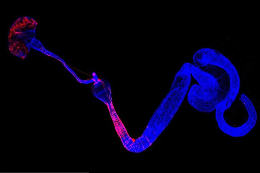
Short report Neuroscience
food intake (Olds and Xu, 2014; Zhang et al., 2014). In contrast, food storage during a meal occurs
primarily in the anterior gut (Lemaitre and Miguel-Aliaga, 2013; Stoffolano and Haselton, 2013).
Enteric neurons of the hypocerebral ganglion innervate the fly crop, foregut, and anterior midgut,
and lesioning of the recurrent nerve (which contains neurons of the hypocerebral ganglion) in Dro-
sophila and blowfly increases feeding duration (Dethier and Gelperin, 1967; Gelperin, 1967;
Pool et al., 2014). Together, these prior studies raise the possibility that a subpopulation of enteric
neurons in Drosophila could be specialized to sense meal-associated gut distension.
Results and discussion
Piezo-expressing enteric neurons innervate the gastrointestinal tract
To explore whether food volume sensation occurs in Drosophila and to investigate underlying mech-
anisms, we first asked whether neurons expressing various mechanosensory ion channels innervated
the anterior gut. Several mechanosensitive ion channels have been reported in Drosophila, including
TRP channels (Nompc, Nanchung, and Inactive), the degenerin/epithelial sodium channel Pickpocket
(Ppk), transmembrane channel-like (Tmc) protein, and Piezo (Coste et al., 2012; Montell, 2005;
Zhang et al., 2016; Zhong et al., 2010). We obtained Gal4 driver lines that mark neurons containing
mechanoreceptor proteins or related family members, induced expression of membrane-tethered
CD8-Green Fluorescent Protein (GFP) or dendritically targeted DenMark fluorescent reporters, and
visualized neuronal innervation of the anterior gut. We observed a small group of Piezo-expressing
enteric neurons located in the hypocerebral ganglion (~5–6 neurons per fly), and a dense network of
Piezo fibers throughout the crop and anterior midgut (Figure 1A and B). Hypocerebral ganglion
neurons were similarly labeled and anterior gut innervation similarly observed in three independent
Piezo-Gal4 driver lines (Figure 1—figure supplement 1A), but not in other Gal4 lines analyzed. We
noted Nanchung expression in some epithelial cells of the crop duct, but not in crop-innervating
neurons. The hypocerebral ganglion and adjacent corpora cardiaca together contain ~35 neurons
per fly based on Elav immunohistochemistry, and Piezo neurons therein were distinct from other
neurons that expressed the fructose receptor Gr43a (~5 neurons per fly) or the glucagon analog adi-
pokinetic hormone (Akh, ~20 neurons per fly) (Figure 1C, D, Figure 1—figure supplement 1B).
Piezo neurites formed a muscle-associated lattice in the gut, and ascending axons contributed to the
recurrent nerve (Figure 1E, F). Using a genetic approach involving the MultiColor FlpOut system
(Nern et al., 2015) for sparse labeling of Piezo cells, flies were obtained with reporter expression in
one or a few hypocerebral ganglion neurons but not in brain structures such as the pars intercerebra-
lis; in these flies, separate Piezo neurons were observed to innervate the crop and/or anterior midgut
(Figure 1—figure supplement 1C). Drosophila Piezo was previously shown to confer mechanically
activated currents when expressed in human cells and to mediate mechanical nociception
(Coste et al., 2012; Kim et al., 2012). Furthermore, vertebrate Piezo homologs play diverse mecha-
nosensory roles, including in internal sensation of airway volume and blood pressure (Min et al.,
2019; Nonomura et al., 2017; Zeng et al., 2018). We hypothesized that Drosophila enteric neurons
that express Piezo and innervate the anterior gut might mediate volumetric control of appetite.
Piezo neurons control feeding behavior
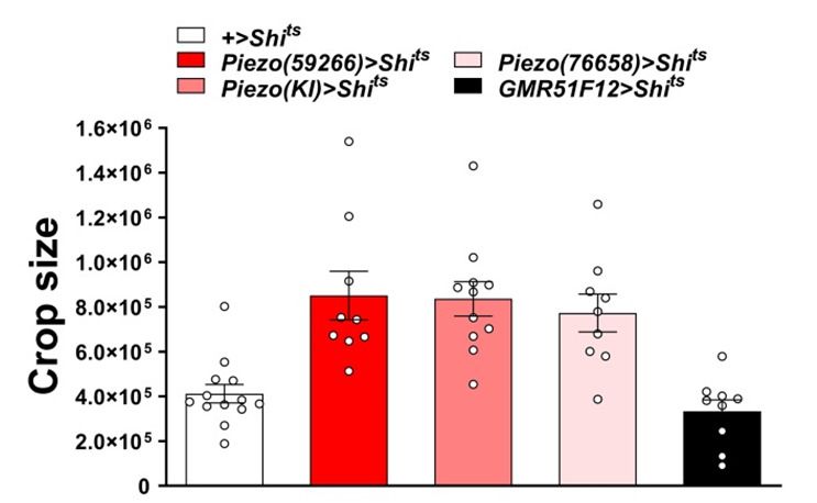
To explore this model, we activated and silenced Piezo neurons using genetic approaches and moni-
tored feeding behavior. We expressed temperature-sensitive Shibire (Shits) that blocks synaptic
transmission at non-permissive temperatures (>32˚C) in Piezo neurons using three independent
Piezo-Gal4 drivers (Piezo>Shits). Piezo>Shits flies were reared at a permissive temperature (18˚C) and
later tested for physiological and behavioral changes at 32˚C. To measure feeding behavior, flies
were fasted for 24 hr, and then given brief access (30 min) to food containing a dye for visualization
and quantification of ingestion (Figure 2A). Piezo>Shits flies from all three genotypes fed ravenously,
and histological examination of the gastrointestinal tract showed gut bloating with increased crop
size (Figure 2B). For comparison, genetic silencing of other gut-innervating neurons labeled in
GMR51F12-Gal4 flies (Figure 2—figure supplement 1) did not impact appetite or cause crop dis-
tension. These findings indicate that disrupting Piezo neurons compromises gut volume homeostasis
and associated control of feeding.
Min et al. eLife 2021;10:e63049. DOI: https://doi.org/10.7554/eLife.63049 2 of 18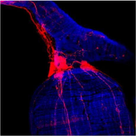
Short report                                                                                                                    Neuroscience

   $                                                                                                %      Piezo-Gal4; UAS-CD8RFP +&*
                                                                            3LH]RQHXURQV
                 &URS
                                                                           9LVFHUDOPXVFOH

                                                                                                                   (ODY          5)3        0HUJH
                                           &URSQHUYH

                                                                                                    (
                                                +&*

                                                                                                           5HFXUUHQW            +&*
                   3URYHQWULFXOXV                                                                           QHUYH

                       $QWHULRUPLGJXW

                                                                         0LGKLQGJXW                      3LH]RQHXURQV
                                                                                                          9LVFHUDOPXVFOH

   &                                    '                                   ) $QWHULRUPLGJXW                        &URS

              &&
                                                                                                            +HDG

                          +&*

                                                                                                            $QXV
                                                    3URYHQWULFXOXV

                 3LH]RQHXURQV                      3LH]RQHXURQV                                                              3LH]RQHXURQV
                  $NK QHXURQV                       *UDQHXURQV                                                             9LVFHUDOPXVFOH

                                                                )LJXUH
Figure 1. Piezo neurons innervate the gastrointestinal tract. (A) Wholemount image of the digestive tract from a Piezo-Gal4 (59266); UAS-DenMark fly
visualized with immunofluorescence for DenMark (red, anti-Red Fluorescent Protein or RFP) and a fluorescent Phalloidin conjugate (blue) to label
visceral muscle. HCG: hypocerebral ganglion, scale bar 100 mm. (B) Immunofluorescence for RFP (red) and Elav (blue) in the HCG from a Piezo-Gal4;
UAS-CD8RFP fly, scale bar 10 mm. (C) Immunofluorescence for GFP (green) and Akh (magenta) in the corpora cardiaca (CC) and HCG from a Piezo-
Gal4; UAS-CD8GFP fly, scale bar 10 mm. (D) Native GFP and RFP fluorescence from the HCG of a Piezo-Gal4; UAS-CD8RFP; Gr43a-LexA; LexAop-
CD8GFP fly, scale bar 10 mm. (E) Image of the recurrent nerve (arrows) labeled by native RFP fluorescence in a Piezo-Gal4; UAS-CD8RFP fly and a
fluorescent Phalloidin conjugate (blue), scale bar 10 mm. (F) The anterior midgut (left) and crop (right) of a Piezo-Gal4; UAS-DenMark fly visualized by
immunofluorescence for DenMark (green) and a fluorescent Phalloidin conjugate (magenta), scale bar 50 mm. See Figure 1—figure supplement 1 and
source data.
The online version of this article includes the following source data and figure supplement(s) for figure 1:
Figure supplement 1. Innervation of the gastrointestinal tract by Piezo neurons.
Figure supplement 1—source data 1. Numerical data to support the graph in Figure 1—figure supplement 1.

                                        To test the effects of activating Piezo neurons on food consumption, we drove expression of the
                                     temperature-regulated ion channel Trpa1 in Piezo neurons using Piezo-Gal4 lines (Piezo>Trpa1).
                                     Thermogenetic activation of Trpa1 in Piezo cells, achieved by transferring Piezo>Trpa1 flies from 18˚
                                     C to 30˚C, suppressed food intake after a 24-hr fast and also blocked meal-associated increases in
                                     crop volume, with similar results observed using three different Piezo-Gal4 drivers (Figure 2C). Since
                                     many cell types express Piezo (Kim et al., 2012), we next used approaches for intersectional genet-
                                     ics involving Gal80, a dominant suppressor of Gal4-mediated gene induction to restrict Trpa1

Min et al. eLife 2021;10:e63049. DOI: https://doi.org/10.7554/eLife.63049                                                                         3 of 18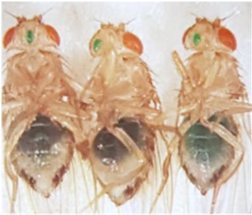
Short report                                                                                                                     Neuroscience

                $

                       )DVWHGIOLHV              *LYHQG\HGIRRG               '\HGIRRGLQDEGRPHQ

   %

                                                   QV

                                                                                                                 QV

   &

                                                    QV                                                            QV

   '                                                               (
                Piezo>CD8RFP;    Piezo>CD8RFP;   Piezo>CD8RFP;
                      +            Elav-Gal80       Cha-Gal80
        %UDLQ

                                                                                           QV
                5)3
                (ODY
        91&

                5)3
                (ODY
        +&*

                5)3
                (ODY

                                                        )LJXUH
Figure 2. Piezo neurons control feeding behavior. (A) Depiction of the colorimetric feeding assay. (B) Fasted flies with Shibire alleles indicated were
given brief access (30 min) to dye-labeled food at 32˚C, and feeding indices and crop sizes were calculated. n (left to right) (feeding index): 16, 11, 13,
10, and 10 trials involving 12 flies per trial. n (crop size): 13, 9, 11, 9, and 9 flies, mean ± SEM, ***p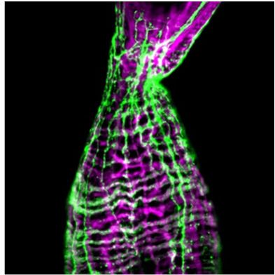
Short report                                                                                                                          Neuroscience

Figure 2 continued
and feeding indices and crop sizes were calculated. n (left to right) (feeding index): 19, 20, 14, 10, and 13 trials involving 12 flies per trial. n (crop size):
12, 6, 11, 10, and 12 flies, mean ± SEM, ***pTrpa1; Elav-
                                       Gal80 flies; to provide additional evidence, we obtained Escargot-Gal4 flies in which Piezo-express-
                                       ing intestinal stem cells (ISCs) are broadly marked (He et al., 2018) and found that thermogenetic
                                       activation of intestinal cells using Escargot-Gal4; UAS-Trpa1 flies also had no effect on feeding (Fig-
                                       ure 2—figure supplement 2B, C). Piezo neurons expressing Dilp2 in the pars intercerebralis are
                                       also reported to innervate the crop and control feeding behavior (Wang et al., 2020), which poten-
                                       tially explain the significant differences we observe in feeding following thermogenetic activation
                                       experiments involving Piezo-Gal4; UAS-Trpa1 and Piezo-Gal4; UAS-Trpa1; Cha-Gal80 flies
                                       (Figure 2E). In control Piezo-Gal4; UAS-CD8RFP flies, we observed reporter expression per fly in
                                       6.2 ± 0.5 hypocerebral neurons and 4.9 ± 1.0 pars intercerebralis neurons, 2.9 ± 0.7 of which express
                                       Dilp2. In Piezo-Gal4; UAS-CD8RFP; Cha-Gal80 flies, we observed reporter expression per fly in
                                       5.2 ± 0.5 hypocerebral neurons and 1.1 ± 0.5 pars intercerebralis neuron, 0.4 ± 0.3 of which express
                                       Dilp2 (about half of flies had one co-labeled neuron and half had zero) (Figure 2—figure supple-
                                       ment 3A–C). In flies that lacked any reporter expression in pars intercerebralis Dilp2 neurons, we still
                                       observed labeled neurites in the anterior midgut and crop nerve, consistent with findings from sto-
                                       chastic labeling (Figure 1—figure supplement 1C) that neurons outside of the pars intercerebralis
                                       innervate these regions. Furthermore, Dilp2-Gal4 does not label Elav-marked hypocerebral neurons
                                       (Figure 2—figure supplement 3D). Additional studies are needed to distinguish the contributions
                                       of hypocerebral and pars intercerebralis Piezo neurons, with data so far suggesting that both sub-
                                       types of Piezo neurons contribute to feeding control.

                                       Piezo enteric neurons respond to crop-distending stimuli
                                       Next, we investigated the response properties of Piezo-expressing enteric neurons. We analyzed
                                       neuronal activity using a transcriptional reporter system involving CaLexA through which sustained
                                       neural activity drives expression of GFP (Masuyama et al., 2012). CaLexA reporter was expressed in
                                       Piezo neurons using Gal4 drivers, along with an orthogonal activity-independent CD8-RFP reporter
                                       for normalization. For validation and determination of response kinetics, Trpa1-induced activation of
                                       Piezo neurons increased CaLexA reporter levels gradually, with maximal induction by 24 hr (Fig-
                                       ure 3—figure supplement 1A). First, we asked whether hypocerebral Piezo neurons, and for com-
                                       parison hypocerebral Gr43a neurons that function as peripheral sugar sensors, changed activity with
                                       feeding state (Figure 3A, B). For both neuron types, we observed that CaLexA-driven GFP

Min et al. eLife 2021;10:e63049. DOI: https://doi.org/10.7554/eLife.63049                                                                                5 of 18
Short report                                                                                                                    Neuroscience

    $
                          *UDQHXURQV                               3LH]RQHXURQV                             Piezo-QXOO3LH]RQHXURQV
                      Gr43a>CaLexA,CD8RFP                          Piezo>CaLexA,CD8RFP                         Piezo>CaLexA,CD8RFP; Piezo KO
    6XFURVH

                   &D/H[$             5)3         0HUJH        &D/H[$              5)3           0HUJH           &D/H[$              5)3          0HUJH
    6XFUDORVH

                   &D/H[$             5)3         0HUJH        &D/H[$              5)3           0HUJH           &D/H[$              5)3           0HUJH
    :DWHU

                   &D/H[$             5)3         0HUJH        &D/H[$              5)3           0HUJH           &D/H[$              5)3           0HUJH
    &RQWURO

                   &D/H[$             5)3         0HUJH        &D/H[$              5)3           0HUJH           &D/H[$              5)3           0HUJH

    %                       *UDQHXURQV                               3LH]RQHXURQV                              PiezoQXOO3LH]RQHXURQV
                                                                                                   QV

                                                                                                                                       QV

    &               Gr43a>CaLexA,CD8RFP
                6XFURVH   6XFUDORVH   :DWHU   &RQWURO
                                                                   Piezo>CaLexA,CD8RFP
                                                            6XFURVH 6XFUDORVH      :DWHU     &RQWURO
                                                                                                            Piezo>CaLexA,CD8RFP;Piezo KO
                                                                                                             6XFURVH   6XFUDORVH    :DWHU     &RQWURO

                                                                   )LJXUH
Figure 3. Piezo mediates enteric neuron responses to crop-distending stimuli. (A) Flies of genotypes indicated were provided solutions of (1) sucrose,
(2) sucralose, (3) water alone after a period of water deprivation (water), or (4) water alone ad libitum for 24 hr (control). Representative images of native
CaLexA-induced GFP reporter (green) and CD8RFP (red) fluorescence visualized in enteric Gr43a neurons (left), Piezo neurons (middle), or Piezo
neurons lacking Piezo (right), scale bar 10 mm. (B) Quantification of CaLexA-induced GFP fluorescence in individual RFP-expressing neurons from flies in
(A). n (from top to bottom): 59, 64, 43, and 67 Gr43a neurons from 13, 14, 9, and 15 flies; 61, 61, 59, and 66 Piezo neurons from 11, 11, 10, and 12 flies;
60, 60, 33, and 37 Piezo-null Piezo neurons from 11, 11, 5, and 6 flies, mean ± SEM, ***p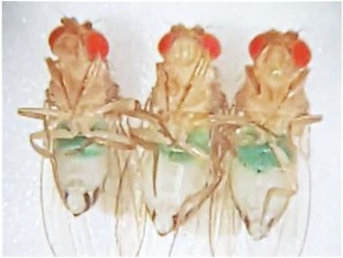
Short report                                                                                                       Neuroscience

Figure 3 continued
Figure supplement 1. Responses and innervation patterns of Piezo neurons in wild-type and Piezo knockout flies.
Figure supplement 1—source data 1. Numerical data to support the graph in Figure 3—figure supplement 1.

                                    expression was low after a fast or in flies fed ad libitum, but was strikingly elevated when flies
                                    engorged themselves on a sucrose diet (Figure 3A, B, Figure 3—figure supplement 1B). Sucrose
                                    consumption could potentially stimulate both gut chemosensors and mechanosensors as an increase
                                    in crop volume was observed compared with flies fed ad libitum (Figure 3—figure supplement 1C).
                                    We next asked whether activity changes in enteric neurons depended on the content of ingested
                                    material. We compared CaLexA-mediated GFP expression levels in flies fed for 24 hr with (1)
                                    sucrose, (2) sucralose, a sweetener that lacks caloric value and stimulates peripheral gustatory recep-
                                    tors but not internal Gr43a neurons, (3) water alone after a period of water deprivation, or (4) water
                                    alone ad libitum. Flies extensively consumed sucrose, sucralose, and water when water-deprived,
                                    resulting in acute increases in crop volume that were not observed in flies given only water ad libi-
                                    tum (Figure 3C). Enteric Gr43a neurons displayed elevated levels of CaLexA-mediated GFP expres-
                                    sion after engorgement on sucrose, which is converted into fructose and glucose, but not sucralose
                                    or water, consistent with a role for these neurons in sensing nutritional carbohydrates
                                    (Miyamoto and Amrein, 2014). In contrast, enteric Piezo neurons were activated more generally by
                                    sucrose, sucralose, and deprivation-induced water ingestion, but not in controls given only water ad
                                    libitum, with responses correlated to the extent of gut distension. The observation that Piezo neu-
                                    rons were similarly activated by water- and sucrose-induced gut distension indicated a sensory
                                    mechanism that does not require chemosensation of particular nutrients. Together, these findings
                                    suggest a model of two segregated sensory pathways through the hypocerebral ganglion, with
                                    Gr43a neurons responding to sugars and Piezo neurons responding to anterior gut
                                    mechanosensation.

                                    Piezo knockout alters enteric neuron responses and fly feeding
                                    behavior
                                    Next, we asked whether the Piezo receptor mediates neuronal responses of hypocerebral neurons.
                                    We obtained Piezo knockout flies and crossed them with flies harboring alleles, enabling the CaLexA
                                    reporter system in Piezo neurons (using Piezo-Gal459266 flies with the Piezo-Gal4 transgene remote
                                    from the endogenous Piezo locus). Remarkably, hypocerebral ganglion neurons marked in Piezo-
                                    Gal4 flies but lacking Piezo expression did not respond to engorgement by sucrose, sucralose, or
                                    water, even though the crops of Piezo knockout flies were distended (Figure 3A, B). (As shown
                                    below, the extent of distension is actually more pronounced in Piezo knockout flies, yet CaLexA-
                                    mediated responses were not observed.) A lack of neuronal responses in Piezo knockout flies is not
                                    due to gross deficits in the ability to produce reporter as Trpa1-mediated activation of Piezo neurons
                                    in Piezo knockout flies was sufficient to induce a CaLexA-mediated response (Figure 3—figure sup-
                                    plement 1D). Furthermore, Piezo neurons still innervated the anterior gut, suggesting that the defi-
                                    cit was not due to coarse developmental miswiring (Figure 3—figure supplement 1E). Instead,
                                    enteric neurons of Piezo knockout flies seemingly fail to respond to crop-distending stimuli due to a
                                    mechanosensory defect.
                                        Next, we asked whether Piezo knockout flies display changes in behavior or physiology. We mea-
                                    sured feeding behavior in Piezo knockout flies and, for comparison, isogenic w1118 flies. For synchro-
                                    nization, flies were fasted for 18 hr and then given ad libitum access to dye-labeled food for 30 min.
                                    Remarkably, Piezo knockout flies increased food intake and had visually observable crop distension
                                    (Figure 4A–C, Figure 4—figure supplement 2A). Moreover, Piezo knockout flies fed ad libitum on
                                    normal fly food for 5–7 days showed an increase in body weight compared to control flies
                                    (Figure 4D). Automated analysis of feeding patterns was performed involving an EXPRESSO plat-
                                    form (Yapici et al., 2016), and Piezo knockout flies displayed an increase in food intake and feeding
                                    bout duration but a similar frequency of feeding bout initiation (Figure 4E). Abnormal gut distension
                                    and feeding behavior were rescued by exogenous expression of Piezo-GFP in Piezo knockout neu-
                                    rons driven by Piezo-Gal4 (Figure 4F, Figure 4—figure supplement 1A). Unlike Drop-dead knock-
                                    out flies that have an enlarged crop due to defective food passage into the intestine (Peller et al.,

Min et al. eLife 2021;10:e63049. DOI: https://doi.org/10.7554/eLife.63049                                                           7 of 18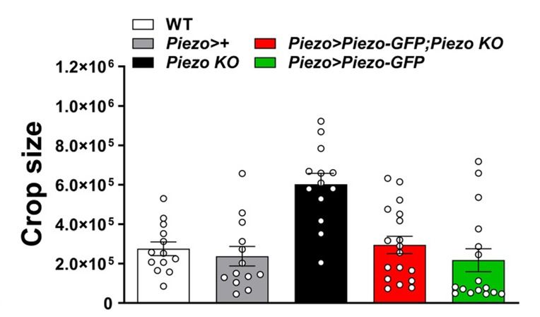
Short report                                                                                                                           Neuroscience

   $                                                                         %
                                   :7                        Piezo KO                                 :7                Piezo KO

    &                                                                        '

                                                                                                   0DOH                 )HPDOH

   (
                                                                                                                     QV

   )
                                                     QV                                                                  QV
                      QV                                                                  QV

                                                          )LJXUH
Figure 4. Piezo knockout alters fly feeding behavior. (A) Fasted wild-type (WT) and Piezo knockout (KO) female flies were given brief access (30 min) to
dye-colored food and imaged, scale bar 0.5 mm. (B) Representative images of the crop (arrow) in WT and Piezo KO flies, scale bar 100 mm, (C)
Calculated feeding indices (left) and crop sizes (right) from flies in (A). n (feeding index: 17 trials involving 204 flies), n (crop size): 14 flies, mean ± SEM,
***p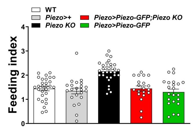
Short report                                                                                                                             Neuroscience

Figure 4 continued
three flies per trial, mean ± SEM, ***p
Short report                                                                                                Neuroscience

Continued

Reagent type
(species) or                                                Source or                                 Additional
resource                        Designation                 reference               Identifiers       information
Genetic reagent                 Piezo(KI)-Gal4              He et al., 2018         PMID:29414942
(D. melanogaster)
Genetic reagent                 Piezo(gene-                 Bloomington             BDSC: 76658
(D. melanogaster)               trap)-Gal4                  Drosophila              RRID:BDSC_76658
                                                            Stock Center
Genetic reagent                 Piezo KO                    Bloomington             BDSC: 58770;      Isogenized with w1118
(D. melanogaster)                                           Drosophila              RRID:BDSC_58770
                                                            Stock Center
Genetic reagent                 UAS-GFP-Piezo               Bloomington             BDSC: 58773;
(D. melanogaster)                                           Drosophila              RRID:BDSC_58773
                                                            Stock Center
Genetic reagent                 UAS-CD8RFP                  Bloomington             BDSC: 32218;
(D. melanogaster)                                           Drosophila              RRID:BDSC_32218
                                                            Stock Center
Genetic reagent                 Hs-Flp, UAS-                Bloomington             BDSC: 64085;
(D. melanogaster)               MCFO                        Drosophila              RRID:BDSC_64085
                                                            Stock Center
Genetic reagent                 UAS-CD8GFP                  Bloomington             BDSC: 5137;
(D. melanogaster)                                           Drosophila              RRID:BDSC_5137
                                                            Stock Center
Genetic reagent                 UAS-Trpa1                   Bloomington             BDSC: 26263;
(D. melanogaster)                                           Drosophila              RRID:BDSC_26263
                                                            Stock Center
Genetic reagent                 UAS-CaLexA                  Bloomington             BDSC: 66542;
(D. melanogaster)                                           Drosophila              RRID:BDSC_66542
                                                            Stock Center
Genetic reagent                 Nanchung-Gal4               Bloomington             BDSC: 24903;
(D. melanogaster)                                           Drosophila              RRID:BDSC_24903
                                                            Stock Center
Genetic reagent                 Inactive-Gal4               Bloomington             BDSC: 36360;
(D. melanogaster)                                           Drosophila              RRID:BDSC_36360
                                                            Stock Center
Genetic reagent                 Painless-Gal4               Bloomington             BDSC: 27894;
(D. melanogaster)                                           Drosophila              RRID:BDSC_27894
                                                            Stock Center
Genetic reagent                 Tmc-Gal4                    Zhang et al., 2016      PMID:27478019
(D. melanogaster)
Genetic reagent                 Gr43a-Gal4                  Miyamoto et al., 2012   PMID:23178127
(D. melanogaster)
Genetic reagent                 Gr43a-LexA                  Fujii et al., 2015      PMID:25702577
(D. melanogaster)
Genetic reagent                 UAS-DenMark                 Bloomington             BDSC: 33061;
(D. melanogaster)                                           Drosophila              RRID:BDSC_33061
                                                            Stock Center
Genetic reagent                 UAS-DenMark                 Bloomington             BDSC: 33062;
(D. melanogaster)                                           Drosophila              RRID:BDSC_33062
                                                            Stock Center
Genetic reagent                 Trp-Gal4                    Bloomington             BDSC: 36359;
(D. melanogaster)                                           Drosophila              RRID:BDSC_36359
                                                            Stock Center
Genetic reagent                 Nompc-Gal4                  Bloomington             BDSC: 36360;
(D. melanogaster)                                           Drosophila              RRID:BDSC_36360
                                                            Stock Center
Genetic reagent                 Drop-dead KO                Bloomington             BDSC: 36360;
(D. melanogaster)                                           Drosophila              RRID:BDSC_36360
                                                            Stock Center
Continued on next page

Min et al. eLife 2021;10:e63049. DOI: https://doi.org/10.7554/eLife.63049                                                     10 of 18
Short report Neuroscience
Continued
Reagent type
(species) or Source or Additional
resource Designation reference Identifiers information
Genetic reagent w1118 Bloomington BDSC: 3605;
(D. melanogaster) Drosophila RRID:BDSC_3605
Stock Center
Genetic reagent Trpa1-Gal4 Bloomington BDSC: 36362;
(D. melanogaster) Drosophila RRID:BDSC_36362
Stock Center
Genetic reagent Ppk-Gal4 Bloomington BDSC: 32078;
(D. melanogaster) Drosophila RRID:BDSC_32078
Stock Center
Genetic reagent GMR51F12-Gal4 Bloomington BDSC: 58685;
(D. melanogaster) Drosophila RRID:BDSC_58685
Stock Center
Genetic reagent Cha-Gal80 Sakai et al., 2009 PMID:19531155
(D. melanogaster)
Genetic reagent UAS-Shibirets Kitamoto, 2001 PMID:11291099
(D. melanogaster)
Genetic reagent Escargot-Gal4 Hayashi et al., 2002 PMID:12324948
(D. melanogaster)
Genetic reagent Elav-Gal80 Yang et al., 2009 PMID:19249273
(D. melanogaster)
Antibody Anti-Dilp2; Veenstra Jan (1:200)
rabbit polyclonal (University of
Bordeaux, France)
Antibody Anti-GFP; Thermo Thermo Fisher (1:200)
chicken Fisher Scientific Scientific
polyclonal Cat# A10262;
RRID:AB_2534023
Antibody Anti-RFP; Rockland Rockland Cat# (1:200)
rabbit 600-401-379;
polyclonal RRID:AB_2209751
Antibody Anti-Elav; Developmental DSHB Cat# (1:200)
mouse monoclonal Studies Elav-9F8A9;
Hydridoma Bank RRID:AB_528217
Antibody Anti-Akh; Kerafast Kerafast (1:200)
rabbit polyclonal Cat# EGA261
Antibody Anti-Flag; Novus Biologicals Novus Cat# NBP1- (1:200)
Rat monoclonal 06712SS;
RRID:AB_1625982
Antibody Anti-HA; Rabbit Cell Signaling Cell Signaling (1:200)
monoclonal Technology Technology
Cat# 3724S;
RRID:AB_1549585
Antibody Anti-V5; Mouse Bio-Rad Bio-Rad Cat# (1:200)
monoclonal MCA2894D549GA
RRID:AB_10845946
Antibody Alexa Fluor-488; Jackson Jackson (1:400)
Chicken ImmunoResearch ImmunoResearch
polyclonal Cat# 703-545-155;
RRID:AB_2340375
Antibody Alexa Fluor-488; Jackson Jackson (1:400)
Rabbit polyclonal ImmunoResearch ImmunoResearch
Cat# 711-545-152;
RRID:AB_2313584
Continued on next page
Min et al. eLife 2021;10:e63049. DOI: https://doi.org/10.7554/eLife.63049 11 of 18Short report Neuroscience
Continued
Reagent type
(species) or Source or Additional
resource Designation reference Identifiers information
Antibody Cy3-AffiniPure; Jackson Jackson (1:400)
Rabbit polyclonal ImmunoResearch ImmunoResearch
Cat# 711-165-152;
RRID:AB_2307443
Antibody Alexa Fluor 647; Jackson Jackson (1:400)
Rabbit polyclonal ImmunoResearch ImmunoResearch
Cat# 711-605-152;
RRID:AB_2492288
Antibody Alexa Fluor 647; Jackson Jackson (1:400)
Mouse polyclonal ImmunoResearch ImmunoResearch
Cat# 715-605-150;
RRID:AB_2340862
Antibody Alexa Fluor 488; Jackson Jackson (1:400)
Mouse polyclonal ImmunoResearch ImmunoResearch
Cat# 715-545-150;
RRID:AB_2340846
Antibody Alexa Fluor 488; Jackson Jackson (1:400)
Rat polyclonal ImmunoResearch ImmunoResearch
Cat# 712-545-153;
RRID:AB_2340684
Chemical Normal goat serum Jackson Jackson (5%)
compound, ImmunoResearch ImmunoResearch
drug Cat# 005-000-121;
RRID:AB_2336990
Chemical Fluoromount-G Southern 0100-01
compound, Biotech
drug
Chemical Phalloidin-FITC Sigma P5282-1MG (1:400)
compound,
drug
Chemical Phalloidin-TRITC Sigma P1951-1MG (1:400)
compound,
drug
Chemical TO-PRO-3 ThermoFisher T3605 (1:400)
compound,
drug
Chemical Green food dye Amazon Amazon standard Manufacturer:
compound, identification McCormick
drug number (ASIN):
B0055AFE5G
Software, Prism 8 GraphPad RRID:SCR_002798
algorithm
Software, Fiji Schindelin et al., PMID:22743772 https://imagej.
algorithm Nature net/Fiji
Methods, 2012
Software, Python-based Samuel C. https://github.
algorithm custom data Whitehead, 2021, com/scw97/
analysis PiezoPaper PiezoPaper
code used for ExpressoCode ExpressoCode;
EXPRESSO assay Min, 2021;
copy archived at swh:1:rev:bd8a
58fa0e4f796e2ed0b72fe
807862305b84b6b
Other Confocal microscope Leica Leica SP5
Flies
Fly stocks were maintained on a regular cornmeal agar diet (Harvard Exelixis facility) at 25˚C, with
mating and collection performed under CO2 anesthesia. For Piezo knockout studies, Piezo knockout
Min et al. eLife 2021;10:e63049. DOI: https://doi.org/10.7554/eLife.63049 12 of 18Short report Neuroscience
flies were isogenized by outcrossing five times into a wild-type w1118 isogenic background. We
obtained Piezo knockout, knock-in (KI) Piezo-Gal4 and UAS-Piezo-GFP flies (Norbert Perrimon),
Tmc-Gal4 (Craig Montell), knock-in Gr43a-LexA and knock-in Gr43a-Gal4 (Hubert Amrein), and from
Bloomington Drosophila Stock Center Piezo-Gal4 (BDSC# 59266), Recombinase-Mediated Cassette
Exchange (RMCE) gene-trap Piezo-Gal4 (BDSC# 76658), UAS-CD8GFP (BDSC# 5137), UAS-CD8RFP
(BDSC# 32218), UAS-Trpa1 (BDSC# 26263), Cha-Gal80 (BDSC# 60321), UAS-CaLexA (BDSC#
66542), Nanchung-Gal4 (BDSC# 24903), Inactive-Gal4 (BDSC# 36360), Painless-Gal4 (BDSC# 27894),
Trp-Gal4 (BDSC# 36359), Trpa1-Gal4 (BDSC# 36362), Nompc-Gal4 (BDSC# 36361), Ppk-Gal4
(BDSC# 32078), UAS-DenMark (BDSC# 33061 and 33062), Drop-dead KO (BDSC# 24901), w1118
(BDSC# 3605), GMR51F12-Gal4 (BDSC# 58685), and Hs-Flp; UAS-MCFO (BDSC# 64085). Escargot-
Gal4, Cha-Gal80, Elav-Gal80, UAS-Shibirets, and Dilp2-Gal4 were as published (Hayashi et al., 2002;
Ikeya et al., 2002; Kitamoto, 2001; Sakai et al., 2009; Yang et al., 2009).
Feeding analysis
Acute feeding assays were performed as previously described with modifications (Albin et al., 2015;
Min et al., 2016). Twelve adult female flies were collected upon eclosion and housed in a vial with
for 5–7 days. Prior to testing, baseline hunger was synchronized by starving flies for 15–18 hr in a
vial containing only on a dampened kimwipe section. The surface of regular fly food (typically ~16.25
ml per 50 ml vial) was dyed with green food coloring (McCormick, 70 ml dye per vial) and dried (24
hr). For testing, starved flies were transferred to vials containing dyed food for 30 min. Trials were
ended by cooling the vials on ice, and a feeding index was scored as described below (see quantifi-
cation). For thermogenetic experiments, flies expressing Trpa1 or Shibire were maintained and
starved at 18˚C prior to testing. Ten minutes prior to testing, starved flies and dye-labeled food
were pre-warmed to 30˚C or 32˚C for experiments with either Trpa1 or Shibire, and then tested as
above. Feeding behavior was scored by visual inspection of ingested dye with scores given from 0
to 5 based on dye intensity, as reported previously (Albin et al., 2015; Min et al., 2016). A feeding
index was expressed by averaging the feeding scores for all flies per vial (~12 flies). For automated
analysis of feeding patterns, fasted male flies (3–5 days old) were individually introduced into cham-
bers connected to an EXPRESSO machine (http://public.iorodeo.com/docs/expresso/hardware_
design_files.html) and feeding bouts were analyzed using EXPRESSO acquisition software (http://
public.iorodeo.com/docs/expresso/device_software.html). Briefly, flies were given access (30 min) to
a 200 mM sucrose solution through a capillary, and capillary fluid volume was measured over time
using the EXPRESSO instrument. Total food consumption, feeding duration, feeding bout numbers,
and feeding latency were then calculated using a Python-based custom data analysis code available
at https://github.com/scw97/PiezoPaperExpressoCode.
Chronic studies of body weight, intestinal transit, fecal rate, and
lifespan
Chronic studies were performed on 5–7-day-old male and female flies fed ad libitum with regular fly
food. Flies were anesthetized (ice, 10 min) and weighed in groups of three in a 1.5-ml Eppendorf
tube, with body weight expressed as the average weight per group of three. Lifespan was analyzed
for a group of 12 flies by counting the number of surviving flies each day. Fecal rates were measured
after feeding flies dye-colored food (dye-colored food is described above) for 1 hr, with visual
inspection of abdominal dye to ensure ingestion. Flies were transferred to an empty vial containing a
1 1 cm filter paper floor for 30 min, and dye-labeled fecal spots on the filter paper were counted.
For analysis of fecal deposition, individual data points reflect the mean behavior of ten flies. Intesti-
nal transit was measured in flies given brief access (30 min) to dye-colored food, with dye location in
the intestine determined visually. A transit index was calculated based on the leading dye edge posi-
tion, with scores of 1, 2, and 3 referring to dye edge in the crop/anterior midgut, middle midgut,
and hindgut/anus, respectively.
Sparse neuronal labeling
Piezo-Gal4 (59266) flies were crossed with MultiColor FlpOut (MCFO) flies (Hs-Flp; UAS-MCFO flies)
that enable multicolor, stochastic, and sparse labeling of Gal4-expressing cells (Nern et al., 2015).
MCFO flies contain multiple Gal4-dependent alleles encoding epitope tags, including HA, FLAG,
Min et al. eLife 2021;10:e63049. DOI: https://doi.org/10.7554/eLife.63049 13 of 18Short report Neuroscience
and V5. Piezo-Gal4; Hs-Flp; UAS-MCFO fly larvae were maintained at 19˚C, and at the third instar,
larvae (wandering stage) were heat-shocked (37˚C, 15 min/day, 3 days) to induce reporter expression
in dispersed neurons, and after eclosion, were collected for dissection of the brain and anterior gut
and immunohistochemistry for HA, Flag, and V5 epitopes.
Immunohistochemistry
Wholemount preparations of the gastrointestinal tract and brain were fixed (4% paraformaldehyde,
phosphate buffered saline or PBS, 20 min, room temperature [RT]), washed (2 5 min, PBS with
0.5% Triton X-100), permeabilized (10 min, PBS with 0.5% Triton X-100), blocked (1 hr, RT, blocking
solution: 5% normal goat serum [Jackson ImmunoResearch, 005-000-121], PBS with 0.1% Triton
X-100), incubated with primary antibody (1:200, blocking solution, 4˚C, overnight), washed (3 10
min, RT, PBS with 0.1% Triton X-100), incubated with secondary antibody (1:200, PBS with 0.1% Tri-
ton X-100, 2 hr, RT), washed (3 10 min, RT, PBS with 0.1% Triton X-100 then 2 5 min, RT, PBS),
mounted on a slide glass with Fluoromount-G mounting medium (Southern Biotech, 0100-01), cov-
ered with a thin coverslip, sealed with nail polish, and analyzed by confocal microscopy (Leica SP5).
Primary antibodies were anti-GFP (Thermo Fisher Scientific, Chicken, A10262), anti-RFP (Rockland,
Rabbit, 600-401-379), anti-Elav (Developmental Studies Hydridoma Bank, Mouse, Elav-9F8A9), anti-
Akh (Kerafast, Rabbit, EGA261), anti-Dilp2 (from Veenstra Jan, University of Bordeaux, France), anti-
Flag (Novus Biologicals, Rat, NBP1-06712SS), anti-HA (Cell Signaling Technology, Rabbit, 3724S),
and anti-V5 (Bio-Rad, Mouse, MCA2894D549GA). Secondary antibodies were anti-Chicken-Alexa
Fluor-488 (Jackson ImmunoResearch, 703-545-155), anti-Rabbit-Alexa Fluor-488 (Jackson ImmunoR-
esearch, 711-545-152), anti-Rabbit-Cy3 (Jackson ImmunoResearch, 711-165-152), anti-Rabbit-Alexa
Fluor-647 (Jackson ImmunoResearch, 711-605-152), anti-Mouse-Alexa Fluor-647 (Jackson ImmunoR-
esearch, 715-605-150), anti-Mouse-Alexa Flour-488 (Jackson ImmunoResearch, 715-545-150), and
anti-Rat-Alexa Fluor 488 (Jackson ImmunoResearch, 712-545-153). For staining of visceral muscle
and nuclei, Phalloidin-FITC (Sigma, P5282-1MG), Phalloidin-TRITC (Sigma, P1951-1MG), and TO-
PRO-3 (ThermoFisher, T3605) were added together with the secondary antibody.
Quantification of crop size and composition
After the feeding assay, flies were fixed (4% paraformaldehyde, PBS, RT, 1 hr) and decapitated. The
anterior gastrointestinal tract was surgically removed after gentle displacement of appendages and
thoracic muscles. Dissected tissue was washed (3 PBS, RT, 5 min) and mounted for bright-field
microscopy using the ‘Analyze-Measure’ tool in Fiji to calculate crop area. Crop muscle and cell den-
sity were quantified as detailed below. For quantification of crop muscle density, the intensity of the
Phalloidin-labeled muscle fibers in a region of interest (ROI) was divided by the total ROI area. For
cell density, the number of nuclei labeled with TO-PRO-3 and counted using ‘Analyse-3D objects
counter’ function in Fiji (https://imagej.net/Fiji) was divided by total ROI area.
Analyzing neuronal responses with CaLexA
CaLexA responses were measured in Piezo-Gal4 or Gr43a-Gal4 flies containing UAS-CaLexA (LexA-
VP16-NFAT, LexAop-rCD2-GFP, and LexAop-CD8GFP-2A-CD8GFP), and UAS-CD8RFP. Responses
of Piezo knockout neurons were measured by introducing Piezo knockout alleles into Piezo-Gal4;
UAS-CaLexA; UAS-CD8RFP flies. For sucrose and sucralose responses, flies were fed ad libitum with
regular food, transferred to vials containing a kimwipe soaked with 10% sucrose solution or 1%
sucralose solution containing green food coloring for 24 hr, and analyzed for crop distension and
CaLexA expression. For water responses, flies were deprived of food and water for 6 hr, and trans-
ferred to vials containing a water-soaked kimwipe. Some flies were harvested after 15 min for analy-
sis of crop distension and others were harvested after 18 hr for analysis of CaLexA expression.
Control flies were placed in a vial containing a water-soaked kimwipe but no food for 24 hr and har-
vested for analysis. For TrpA1-mediated neuron stimulation, WT and Piezo KO flies bearing a Piezo-
Gal4, UAS-CaLexA (LexA-VP16-NFAT, LexAop-rCD2GFP, and LexAop-CD8GFP-2A-CD8GFP), and
UAS-Trpa1 were placed in a 30˚C incubator for 24 hr prior to analysis. For analysis of CaLexA expres-
sion, flies were anesthetized (ice, 10 min), and the anterior gastrointestinal tract was surgically
removed. Dissected tissue was fixed (4% paraformaldehyde, PBS, 20 min, RT), washed (3 5 min,
PBS), and slide mounted with Fluoromount-G mounting medium and a coverslip. Native GFP
Min et al. eLife 2021;10:e63049. DOI: https://doi.org/10.7554/eLife.63049 14 of 18Short report Neuroscience
(derived from CaLexA activation) and RFP (constitutive from a Gal4-dependent reporter) fluores-
cence was analyzed by confocal microscopy (Leica SP5).
For quantification of CaLexA-dependent reporter in Figure 3B and S3B, intensity of GFP and RFP
fluorescence was calculated per neuron and a CaLexA index expressed as GFP fluorescence divided
by RFP fluorescence. For quantification of CaLexA-dependent reporter in Figure 2—figure supple-
ment 2A, D, which involved flies lacking an RFP allele for neuron identification and normalization,
GFP intensity was measured in the whole hypocerebral ganglion and a background subtraction was
performed involving a comparably sized region of the proventriculus lacking Gal4-positive cell bod-
ies. For S3A and S3D, background-subtracted GFP fluorescence was divided by RFP fluorescence
from a control Piezo-Gal4; UAS-CD8RFP fly to generate a CaLexA index.
Statistical analysis
Data in graphs are represented as means ± SEM, with sample sizes provided in figure legends. Sta-
tistical significance was analyzed by ANOVA Dunnett’s multiple comparison test or unpaired t-test
using Prism 8 software (GraphPad), as indicated in figure legends.
Acknowledgements
We thank Norbert Perrimon, Veenstra Jan, Bryan Song, and Dragana Rogulja for reagents and
advice, Jinfei Ni for blinded analysis of behavior, Norbert Perrimon, Craig Montell, Julie Simpson,
Hubert Amrein, and Bloomington Drosophila Stock Center for flies, and Hansine Heggeness and
Exelixis facility at Harvard Medical School for fly food and stock maintenance.
Additional information
Competing interests
Stephen Liberles: Reviewing editor, eLife. The other authors declare that no competing interests
exist.
Funding
Funder Grant reference number Author
American Heart Association 20POST35210914 Soohong Min
National Institutes of Health NS090994 David Van Vactor
National Institutes of Health RO1DK116294 Greg SB Suh
National Institutes of Health RO1DK106636 Greg SB Suh
Samsung Science and Tech- SSTF-BA-1802-11 Greg SB Suh
nology Foundation
Howard Hughes Medical Insti- Stephen Liberles
tute
National Institutes of Health R35 GM133698-01 Nilay Yapici
The funders had no role in study design, data collection and interpretation, or the
decision to submit the work for publication.
Author contributions
Soohong Min, Conceptualization, Data curation, Formal analysis, Funding acquisition, Validation,
Investigation, Visualization, Methodology, Writing - original draft, Writing - review and editing; Yang-
kyun Oh, Data curation, Formal analysis, Investigation, Visualization, Methodology; Pushpa Verma,
Resources; Samuel C Whitehead, Resources, Software, Funding acquisition; Nilay Yapici, Greg SB
Suh, Conceptualization, Resources, Software, Supervision, Funding acquisition, Project administra-
tion, Writing - review and editing; David Van Vactor, Conceptualization, Resources, Supervision,
Funding acquisition, Writing - original draft, Project administration, Writing - review and editing;
Min et al. eLife 2021;10:e63049. DOI: https://doi.org/10.7554/eLife.63049 15 of 18Short report Neuroscience
Stephen Liberles, Conceptualization, Resources, Software, Supervision, Funding acquisition, Writing
- original draft, Project administration, Writing - review and editing
Author ORCIDs
Soohong Min https://orcid.org/0000-0003-0683-2935
Stephen Liberles https://orcid.org/0000-0002-2177-9741
Decision letter and Author response
Decision letter https://doi.org/10.7554/eLife.63049.sa1
Author response https://doi.org/10.7554/eLife.63049.sa2
Additional files
Supplementary files
. Transparent reporting form
Data availability
All datapoints used are provided in figures and in a source data file.
References
Abraira VE, Ginty DD. 2013. The sensory neurons of touch. Neuron 79:618–639. DOI: https://doi.org/10.1016/j.
neuron.2013.07.051, PMID: 23972592
Albin SD, Kaun KR, Knapp JM, Chung P, Heberlein U, Simpson JH. 2015. A subset of serotonergic neurons
evokes hunger in adult Drosophila. Current Biology 25:2435–2440. DOI: https://doi.org/10.1016/j.cub.2015.08.
005, PMID: 26344091
Bjordal M, Arquier N, Kniazeff J, Pin JP, Léopold P. 2014. Sensing of amino acids in a dopaminergic circuitry
promotes rejection of an incomplete diet in Drosophila. Cell 156:510–521. DOI: https://doi.org/10.1016/j.cell.
2013.12.024, PMID: 24485457
Coste B, Xiao B, Santos JS, Syeda R, Grandl J, Spencer KS, Kim SE, Schmidt M, Mathur J, Dubin AE, Montal M,
Patapoutian A. 2012. Piezo proteins are pore-forming subunits of mechanically activated channels. Nature 483:
176–181. DOI: https://doi.org/10.1038/nature10812, PMID: 22343900
Dethier VG, Gelperin A. 1967. Hyperphagia in the blowfly. The Journal of Experimental Biology 191:200.
Dus M, Lai JS, Gunapala KM, Min S, Tayler TD, Hergarden AC, Geraud E, Joseph CM, Suh GS. 2015. Nutrient
sensor in the brain directs the action of the Brain-Gut Axis in Drosophila. Neuron 87:139–151. DOI: https://doi.
org/10.1016/j.neuron.2015.05.032, PMID: 26074004
Fujii S, Yavuz A, Slone J, Jagge C, Song X, Amrein H. 2015. Drosophila sugar receptors in sweet taste
perception, olfaction, and internal nutrient sensing. Current Biology 25:621–627. DOI: https://doi.org/10.1016/
j.cub.2014.12.058, PMID: 25702577
Gelperin A. 1967. Stretch receptors in the foregut of the blowfly. Science 157:208–210. DOI: https://doi.org/10.
1126/science.157.3785.208, PMID: 17806269
Hayashi S, Ito K, Sado Y, Taniguchi M, Akimoto A, Takeuchi H, Aigaki T, Matsuzaki F, Nakagoshi H, Tanimura T,
Ueda R, Uemura T, Yoshihara M, Goto S. 2002. GETDB, a database compiling expression patterns and
molecular locations of a collection of Gal4 enhancer traps. Genesis 34:58–61. DOI: https://doi.org/10.1002/
gene.10137, PMID: 12324948
He L, Si G, Huang J, Samuel ADT, Perrimon N. 2018. Mechanical regulation of stem-cell differentiation by the
stretch-activated piezo channel. Nature 555:103–106. DOI: https://doi.org/10.1038/nature25744, PMID: 29414
942
Ikeya T, Galic M, Belawat P, Nairz K, Hafen E. 2002. Nutrient-dependent expression of insulin-like peptides from
neuroendocrine cells in the CNS contributes to growth regulation in Drosophila. Current Biology 12:1293–
1300. DOI: https://doi.org/10.1016/S0960-9822(02)01043-6, PMID: 12176357
Kim SE, Coste B, Chadha A, Cook B, Patapoutian A. 2012. The role of Drosophila piezo in mechanical
nociception. Nature 483:209–212. DOI: https://doi.org/10.1038/nature10801, PMID: 22343891
Kitamoto T. 2001. Conditional modification of behavior in Drosophila by targeted expression of a temperature-
sensitive shibire allele in defined neurons. Journal of Neurobiology 47:81–92. DOI: https://doi.org/10.1002/
neu.1018, PMID: 11291099
Lemaitre B, Miguel-Aliaga I. 2013. The digestive tract of Drosophila melanogaster. Annual Review of Genetics
47:377–404. DOI: https://doi.org/10.1146/annurev-genet-111212-133343, PMID: 24016187
Masuyama K, Zhang Y, Rao Y, Wang JW. 2012. Mapping neural circuits with activity-dependent nuclear import
of a transcription factor. Journal of Neurogenetics 26:89–102. DOI: https://doi.org/10.3109/01677063.2011.
642910, PMID: 22236090
Min et al. eLife 2021;10:e63049. DOI: https://doi.org/10.7554/eLife.63049 16 of 18Short report Neuroscience
Min S, Chae HS, Jang YH, Choi S, Lee S, Jeong YT, Jones WD, Moon SJ, Kim YJ, Chung J. 2016. Identification of
a peptidergic pathway critical to satiety responses in Drosophila. Current Biology 26:814–820. DOI: https://doi.
org/10.1016/j.cub.2016.01.029, PMID: 26948873
Min S, Chang RB, Prescott SL, Beeler B, Joshi NR, Strochlic DE, Liberles SD. 2019. Arterial baroreceptors sense
blood pressure through decorated aortic claws. Cell Reports 29:2192–2201. DOI: https://doi.org/10.1016/j.
celrep.2019.10.040
Min S. 2021. PiezoPaperExpressoCode. Software Heritage. swh:1:rev:
bd8a58fa0e4f796e2ed0b72fe807862305b84b6b. https://archive.softwareheritage.org/swh:1:dir:
f32cf0672242df570a30ab652ada1e0a3b649675;origin=https://github.com/scw97/PiezoPaperExpressoCode;visit=
swh:1:snp:bf365259c4154f33a64ebf660c5b4a36078ec9d4;anchor=swh:1:rev:
bd8a58fa0e4f796e2ed0b72fe807862305b84b6b/
Miyamoto T, Slone J, Song X, Amrein H. 2012. A fructose receptor functions as a nutrient sensor in the
Drosophila brain. Cell 151:1113–1125. DOI: https://doi.org/10.1016/j.cell.2012.10.024, PMID: 23178127
Miyamoto T, Amrein H. 2014. Diverse roles for the Drosophila fructose sensor Gr43a. Fly 8:19–25. DOI: https://
doi.org/10.4161/fly.27241, PMID: 24406333
Montell C. 2005. Drosophila TRP channels. Pflügers Archiv - European Journal of Physiology 451:19–28.
DOI: https://doi.org/10.1007/s00424-005-1426-2
Nern A, Pfeiffer BD, Rubin GM. 2015. Optimized tools for multicolor stochastic labeling reveal diverse
stereotyped cell arrangements in the fly visual system. PNAS 112:E2967–E2976. DOI: https://doi.org/10.1073/
pnas.1506763112, PMID: 25964354
Nonomura K, Woo SH, Chang RB, Gillich A, Qiu Z, Francisco AG, Ranade SS, Liberles SD, Patapoutian A. 2017.
Piezo2 senses airway stretch and mediates lung inflation-induced apnoea. Nature 541:176–181. DOI: https://
doi.org/10.1038/nature20793, PMID: 28002412
Olds WH, Xu T. 2014. Regulation of food intake by mechanosensory ion channels in enteric neurons. eLife 3:
e04402. DOI: https://doi.org/10.7554/eLife.04402
Peller CR, Bacon EM, Bucheger JA, Blumenthal EM. 2009. Defective gut function in drop-dead mutant
Drosophila. Journal of Insect Physiology 55:834–839. DOI: https://doi.org/10.1016/j.jinsphys.2009.05.011,
PMID: 19500585
Phillips RJ, Powley TL. 1996. Gastric volume rather than nutrient content inhibits food intake. American Journal
of Physiology-Regulatory, Integrative and Comparative Physiology 271:R766–R769. DOI: https://doi.org/10.
1152/ajpregu.1996.271.3.R766
Pool AH, Kvello P, Mann K, Cheung SK, Gordon MD, Wang L, Scott K. 2014. Four GABAergic interneurons
impose feeding restraint in Drosophila. Neuron 83:164–177. DOI: https://doi.org/10.1016/j.neuron.2014.05.
006, PMID: 24991960
Ranade SS, Syeda R, Patapoutian A. 2015. Mechanically activated ion channels. Neuron 87:1162–1179.
DOI: https://doi.org/10.1016/j.neuron.2015.08.032, PMID: 26402601
Rolls BJ, Castellanos VH, Halford JC, Kilara A, Panyam D, Pelkman CL, Smith GP, Thorwart ML. 1998. Volume of
food consumed affects satiety in men. The American Journal of Clinical Nutrition 67:1170–1177. DOI: https://
doi.org/10.1093/ajcn/67.6.1170, PMID: 9625090
Sakai T, Kasuya J, Kitamoto T, Aigaki T. 2009. The Drosophila TRPA channel, painless, regulates sexual
receptivity in virgin females. Genes, Brain and Behavior 8:546–557. DOI: https://doi.org/10.1111/j.1601-183X.
2009.00503.x, PMID: 19531155
Sánchez-Alcañiz JA, Zappia G, Marion-Poll F, Benton R. 2017. A mechanosensory receptor required for food
texture detection in Drosophila. Nature Communications 8:14192. DOI: https://doi.org/10.1038/ncomms14192,
PMID: 28128210
Stoffolano JG, Haselton AT. 2013. The adult dipteran crop: a unique and overlooked organ. Annual Review of
Entomology 58:205–225. DOI: https://doi.org/10.1146/annurev-ento-120811-153653, PMID: 23317042
Umans BD, Liberles SD. 2018. Neural sensing of organ volume. Trends in Neurosciences 41:911–924.
DOI: https://doi.org/10.1016/j.tins.2018.07.008, PMID: 30143276
Wang P, Jia Y, Liu T, Jan Y-N, Zhang W. 2020. Visceral Mechano-sensing neurons control Drosophila Feeding by
Using Piezo as a Sensor. Neuron 108:640–650. DOI: https://doi.org/10.1016/j.neuron.2020.08.017
Williams EK, Chang RB, Strochlic DE, Umans BD, Lowell BB, Liberles SD. 2016. Sensory neurons that detect
stretch and nutrients in the digestive system. Cell 166:209–221. DOI: https://doi.org/10.1016/j.cell.2016.05.011
Yang CH, Rumpf S, Xiang Y, Gordon MD, Song W, Jan LY, Jan YN. 2009. Control of the postmating behavioral
switch in Drosophila Females by Internal Sensory Neurons. Neuron 61:519–526. DOI: https://doi.org/10.1016/j.
neuron.2008.12.021, PMID: 19249273
Yapici N, Cohn R, Schusterreiter C, Ruta V, Vosshall LB. 2016. A taste circuit that regulates ingestion by
integrating food and hunger signals. Cell 165:715–729. DOI: https://doi.org/10.1016/j.cell.2016.02.061,
PMID: 27040496
Zagorodnyuk VP, Chen BN, Brookes SJ. 2001. Intraganglionic laminar endings are mechano-transduction sites of
vagal tension receptors in the guinea-pig stomach. The Journal of Physiology 534:255–268. DOI: https://doi.
org/10.1111/j.1469-7793.2001.00255.x, PMID: 11433006
Zeng WZ, Marshall KL, Min S, Daou I, Chapleau MW, Abboud FM, Liberles SD, Patapoutian A. 2018. PIEZOs
mediate neuronal sensing of blood pressure and the baroreceptor reflex. Science 362:464–467. DOI: https://
doi.org/10.1126/science.aau6324, PMID: 30361375
Min et al. eLife 2021;10:e63049. DOI: https://doi.org/10.7554/eLife.63049 17 of 18Short report Neuroscience
Zhang W, Yan Z, Li B, Jan LY, Jan YN. 2014. Identification of motor neurons and a mechanosensitive sensory
neuron in the defecation circuitry of Drosophila larvae. eLife 3:e03293. DOI: https://doi.org/10.7554/eLife.
03293
Zhang YV, Aikin TJ, Li Z, Montell C. 2016. The basis of food texture sensation in Drosophila. Neuron 91:863–
877. DOI: https://doi.org/10.1016/j.neuron.2016.07.013, PMID: 27478019
Zhong L, Hwang RY, Tracey WD. 2010. Pickpocket is a DEG/ENaC protein required for mechanical nociception in
Drosophila larvae. Current Biology 20:429–434. DOI: https://doi.org/10.1016/j.cub.2009.12.057,
PMID: 20171104
Min et al. eLife 2021;10:e63049. DOI: https://doi.org/10.7554/eLife.63049 18 of 18You can also read