Characterization of a Mouse Model of Alzheimer's Disease Expressing Aβ4-42 and Human Mutant Tau
←
→
Page content transcription
If your browser does not render page correctly, please read the page content below
International Journal of
               Molecular Sciences

Article
Characterization of a Mouse Model of Alzheimer’s Disease
Expressing Aβ4-42 and Human Mutant Tau
Silvia Zampar             and Oliver Wirths *

                                          Department of Psychiatry and Psychotherapy, University Medical Center (UMG), Georg-August-University,
                                          D-37075 Göttingen, Germany; silvia.zampar@med.uni-goettingen.de
                                          * Correspondence: oliver.wirths@medizin.uni-goettingen.de

                                          Abstract: The relationship between the two most prominent neuropathological hallmarks of
                                          Alzheimer’s Disease (AD), extracellular amyloid-β (Aβ) deposits and intracellular accumulation of
                                          hyperphosphorylated tau in neurofibrillary tangles (NFT), remains at present not fully understood.
                                          A large body of evidence places Aβ upstream in the cascade of pathological events, triggering NFTs
                                          formation and the subsequent neuron loss. Extracellular Aβ deposits were indeed causative of an
                                          increased tau phosphorylation and accumulation in several transgenic models but the contribution
                                          of soluble Aβ peptides is still controversial. Among the different Aβ variants, the N-terminally
                                          truncated peptide Aβ4–42 is among the most abundant. To understand whether soluble Aβ4–42
                                          peptides impact the onset or extent of tau pathology, we have crossed the homozygous Tg4–42
                                          mouse model of AD, exclusively expressing Aβ4–42 peptides, with the PS19 (P301S) tau transgenic
                                          model. Behavioral assessment showed that the resulting double-transgenic line presented a partial

                                   worsening of motor performance and spatial memory deficits in the aged group. While an increased
Citation: Zampar, S.; Wirths, O.          loss of distal CA1 pyramidal neurons was detected in young mice, no significant alterations in
Characterization of a Mouse Model of      hippocampal tau phosphorylation were observed in immunohistochemical analyses.
Alzheimer’s Disease Expressing
Aβ4-42 and Human Mutant Tau. Int.         Keywords: Alzheimer’ disease; amyloid β; tau; behavior; neuron loss; transgenic mice; Aβ4-42
J. Mol. Sci. 2021, 22, 5191. https://
doi.org/10.3390/ijms22105191

Academic Editors: Agueda                  1. Introduction
A. Rostagno, Agnieszka
                                               Alzheimer’s disease (AD) is a progressive neurodegenerative disorder that is
Baranowska-Bik and
                                          histopathologically characterized by the deposition of extracellular senile plaques contain-
Arkadiusz Orzechowski
                                          ing amyloid-β (Aβ) protein [1], as well as the intracellular accumulation of so-called neu-
                                          rofibrillary tangles (NFTs) consisting of hyperphosphorylated protein tau [2].
Received: 20 April 2021
Accepted: 11 May 2021
                                          While Aβ peptides are derived from proteolytical cleavage events of the transmembrane
Published: 14 May 2021
                                          amyloid precursor protein (APP), tau proteins are brain-specific microtubule-associated
                                          molecules enriched in axons. In recent years, a variety of studies employing mainly trans-
Publisher’s Note: MDPI stays neutral
                                          genic AD mouse models investigated the relationship between both pathological hallmarks.
with regard to jurisdictional claims in
                                          According to the amyloid cascade hypothesis, a disequilibrium of Aβ production and clear-
published maps and institutional affil-   ance is regarded as an upstream event entailing the formation of NFTs and the subsequent
iations.                                  loss of synapses and neurons [3]. The common belief is that Aβ acts as a pathological
                                          trigger, with tau being the required executor [4,5]. APP transgenic mice expressing the
                                          E693∆ mutation display age-dependent accumulation of intraneuronal Aβ oligomers in the
                                          absence of amyloid deposits but show abnormal tau phosphorylation from 8 months [6].
Copyright: © 2021 by the authors.
                                          Such data is corroborated by studies from double-transgenic mouse models expressing both
Licensee MDPI, Basel, Switzerland.
                                          mutant APP and tau transgenes, showing in general increased levels of phosphorylated
This article is an open access article
                                          tau as well as accelerated tau pathology compared to their single tau transgenic parental
distributed under the terms and           strains [7–13]. However, the importance of tau on the detrimental effect of Aβ is a matter
conditions of the Creative Commons        of debate. In many experimental models, tau appears to be essential for the execution
Attribution (CC BY) license (https://     of Aβ toxicity as the absence of the endogenous protein seems to confer protection from
creativecommons.org/licenses/by/          Aβ-induced neurodegeneration. Indeed, primary neurons isolated from tau knock-out
4.0/).                                    mice resist exposure to an excess of fibrillary Aβ peptides [14]. Hippocampal neuron

Int. J. Mol. Sci. 2021, 22, 5191. https://doi.org/10.3390/ijms22105191                                       https://www.mdpi.com/journal/ijms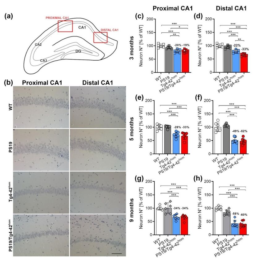
Int. J. Mol. Sci. 2021, 22, 5191                                                                                          2 of 17

                                   loss observed in 5XFAD transgenic mice could be rescued 5XFAD on a tau knock-out
                                   background, together with an ~50% reduction in amyloid plaque burden [15]. In good
                                   agreement, the absence of tau prevented Aβ-induced impairment of long-term potenti-
                                   ation (LTP) in acute slice preparations, suggesting that tau is required for Aβ to impair
                                   hippocampal synaptic plasticity [16]. On the other hand, a more recent finding points to
                                   the opposite direction. In TgAPP mice, the lack of endogenous tau indicated that synaptic
                                   and memory impairments induced by Aβ were tau-independent, as tau suppression did
                                   not protect against Aβ-induced deficits in long-term synaptic plasticity and memory or
                                   amyloid deposition [17].
                                        While there is convincing evidence that the presence of extracellular Aβ deposits
                                   contributes to an increased tau phosphorylation and accumulation, it is less clear if soluble
                                   Aβ peptides influence tau pathology in a comparable manner. It has been demonstrated
                                   that soluble Aβ dimers isolated from human AD brain induce tau hyperphosphorylation at
                                   AD-relevant epitopes when they were applied to hippocampal neurons in sub-nanomolar
                                   concentrations [18]. Intracerebral injection of either Aβ1–40 or Aβ1–42 into P301S tau
                                   transgenic mice induced tau phosphorylation and aggregation only in the latter case,
                                   while injections of Aβ1–40 even decreased tau phosphorylation at certain epitopes [19].
                                   On the other hand, a mouse model expressing human 4-repeat tau protein did not show
                                   significant amounts of tau pathology at 6 months of age and co-expression of Aβ1–42 in the
                                   absence of APP overexpression did not lead to an aggravated phenotype [20]. We have
                                   recently generated a transgenic mouse model (Tg4–42) that overexpresses Aβ4–42 peptides
                                   under the control of the murine neuron-specific Thy1-promoter [21]. Aβ4–42 peptides are
                                   among the first Aβ peptides that have been identified in human AD brain and represent a
                                   highly abundant peptide species [1,22,23]. Tg4–42 mice do not form overt extracellular Aβ
                                   deposits but present robust behavioral deficits together with abundant CA1 neuron loss at
                                   an age of 6 months [24,25].
                                        Here, we determined whether soluble N-terminally truncated Aβ4–42 peptides have
                                   an impact on the onset or extent of tau pathology in the absence of confounding human
                                   APP overexpression. We generated double-transgenic mice by crossbreeding homozygous
                                   Tg4–42 mice with the widely used PS19 tau transgenic model overexpressing tau with the
                                   P301S mutation [26] and performed behavioral and neuropathological analysis at different
                                   time points.

                                   2. Results
                                   2.1. Transgene Expression and Weight Assessment in PS19/Tg4–42hom
                                         Transgene mRNA expression levels in the bigenic PS19/Tg4–42hom line were mea-
                                   sured and compared to the parental lines at 3, 5 and 9 months of age to exclude an impact
                                   of altered transgene expression as a consequence of the crossing (primers listed in Table 1).
                                   As Aβ4–42 is predominantly expressed in the CA1 region in Tg4–42hom mice [21], hip-
                                   pocampal mRNA extracts were used for the analysis of both transgenes. No significant
                                   differences were found in either Aβ4–42 transgene expression between Tg4–42hom and
                                   PS19/Tg4–42hom (Figure 1a,d,g), or in human MAPT mRNA levels between PS19 and
                                   PS19/Tg4–42hom (Figure 1b,e,h) at any time point. Body weight of the transgenic lines did
                                   not differ compared to WT littermates, and no significant changes were observed between
                                   PS19/Tg4–42hom and the two parental lines at 3 and 5 months (Figure 1c,f). In the aged
                                   group, both PS19 and PS19/Tg4–42hom mice revealed a significantly reduced body weight
                                   compared to WT mice (p < 0.05 for PS19, p < 0.001 for PS19/Tg4–42hom ) (Figure 1i).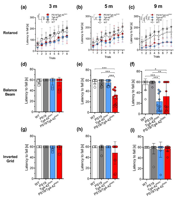
Int. J. Mol. Sci. 2021, 22, 5191                                                                                                           3 of 17

                                   Table 1. Sequences of Forward and Reverse primers and probes used in qRT-PCR analyses.

                                     Transgene/Gene                    Forward Primer                             Reverse Primer
                                      Human Aβ4–42            TCCGGCCAGAACGTCGATTC                        GGAGAAGCAAGACCTCTG
    Int. J. Mol. Sci. 2021, 22, x FOR PEER REVIEW
                                           Human MAPT        CCAAGTGTGGCTCATTAGGCA                      CCAATCTTCGACTGGACTCTGT3 of 17
                                      Murine β-Actin          ATGGAGGGGAATACAGCCC                         ATGGAGGGGAATACAGCCC

                                        Figure 1. Transgene expression and body weight in PS19/Tg4–42hom mice. Hippocam-
                                      Figure   1. Transgene
                                        pal mRNA              expression
                                                      expression   of the and  body
                                                                          Aβ4–42      weight in(a,d,g)
                                                                                   transgene     PS19/Tg4–42    hom mice. Hippocampal mRNA ex-
                                                                                                        and human       PS19 (MAPT) gene
                                      pression of the Aβ4–42 transgene (a,d,g) hom  and human PS19 (MAPT) gene (b,e,h) were measured in
                                        (b,e,h) werehom
                                                      measured in PS19/Tg4–42          mice and compared to the ones of the parental
                                      PS19/Tg4–42        mice and compared to the ones of the parental lines (n = 5–6). At the considered
                                        lines (n = 5–6). At the considered ages of 3 (c) or at 5 months (f), no changes in body weight
                                      ages of 3 (c) or at 5 months (f), no changes in body weight were detected in the transgenic         hom
                                                                                                                                                 lines
                                        were detected
                                      compared     to WTincontrols,
                                                            the transgenic  lines compared
                                                                     nor in PS19/Tg4–42   hom to WT controls,
                                                                                              compared     to PS19 nororinTg4–42
                                                                                                                           PS19/Tg4–42
                                                                                                                                 hom mice (n = 12–14).
                                                                        hom mice (n = 12–14). At 9 months, the body weight of PS19
                                      Atcompared
                                          9 months,tothe
                                                       PS19
                                                         bodyorweight
                                                                Tg4–42of  PS19 and PS19/Tg4–42hom mice was significantly reduced compared
                                                             hom
                                      toand
                                         WTPS19/Tg4–42            mice
                                              controls (i). All data arewas  significantly
                                                                         given  as mean ± reduced    compared
                                                                                           SD. (a,b,d,e,g,h)        to WT controls
                                                                                                                Mann–Whitney         (i).(c,f)
                                                                                                                                   test,   AllOne-way
                                        data arefollowed
                                      ANOVA        given asbymean  ± SD. (a,b,d,e,g,h)
                                                               Bonferroni’s              Mann–Whitney
                                                                              multiple comparison:     * p < test,
                                                                                                             0.05, (c,f)
                                                                                                                    *** pOne-way
                                                                                                                          < 0.001. ANOVA
                                        followed by Bonferroni’s multiple comparison: * p < 0.05, *** p < 0.001.
                                      2.2. PS19/Tg4–42hom Mice Display a Partial Worsening of Motor Performance at 5 Months of Age
                                           Motor performances of 3-, 5- and 9-month-old animals were tested in the accelerating
                                      rotarod, balance beam and inverted grid tasks (Figure 2). The accelerating rotarod task
                                      allows for the assessment of motor skill learning, motor coordination and balance. Al-
                                      ready at 3 months of age, Tg4–42hom and PS19/Tg4–42hom displayed a deficit in the rotarod
                                      test with significantly reduced fall latencies compared to WT (p < 0.05) and PS19 (p < 0.001)
                                      groups (Figure 2a). At 5 months, these deficits remained consistent but significant differ-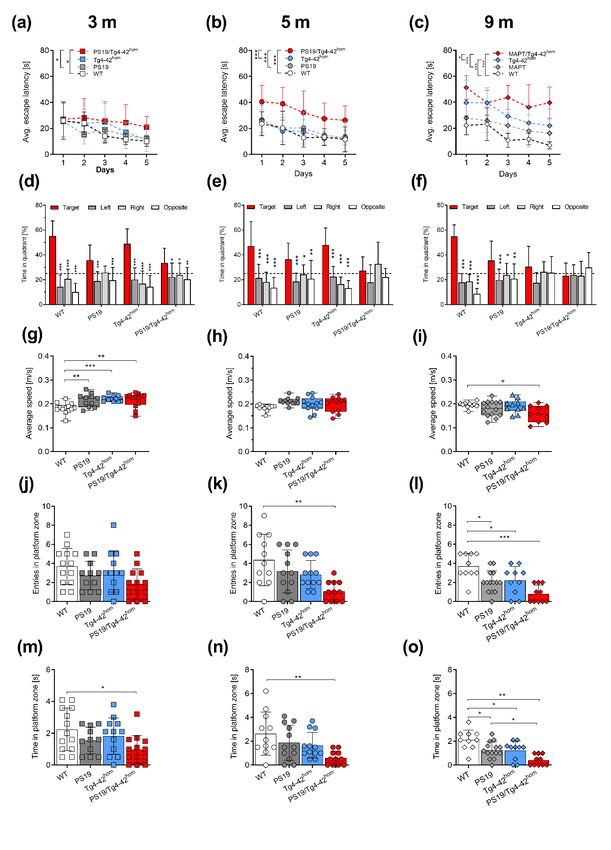
Int. J. Mol. Sci. 2021, 22, 5191                                                                                          4 of 17

                                   2.2. PS19/Tg4–42hom Mice Display a Partial Worsening of Motor Performance at 5 Months of Age
                                        Motor performances of 3-, 5- and 9-month-old animals were tested in the accelerating
                                   rotarod, balance beam and inverted grid tasks (Figure 2). The accelerating rotarod task
                                   allows for the assessment of motor skill learning, motor coordination and balance. Already
                                   at 3 months of age, Tg4–42hom and PS19/Tg4–42hom displayed a deficit in the rotarod test
                                   with significantly reduced fall latencies compared to WT (p < 0.05) and PS19 (p < 0.001)
                                   groups (Figure 2a). At 5 months, these deficits remained consistent but significant dif-
                                   ferences were found only in comparison to the PS19 line (p < 0.05 for PS19/Tg4–42hom ,
                                   p < 0.001 for Tg4–42hom ) (Figure 2b). Tg4–42hom and PS19/Tg4–42hom performances in
                                   the accelerating rotarod task progressively worsened over time, with a further reduction
                                   in fall latencies at 9 months, resulting in significantly reduced values compared to both
                                   the WT and PS19 groups (p < 0.001) (Figure 2c). Young mice performed comparably in
                                   the balance beam task (Figure 2d), while at the 5-months’ time point, PS19/Tg4–42hom
                                   displayed a deficit, presenting a significantly reduced latency to fall when compared to
                                   WT, PS19 and Tg4–42hom mice (p < 0.001) (Figure 2e). Aged Tg4–42hom , in addition to
                                   PS19/Tg4–42hom mice, showed a significantly reduced fall latency compared to WT and
                                   PS19 animals (Figure 2f), though no differences were observed between the two impaired
                                   lines. In the inverted grid task, no differences among the groups were revealed at any time
                                   point (Figure 2g–i).

                                   2.3. Co-Expression of Aβ4-42 and Mutant Tau Leads to Spatial Memory Deficits in
                                   PS19/Tg4–42hom Mice
                                         Spatial reference memory was assessed using the MWM task. During the cued
                                   training, all analyzed groups showed progressive decrease in escape latencies at 3 and
                                   5 months of age, however, PS19/Tg4–42hom displayed a significant increase in latency
                                   compared to Tg4–42hom mice (p < 0.05) at 3 months, as well as to PS19 (p < 0.001) and WT
                                   (p < 0.01) mice at 5 months of age (Supplementary Figure S1). At 9 months of age, all
                                   three transgenic lines showed an increased escape latency compared to WT controls (PS19:
                                   p < 0.05, Tg4–42hom and PS19/Tg4–42hom : p < 0.001). While the two parental lines presented
                                   a progressive decrease over the three days of testing, PS19/Tg4–42hom mice displayed
                                   the poorest performance, significantly different from the two parental lines (p < 0.001)
                                   (Supplementary Figure S1). In the subsequent acquisition training, PS19/Tg4–42hom mice
                                   already at 3 months presented a significantly increased escape latency compared to WT
                                   and PS19 mice (p < 0.05), indicative of the development of a spatial learning deficit in
                                   this line (Figure 3a). The deficits observed in PS19/Tg4–42hom mice worsened over time,
                                   with a poorer performance at 5 months in the acquisition training and a significantly
                                   increased latency to reach the platform compared to all other investigated lines (p < 0.001)
                                   (Figure 3b). As expected, Tg4–42hom mice displayed spatial learning deficits at 9 months,
                                   needing significantly more time to reach the hidden platform compared to WT mice
                                   (p < 0.001; Figure 3c). The spatial learning impairment observed in 5-month-old
                                   PS19/Tg4–42hom mice worsened as time progressed, as the aged group presented an
                                   increased escape latency compared to both WT (p < 0.001) and the two parental lines (PS19:
                                   p < 0.001, Tg4–42hom : p < 0.05) (Figure 3c). Significantly different average speeds were
                                   noticed among the groups during both cued and acquisition trainings (Supplementary
                                   Figures S1 and S2). While the WT group was the one usually swimming most slowly at
                                   3 and 5 months, at 9 months, Tg4–42hom and PS19/Tg4–42hom lines showed a reduced
                                   average speed in the cue training, but no differences were observed in the acquisition
                                   training session (Supplementary Figure S2).
and PS19 groups (p < 0.001) (Figure 2c). Young mice performed comparably in the balance
beam task (Figure 2d), while at the 5-months’ time point, PS19/Tg4–42hom displayed a deficit,
presenting a significantly reduced latency to fall when compared to WT, PS19 and Tg4–42hom
mice (p < 0.001) (Figure 2e). Aged Tg4–42hom, in addition to PS19/Tg4–42hom mice, showed a
significantly reduced fall latency compared to WT and PS19 animals (Figure 2f), though no
Int. J. Mol. Sci. 2021, 22, 5191 5 of 17
differences were observed between the two impaired lines. In the inverted grid task, no dif-
ferences among the groups were revealed at any time point (Figure 2g–i).
Figure 2. Partial worsening of motor performances in 5-month-old PS19/Tg4–42hom mice. Female and male WT, PS19,
Figure
Tg4–42 2.and
hom Partial worseninghom
PS19/Tg4–42 of motor performances
mice were in 5-month-old
tested in the acceleratingPS19/Tg4–42
hom mice. Female and male WT, PS19, Tg4–
rotarod (a–c), balance beam (d–f) and inverted grid
42 hom and PS19/Tg4–42hom mice were tested in the accelerating rotarod (a–c), balance beam (d–f) and inverted grid (g–i)
(g–i) tasks at 3, 5 and 9 months of age (n = 12–14). Motor deficits in the accelerating rotarod task were observed in both
taskshom
at 3, 5 and 9 monthshom
of age (n = 12–14). Motor deficits in the accelerating rotarod task were observed in both Tg4–
Tg4–42 and PS19/Tg4–42 mice (a–c), while only the bigenic line showed a significant reduced latency to fall from the
42hom and PS19/Tg4–42hom mice (a–c), while only thehom bigenic line showedhoma significant reduced latency to fall from the
balance beam at 5 months (e). At 9 months, both Tg4–42 and PS19/Tg4–42 mice revealed a deficit (f). No significant
balance beam at 5 months (e). At 9 months, both Tg4–42hom and PS19/Tg4–42hom mice revealed a deficit (f). No significant
differences were observed in the inverted grid task at any age (g–i). All data are given as mean ± SD. (a–c) Two-way
ANOVA RM, followed by Bonferroni’s multiple comparison, (d–i) One-way ANOVA, followed by Bonferroni’s multiple
comparison: * p < 0.05, ** p < 0.01, *** p < 0.001.Int. J. Mol. Sci. 2021, 22, 5191 6 of 17
Int. J. Mol. Sci. 2021, 22, x FOR PEER REVIEW 6 of 17
Figure 3. Spatial memory deficits in PS19/Tg4–42hom mice. Female and male WT, PS19, Tg4–42hom and PS19/Tg4–42hom
mice were tested
Figure 3.in the Morris
Spatial memory Water Maze
deficits in test (nhom
PS19/Tg4–42 = mice.
10–12) at 3and
Female (a,d,g,j,m), 5 (b,e,h,k,n)
male WT, PS19, Tg4–42hom and andPS19/Tg4–42
9 (c,f,i,l,o)
hommonths
mice of age.
were tested
Spatial learning in the
deficits Morris
were Water Maze
detected test the
during (n = acquisition
10–12) at 3 (a,d,g,j,m),
training5 in(b,e,h,k,n) and 9 (c,f,i,l,o)
PS19/Tg4–42 months at
hom already of 3age. Spatial (a), which
months
learning deficits were detected during the acquisition training in PS19/Tg4–42hom already at 3 months (a), which worsened
worsened with withaging (b,c).NoNo
aging (b,c). spatial
spatial reference
reference memorymemory
deficitsdeficits were observed
were observed in young
in young animals duringanimals during
the probe thewhile
trial (d), probe trial (d),
while 5-month-old mice co-expressing Aβ4-42 and mutant human tau failed to remember the position of the goal quadrant
(e). Aged Tg4–42hom and PS19/Tg4–42hom mice both displayed deficits in the probe trial (f). The transgenic lines swam
significantly faster than WT littermates at young age (g), while no differences in average speed were observed at 5 months
of age (j). Aged PS19/Tg4–42hom swam significantly slower than WT controls (i). Compared to WT mice, PS19/Tg4–42hom
mice showed a reduced time in the platform zone at 3 (m) and 5 months (l), as well as a reduced number of platform zone
entries at 5 (k) but not at 3 months of age (j). All three transgenic lines showed reduced entries and time spent in platform
zone at 9 months (l, o). All data are expressed as mean ± SD. (a–c) Two-way RM ANOVA or (d–f) Two-way ANOVA
followed by Bonferroni’s multiple comparison test. (g–o) One-way ANOVA followed by Bonferroni’s multiple comparison:
* p < 0.05, ** p < 0.01, *** p < 0.001.Int. J. Mol. Sci. 2021, 22, 5191 7 of 17
In the probe trial, preference for the goal quadrant was analyzed to determine the
presence of spatial reference memory deficits. All groups at 3 months showed a significant
goal quadrant preference compared to the other quadrants (Figure 3d). Interestingly, this
preference was not observed in 5-month-old PS19/Tg4–42hom mice (Figure 3e), indicating
a spatial reference memory deficit at this time point, which could not be explained by
differences in swimming speed (Figure 3g,h). In the aged groups, both Tg4–42hom and
PS19/Tg4–42hom mice displayed spatial memory deficits, swimming a comparable amount
of time among the four quadrants, while PS19 and WT mice showed a preference for the
goal quadrant (Figure 3f). At this time point, PS19/Tg4–42hom mice swam significantly
slower in the probe trial compared to WT mice (p < 0.05) (Figure 3i). A direct comparison
of time spent in the goal quadrant during the probe trial among the groups revealed
a significant reduction in PS19/Tg4–42hom compared to WT (p < 0.001) and Tg4–42hom
(p < 0.05) mice, starting already at 3 months of age and persisting at 5 months, while no
differences were observed compared to PS19 littermates (Supplementary Figure S3). At
9 months, all three transgenic lines spent significantly less time in the target quadrant
compared to WT controls (Supplementary Figure S3). Additionally, PS19/Tg4–42hom mice
showed significantly less entries into the platform zone at 5 (Figure 3k) but not at 3 months
(Figure 3j) and spent significantly less time in the platform zone during the probe trial
compared to the WT group, both at 3 and 5 months (p < 0.05 and p < 0.01, respectively)
(Figure 3m,n). Aged transgenic lines displayed reduced entries and time spent in the
platform zone when compared to non-transgenic mice (Figure 3l,o), and PS19/Tg4–42hom
spent significantly less time in the platform zone than PS19 littermates (p < 0.05) (Figure 3o).
In contrast to spatial reference memory, no significant alteration was observed in
PS19/Tg4–42hom mice with regard to recognition memory (Supplementary Figure S4). In
young animals, all transgenic lines showed a preference towards the novel object and
showed comparable discrimination index (DI) values, although Tg4–42hom DI resulted
significantly lower than WT controls (p < 0.05), which might be attributed to the small
sample variability. Though PS19/Tg4–42hom mice showed the lowest DI of all groups at
the 5-month time point, they did not perform significantly worse compared to the other
genotypes. At 9 months, both Tg4–42hom and PS19/Tg4–42hom mice showed an impaired
recognition memory, being unable to discriminate between the novel and familiar objects.
These two lines showed a DI close to zero, and significantly lower than WT controls.
Moreover, the PS19/Tg4–42hom group showed a reduced DI compared to PS19 littermates
(Supplementary Figure S4).
2.4. Increased Distal CA1 Neuron Loss in Young PS19/Tg4–42hom Mice
Homozygous Tg4–42 mice display an age-dependent neuron loss in the CA1 region
of the hippocampus, reaching a plateau after 6 months of age, a time point when deficits
become evident in a variety of behavioral tasks [27]. Hematoxylin staining was performed
to assess the effects of the co-expression of transgenic Aβ4-42 and mutant tau on CA1
neuron numbers in 3-, 5- and 9-month-old bigenic animals (Figure 4b and Supplementary
Figure S5). The CA1 pyramidal layer was analyzed separately for the distal and proxi-
mal region, as these two areas are believed to be involved in different types of memory
(Figure 4a). Tg4–42hom mice presented the expected age-dependent neuron loss with a
total CA1 (calculated as distal + proximal) neuron loss of approximately 20%, 40% and
45% compared to WT mice at 3, 5 and 9 months, respectively (Supplementary Figure S6),
confirming previous data obtained with stereological analyses [27]. At the considered time
points, the PS19 line did not show any difference compared to WT mice, neither in the
distal nor proximal region of CA1 (Figure 4c–h), in good agreement with previous findings
showing no hippocampal neuron loss in this model until 9–12 months [26]. In contrast,
Tg4–42hom and PS19/Tg4–42hom mice displayed a significant age-dependent neuron loss
in the distal as well as the proximal CA1 at both time points when compared to the WT
and PS19 groups (p < 0.001) (Figure 4c–h). At all considered time points, the neuron loss is
more prominent in the distal part (Figure 4b), the CA1 region where the Tg4–42hom lineing previous data obtained with stereological analyses [27]. At the considered time points,
the PS19 line did not show any difference compared to WT mice, neither in the distal nor
proximal region of CA1 (Figure 4c–h), in good agreement with previous findings showing
no hippocampal neuron loss in this model until 9–12 months [26]. In contrast, Tg4–42hom
Int. J. Mol. Sci. 2021, 22, 5191 and PS19/Tg4–42hom mice displayed a significant age-dependent neuron loss in the 8distal of 17
as well as the proximal CA1 at both time points when compared to the WT and PS19
groups (p < 0.001) (Figure 4c–h). At all considered time points, the neuron loss is more
prominent in the distal part (Figure 4b), the CA1 region where the Tg4–42hom line prefer-
preferentially accumulates hom
entially accumulates Aβ4-42Aβ 4-42 peptides
peptides (Figure
(Figure 5b). 5b). PS19/Tg4–42
PS19/Tg4–42 hom mice,mice, expressing
expressing both
both transgenic tau and Aβ , presented an increased neuron loss compared to Tg4–42 hom
4–42
transgenic tau and Aβ4–42, presented an increased neuron loss compared to Tg4–42 hom an-
animals
imals (p(pwere taken at 400x magnification from the distal and proximal part of the hippocampal CA1 pyramidal layer. (b) Example
images of the CA1 pyramidal layer at 5 months of age. Tg4–42hom and PS19/Tg4–42hom mice displayed a comparable neuron
loss at 5 and 9 months of age in both distal and proximal CA1 (e–h). In young PS19/Tg4–42hom mice, a significant reduction
Int. J. Mol. Sci. 2021, 22, 5191 9 of 17
in neuron numbers was observed compared to Tg4–42hom in the distal (d), but not in the proximal CA1 area (c). All data
are given as mean ± SD. One-way ANOVA followed by Bonferroni’s multiple comparison test: * p < 0.05, ** p < 0.01, *** p
< 0.001. Scale bar: 50 μm.
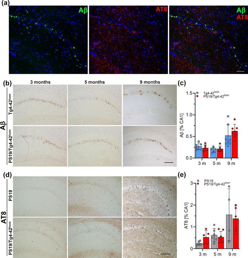
Figure 5. Phosphorylated tau and Aβ4–42 immunoreactivity. (a) Fluorescent staining of Aβ4–42 (green) and phosphorylated
Figure 5. Phosphorylated tau and Aβ4–42 immunoreactivity. (a) Fluorescent staining of Aβ4–42 (green) and phosphorylated
tau protein (AT8—red) in the CA1 pyramidal layer from a 3-month-old PS19/Tg4–42hom mouse did not reveal a major co-
tau protein (AT8—red) in the hom
localization. Representative AβCA1(b) andpyramidal
AT8 (d) layer from
staining a 3-month-old
in CA1 PS19/Tg4–42
of PS19, Tg4–42 mouse hom
hom and PS19/Tg4–42 didmice.
not Immuno-
reveal a major co-
localization. hom and PS19/Tg4–42hom mice. Immunohisto-
histochemicalRepresentative Aβper
analysis (n = 5–7 (b) time
and AT8
point,(d) staining per
3 sections in CA1 of PS19,
animal) showedTg4–42
no difference in Aβ immunoreactivity be-
chemical analysis
tween Tg4–42 (n = PS19/Tg4–42
hom and 5–7 per time hom
point,
mice3 sections perpoint
at any time animal)
(c).showed no difference
A tendency towards anin increase
Aβ immunoreactivity withTg4–42hom
between
in the area covered
phosphorylated
and PS19/Tg4–42 hom
tau in mice
3-month-old
at any PS19/Tg4–42
time point (c). mice,
hom while notowards
A tendency differences
an in tau pathology
increase in the were
area observed at 5 months
covered with phosphorylated
between
tau PS19 and PS19/Tg4–42
in 3-month-old PS19/Tg4–42 hom animals
hom mice, (e). Allno
while data are given in
differences as tau
mean ± SD. Unpaired
pathology t-test. Scale
were observed at 5bars: (a): 50
months µ m, (b, PS19 and
between
d): 100 μm. hom animals (e). All data are given as mean ± SD. Unpaired t-test. Scale bars: (a): 50 µm, (b,d): 100 µm.
PS19/Tg4–42Int. J. Mol. Sci. 2021, 22, 5191 10 of 17
2.5. Hippocampal Aβ Ppathology and Tau Phosphorylation in PS19/Tg4–42hom Mice
Fluorescent immunohistochemistry was performed on paraffin brain sections to
investigate if Aβ4–42 and tau co-localize within the same neurons in PS19/Tg4–42hom
mice (Figure 5a). The pyramidal layer was analyzed specifically as it is the main region
expressing Aβ4–42 in Tg4–42hom mice. No major co-localizations of Aβ4–42 and tau im-
munoreactivity were observed within the same pyramidal neuron. Next, Aβ4–42 pathology
was quantified in the CA1 region of the hippocampus and compared between Tg4–42hom
and PS19/Tg4–42hom (Figure 5b,c) mice after staining with the 24311 antibody. No sig-
nificant differences in Aβ load were detected in PS19/Tg4–42hom compared to single
transgenic Tg4–42hom mice at either 3, 5 or 9 months of age (Figure 5c). To determine
whether transgenic Aβ4–42 accumulation aggravates tau pathology, hyperphosphorylated
tau, immunohistochemically stained with the AT8 antibody, was quantified in the CA1
region of the hippocampus comparing PS19 and PS19/Tg4–42hom mice (Figure 5d,e). At 3
months of age, an increased immunoreactivity of phosphorylated tau was detected upon
Aβ4–42 expression in PS19/Tg4–42hom mice, with a trend towards statistical significance
(p = 0.0573). In contrast, no significant difference was observed at 5 months of age be-
tween PS19 and PS19/Tg4–42hom mice (Figure 5e). At 3 and 5 months of age, phospho-tau
immunoreactivity presented mostly as a diffuse staining with very few intracellular ac-
cumulations. This was predominantly observed in the bigenic line, but without a clear
aggravation in tau pathology. At 9 months, abundant tau pathology with NFTs could be
observed in the CA1 pyramidal layer, but with no significant difference between PS19 and
PS19/Tg4–42hom mice.
3. Discussion
In general, PS19 mice did not show overt deficits compared to age-matched WT mice
in terms of motor or memory performance at younger ages. With regard to the accelerating
rotarod task, they even seemed to perform slightly better than WT littermates, however
without reaching statistical significance. This is consistent with a hyperactivity phenotype
in 6-month-old PS19 mice, presenting with better, albeit not statistically significant, per-
formance on the rotarod compared to WT control animals [28]. A related observation was
reported recently in 3.5- to 12-month-old PS19 mice, showing a ~10% prolonged latency in
the accelerating rotarod test [29].
Potential effects of excess Aβ peptides on an already existing tau pathology have been
studied in several experimental paradigms. A substantially increased number of NFTs was
detected in P301L tau transgenic mice following injection of fibrillary Aβ42 peptides [30]
and an induction of tau pathology was observed in the same line of tau transgenic mice after
infusion of an Aβ-containing brain extract from a 24-month-old APP23 mouse [31]. Related
findings were reported by Vasconselos and colleagues, showing that pre-aggregated Aβ is
able to induce fibrillization of tau both in vitro and in vivo [32], and interestingly, passive
immunization against Aβ in the 3xTg mouse model not only reduced extracellular amyloid
plaques but also resulted in decreased tau pathology [33].
The lack of an influence of mutant tau on Aβ accumulation, as observed in the present
report, confirms previous studies in APP/Tau transgenic lines with robust extracellular
Aβ deposition. Using the same line of P301S tau transgenic mice, a trend towards a higher
extracellular amyloid plaque load was detected in PDAPP/Tau bigenic mice [8], while no
evidence for increased Aβ pathology was evident in 5XFAD/PS19 mice at either 3 or 9
months of age [9]. However, it has to be noted that an up to 5-fold increased amyloid load
was observed in 16-month-old Tg2576 mice that have been crossed with a tau transgenic
line harboring a triple-mutant tau (G272V, P310L, R406W) [34].
The Tg4–42 mouse model used in the present study primarily shows an intraneuronal
accumulation of Aβ peptides, in particular in CA1 hippocampal neurons, and does not
form overt extracellular plaques [21]. A related model expressing only Aβ1–42 peptides with
the rat preproenkephalin signal peptide (APP48) in the absence of APP overexpression
developed intracellular Aβ lesions and presented with reduced hippocampal neuronInt. J. Mol. Sci. 2021, 22, 5191 11 of 17
numbers already at young age [35]. This line had been crossed with tau transgenic mice
overexpressing human 4-repeat tau with the P301S mutation (TAU58). Double transgenic
mice showed neither evidence of increased levels of soluble Aβ, albeit data on Aβ levels in
single transgenic APP48 mice were not reported, nor significant amounts of phospho-tau
pathology [20]. Similarly, we observed no significant increase of phosphorylated tau in
PS19/ Tg4–42hom mice except for a trend in young animals. These findings in models
harboring transgenic Aβ peptides in the absence of APP overexpression strikingly contrast
the obvious exacerbation of tau pathology observed in tau transgenic mice when crossed
with mouse models of AD overexpressing mutant APP [7–13,36]. The overexpression of
APP in the studied mouse models could be a determining factor in the exacerbation of
tau pathology. Takahashi and colleagues [37] showed that the presence of APP induced
intracellular phosphorylated tau aggregation in cell culture exposed to tau fibrils in a dose-
dependent manner. In addition, recent data suggested that the toxic effects of oligomeric tau
on memory and long-term potentiation (LTP) in WT mice appear to be APP-dependent [38].
With regard to hippocampal neuron loss, a significantly decreased number of CA1
pyramidal cells has been detected in 9-month-old 5XFAD/PS19 mice in comparison to their
parental single transgenic 5XFAD or PS19 lines [9]. A related observation was made in
3-month-old PS19/Tg4–42hom mice, showing a significantly reduced CA1 pyramidal cell
number in the distal part of the CA1 region. The lack of such a difference in 5-month-old
mice might be attributed to the fact that single Tg4–42hom mice already show a profound
CA1 neuron loss, reaching a kind of plateau at that time point [27]. Indeed, between 5 and 9
months, the neuron loss in Tg4–42hom mice worsens by less than 10%. While the distal part
of the CA1 region appears to be mainly involved in non-spatial memory, the proximal part
is supposed to play a more important role in tasks depending on spatial information [39,40].
Though Tg4–42hom as well as PS19/Tg4–42hom mice showed a significant loss of distal CA1
pyramidal neurons already at 3 and 5 months, this is not reflected in a major impairment
of recognition memory, at least not detectable in the NOR task carried out in the present
study. A deficit in this task became obvious in aged mice, which was accompanied by
the loss of ~60% of neurons in the distal CA1. Neuron loss in the proximal CA1 part was
most pronounced in 5-month-old PS19/Tg4–42hom mice. At this time point, bigenic mice
showed spatial memory deficits in the MWM task, however, it has to be noted that neuron
numbers did not differ significantly from single transgenic Tg4–42hom mice still learning
the task, confirming results from a previous study employing 5-month-old Tg4–42hom
mice [27]. By 9 months of age, Tg4–42hom mice display obvious spatial learning deficits.
Despite comparable loss of proximal pyramidal neurons with Tg4–42hom at this time
point, PS19/Tg4–42hom displayed the worst performances in goal quadrant and platform
parameters among the transgenic lines. The presence of spatial memory deficits in the PS19
line is controversial, with some studies pointing towards the development of deficits in
the MWM test from a relatively young age of 5 [28] or 6.5 months [41], while others report
initial deficits in spatial memory at 10 [42] or even 12 months of age [29], respectively. The
tau transgenic line did not present with loss of CA1 pyramidal neurons at the considered
time points, but neuron loss in the CA3 and reduced DG volume have been described in
P301S mice starting at 8 months [26,43].
Although the distal CA1 neuron loss observed in young PS19/ Tg4–42hom mice
was accompanied by a tendency towards increased phospho-tau immunoreactivity, no
obvious behavioral alterations were observed at this time point, while in aged mice, the
behavioral deficits observed in the bigenic line could not be related to an aggravation of
neuron loss or tau pathology. The absence of obvious aggravation of tau pathology might
suggest that the co-presence alone of Aβ4–42 and transgenic human tau and their additive
singular detrimental effects could lead to the observed phenotypes in young mice. As
an aggravation of tau pathology has been reported repeatedly in a variety of APP/tau
transgenic lines, our data might support an important confounding role of transgenic
human APP overexpression.Int. J. Mol. Sci. 2021, 22, 5191 12 of 17
4. Materials and Methods
4.1. Transgenic Mice
The generation of the Tg4–42 line was previously described [21]. In brief, the human
Aβ4-42 sequence is fused to the signal peptide sequence of the thyrotropin-releasing hor-
mone and the expression is under the control of the Thy1 promoter. The Tg4–42 mice were
bred to homozygosity (Tg4–42hom ). The line was generated and maintained on a C57BL/6J
genetic background.
PS19 mice overexpress human tau with the P310S mutation under the control of the
murine prion protein promoter [26] and were purchased from Jackson laboratories (Bar
Harbor, ME, USA) (B6;C3-Tg(Prnp-MAPT*P301S)PS19Vle/J). Mice were backcrossed to
C57Bl/6J for more than 5 generations.
Bigenic mice (PS19/Tg4–42hom ) were generated by breeding transgene-positive
Tg4–42hom and PS19 mice and were maintained on a C57BL/6J genetic background. Ac-
cordingly, littermates were only obtained for Tg4-42hom and PS19/Tg4-42hom (with an
additional tau transgene). In a second line of breeding, WT mice were bred with heterozy-
gous PS19 mice to obtain WT and PS19 littermates. An equal number of female and male
transgenic and age-matched C57Bl/6J (WT) mice at 3, 5 and 9 months of age were used in
the present study. All animals were handled according to the German guidelines for animal
care and all experiments have been approved by the local animal care and use committee
(LAVES, Lower Saxony, Germany). Food and water were provided ad libitum.
4.2. Reverse Transcription and qRT-PCRs
Deep-frozen hippocampus samples were used to isolate mRNA. The tissues were
weighted, supplied with 1 mL of Trifast® reagent (PegLab, Wilmington, DE, USA) per
100 mg of sample and homogenized using a glass-Teflon homogenizer with 15 strokes
at 800 rpm. RNA isolation was performed following the manufacturer’s protocol. RNA
samples were subjected to digestion with DNAse I, followed by reverse transcription using
the RevertAid RT Kit (Thermo Fisher Scientific, Waltham, MA, USA) according to the
protocol of the supplier. The Biozym Blue S’Green qPCR Mix, containing SYBR Green
as the intercalating fluorescent dye, was used to perform gene expression analysis. Raw
data were collected using the MxPro Mx3000P software (Stratagene, Bellingham, WA,
USA) and the average Ct value was calculated from the duplicate for each sample. The
relative expression of the genes of interest (GOIs) was performed using murine β-Actin as
a reference gene for normalization, and were calibrated to a selected control group using
the ∆∆Ct method [44]. The following primer sets were employed, as in Table 1.
4.3. Behavioral Tasks
4.3.1. Accelerating Rotarod
A computer-controlled rotarod system (TSE, Technical and Scientific Equipment) was
used to assess motor performance and motor learning in the rotarod task [45]. During 2
days of testing, each animal performed 4 trials, at least 10 min apart, per day. In each trial,
the rod accelerated from 4 to 40 rpm over a maximum trial time of 300 s and the latency to
fall was recorded. The task was performed under red light condition and the apparatus
was cleaned with 70% ethanol between each trial to avoid odor cues.
4.3.2. Balance Beam
In order to assess balance and fine motor coordination, the balance beam test was
conducted [46]. Mice were placed on the center of a wooden beam (1 cm wide, 50 cm long,
44 cm high), at both ends of which a 9 × 15 cm escape platform was attached. The ground
surface underneath the beam was padded to avoid possible injures. Each mouse performed
three consecutive 60 s trials with at least 10 min intervals in between in one single day of
testing. The latency to fall from the beam was recorded, while if a mouse remained on the
beam for the whole 60 s trial or escaped to one of the platforms, the maximum time of 60 s
was given.Int. J. Mol. Sci. 2021, 22, 5191 13 of 17
4.3.3. Inverted Grid
Neuromuscular abilities, vestibular function and muscle strength were tested with the
inverted grid task [47]. Each mouse was positioned in the center of a metallic wire grid
(45 cm in length, 30 cm in width, with a grid spacing of 1 cm), which was inverted and
suspended 40 cm above a padded surface. The latency to fall was recorded during a single
60 s trial.
4.3.4. Morris Water Maze (MWM)
The Morris Water Maze (MWM) was used to assess spatial reference memory [48]
and was performed as previously described [21]. In brief, mice were at first subjected
to 3 days of cued training, each consisting of four 60 s trials 10 min apart. During each
trial of the cue training, the platform was marked with a triangular flag. The location
of the platform and the starting point for the mice always changed between the four
quadrants. A 5-day acquisition training (4 trials/day) followed 24 h after the end of the cue
training. In this phase, the flag was removed from the platform that remained stationary
for each mouse and in each trial. Proximal and distal visual cues were present during the
acquisition training. During both cue and acquisition training, the latency to reach the
platform, average speed and distance were recorded with an automated video tracking
system (ANY-Maze, Stoelting, Wood Dale, IL, USA). Twenty-four hours after the last day of
acquisition training, a probe trial was performed to address spatial memory. The platform
was removed from the pool and mice were introduced from a novel entry point. Mice were
allowed to freely swim for the 60 s trial duration and abidance in the different quadrants
was recorded.
4.3.5. Novel Object Recognition (NOR)
The novel object recognition (NOR) task, based on the innate preference of rodents
for novelty, was performed to test for recognition memory. During the first day of testing,
two identical objects were placed in the arena and presented to the mice, which could
freely explore during a single trial session. On the second day of testing, one of the
identical objects was exchanged to a novel object. On both days, exploration time of each
object was recorded manually for every mouse during the single 5 min trial sessions. The
recognition performances was quantified using the Discrimination Index (DI), measured
as the differences between novel (Tnovel ) and familiar (Tfamiliar ) object exploration times in
proportion to the animal’s total exploration time (Ttotal ) [25].
4.4. Tissue Collection and Preservation
Mice were deeply anesthetized through an intraperitoneal injection of a mixture of
ketamine and xylazine and were transcardially perfused using ice-cold 0.01 M phosphate-
buffered saline (PBS). The brains were rapidly and carefully removed from the skull. The
right hemisphere was drop-fixed in 4% formalin solution at 4 ◦ C for at least 72 h protected
from light and subsequently embedded in paraffin. The left hemisphere was dissected
to obtain hippocampi samples that were deep-frozen on dry-ice and stored at −80 ◦ C
until processing.
4.5. Quantification of CA1 Neuron Number
Neuron loss was assessed in the CA1 region of the hippocampus on sagittal brain
sections (bregma 0.72–1.08) of 3- and 5-month-old mice (n = 6–7 per time point) as previ-
ously described [25]. Paraffin sections of 4 µm thickness (3 sections per animal, at least
30 µm apart) were stained with hematoxylin to identify the nuclei. Neuronal nuclei were
distinguished from glia cells based on their size and characteristic appearance. Images
from the distal (towards subiculum) and proximal (extending to CA2) part of the CA1 were
acquired using an Olympus BX-51 microscope equipped with a Moticam pro 282 camera
(Motic, Wetzlar, Germany) at 400× magnification. CA1 pyramidal neurons in a definedInt. J. Mol. Sci. 2021, 22, 5191 14 of 17
area were counted using the manual cell counting tool implemented in ImageJ1.51 (NIH,
Bethesda, MD, USA).
4.6. Immunohistochemistry
Sagittal paraffin brain samples were cut at 4 µm and used in immunohistochemical
staining. Sections were processed as previously described [49]. In brief, paraffin was
removed, incubating the slides in xylol and the sections were rehydrated with an ascending
ethanol series. Endogenous peroxidases were blocked with a 30 min treatment of 0.3%
H2 O2 in 0.01 M phosphate-buffered saline (PBS), and antigens were retrieved by boiling
sections in 0.01 M citrate buffer (pH 6.0). In case of amyloid-β staining, the epitopes were
exposed to an additional 3 min treatment with 88% formic acid. An incubation of 4%
skim milk in 0.01M PBS with 10% fetal cow serum (FCS) was applied for 1 h to block
unspecific binding sites. The following primary antibodies, diluted to the desired concen-
tration in 0.01 M PBS including 10% FCS, were applied overnight at room temperature
in a humid chamber: 24311 (pan-Aβ, 1:500, rabbit pAb [9]) and AT8 (phosphorylated
Tau pSer202/pThr205, 1:500, mouse mAb, Thermo Fisher Scientific, Dreieich, Germany).
Biotinylated secondary antibodies (1:200, Dianova, Hamburg, Germany) or fluorescent-
labelled secondary antibodies (1:750, Thermo Fisher Scientific, Dreieich, Germany) were
applied for 1 h. Staining was visualized via the ABC method using the Vectastain kit (Vector
Laboratories, Burlingame, CA, USA) with diaminobenzidine (DAB) as a chromogen and
hematoxylin counterstaining. When a fluorescent immunohistochemistry was performed,
4’,6-diamidin-2-phenylindol (DAPI) was used to label the nuclei. Fluorescent images
were taken using a Nikon TiE microscope (Nikon, Tokyo, Japan) and analyzed with NIS
Elements imaging software (Nikon, Tokyo, Japan).
4.7. Quantification of Aβ and Tau Immunoreactivity
Serial images of the CA1 region of the hippocampus from DAB-stained sections were
taken (n = 3 sections per animal), at least 30 µm apart from each other, using an Olympus
BX-51 microscope equipped with a Moticam pro 282 camera (Motic, Wetzlar, Germany)
with a 100x magnification lens (n = 5–6 per time point). The captured images were analyzed
using the ImageJ software. A fixed intensity threshold was applied to define the DAB
staining, after binary transformation to 8-bit black and white images. The percentage of
the area covered by DAB staining was measured and compared between the different
genotypes. Aβ load (24311) was quantified in Tg4–42hom and PS19/Tg4–42hom , and tau
immunoreactivity (AT8) in PS19 and PS19/Tg4–42hom mice.
4.8. Statistical Analyses
All data have been analyzed using the Shapiro–Wilk test for normality to ensure that
parametric tests can be applied. When parametric testing was possible, differences between
groups were tested with unpaired t-test, one-way analysis of variance (ANOVA) followed
by Bonferroni’s post-hoc, or repeated measure two-way ANOVA followed by Bonferroni’s
post-hoc, as indicated. In case of non-parametric testing, the Mann–Whitney test was
performed. All data were presented as means ± standard deviation (SD). Significance
levels were as follows: * p < 0.05, ** p < 0.01, *** p < 0.001. All calculations were performed
using GraphPad Prism version 8 for Windows (Graph Pad Software, San Diego, CA, USA).
5. Conclusions
As it has been shown previously that the presence of Aβ peptides leads to an aggrava-
tion of an existing tau pathology in a variety of transgenic AD mouse models, we evaluated
the impact of soluble N-terminally truncated Aβ4-42 peptides in the widely used PS19
mouse model of AD. Though we detected a partial worsening of motor and spatial memory
performance, as well as an aggravated CA1 neuron loss at young age, we did not observe
the accelerated formation of tau aggregates reported in models with overt extracellular
Aβ plaque pathology. Hence, the presence of extracellular amyloid plaques might be aInt. J. Mol. Sci. 2021, 22, 5191 15 of 17
prerequisite for enhanced neurofibrillary tangle formation in transgenic mice co-expressing
human mutant APP and Tau.
Supplementary Materials: The following are available online at https://www.mdpi.com/article/
10.3390/ijms22105191/s1, Figure S1: Average escape latency and speed in cued training of MWM,
Figure S2: Water Maze—Speed in acquisition training, Figure S3: Water Maze—Time in target
quadrant, Figure S4: Novel object recognition task, Figure S5: Examples of proximal and distal CA1,
Figure S6: Neuron loss in total CA1.
Author Contributions: Conceptualization, O.W.; Formal analysis, O.W. and S.Z.; Investigation, S.Z.;
Data curation, S.Z.; Writing—Original draft, O.W. and S.Z.; Supervision, O.W. All authors have read
and agreed to the published version of the manuscript.
Funding: O.W. is supported by Gerhard Hunsmann Stiftung, Alzheimer Forschung Initiative e.V.
and Alzheimer Stiftung Göttingen. We acknowledge support by the Open Access Publication Funds
of the Göttingen University.
Institutional Review Board Statement: All animals were handled according to the German guide-
lines for animal care, and all experiments have been approved by the local animal care and use
committee (Landesamt für Verbraucherschutz und Lebensmittelsicherheit (LAVES), Lower Sax-
ony, Germany).
Data Availability Statement: Original data is available from the authors upon reasonable request.
Acknowledgments: The expert technical assistance of Petra Tucholla and Petra Rieper is grate-
fully acknowledged.
Conflicts of Interest: The authors declare no conflict of interest.
References
1. Masters, C.L.; Simms, G.; Weinman, N.A.; Multhaup, G.; McDonald, B.L.; Beyreuther, K. Amyloid plaque core protein in
Alzheimer disease and Down syndrome. Proc. Natl. Acad. Sci. USA 1985, 82, 4245–4249. [CrossRef] [PubMed]
2. Grundke-Iqbal, I.; Iqbal, K.; Quinlan, M.; Tung, Y.C.; Zaidi, M.S.; Wisniewski, H.M. Microtubule-associated protein tau. A
component of Alzheimer paired helical filaments. J. Biol. Chem. 1986, 261, 6084–6089. [CrossRef]
3. Hardy, J.; Selkoe, D.J. The amyloid hypothesis of Alzheimer’s disease: Progress and problems on the road to therapeutics. Science
2002, 297, 353–356. [CrossRef] [PubMed]
4. Bloom, G.S. Amyloid-β and tau: The trigger and bullet in alzheimer disease pathogenesis. JAMA Neurol. 2014, 71, 505–508.
[CrossRef]
5. Nisbet, R.M.; Polanco, J.C.; Ittner, L.M.; Gotz, J. Tau aggregation and its interplay with amyloid-beta. Acta Neuropathol. 2015, 129,
207–220. [CrossRef] [PubMed]
6. Tomiyama, T.; Matsuyama, S.; Iso, H.; Umeda, T.; Takuma, H.; Ohnishi, K.; Ishibashi, K.; Teraoka, R.; Sakama, N.;
Yamashita, T.; et al. A mouse model of amyloid-β oligomers: Their contribution to synaptic alteration, abnormal tau phosphory-
lation, glial activation, and neuronal loss in vivo. J. Neurosci. 2010, 30, 4845–4856. [CrossRef]
7. Grueninger, F.; Bohrmann, B.; Czech, C.; Ballard, T.M.; Frey, J.R.; Weidensteiner, C.; von Kienlin, M.; Ozmen, L. Phosphorylation
of Tau at S422 is enhanced by Abeta in TauPS2APP triple transgenic mice. Neurobiol. Dis. 2010, 37, 294–306. [CrossRef]
8. Hurtado, D.E.; Molina-Porcel, L.; Iba, M.; Aboagye, A.K.; Paul, S.M.; Trojanowski, J.Q.; Lee, V.M. Aβ accelerates the spatiotemporal
progression of tau pathology and augments tau amyloidosis in an Alzheimer mouse model. Am. J. Pathol. 2010, 177, 1977–1988.
[CrossRef]
9. Saul, A.; Sprenger, F.; Bayer, T.A.; Wirths, O. Accelerated tau pathology with synaptic and neuronal loss in a novel triple transgenic
mouse model of Alzheimer’s disease. Neurobiol. Aging 2013, 34, 2564–2573. [CrossRef]
10. Stancu, I.-C.; Ris, L.; Vasconcelos, B.; Marinangeli, C.; Goeminne, L.; Laporte, V.; Haylani, L.E.; Couturier, J.; Schakman, O.;
Gailly, P.; et al. Tauopathy contributes to synaptic and cognitive deficits in a murine model for Alzheimer’s disease. FASEB J.
2014, 28, 2620–2631. [CrossRef]
11. Lewis, J.; Dickson, D.W.; Lin, W.L.; Chisholm, L.; Corral, A.; Jones, G.; Yen, S.H.; Sahara, N.; Skipper, L.; Yager, D.; et al. Enhanced
neurofibrillary degeneration in transgenic mice expressing mutant tau and APP. Science 2001, 293, 1487–1491. [CrossRef]
12. Kang, S.; Kim, J.; Chang, K.A. Spatial memory deficiency early in 6xTg Alzheimer’s disease mouse model. Sci. Rep. 2021, 11, 1334.
[CrossRef]
13. Héraud, C.; Goufak, D.; Ando, K.; Leroy, K.; Suain, V.; Yilmaz, Z.; De Decker, R.; Authelet, M.; Laporte, V.; Octave, J.-N.; et al.
Increased misfolding and truncation of tau in APP/PS1/tau transgenic mice compared to mutant tau mice. Neurobiol. Dis. 2014,
62, 100–112. [CrossRef] [PubMed]Int. J. Mol. Sci. 2021, 22, 5191 16 of 17
14. Rapoport, M.; Dawson, H.N.; Binder, L.I.; Vitek, M.P.; Ferreira, A. Tau is essential to beta-amyloid-induced neurotoxicity. Proc.
Natl. Acad. Sci. USA 2002, 99, 6364–6369. [CrossRef] [PubMed]
15. Leroy, K.; Ando, K.; Laporte, V.; Dedecker, R.; Suain, V.; Authelet, M.; Heraud, C.; Pierrot, N.; Yilmaz, Z.; Octave, J.N.; et al. Lack
of Tau Proteins Rescues Neuronal Cell Death and Decreases Amyloidogenic Processing of APP in APP/PS1 Mice. Am. J. Pathol.
2012, 181, 1928–1940. [CrossRef] [PubMed]
16. Shipton, O.A.; Leitz, J.R.; Dworzak, J.; Acton, C.E.; Tunbridge, E.M.; Denk, F.; Dawson, H.N.; Vitek, M.P.; Wade-Martins, R.;
Paulsen, O.; et al. Tau protein is required for amyloid {beta}-induced impairment of hippocampal long-term potentiation. J.
Neurosci. 2011, 31, 1688–1692. [CrossRef]
17. Puzzo, D.; Argyrousi, E.K.; Staniszewski, A.; Zhang, H.; Calcagno, E.; Zuccarello, E.; Acquarone, E.; Fa’, M.; Li Puma, D.D.;
Grassi, C.; et al. Tau is not necessary for amyloid-β–induced synaptic and memory impairments. J. Clin. Investig. 2020, 130,
4831–4844. [CrossRef]
18. Jin, M.; Shepardson, N.; Yang, T.; Chen, G.; Walsh, D.; Selkoe, D.J. Soluble amyloid beta-protein dimers isolated from Alzheimer
cortex directly induce Tau hyperphosphorylation and neuritic degeneration. Proc. Natl. Acad. Sci. USA 2011, 108, 5819–5824.
[CrossRef]
19. Hu, X.; Li, X.; Zhao, M.; Gottesdiener, A.; Luo, W.; Paul, S. Tau pathogenesis is promoted by Abeta1-42 but not Abeta1-40. Mol.
Neurodegen. 2014, 9, 52. [CrossRef]
20. Gomes, L.A.; Hipp, S.A.; Upadhaya, A.R.; Balakrishnan, K.; Ospitalieri, S.; Koper, M.J.; Largo-Barrientos, P.; Uytterhoeven, V.;
Reichwald, J.; Rabe, S.; et al. Aβ-induced acceleration of Alzheimer-related τ-pathology spreading and its association with prion
protein. Acta Neuropathol. 2019, 138, 913–941. [CrossRef]
21. Bouter, Y.; Dietrich, K.; Wittnam, J.L.; Rezaei-Ghaleh, N.; Pillot, T.; Papot-Couturier, S.; Lefebvre, T.; Sprenger, F.; Wirths, O.;
Zweckstetter, M.; et al. N-truncated amyloid β (Aβ) 4–42 forms stable aggregates and induces acute and long-lasting behavioral
deficits. Acta Neuropathol. 2013, 126, 189–205. [CrossRef]
22. Portelius, E.; Bogdanovic, N.; Gustavsson, M.K.; Volkmann, I.; Brinkmalm, G.; Zetterberg, H.; Winblad, B.; Blennow, K. Mass
spectrometric characterization of brain amyloid beta isoform signatures in familial and sporadic Alzheimer’s disease. Acta
Neuropathol. 2010, 120, 185–193. [CrossRef]
23. Wirths, O.; Zampar, S. Emerging roles of N- and C-terminally truncated Aβ species in Alzheimer’s disease. Expert Opin. Ther.
Targets 2019, 23, 991–1004. [CrossRef] [PubMed]
24. Hüttenrauch, M.; Brauss, A.; Kurdakova, A.; Borgers, H.; Klinker, F.; Liebetanz, D.; Salinas-Riester, G.; Wiltfang, J.; Klafki, H.W.;
Wirths, O. Physical activity delays hippocampal neurodegeneration and rescues memory deficits in an Alzheimer disease mouse
model. Transl. Psych. 2016, 6, e800. [CrossRef] [PubMed]
25. Stazi, M.; Wirths, O. Chronic Memantine Treatment Ameliorates Behavioral Deficits, Neuron Loss, and Impaired Neurogenesis in
a Model of Alzheimer’s Disease. Mol. Neurobiol. 2021, 58, 204–216. [CrossRef] [PubMed]
26. Yoshiyama, Y.; Higuchi, M.; Zhang, B.; Huang, S.M.; Iwata, N.; Saido, T.C.; Maeda, J.; Suhara, T.; Trojanowski, J.Q.; Lee, V.M.
Synapse loss and microglial activation precede tangles in a P301S tauopathy mouse model. Neuron 2007, 53, 337–351. [CrossRef]
27. Antonios, G.; Borgers, H.; Richard, B.C.; Brauß, A.; Meißner, J.; Weggen, S.; Pena, V.; Pillot, T.; Davies, S.L.; Bakrania, P.; et al.
Alzheimer therapy with an antibody against N-terminal Abeta 4-X and pyroglutamate Abeta 3-X. Sci. Rep. 2015, 5, 17338.
[CrossRef]
28. Takeuchi, H.; Iba, M.; Inoue, H.; Higuchi, M.; Takao, K.; Tsukita, K.; Karatsu, Y.; Iwamoto, Y.; Miyakawa, T.; Suhara, T.; et al. P301S
Mutant Human Tau Transgenic Mice Manifest Early Symptoms of Human Tauopathies with Dementia and Altered Sensorimotor
Gating. PLoS ONE 2011, 6, e21050. [CrossRef]
29. Sun, Y.; Guo, Y.; Feng, X.; Jia, M.; Ai, N.; Dong, Y.; Zheng, Y.; Fu, L.; Yu, B.; Zhang, H.; et al. The behavioural and neuropathologic
sexual dimorphism and absence of MIP-3α in tau P301S mouse model of Alzheimer’s disease. J. Neuroinflamm. 2020, 17, 72.
[CrossRef]
30. Götz, J.; Chen, F.; van Dorpe, J.; Nitsch, R.M. Formation of neurofibrillary tangles in P301l tau transgenic mice induced by Abeta
42 fibrils. Science 2001, 293, 1491–1495. [CrossRef]
31. Bolmont, T.; Clavaguera, F.; Meyer-Luehmann, M.; Herzig, M.C.; Radde, R.; Staufenbiel, M.; Lewis, J.; Hutton, M.; Tolnay, M.;
Jucker, M. Induction of tau pathology by intracerebral infusion of amyloid-beta -containing brain extract and by amyloid-beta
deposition in APP x Tau transgenic mice. Am. J. Pathol. 2007, 171, 2012–2020. [CrossRef] [PubMed]
32. Vasconcelos, B.; Stancu, I.-C.; Buist, A.; Bird, M.; Wang, P.; Vanoosthuyse, A.; Van Kolen, K.; Verheyen, A.; Kienlen-Campard, P.;
Octave, J.-N.; et al. Heterotypic seeding of Tau fibrillization by pre-aggregated Abeta provides potent seeds for prion-like seeding
and propagation of Tau-pathology in vivo. Acta Neuropathol. 2016, 131, 549–569. [CrossRef]
33. Oddo, S.; Billings, L.; Kesslak, J.P.; Cribbs, D.H.; LaFerla, F.M. Abeta Immunotherapy Leads to Clearance of Early, but Not Late,
Hyperphosphorylated Tau Aggregates via the Proteasome. Neuron 2004, 43, 321–332. [CrossRef] [PubMed]
34. Ribe, E.M.; Perez, M.; Puig, B.; Gich, I.; Lim, F.; Cuadrado, M.; Sesma, T.; Catena, S.; Sanchez, B.; Nieto, M.; et al. Accelerated
amyloid deposition, neurofibrillary degeneration and neuronal loss in double mutant APP/tau transgenic mice. Neurobiol. Dis.
2005, 20, 814–822. [CrossRef] [PubMed]
35. Abramowski, D.; Rabe, S.; Upadhaya, A.R.; Reichwald, J.; Danner, S.; Staab, D.; Capetillo-Zarate, E.; Yamaguchi, H.; Saido, T.C.;
Wiederhold, K.-H.; et al. Transgenic Expression of Intraneuronal Aβ42 But Not Aβ40 Leads to Cellular Aβ Lesions, Degeneration,
and Functional Impairment without Typical Alzheimer’s Disease Pathology. J. Neurosci. 2012, 32, 1273–1283. [CrossRef] [PubMed]You can also read